sucrose loading in isolated veins of pisum sativum - plant physiology
TRANSCRIPT
Plant Physiol. (1989) 91, 259-2650032-0889/89/91/0259/07/$01 .00/0
Received for publication December 31, 1988and in revised form May 11, 1989
Sucrose Loading in Isolated Veins of Pisum sativum:Regulation by Abscisic Acid, Gibberellic Acid, and
Cell Turgor'
Juan Jose Estruch, Juli G. Pereto2, Yolanda Vercher, and Jose Pio Beltran*3Institut d'Agroquimica i Tecnologia d'Aliments, CSIC, Jaume Roig 11, E-46010 Valencia, Spain (J.J.E., Y.V.,
J.P.B.); and Departament de Bioquimica i Biologia Molecular, Universitat de Valencia, Dr. Moliner 50, E-46100Bur/assot, Spain (J.G.P.)
ABSTRACT
Enzymatically isolated vein networks from mature pea (Pisumsativum L. cv Alaska) leaves were employed to investigate theproperties of sucrose loading and the effect of phytohormonesand cell turgor on this process. The sucrose uptake showed twocomponents: a saturable and a first-order kinetics system. Thehigh affinity system (Km, 3.3 millimolar) was located at the plas-malemma (p-chloromercuriphenylsulfonic acid and orthovan-adate sensitivity). Further characterization of this system, includ-ing pH dependence and effects of energy metabolism inhibitors,supported the Hf-sugar symport concept for sucrose loading.Within a physiological range (0.1-100 micromolar) and after 90min, abscisic acid (ABA) inhibited and gibberellic acid (GA3)promoted 1 millimolar sucrose uptake. These responses werepartially (ABA) or totally (GA3) turgor-dependent. In experimentsof combined hormonal treatments, ABA counteracted the GA3positive effects on sucrose uptake. The abolishment of theseresponses by p-chloromercuriphenylsulfonic acid and experi-ments on proton flux suggest that both factors (cell turgor andhormones) are modulating the H+ATPase plasmalemma activity.The results are discussed in terms of their physiologicalrelevance.
Assimilate distribution appears to be under sink controland may be hormonally regulated (1 1, 24). Although manydifferent sites and processes have been described as possibletargets of hormonal control, much attention has been paid tothe effects of hormones on both phloem loading and unload-ing of sucrose ( 17). We developed an experimental system tostudy the hormone-directed transport in intact pea plantsduring parthenocarpic fruit set induced by GAs (18). Sucrosetransport from source to sink was activated by GA, and GA3
'This work was supported by Consejo Superior de InvestigacionesCientificas (grant 1-215), Ministerio de Educaci6n y Ciencia C-Acci6nIntegrada Hispano-Alemana (B23/9, 1987), and Direcci6n Generalde Investigaci6n Cientifica y Tecnia T (grant PB-0402). J.J.E. was therecipient of a Ministerio de Educaci6n y Ciencia-Formaci6n dePersonal Investigador fellowship.
2 Present address: Plant Science Institute, University of Pennsyl-vania, Philadelphia, PA 19104.
3Present address: Molekulare Pflanzengenetik, Max-Planck-Insti-tut fur Zuchtungsforschung, D 5000 Koln 30, Federal Republic ofGermany.
increasing phloem unloading at the sink (ovary). Moreover, astrong increase in sucrose exported from the source leaf wasobserved when the GAs reached the leafadjacent to the ovary,suggesting that sucrose loading at the source could also beactivated by GAs.The current knowledge on the mechanism of phloem load-
ing is based, initially, on studies of sucrose uptake kineticscarried out with leaf discs and other complex plant materials.Attempts have been made to develop new experimental ap-proaches to obtain more homogeneous, phloem-enriched, andstill functional tissues. Thus, it is possible in some plants todissect surgically the vascular components from mesophyllcells, and even to separate phloem from xylem tissue (2, 4).Daie et al. (5), using surgically isolated phloem tissue fromcelery as well as intact plants reported that GA3 and IAAincrease sucrose loading and that the hormonal effects werecell turgor-dependent and H+ATPase-mediated (3).To remove the mesophyll cells of fully expanded leaves,
the alternative of choice for most plant systems should be toresort to enzymic maceration. Although sucrose uptake hasbeen characterized using this approach (1, 22, 27), hormonalregulation of this process has not been reported.The aim of the present work was to use enzymatically
isolated functional phloem-enriched tissue from mature leavesof Pisum sativum to study the effects of GA3, ABA, and cellturgor on sucrose uptake. Our results show the suitability ofthis simplified experimental system to investigate factors con-trolling the process of sucrose uptake.
MATERIALS AND METHODS
Plant Material
Plants of Pisum sativum L. cv Alaska No. 7 were grownfrom seeds as described previously (18). Several days beforethe experiment, plants were transferred from a greenhouse toa controlled cabinet and maintained on a photoperiod of 16h light (Philips TLD 1 8W-33 fluorescent tubes, 20 W.m2 atthe level of the first flower) and 8 h dark with temperatureranges of 22 to 24°C and 16 to 1 8°C, respectively.
Vein Network Isolation
For the isolation of the vein networks, leaves adjacent tothe first flower (seventh node) were employed 48 h after
259
Dow
nloaded from https://academ
ic.oup.com/plphys/article/91/1/259/6085296 by guest on 02 D
ecember 2021

Plant Physiol. Vol. 91,1989
anthesis. Leaves were cut off and their abaxial epidermis wasstripped. The resulting leaves were floated on 5 mL of basesolution (BS)4 consisting of 0.5 mM CaC12, 0.5 mM MgCl2, 5mM KCl, and 5 mm Mes (pH 6.0), adjusted to the appropriateosmolality with mannitol (Sigma). After 30 min, the BS wasreplaced by a maceration medium containing BS supple-mented with 2% cellulose Onozuka R-10 (Yakult Co Ltd.)and 0.05% pectolyase Y-23 (Seishin Pharmaceuticals), andgently shaken (50 rpm) for 90 min at 28TC. The adaxialepidermis and, with it, all the mesophyll cells, were thenremoved with a fine forceps. The resulting vein network wasrinsed twice in BS.Image analysis (IBAS 2000, Kontron) of pictures of the
isolated vein networks showed that the surface area of thevein system represented 48% of the total surface area of thepea leaf. The dry weight of the isolated veins accounted for17.1% of the fresh weight of the leaves (13.7 ± 0.1 mg.cm-2).The viability of the vein system was determined with fluo-
rescein diacetate as described by Wildhom (25).
Light and Electron Microscopy
For morphological studies, tissue samples (vein networksand whole leaves) were prepared as described by Vercher etal. (23) for the pea ovaries, except that the fixation and washbuffer was 0.05 M phosphate instead of 0.1 M. Ultrathinsections were observed under a Jeol lOOS electron microscope.
Sugar UptakeThe vein networks were incubated for 1 h in 5 mL ofbuffer-
free BS (pH adjusted to 6.0 with NaOH) BS containing variousconcentrations of unlabeled sugars and [U-'4C]sucrose or D-[U-'4C]sorbitol (Radiochemical Center, Amersham; specificactivity, 25.9 MBq * mol-' to 77.7 GBq * mol-'). All incubationsolutions were isoosmotic (unless otherwise stated, adjustedto 400 mOsm). When used, inhibitors were applied in apreincubation period of 30 min and, except for PCMPS, alsoduring the incubation with sugars. Controls contained thesame volume ofthe inhibitor's solvent. In experiments dealingwith hormonal effects on sucrose uptake, growth substanceswere added to a buffer-free BS, present during both preincu-bation and incubation periods. After the incubation, excessradioactivity from the surface was completely removed bytwo successive, 2-min washes with 5 mL of the BS. The tissuewas then killed with 80% (v/v) aqueous ethanol at 80°C for10 min and homogenized. After centrifugation (1600g, 10min) the ethanol-soluble radioactivity was measured by liquidscintillation. For the pH experiments, 5 mm nonpenetratingbuffers were used: Mes (pH 4.5 and 5.5), Mops (pH 6.5),Hepes (pH 7.5), and Tricine (pH 8.5). The sugar uptakeperiod was performed with gentle shaking at 30°C underillumination (Osram nitraphot 500-W lamps, 380 W m-2).
Determination of the Apoplastic Concentration ofSucrose
Apoplastic sucrose of the pea leafwas collected as describedby Delrot et al. (9). Leaf excision was carried out 3 to 4 h
4Abbreviations: BS, base solution; PCMPS, para-chloromercuri-phenylsulfonic acid; FC, fusicoccin.
after the beginning of the photoperiod. After quick removalof the lower epidermis, leaves were floated on 5 mL of BSwith 250 mm mannitol in the dark at 4°C. At several times,aliquots of 0.3 mL were withdrawn, mixed with 1 uL of 2-mercaptoethanol, and boiled for 10 min. Sucrose concentra-tion was determined spectrophotometrically by enzymaticmethods adapted from ref. 13. Leaf apoplastic volume wasevaluated according to Morrod (16). Peeled leaves werefloated on 50 mL of BS containing 250 mM mannitol, 0.1%inulin (Merck), and [3H]inulin (Radiochemical Center; spe-cific activity, 18.5 kBq *mol-'). After different times, tissuewas rapidly rinsed in cold medium and extracted with 80%ethanol, and the radioactivity was counted.
Proton Flux
Proton extrusion activity was continuously monitored usinga pH meter (Orion Research Ion Analyzer EA920) connectedto an IBM-PC. As standard conditions, about 55 to 60 cm2of vein networks (preincubated in BS for 2 h) were placed ina vial containing 7 mL of BS continuously mixed with amagnetic stirrer. Measurements were made under room lightand temperature.
RESULTS
The isolated vein networks maintained their original mac-roscopic structure (Fig. IA). The use of the fluorescent dyefluorescein diacetate provided additional evidence that thevein networks were largely intact, indicating the viability ofthis system.
Ultrastructure of Phloem-Enriched Leaf Tissue of P.sativum
Transverse sections of isolated veins were studied by lightmicroscopy to ascertain the integrity of the vascular tissue.The isolated central vein showed a structure similar to thecentral vein of the intact pea leaf (Fig. 1, B and C), exceptthat the mesophyll cells adjacent to the vascular bundle hadtheir walls disrupted. Xylem and phloem cells did not seem
Figure 1. Vein networks enzymatically isolated from mature leavesof P. sativum. A, Leaf with stripped abaxial epidermis and treated (90min) with cellulose and pectolyase. B and C, Light micrographs oftransverse sections of the central vein of pea leaf. B, intact leaf; C,enzymatically isolated vein. Bar = 50 ,im. m, mesophyll; p, phloem;x, xylem.
260 ESTRUCH ET AL.
Dow
nloaded from https://academ
ic.oup.com/plphys/article/91/1/259/6085296 by guest on 02 D
ecember 2021

REGULATION OF SUCROSE LOADING IN PISUM
affected by the enzymatic treatment, not even the cells of thebundlesheath.The ultrastructural characteristics of the phloem cells in the
isolated veins were essentially the same as the intact leaf.
Sugar Uptake
The measurement of the sucrose uptake by the isolatedvein networks of pea leaves was essentially not affected by themetabolism of this sugar in the tissue. Thus, in the uptakeexperiments using asymmetrically labeled sucrose (['4C]fruc-tosyl sucrose) followed by HPLC analyses of the resultingextracts, we found all the label in the fraction with theretention time corresponding to sucrose. On the other hand,after treatment of extracts with yeast invertase (Boehringer-Mannheim), all the label was found in the fraction with theretention time corresponding to fructose. This total absenceof "'C label in the glucose moiety indicated that sucrose wastaken up by the isolated veins without extracellular hydrolysis.As can be observed (Fig. 2A) the uptake up to 90 mM
sucrose by the isolated veins showed a two-component kinet-ics: a saturable system and an apparently nonsaturable or lowaffinity system. In the presence of PCMPS, a nonpenetratingthiol binding agent, the kinetics were linear with the sameslope as the nonsaturable system (Fig. 2A). The contributionof the saturable component with highest affinity was ascer-tained by substracting sucrose uptake in the presence ofPCMPS from total uptake, using sucrose concentrations be-
10_ B
8 o
6 -
4
2
00 2 4 6 8 10Isucrose], mM
v C'aEE 6 - r=0.998 a
4 Km=3.3mME 4 -
x -
-L2 #0X - o0~
° 2 4 6 8 10-. Isucroserl, mM' I
Figure 2. Concentration dependence (up to 90 mM) of the sucrose
uptake by isolated veins of pea leaves. A, Effect of the presence (0)or the absence (0) of 2 mM PCMPS on sucrose uptake. B, Apparentlinear component (0, +PCMPS) is subtracted from the total uptake(0, -PCMPS) for obtaining the saturable component (O). C, Line-weaver-Burk plot for the calculated values in B. Bars are the SE ofthe mean of three replicates, two leaves per replicate.
low 10 mm (Fig. 2B). The saturable component exhibited anapparent Km of 3.3 mM (Fig. 2C).
Sucrose uptake by isolated vein networks was dependenton cell turgor. Figure 3 shows the variation in sucrose uptakeat different concentrations of mannitol, a nonpenetratingosmoticum in this system (data not shown). Highest sucroseuptake occurred at 500 mOsm. In addition, the presence of 5mM K+ in the medium promoted the sucrose uptake in therange of osmolality tested, the maximum being at 300 mOsm(Fig. 3). According to these results we used 5 mM KCI and400 mOsm mannitol as standard incubation conditions.
Secondary Transport of Sucrose
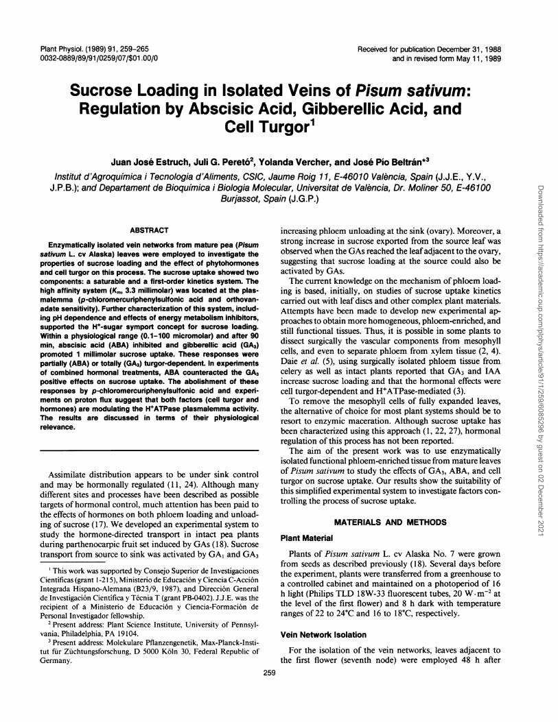
Sucrose uptake in isolated vein networks appears to occuraccording to the H+-cotransport concept for the secondarytransport of sucrose during phloem loading (10). Maximalsucrose uptake in this system was at pH 5.0 to 6.0 (Fig. 4),and the pH dependence was abolished by PCMPS, showing aprofile similar to that of the uptake of sorbitol (a diffusion-like process) (Fig. 4). The presence of 10 /M FC (a potentenhancer of the plasmalemma H+ATPase) in the mediumresulted in 68% increase in uptake at 1 mm sucrose. The H+-cotransport hypothesis is further substantiated by the obser-vation that several agents that interfere with the generation ormaintenance of an electrochemical gradient of H+ inhibitedsucrose uptake. Protonophores like 2,4-dinitrophenol (1 mM)and carbonylcyanide m-chlorophenylhydrazone (50 jLM) dra-matically reduced sucrose uptake (88% and 90% inhibition,respectively). The use of selective inhibitors of the proton-pumping activities (20) like orthovanadate (affecting plasma-lemma H+ATPase), diminished sucrose uptake rate (32%).However, in the presence ofPCMPS that inhibits 64% of totalsucrose uptake (Table I), no additional inhibition was pro-duced by orthovanadate. Azide (a selective inhibitor of mi-
160 -
a) 14012020
0
80
0 200 400 600 800osmolality, mOsm
Figure 3. Effect of mannitol concentration in the incubation solutionon uptake of sucrose (1 mM) by isolated veins of pea leaves. Uptakewas in the presence (Cl) or absence (0) of 5 mm KCI. Bars are the SEof the mean of three replicates, two leaves per replicate.
C4
x
x05E
CL
0.
IDu40
(A
261
Dow
nloaded from https://academ
ic.oup.com/plphys/article/91/1/259/6085296 by guest on 02 D
ecember 2021
Plant Physiol. Vol. 91,1989
'IIxr_X
-Vd
C
L4
El 4 . . . .'4.5 5.5 6.5 7.5 8.5
pH
Figure 4. Effect of pH on uptake of 1 mm sucrose by isolated veinsof pea leaves treated (@) and nontreated (0) with 2 mm PCMPS, andon uptake of 1 mm sorbitol (U). Bars are the SE of the mean of threereplicates, two leaves per replicate.
Table I. Effect of FC, ABA, and GA3 on Uptake of Sucrose (1 mM)by Isolated Veins of Pea Treated and Nontreated with 2 mM PCMPS
Tissue was incubated in buffer-free base medium (400 mOsm).Values are relative to sucrose uptake in controls (2.1 ± 0.7 nmol-h-*cm-2, and are the mean of two replicates ± SE, two leaves per
replicate.
Sucrose Uptake (Relative to
Substance Added Control)-PCMPS +2 mm PCMPS
None (control) 1.00 0.36 ± 0.0510,gM FC 1.67 ± 0.01 0.30 ± 0.021 zlM ABA 0.75 ± 0.04 0.40 ± 0.0210MmGA3 1.40±0.01 0.34±0.0210 MmFC + 1 Mm ABA 1.25 ± 0.02 -
1OMmFC + 10 Mm ABA 1.08 ± 0.02 -
10,gM GA3+ 1 lM ABA 1.00 ± 0.11 -
10MmGA3+ 10MmABA 0.84±0.02 -
tochondrial ATP synthase) and dicyclohexylcarbodiimide (ablocking agent of all known H+ATPases) strongly reducedsucrose uptake by 90% and 73%, respectively.
Hormonal Effects on Sucrose Uptake
After a relatively short period of time (a maximum of 90min), ABA inhibits (25%) and GA3 promotes (40%) sucrose
uptake by the isolated veins of pea (Table I). However, GA3-stimulated sucrose uptake ranged from 26% to 50% in exper-iments with a different set of plants.The treatment of the vein networks with PCMPS negates
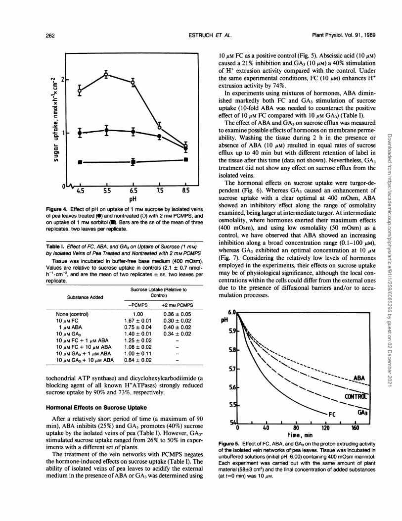
the hormone-induced effects on sucrose uptake (Table I). Theability of isolated veins of pea leaves to acidify the externalmedium in the presence ofABA or GA3 was determined using
10 Mm FC as a positive control (Fig. 5). Abscissic acid (10 MM)caused a 21% inhibition and GA3 (10 Mm) a 40% stimulationof H+ extrusion activity compared with the control. Underthe same experimental conditions, FC (10 Mm) enhances H'extrusion activity by 74%.
In experiments using mixtures of hormones, ABA dimin-ished markedly both FC and GA3 stimulation of sucrose
uptake (10-fold ABA was needed to counteract the positiveeffect of 10 Mm FC compared with 10 Mm GA3) (Table I).The effect ofABA and GA3 on sucrose efflux was measured
to examine possible effects ofhormones on membrane perme-
ability. Washing the tissue during 2 h in the presence orabsence of ABA (10 MM) resulted in equal rates of sucrose
efflux up to 40 min but with different retention of label inthe tissue after this time (data not shown). Nevertheless, GA3treatment did not show any effect on sucrose efflux from theisolated veins.The hormonal effects on sucrose uptake were turgor-de-
pendent (Fig. 6). Whereas GA3 caused an enhancement ofsucrose uptake with a clear optimal at 400 mOsm, ABAshowed an inhibitory effect along the range of osmolalityexamined, being larger at intermediate turgor. At intermediateosmolality, where hormones exerted their maximum effects(400 mOsm), and using low osmolality (50 mOsm) as a
control, we have observed that ABA showed an increasinginhibition along a broad concentration range (0.1-100 gM),whereas GA3 exhibited an optimal concentration at 10 Mm
(Fig. 7). Considering the relatively low levels of hormonesemployed in the experiments, their effects on sucrose uptakemay be of physiological significance, although the local con-
centrations within the cells could differ from the external onesdue to the presence of diffusional barriers and/or to accu-
mulation processes.
6.0
pH
5,9
5.8
~~~~ABA
5.6
\~._ C0NTRT
0 40 80 120 160time, min
Figure 5. Effect of FC, ABA, and GA3 on the proton extruding activityof the isolated vein networks of pea leaves. Tissue was incubated inunbuffered solutions (initial pH, 6.00) containing 400 mOsm mannitol.Each experiment was carried out with the same amount of plantmaterial (58+3 cm2) and the final concentration of added substances(at t=0 min) was 10 Am.
2-
1p.
*_ -U
A . I I I
ESTRUCH ET AL.262
Dow
nloaded from https://academ
ic.oup.com/plphys/article/91/1/259/6085296 by guest on 02 D
ecember 2021

REGULATION OF SUCROSE LOADING IN PISUM
ICA
140
130 -
120
100 0o-0o -
100
90
50 - %%%,70-l60
0 20 I 40I 6 80O 200 400 600 aoo
osmolality, mOsmFigure 6. Turgor dependence of the ABA and GA3 effects on sucroseuptake by isolated vein networks of pea leaves. Cell turgor was
varied with mannitol. Uptake of 1 mm sucrose was in the absence(control = 100%) or presence of 10 jLM GA3 (0) or 10 uM ABA (El).
Data are the mean of two replicates ± SE, three leaves per replicate.
DISCUSSION
Isolated vein networks display two components in thekinetics of sucrose uptake. The inhibition of the high affinitysaturable system (Ki, 3.3 mM) by PCMPS, which binds to theextracellular side of the binding center of the sucrose carrier(15), suggests that it is located at the plasmalemma. Althoughit has been reported that plasmalemma H+ATPase is sensitiveto mercurials (20), the H+ extruding activity of isolated veinnetworks of pea leaves is not affected by PCMPS (data notshown). Similarly, mM PCMBS exerted a strong inhibitionon sucrose uptake, but did not inhibit proton extrusion inViciafaba (8).Further characterization of the carrier-mediated transport
in our system came from studies on its pH dependence andproton efflux, and studies on the effect of energy metabolisminhibitors. Assuming that PCMPS is affecting the carrier butnot the H+ATPase of the plasmalemma, an outstanding ob-servation is that, in the presence of PCMPS, orthovanadatedid not cause any further inhibition. All evidence takentogether indicates that sucrose uptake across the plasma-lemma is a secondary transport mechanism based on a H+-sugar symport.The concentration of sucrose in the apoplast of pea leaves
(9.8 mM) was on the order of the one reported by Delrot etal. (9) in V. faba leaves (1-5 mM). It is worth noting that theKm of the plasmalemma sucrose carrier is in the range of thesucrose concentration at the apoplast. Therefore, this carrier
160 I
1401
°- 120.4-C0
-100
GIu
NOe,
, 100In
0.1 1.0 10 100[ GA3 ]J,.M
I
80
60
0.1 1.0 10 100[ABA L,OM
Figure 7. Effect of GA3 or ABA on uptake of sucrose (1 mM) byisolated vein networks of pea leaves. Incubation was for 1 h inunbuffered solutions at pH 6 adjusted to 50 mOsm (0, El) or 400mOsm (H,O) with mannitol. In each experiment, various concentra-tions of hormones were added to preincubation (30 min) and incu-bation solutions (60 min). Data are the mean of two replicates ± SE,three leaves per replicate.
would not be working at maximum activity whereby it couldbe significant from a regulatory point of view.On the other hand, taking into account the levels of sucrose
in the apoplast of pea leaves, the relative contribution of thelinear component of the sucrose uptake in vivo should be oflittle importance. Wilson et al. (27), working with strippedand enzymatically digested leaves of Allium cepa, and Daie(2), working with surgically obtained vascular bundles ofcelery, have suggested that a low affinity component repre-sented the contribution of the phloem parenchyma to sugaruptake. Daie (4) has also suggested that the linear componentof sucrose uptake by excised celery phloem is the diffusioncontribution of xylem parenchyma mainly.The existence of an active and selective sucrose uptake by
the veins, together with the absence of plasmodesmatal con-
nections between mesophyll cells and the vascular system,
a
In
4-Liw
as44--
a)
IF/g
§,o
I-
L
a
263
I I I a
Dow
nloaded from https://academ
ic.oup.com/plphys/article/91/1/259/6085296 by guest on 02 D
ecember 2021

Plant Physiol. Vol. 91,1989
points out an apoplastic mechanism (7) for sucrose loadinginto the phloem of P. sativum. In this sense, Turgeon andWimmers (21), using leaf discs and autoradiographic tech-niques, have shown recently that an apoplastic pathway is aplausible explanation for the loading of sucrose in pea leaves.
Sucrose uptake rates were higher at low cell turgor than theones at high cell turgor (see Fig. 5). According to the massflow theory, high turgor is required to support the hydrostaticpressure gradient between source and sink to facilitate assim-ilate translocation (12). Thus, below a turgor set point, thesieve element-companion cell complex would respond byincreasing the sugar uptake to maintain constant assimilateflow to sink tissues. Some studies (3, 6, 19) support the viewthat changes in cell turgor are occurring at the plasmalemmaof the sieve element-companion cell complex, and suggestthat H+ATPase might play a key role in the mechanism ofosmotic adaptation. Obviously, this osmoregulatory phenom-enon should be transient, if it is occurring in vivo.
Sucrose uptake by isolated vein networks was affected bythe presence of phytohormones (Table I). The disappearanceof the hormonal effects after PCMPS treatment (see Table I)suggests that both ABA and GA3 could affect sucrose loadingat the level of the plasma membrane.
Since sugar-H+ cotransport is a plausible explanation ofphloem loading in this tissue, at least two possible targets forthe hormonal action can be envisaged, namely the sucrosecarrier and the H+ATPase, both located at the plasmalemma.A direct effect ofthese hormones on the sucrose carrier shouldbe excluded because ABA and GA3 did not affect the 1 mMsucrose uptake in buffered solutions (5 mM Mes-OH, pH 6).Therefore, the hormonal action could be focused on theplasmalemma H+ATPase. This supports previous findings ofMalek and Baker (14) for ABA acting on sucrose loading inRicinus communis, and by Daie (3) for IAA and GA3 effectson sucrose uptake by isolated phloem segments of celery.
Furthermore, the hormonal response of the vein systemwas dependent on the osmotic concentration of the solution,indicating an interaction between hormonal effects and cellturgor. We could interpret the observed hormonal effects onsucrose loading in two different physiological situations. Un-der a normal water supply, GA3 may play a role as a positivesignal, coming from demanding areas, i.e. sinks like devel-oping fruits (18), increasing phloem loading of sucrose at thesource leaf. The GA3 effects would be mediated by the plas-malemma H+ATPase, an activity dependent ofthe cell turgor.In contrast, under a water deficit situation, to which peaplants are particularly sensitive (26), the reduction in cellturgor would be a major factor responsible for triggering thechange both in the sucrose uptake activity and in the hor-monal balance, mainly increasing the ABA level. The appar-ent hierarchy of the hormonal effects on sucrose loading isoutstanding, ABA (negative signal, partially turgor-independ-ent) playing a predominant role over GA3.
This paper shows that, despite the enzymatic treatment,isolated vein networks of pea leaves are suitable to study notonly sucrose loading but hormonal regulation of this process.However, further studies are necessary (i.e. high-resolutionmicroautoradiography) to show the specific type of cells in-volved in sucrose loading.
ACKNOWLEDGMENTS
Thanks are due to M. Marti and R. Martinez Pardo for theirtechnical assistance. W. Regh (Botanisches Institut, Universitat Essen,Federal Republic of Germany) performed the HPLC analyses ofsugars, J.M. Soria (Department of Microbiology, Universitat de Val-encia) helped us with computer work, and R. Sendra and M.S. delPino (Department of Biochemistry and Molecular Biology, Univer-sitat de Valencia) designed the program of the image analyzer. Wealso thank Drs. V. Conejero (Department of Biotechnology, Univer-sitat Politenica de Valencia) and J. Segura (Department of PlantBiology, Universitat de Valencia) for their critical review of themanuscript.
LITERATURE CITED
1. Cataldo DA (1974) Vein loading: The role of the symplastintracellular transport of carbohydrate between the mesophylland minor veins of tobacco leaves. Plant Physiol 53: 912-917
2. Daie J (1986) Kinetics of sugar transport in isolated vascularbundles and phloem tissue of celery. J Am Soc Hort Sci 111:2 16-220
3. Daie J (1987) Interaction of cell turgor and hormones on sucroseuptake in isolated phloem of celery. Plant Physiol 84: 1033-1037
4. Daie J (1987) Sucrose uptake in isolated phloem of celery is asingle saturable transport system. Planta 171: 474-482
5. Daie J, Watts M, Aloni B, Wyse RE (1986) In vitro and in vivomodification of sugar transport and translocation in celery byphytohormones. Plant Sci 46: 35-41
6. Daie J, Wyse RE (1985) Evidence on the mechanism of en-hanced sucrose uptake at low cell turgor in leaf discs ofPhaseolus coccineus. Physiol Plant 64: 547-552
7. Delrot S (1987) Phloem loading: Apoplastic or symplastic? PlantPhysiol Biochem 25: 667-676
8. Delrot S, Despeghel JP, Bonnemain JL (1980) Phloem loadingin Vicia faba leaves: Effect of N-ethylmaleimide and parach-loromercuribenzenesulfonic acid on H+extrusion, K+ and su-crose uptake. Planta 149: 144-148
9. Delrot S, Faucher M, Bonnemain JL, Bonmort J (1983) Nycth-emeral changes in intracellular and apoplastic sugars in Viciafaba leaves. Physiol Veg 21: 459-467
10. Giaquinta RT (1983) Phloem loading of sucrose. Annu Rev PlantPhysiol 34: 347-387
1 1. Gifford RM, Evans LT (1981) Photosynthesis, carbon partition-ing and yield. Annu Rev Plant Physiol 32: 485-509
12. Hitz WD, Giaquinta RT (1987) Sucrose transport in plants.BioEssays 6: 217-221
13. Jones MGK, Outlaw WH, Lowry OH (1977) Enzymatic assayof 10-7 to 10-'4 moles of sucrose in plant tissues. Plant Physiol60: 379-383
14. Malek L, Baker DA (1978) Effect of fusicoccin on proton co-transport of sugars in the phloem loading ofRicinus communisL. Plant Sci Lett 11: 233-239
15. M'Batchi B, Delrot S (I1984) Parachloromercuribenzenesulfonicacid. A potential tool for differential labelling of the sucrosetransporter. Plant Physiol 75: 154-160
16. Morrod RS (1974) A new method for measuring the permeabilityof plant cell membranes using epidermis-free leaf discs. J ExpBot 25: 521-533
17. Patrick JW (1987) Are hormones involved in assimilate trans-port? In GV Hoad, JP Lenton, MB Jackson, RK Atkin, eds,Hormone Action in Plant Development. A Critical Appraisal.Butterworths, London, pp 175-187
18. Pereto JG, Beltran JP (1987) Hormone-directed sucrose trans-port during fruit set induced by gibberellins in Pisum sativum.Physiol Plant 69: 356-360
19. Reinhold L, Seiden A, Volokita M (1984) Is modulation of therate of proton pumping a key event in osmoregulation? PlantPhysiol 75: 846-849
20. Serrano R (1984) Plasma membrane ATPase of fungi and plants
ESTRUCH ET AL.264
Dow
nloaded from https://academ
ic.oup.com/plphys/article/91/1/259/6085296 by guest on 02 D
ecember 2021

REGULATION OF SUCROSE LOADING IN PISUM
as a novel type of proton pump. Curr Top Cell Regul 23: 87-126
21. Turgeon R, Wimmers LE (1988) Different patterns of vein load-ing of exogenous ['4C]sucrose in leaves of Pisum sativum andColeus blumei. Plant Physiol 87: 179-182
22. Van Bet AJE, Koops AJ (1985) Uptake of ['4C]sucrose in isolatedminor-vein networks of Commelina benghalensis L. Planta164: 362-369
23. Vercher Y, Molowny A, Carbonell J (1987) Gibberellic acideffects on the ultrastructure of endocarp cells of unpollinatedovaries of Pisum sativum. Physiol Plant 71: 302-308
24. Weaver RJ, Johnson JO (1985) Relation ofhormones to nutrient
mobilization and the internal environment of the plant: Thesupply of mineral nutrients and photosynthate. In RP Pharis,DM Reid, eds, Encyclopedia of Plant Physiology (New Series),Vol 11. Springer-Verlag, Berlin, pp 3-36
25. Wildhom J (1972) The use of fluorescein diacetate and pheno-safranine for determining viability of cultured plant cells. StainTechnol 47: 189-194
26. Wilson DR, Jamieson PD, Jermyn WA, Hanson R (1985) Modelsof growth and water use of field peas (Pisum sativum L.). InPD Hebblethwaite, MC Heath, TCK Dawkins, eds, The PeaCrop. Butterworths, London, pp 139-151
27. Wilson C, Oross JW, Lucas WJ (1985) Sugar uptake into Alliumcepa leaf tissue: An integrated approach. Planta 164: 227-240
265
Dow
nloaded from https://academ
ic.oup.com/plphys/article/91/1/259/6085296 by guest on 02 D
ecember 2021