computational modelling of pisum sativum l superoxide
TRANSCRIPT
INT. J. BIOAUTOMATION, 2014, 18(2), 75-88
75
Computational Modelling of Pisum Sativum L. Superoxide Dismutase and Prediction of Mutational Variations through in silico Methods Nathan Vinod Kumar1*, Lizzy Mathew2, Edward Gnanaraj Wesely3 1Department of Botany and Microbiology Lady Doak College Madurai 625 002, Tamil Nadu, India E-mail: [email protected] 2Department of Botany St. Teresa’s College Cochin 682 017, Ernakulam, Kerala, India E-mail: [email protected] 3Department of Botany A. A. Government Arts College Namakkal 637 002, Tamil Nadu, India E-mail: [email protected] *Corresponding author
Received: January 4, 2013 Accepted: March 23, 2014 Published: June 30, 2014 Abstract: Superoxide dismutase (SOD) is one of the major enzymes expressed in the oxidative stress pathway in plants. Its expression is also evident in other taxonomic group in oxidative reactions. Pisum sativum a common plant is being studied in the present work where SOD is characterized using computational tools. SOD sequence of P. sativum [CAA42737.1] Ala and Leu rich protein with alkaline pI value was used as query sequence and used to obtain nine similar sequences through BLASTp. Phylogenetic tree was constructed using MEGA 5.0 based on neighbour joining method. Physiochemical parameters and amino acid composition was studied and compared with query sequences and other similar sequences. Secondary structures were predicted to understand the dominant components. Homology modeling of P. sativum SOD was done using SWISS MODEL and quality was evaluated using standard methods. 27 active sites were detected in SOD predicted model which were Lys rich. Keywords: BLASTp, Neighbour joining tree, Ramachandran plot, SOPMA, SWISS MODEL.
Introduction Oxidative stress is a major biotic stress seen in plant as well as animal cells which hampers its productivity. Higher plants employ defense strategies to cope up with various environmental stresses [21, 27]. Reactive oxygen species (ROS) are produced in both stressed and unstressed cells [1]. The univalent reduction of di-oxygen occurs in almost all aerobic cells [6, 21]. Free radicals are the chemicals which have an uncoupled electron and are capable of reacting rapidly [2]. The first product is superoxide radical anion, O2
-. Subsequently, other toxic chemical entities such as H2O2 and hydroxyl radicals, OH-, are formed. Superoxide radicals resulting in formation of H2O2 formation are detoxified by superoxide dismutase (SOD) [1, 21]. Within a cell, the SOD constitutes the first line of defense against ROS. Apart from SOD, plant cells equip some other major oxygen radical detoxifying enzymes such as ascorbate peroxidase (APX), glutathione reductase (GR) and catalase (CAT) [1, 21].
INT. J. BIOAUTOMATION, 2014, 18(2), 75-88
76
SODs are found in different sub-cellular locations. Based on the co-factor used by the enzyme, they are classified into three groups namely Fe-SOD, Mn-SOD and Cu-Zn-SOD. Mn-SOD occurs in mitochondria and peroxisomes. It carries a metal atom per subunit. Even though Mn- and Fe-SODs have similar primary, secondary and tertiary structure, these enzymes have diverged sufficiently that Fe (II) could not restore the function of Mn-SOD and vice versa [13]. Mn-SOD is either a hetero-dimeric or a homo-dimeric structure with one Mn (III) atom per subunit. It is not deactivated by hydrogen peroxide and is present in both prokaryotes as well as eukaryotes [1]. Once the superoxide radicals are formed, Mn-SOD dismutates it to H2O2 due to its enzyme activity [24]. Since these enzymes are playing a major role in oxidative stress control in plants needs to be characterized well. Apart from oxidative stress, expression of SOD is also reported in case of drought and metal toxicity also [23, 25]. There are many studies focused on the structural and functional characterization of SODs of plants. But, not many reports are available on application of computational tools for its characterization. The present work focuses on the computational characterization and structure evaluation of P. sativum SOD which could contribute in engineering of a plant with resistance under various stress environments especially in oxidative stress. As the stress tolerant plants are the need of the hour for increased productivity and better quality, its creation is only possible through better understanding of such stress related enzymes and the mechanisms involved in it. Materials and methods Sequence retrieval and BLASTp Superoxide dismutase protein sequence of P. sativum [CAA42737.1] was retrieved from NCBI databank and BLASTp was performed to retrieve nine similar sequences of different origins. The retrieved sequences are:
• AAF50095.1 (Drosophila melanogaster), • AAA29934.1 (Schistosoma mansoni), • CAA43859.1 (Chymomyza amoena), • YP_001176531.1 (Enterobacter sp. 638), • BAA02655.1 (Thermus aquaticus), • YP_004787678.1 (Muricauda ruestringensis), • YP_004272108.1 (Planctomyces brasiliensis DSM 5305), • ADY29074.1 (Cellulophaga lytica DSM 7489), • ADV48601.1 (Cellulophaga algicola DSM 14237).
Sequences obtained from BLASTp were aligned using CLUSTAL W multiple sequence alignment in BioEdit 5.0 [17]. Neighbour joining tree was constructed for the same using MEGA 5.0 software [29, 37]. Physicochemical characterization SOD sequences were subjected to ProtParam analysis to obtain molecular weight, pI, aliphatic index, GRAVY and Instability index (II) of all sequences [15]. Percentage of amino acid occurrence was compared for these sequences. Transmembrane probability for SOD of P. sativum was analysed using TmPred and DAS TM segment prediction server. Secondary structure prediction and homology modeling Secondary structure of SODs was analyzed using SOPMA analysis [16] and the homology modeling of SOD of P. sativum was done on SWISS MODEL based on the template 3dc6C
INT. J. BIOAUTOMATION, 2014, 18(2), 75-88
77
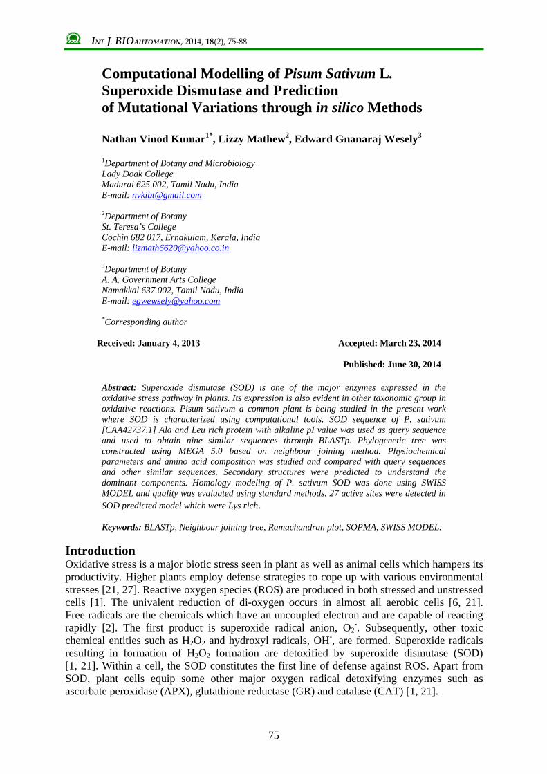
[3, 22, 28]. Ramachandran plot was constructed using RAMPAGE software and quality of the model was predicted using VMD software [18]. PROSA analysis was also performed for the same. Using Swiss pdb Viewer, electron density map for the SOD was made. Active site prediction and sub cellular localization Active sites of SOD of P. sativum were predicted using automated active site prediction server of SCFBio. Amino acid composition of each cavity was studied and tabulated. As SODs are classified based on co-factors and are distributed to different sub-cellular locations. Target P analysis was performed to classify the retrieved sequences and classify them based on their possible locations. Results and discussion Phylogenetic analysis Ten similar sequences were retrieved after performing BLASTp and CLUSTAL W multiple sequence alignment was done using BioEdit software. Phylogenetic tree was constructed based on Thermus aquaticus [BAA02655.1] SOD sequence as root using Neighbour joining method with boot strap of 1000 replications (Fig. 1). P. sativum SOD was placed as distinct branch with zero boot strap value showing least divergence from the root. SOD of Enterobacter sp. and Drosophila melanogaster exhibited highest divergence supported by boot strap value of 100.
AAF50095.1 Drosophila melanogaster
CAA43859.1 Chymomyza amoena
AAA29934.1 Schistosoma mansoni
YP 001176531.1 Enterobacter sp. 638
YP 004272108.1 Planctomyces brasiliensis
YP 004787678.1 Muricauda ruestringensis
ADY29074.1 Cellulophaga lytica DSM 7489
ADV48601.1 Cellulophaga algicola DSM 142
CAA42737.1 Pisum sativum
BAA02655.1 Thermus aquaticus
100
60
100
78
96
72
0.8106
0.0771
0.4416
0.0771
0.6448
0.6422
0.1042
0.1773
0.0815
0.0815
0.3645
0.2032
0.4729
0.0227
0.0731
0.9404-0.1684
Fig. 1 Neighbour joining tree showing evolutionary relationship
between various SOD sequences
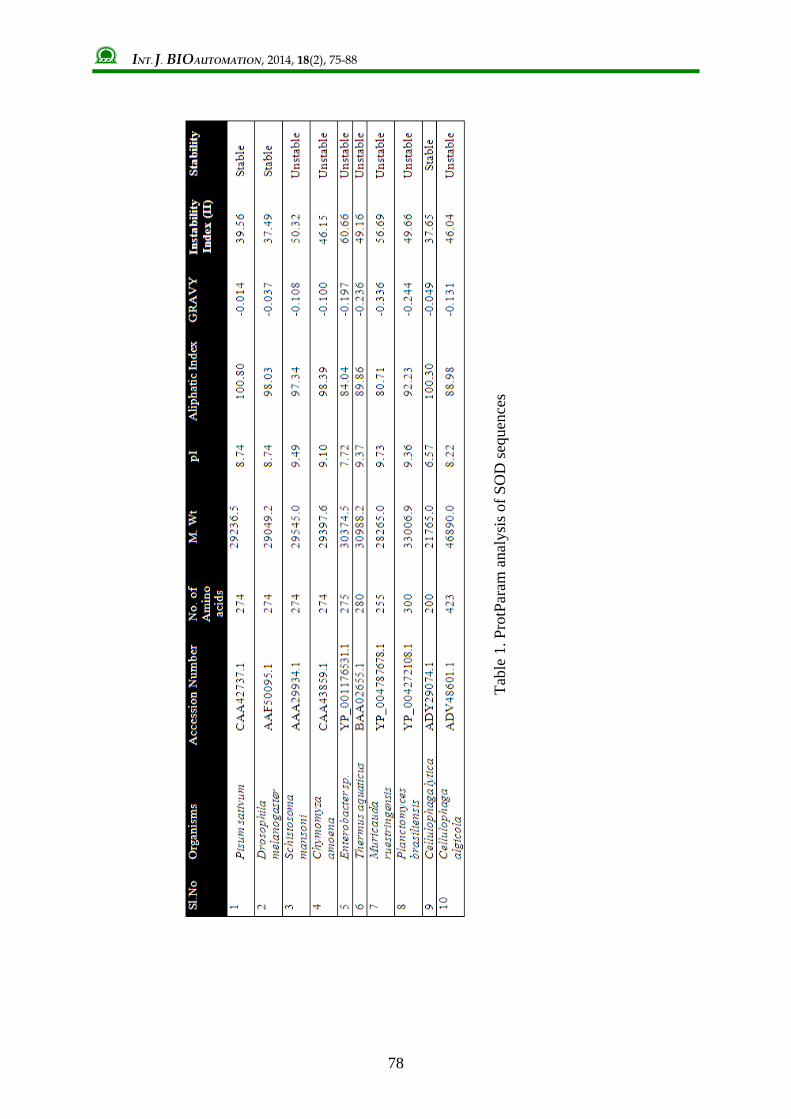
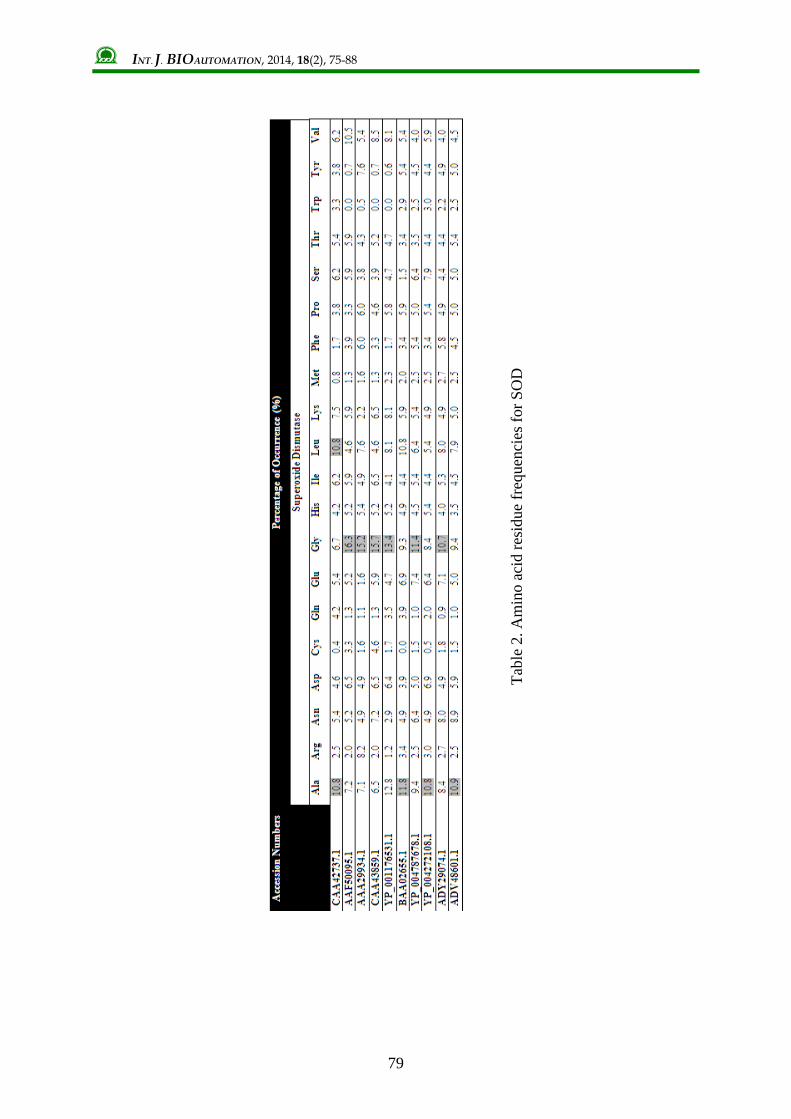
Physiochemical characterization Various physiochemical parameters like molecular weight, theoretical pI, aliphatic index, instability index, and GRAVY were predicted using ProtParam analysis in ExPASy server (Table 1). It was notable that SOD exhibited an alkaline pI which ranged from 7.72 to 9.73. Cellulophaga lytica SOD [ADY29074.1] showed a pI value of 6.57 which was acidic. A higher aliphatic index indicated its stability in wide range of temperatures. GRAVY value of P. sativum SOD was the least which ensures better interaction with water molecules. SODs of P. sativum, D. melanogaster and Cellulophaga lytica was classified as stable one by its Instability index (II) values. When amino acid composition of the SOD sequences were analysed, it was found that 6 SOD sequences were rich in Gly whereas for the rest of the sequences, Ala was the dominant residue (Table 2). P. sativum SOD had Ala and Leu dominance with a percentage of 10.8.
INT. J. BIOAUTOMATION, 2014, 18(2), 75-88
78
Tabl
e 1.
Pro
tPar
am a
naly
sis o
f SO
D se
quen
ces
INT. J. BIOAUTOMATION, 2014, 18(2), 75-88
79
Tabl
e 2.
Am
ino
acid
resi
due
freq
uenc
ies f
or S
OD
INT. J. BIOAUTOMATION, 2014, 18(2), 75-88
80
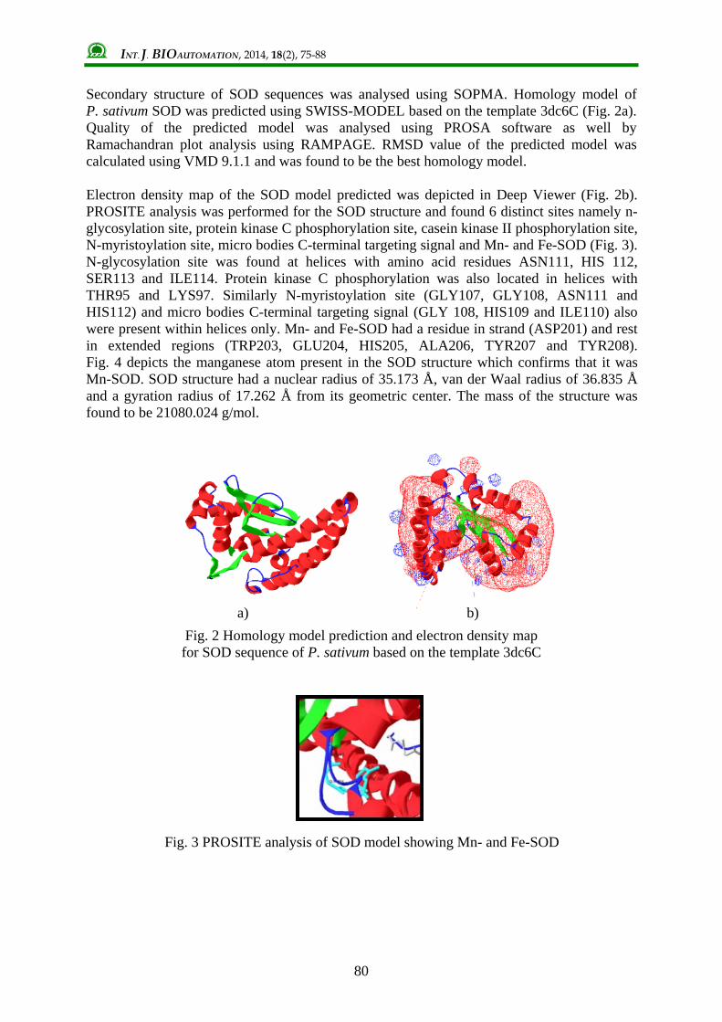
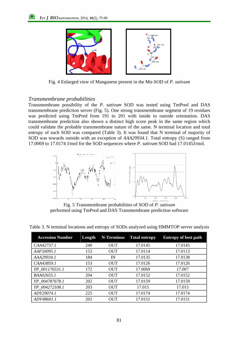
Secondary structure of SOD sequences was analysed using SOPMA. Homology model of P. sativum SOD was predicted using SWISS-MODEL based on the template 3dc6C (Fig. 2a). Quality of the predicted model was analysed using PROSA software as well by Ramachandran plot analysis using RAMPAGE. RMSD value of the predicted model was calculated using VMD 9.1.1 and was found to be the best homology model. Electron density map of the SOD model predicted was depicted in Deep Viewer (Fig. 2b). PROSITE analysis was performed for the SOD structure and found 6 distinct sites namely n-glycosylation site, protein kinase C phosphorylation site, casein kinase II phosphorylation site, N-myristoylation site, micro bodies C-terminal targeting signal and Mn- and Fe-SOD (Fig. 3). N-glycosylation site was found at helices with amino acid residues ASN111, HIS 112, SER113 and ILE114. Protein kinase C phosphorylation was also located in helices with THR95 and LYS97. Similarly N-myristoylation site (GLY107, GLY108, ASN111 and HIS112) and micro bodies C-terminal targeting signal (GLY 108, HIS109 and ILE110) also were present within helices only. Mn- and Fe-SOD had a residue in strand (ASP201) and rest in extended regions (TRP203, GLU204, HIS205, ALA206, TYR207 and TYR208). Fig. 4 depicts the manganese atom present in the SOD structure which confirms that it was Mn-SOD. SOD structure had a nuclear radius of 35.173 Å, van der Waal radius of 36.835 Å and a gyration radius of 17.262 Å from its geometric center. The mass of the structure was found to be 21080.024 g/mol.
Fig. 2 Homology model prediction and electron density map for SOD sequence of P. sativum based on the template 3dc6C
Fig. 3 PROSITE analysis of SOD model showing Mn- and Fe-SOD
a) b)
INT. J. BIOAUTOMATION, 2014, 18(2), 75-88
81
Fig. 4 Enlarged view of Manganese present in the Mn-SOD of P. sativum
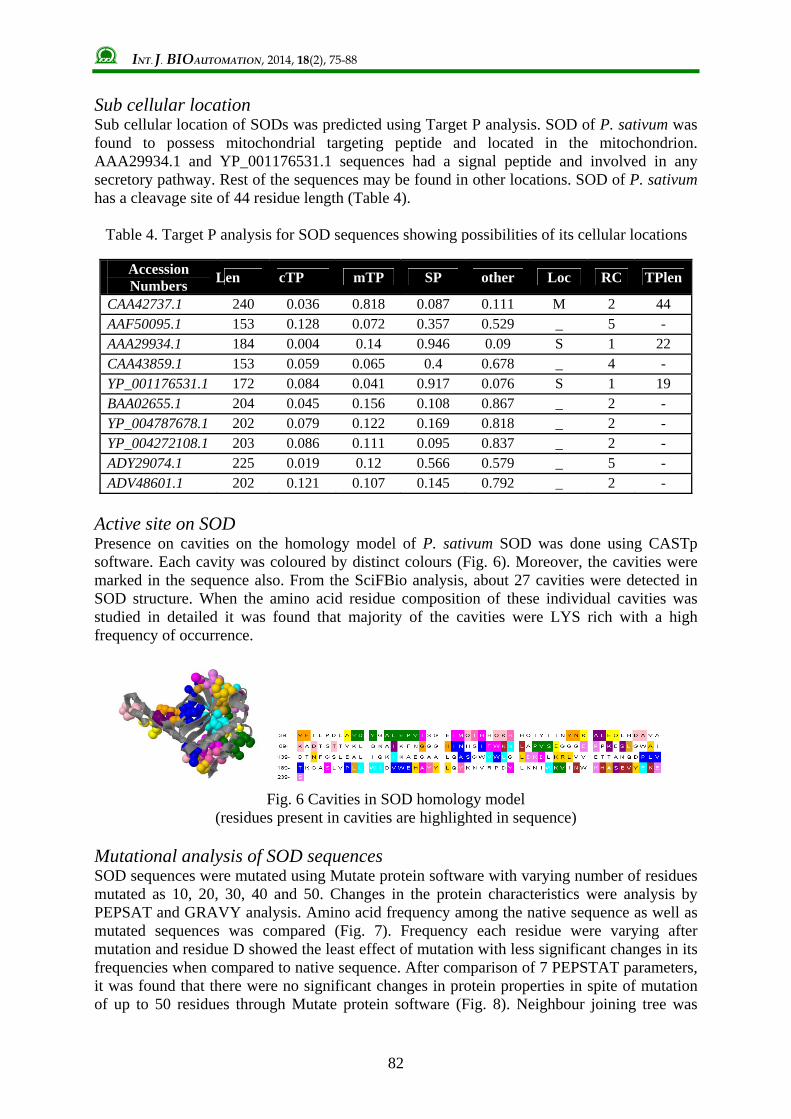
Transmembrane probabilities Transmembrane possibility of the P. sativum SOD was tested using TmPred and DAS transmembrane prediction server (Fig. 5). One strong transmembrane segment of 19 residues was predicted using TmPred from 191 to 201 with inside to outside orientation. DAS transmembrane prediction also shown a distinct high score peak in the same region which could validate the probable transmembrane nature of the same. N terminal location and total entropy of each SOD was compared (Table 3). It was found that N terminal of majority of SOD was towards outside with an exception of AAA29934.1. Total entropy (S) ranged from 17.0069 to 17.0174 J/mol for the SOD sequences where P. sativum SOD had 17.0145J/mol.
Fig. 5 Transmembrane probabilities of SOD of P. sativum
performed using TmPred and DAS Transmembrane prediction software
Table 3. N terminal locations and entropy of SODs analyzed using HMMTOP server analysis
Accession Number Length N-Terminus Total entropy Entropy of best path
CAA42737.1 240 OUT 17.0145 17.0145 AAF50095.1 153 OUT 17.0114 17.0113 AAA29934.1 184 IN 17.0135 17.0138 CAA43859.1 153 OUT 17.0126 17.0126 YP_001176531.1 172 OUT 17.0069 17.007 BAA02655.1 204 OUT 17.0152 17.0152 YP_004787678.1 202 OUT 17.0159 17.0159 YP_004272108.1 203 OUT 17.015 17.015 ADY29074.1 225 OUT 17.0174 17.0174 ADV48601.1 202 OUT 17.0151 17.0151
INT. J. BIOAUTOMATION, 2014, 18(2), 75-88
82
Sub cellular location Sub cellular location of SODs was predicted using Target P analysis. SOD of P. sativum was found to possess mitochondrial targeting peptide and located in the mitochondrion. AAA29934.1 and YP_001176531.1 sequences had a signal peptide and involved in any secretory pathway. Rest of the sequences may be found in other locations. SOD of P. sativum has a cleavage site of 44 residue length (Table 4).
Table 4. Target P analysis for SOD sequences showing possibilities of its cellular locations
Active site on SOD Presence on cavities on the homology model of P. sativum SOD was done using CASTp software. Each cavity was coloured by distinct colours (Fig. 6). Moreover, the cavities were marked in the sequence also. From the SciFBio analysis, about 27 cavities were detected in SOD structure. When the amino acid residue composition of these individual cavities was studied in detailed it was found that majority of the cavities were LYS rich with a high frequency of occurrence.
Fig. 6 Cavities in SOD homology model
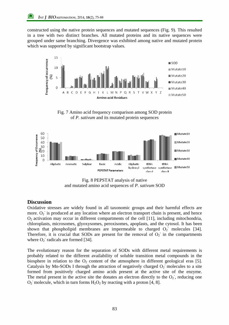
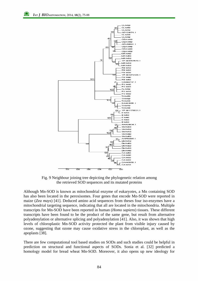
(residues present in cavities are highlighted in sequence) Mutational analysis of SOD sequences SOD sequences were mutated using Mutate protein software with varying number of residues mutated as 10, 20, 30, 40 and 50. Changes in the protein characteristics were analysis by PEPSAT and GRAVY analysis. Amino acid frequency among the native sequence as well as mutated sequences was compared (Fig. 7). Frequency each residue were varying after mutation and residue D showed the least effect of mutation with less significant changes in its frequencies when compared to native sequence. After comparison of 7 PEPSTAT parameters, it was found that there were no significant changes in protein properties in spite of mutation of up to 50 residues through Mutate protein software (Fig. 8). Neighbour joining tree was
Accession Numbers Len cTP mTP SP other Loc RC TPlen
CAA42737.1 240 0.036 0.818 0.087 0.111 M 2 44 AAF50095.1 153 0.128 0.072 0.357 0.529 _ 5 - AAA29934.1 184 0.004 0.14 0.946 0.09 S 1 22 CAA43859.1 153 0.059 0.065 0.4 0.678 _ 4 - YP_001176531.1 172 0.084 0.041 0.917 0.076 S 1 19 BAA02655.1 204 0.045 0.156 0.108 0.867 _ 2 - YP_004787678.1 202 0.079 0.122 0.169 0.818 _ 2 - YP_004272108.1 203 0.086 0.111 0.095 0.837 _ 2 - ADY29074.1 225 0.019 0.12 0.566 0.579 _ 5 - ADV48601.1 202 0.121 0.107 0.145 0.792 _ 2 -
INT. J. BIOAUTOMATION, 2014, 18(2), 75-88
83
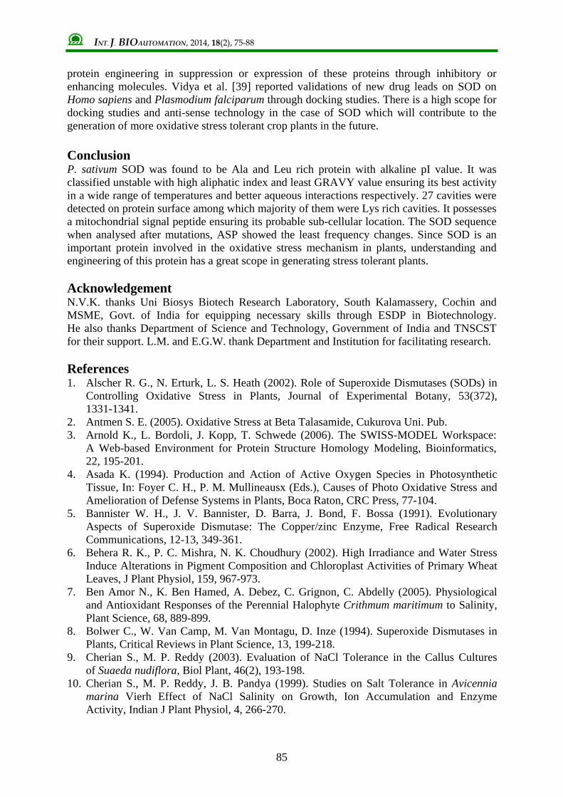
constructed using the native protein sequences and mutated sequences (Fig. 9). This resulted in a tree with two distinct branches. All mutated proteins and its native sequences were grouped under same branching. Divergence was exhibited among native and mutated protein which was supported by significant bootstrap values.
Fig. 7 Amino acid frequency comparison among SOD protein of P. sativum and its mutated protein sequences
Fig. 8 PEPSTAT analysis of native
and mutated amino acid sequences of P. sativum SOD Discussion Oxidative stresses are widely found in all taxonomic groups and their harmful effects are more. O2
- is produced at any location where an electron transport chain is present, and hence O2 activation may occur in different compartments of the cell [11], including mitochondria, chloroplasts, microsomes, glyoxysomes, peroxisomes, apoplasts, and the cytosol. It has been shown that phospholipid membranes are impermeable to charged O2
- molecules [34]. Therefore, it is crucial that SODs are present for the removal of O2
- in the compartments where O2
- radicals are formed [34]. The evolutionary reason for the separation of SODs with different metal requirements is probably related to the different availability of soluble transition metal compounds in the biosphere in relation to the O2 content of the atmosphere in different geological eras [5]. Catalysis by Mn-SODs I through the attraction of negatively charged O2
- molecules to a site formed from positively charged amino acids present at the active site of the enzyme. The metal present in the active site the donates an electron directly to the O2
-, reducing one O2
- molecule, which in turn forms H2O2 by reacting with a proton [4, 8].
INT. J. BIOAUTOMATION, 2014, 18(2), 75-88
84
Fig. 9 Neighbour joining tree depicting the phylogenetic relation among the retrieved SOD sequences and its mutated proteins
Although Mn-SOD is known as mitochondrial enzyme of eukaryotes, a Mn containing SOD has also been located in the peroxisomes. Four genes that encode Mn-SOD were reported in maize (Zea mays) [41]. Deduced amino acid sequences from theses four iso-enzymes have a mitochondrial targeting sequence, indicating that all are located in the mitochondria. Multiple transcripts for Mn-SOD have been reported in human (Homo sapiens) tissues. These different transcripts have been found to be the product of the same gene, but result from alternative polyadenylation or alternative splicing and polyadenylation [41]. Also, it was shown that high levels of chloroplastic Mn-SOD activity protected the plant from visible injury caused by ozone, suggesting that ozone may cause oxidative stress in the chloroplast, as well as the apoplasts [38]. There are few computational tool based studies on SODs and such studies could be helpful in prediction on structural and functional aspects of SODs. Sonia et al. [32] predicted a homology model for bread wheat Mn-SOD. Moreover, it also opens up new ideology for
INT. J. BIOAUTOMATION, 2014, 18(2), 75-88
85
protein engineering in suppression or expression of these proteins through inhibitory or enhancing molecules. Vidya et al. [39] reported validations of new drug leads on SOD on Homo sapiens and Plasmodium falciparum through docking studies. There is a high scope for docking studies and anti-sense technology in the case of SOD which will contribute to the generation of more oxidative stress tolerant crop plants in the future. Conclusion P. sativum SOD was found to be Ala and Leu rich protein with alkaline pI value. It was classified unstable with high aliphatic index and least GRAVY value ensuring its best activity in a wide range of temperatures and better aqueous interactions respectively. 27 cavities were detected on protein surface among which majority of them were Lys rich cavities. It possesses a mitochondrial signal peptide ensuring its probable sub-cellular location. The SOD sequence when analysed after mutations, ASP showed the least frequency changes. Since SOD is an important protein involved in the oxidative stress mechanism in plants, understanding and engineering of this protein has a great scope in generating stress tolerant plants. Acknowledgement N.V.K. thanks Uni Biosys Biotech Research Laboratory, South Kalamassery, Cochin and MSME, Govt. of India for equipping necessary skills through ESDP in Biotechnology. He also thanks Department of Science and Technology, Government of India and TNSCST for their support. L.M. and E.G.W. thank Department and Institution for facilitating research. References 1. Alscher R. G., N. Erturk, L. S. Heath (2002). Role of Superoxide Dismutases (SODs) in
Controlling Oxidative Stress in Plants, Journal of Experimental Botany, 53(372), 1331-1341.
2. Antmen S. E. (2005). Oxidative Stress at Beta Talasamide, Cukurova Uni. Pub. 3. Arnold K., L. Bordoli, J. Kopp, T. Schwede (2006). The SWISS-MODEL Workspace:
A Web-based Environment for Protein Structure Homology Modeling, Bioinformatics, 22, 195-201.
4. Asada K. (1994). Production and Action of Active Oxygen Species in Photosynthetic Tissue, In: Foyer C. H., P. M. Mullineausx (Eds.), Causes of Photo Oxidative Stress and Amelioration of Defense Systems in Plants, Boca Raton, CRC Press, 77-104.
5. Bannister W. H., J. V. Bannister, D. Barra, J. Bond, F. Bossa (1991). Evolutionary Aspects of Superoxide Dismutase: The Copper/zinc Enzyme, Free Radical Research Communications, 12-13, 349-361.
6. Behera R. K., P. C. Mishra, N. K. Choudhury (2002). High Irradiance and Water Stress Induce Alterations in Pigment Composition and Chloroplast Activities of Primary Wheat Leaves, J Plant Physiol, 159, 967-973.
7. Ben Amor N., K. Ben Hamed, A. Debez, C. Grignon, C. Abdelly (2005). Physiological and Antioxidant Responses of the Perennial Halophyte Crithmum maritimum to Salinity, Plant Science, 68, 889-899.
8. Bolwer C., W. Van Camp, M. Van Montagu, D. Inze (1994). Superoxide Dismutases in Plants, Critical Reviews in Plant Science, 13, 199-218.
9. Cherian S., M. P. Reddy (2003). Evaluation of NaCl Tolerance in the Callus Cultures of Suaeda nudiflora, Biol Plant, 46(2), 193-198.
10. Cherian S., M. P. Reddy, J. B. Pandya (1999). Studies on Salt Tolerance in Avicennia marina Vierh Effect of NaCl Salinity on Growth, Ion Accumulation and Enzyme Activity, Indian J Plant Physiol, 4, 266-270.
INT. J. BIOAUTOMATION, 2014, 18(2), 75-88
86
11. Elstner E. F. (1991). Mechanisms of Oxygen Activation in Different Compartments of Plant Cells, In: E. J. Pell, K. L. Steffens (Eds.), Active Oxygen/oxidative Stress and Plant Metabolism, Rockville MD, American Society of Plant Physiologists, 13-25.
12. Fang Z. Q., L. Y. Yuan, P. C. Hong, L. C. Ming, W. B. Shan (2005). NaCl Enhances Thylakoid-bound SOD Activity in the Leaves of C3 Halophytes Suaeda salsa L., Plant Science, 168, 423-430.
13. Fridovich I. (1986). Superoxide Dismutases, Advances in Enzymology and Related Areas of Molecular Biology, 58, 61-97.
14. Garnier J., J. F. Gibrat, B. Robson (1996). GOR Secondary Structure Prediction Method Version IV, Methods in Enzymology, R. F. Doolittle (Ed.), 266, 540-553.
15. Gasteiger E., C. Hoogland, A. Gattiker, S. Duvaud, M. R. Wilkins, R. D. Appel, A. Bairoch (2005). Protein Identification and Analysis Tools on the ExPASy Server, In: John M. Walker (Ed.), The Proteomics Protocols Handbook, Humana Press, 571-607.
16. Geourjon C., G. Deleage (1995). SOPMA: Significant Improvements in Protein Secondary Structure Prediction by Consensus Prediction from Multiple Alignments, Comput Appl Biosci, 11(6), 681-684.
17. Hompson J., D. Higgins, T. Gibson (1994). CLUSTAL W: Improving the Sensitivity of Progressive Multiple Sequence Alignment through Sequence Weighting Position Specific Gap Penalties and Weight Matrix Choice, Nucleic Acids Res, 22(22), 4673-4690.
18. Humphrey W., A. Dalke, K. Schulten (1996). VMD: Visual Molecular Dynamics, J Mol Graphics, 14(1), 33-38.
19. Hurst A. C., T. E. E. Grams, R. Ratajczeak (2004). Effects of Salinity, High Irradiance, Ozone and Ethylene on Mode of Photosynthesis, Oxidative Stress and Oxidative Damage in the C3/CAM Intermediate Plant Mesembryanthemum crystallinum L., Plant Cell Environ, 27, 187-197.
20. Jithesh M. N., S. R. Prashanth, K. R. Sivaprakash, A. K. Parida (2006). Monitoring Expression Profiles of Antioxidant Genes to Salinity, Iron, Oxidative, Light and Hyperosmotic Stresses in the Highly Salt Tolerant Grey Mangrove, Avicennia marina (Forsk.) Vierh. by mRNA Analysis, Plant Cell Rep, 25, 865-876.
21. Keles Y., S. Unyayar (2004). Responses of Antioxidant Defense System of Helianthus annus to Absisic Acid Under Drought and Water Logging, Acta Physiol Planta, 26, 149-156.
22. Kiefer F., K. Arnold, M. Künzli, L. Bordoli, T. Schwede (2009). The SWISS-MODEL Repository and Associated Resources, Nucleic Acids Res, 37, D387-D392.
23. Mishra S., A. B. Jha, R. S. Dubey (2011). Arsenite Treatment Induces Oxidative Stress, Upregulates Antioxidant System, and Causes Phytochelatin Synthesis in Rice Seedlings, Protoplasma, 248(3), 565-577.
24. Navrot N., N. Rouhier, E. Gelhaye, J. P. Jacquot (2007). Reactive Oxygen Species Generation and Antioxidant Systems in Plant Mitochondria, Physiol Plant, 129, 185-195.
25. Sharma P., R. S. Dubey (2005). Drought Induces Oxidative Stress and Enhances the Activities of Antioxidant Enzymes in Growing Rice Seedlings, Plant Growth Regulation, 46(3), 209-221.
26. Parida A. K., A. B. Das, P. Mohanty (2004). Defense Potential to NaCl in a Mangrove, Bruguiera parviflora: Differential Changes of Isoforms of Some Antioxidative Enzymes, J Plant Physiol, 161, 531-542.
27. Pastori G. M., C. H. Foyer (2002). Common Components, Networks and Pathways of Cross Tolerance to Stress. The Central Role of “Redox” and Abscisic Acid Mediated Controls, Plant Physiol, 129(2), 460-468.
28. Peitsch M. C. (1995). Protein Modeling by E-mail, Nature Biotechnology, 13, 658-660.
INT. J. BIOAUTOMATION, 2014, 18(2), 75-88
87
29. Saitou N., M. Nei (1987). The Neighbor-joining Method: A New Method for Reconstructing Phylogenetic Trees, Mol Biol and Evol, 4, 406-425.
30. Seyed M. S. J.-e-E., B. Alizadeh, M. Zaefizadeh, R. A. Zakarya, M. Khayatnezhad (2011). Superoxide Dismutase (SOD) Activity in NaCl Stress in Salt-sensitive and Salt-tolerance Genotypes of Colza (Brassica napus L.), Middle East Journal of Scientific Research, 7(1), 7-11.
31. Slesak I., Z. Miszalski, B. Karpinska, E. Niewiadomska, R. Ratajczak, S. Karpinski (2002). Redox Control of Oxidative Stress Responses in the C3-CAM Intermediate Plant Mesembryanthemum crystallinum, Plant Physiol Biochem, 40, 669-677.
32. Sheoran S., B. Pandey, R. Singh, P. Sharma, R. Chatrath (2011). Modelling and Phylogeny Analysis of Bread Wheat MnSOD, Bioinformation, 6(6), 209-211.
33. Taji T., M. Seki, M. Satou, T. Sakurai, M. Kobayashi, K. Ishiyama (2004). Comparative Genomics in Salt Tolerance between Arabidopsis and Arabidopsis-related Halophyte Salt Cress using Arabidopsis Microarray, Plant Physiol, 135, 1-13.
34. Takahashi M. A., K. Asada (1983). Superoxide Anion Permeability of Phospholipid Membranes and Chloroplast Thylakoids, Archives of Biochemistry and Biophysics, 226, 558-566.
35. Takemura T., N. Hanagata, Z. Dubinsky, I. Karube (2002). Molecular Characterization and Response to Salt Stress of mRNAs Encoding Cytosolic Cu/Zn Superoxide Dismutase and Catalase from Bruguiera gymnorrhiza, Trees, 16, 94-99.
36. Takemura T., N. Hanagata, K. Sugihara, S. Baba, I. Karube, Z. Dubinsky (2000). Physiological and Biochemical Responses to Salt Stress in the Mangrove, Bruguiera gymnorrhiza, Aquat Bot, 68, 15-28.
37. Tamura K., J. Dudley, M. Nei, S. Kumar (2007). MEGA4: Molecular Evolutionary Genetics Analysis (MEGA) Software Version 4.0, Mol Biol and Evol, 24, 1596-1599.
38. Van Camp W., H. Willekens, C. Bowler, M. Van Montagu, D. Inze, C. Langebartels, H. Sandermann (1994). Elevated Levels of Superoxide Dismutase Protect Transgenic Plants Against Ozone Damage, Nature Biotechnology, 12, 165-168.
39. Vidya G. B., J. R. T. Pinto, V. R. Pai (2010). In silico Docking for Validation of Drug Leads on Superoxide Dismutase of Homo sapiens and Plasmodium falciparum, Biomedical Research, 21(2), 214-220.
40. Wang B., U. Luttge, R. Ratajczak (2004). Specific Regulation of SOD Isoforms by NaCl and Osmotic Stress in Leaves of C3 Halophyte Suaeda salsa L., J Plant Physiol, 161, 285-293.
41. Zhu D., J. G. Scandalios (1993). Maize Mitochondrial Manganese Superoxide Dismutases are Encoded by a Differentially Expressed Multigene Family, Proceedings of the National Academy of Science USA, 90(20), 9310-9314.
INT. J. BIOAUTOMATION, 2014, 18(2), 75-88
88
Nathan Vinod Kumar, M.Sc. Biotechnology
E-mail: [email protected]
Vinod Kumar Nathan completed M.Sc. Biotechnology from Periyar University, Salem, India. Presently he is a research scholar of Madurai Kamaraj University, India. His field of interest is plant biotechnology and plant stress biology. He has command over many bioinformatics tools. Currently he is associated with a Department of Science and Technology, Government of India funded research project on Ezymology.
Lizzy Mathew, Ph.D. E-mail: [email protected]
Dr. Lizzy Mathew obtained her M.Phil. degree from Madurai Kamaraj University followed by Ph.D. from Mahatma Gandhi University, Kottayam, Kerala. She is presently working as Reader in Department of Botany, St. Teresa’s college, Ernakulam, Kerala. Her fields of interests are Plant Taxonomy and Phytochemistry.
Edward Gnanaraj Wesely, Ph.D. E-mail: [email protected]
Dr. Edward Gnanaraj Wesely got his Ph.D. from Madurai Kamaraj University and served as Assistant Professor in Department of Biotechnology, Muthayammal College of Arts and Science, Namakkal Dist., Tamil Nadu, India. Later he served as the Department Head. Presently he is working as Assistant Professor in A. A. Government Arts College, Namakkal. His fields of interests are Plant Taxonomy and Phytochemistry.