substrate-selective inhibition of cyclooxygenase-2 ... · substrate-selective inhibition of...
TRANSCRIPT
Substrate-Selective Inhibition of Cyclooxygenase-2: Development and Evaluation of Achiral Profen Probes Matthew A. Windsor,† Daniel J. Hermanson,† Philip J. Kingsley,† Shu Xu,† Brenda C. Crews,† Winnie Ho,‡ Catherine M. Keenan,‡ Surajit Banerjee,§ Keith A. Sharkey‡ and Lawrence J. Marnett*,† †A.B. Hancock Jr. Memorial Laboratory for Cancer Research, Departments of Biochemistry, Chemistry, and Pharmacology, Vanderbilt Institute of Chemical Biology, Center in Molecular Toxicology, and Vanderbilt-‐Ingram Cancer Center, Vanderbilt University School of Medicine, Nashville TN, United States. ‡Hotchkiss Brain Institute and Snyder Institute for Chronic Diseases, Department of Physiology and Pharmacology, University Calgary, Calgary, AB, Canada.
§Northeastern Collaborative Access Team and Department of Chemistry and Chemical Biology, Cornell University, Ithaca, NY, United States, and Argonne National Laboratory, Argonne, IL, United States.
SUPPORTING INFORMATION
Table of Contents
Detailed Funding Sources Information……………………………………………………………………………………….2
Supplimental Figures (Table S1, Figures S1, S2)………………………………………………………………………….3
Raw material supplies and instrumental…………………………………………………………………………………….5
General synthetic procedures…………………………………………………………………………………………………….6
Characterization of compounds………………………………………………………………………………………………….8
Separation of compounds 7d, 7e……………………………………………………………………………………………...13
General procedure for in vitro assay…………………………………………………………………………………………14
General procedure for RAW cells assay…………………………………………………………………………………….15
General procedure for in vivo mouse assay……………………………………………………………………………….15
General procedure for crystallization, X-‐ray data collection, structure
determination and refinement (Table S2)……………………………………………………………………………16
References………………………………………………………………………………………………………………………………19
2
Detailed Funding Sources Information:
This work was supported by grants from the National Institutes of Health (to LJM GM15431 and
CA89450) and from the Canadian Institutes of Health Research (to KAS). Research conducted at
the Advanced Photon Source on the Northeastern Collaborative Access Team beamlines (SB) was
supported by grants from the National Center for Research Resources (5P41RR015301-‐10) and
the National Institute of General Medical Sciences (8 P41 GM103403-‐10) from the National
Institutes of Health. Use of the Advanced Photon Source, an Office of Science User Facility operated
for the U.S. Department of Energy (DOE) Office of Science by Argonne National Laboratory, was
supported by the U.S. DOE under Contract No. DE-‐AC02-‐06CH11357. KAS is an Alberta Heritage
Foundation for Medical Research Medical Scientist and the Crohn’s and Colitis Foundation of
Canada Chair in the Inflammatory Bowel Disease Research at the University of Calgary.
3
Supplimental Figures:
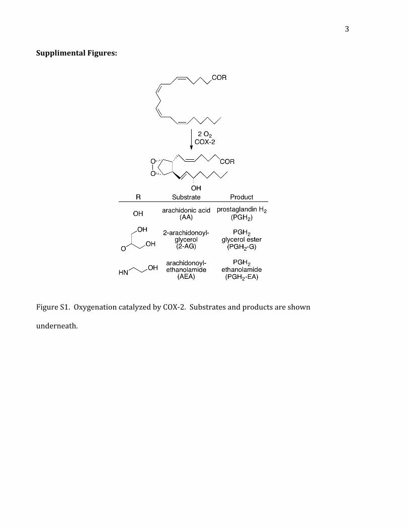
Figure S1. Oxygenation catalyzed by COX-‐2. Substrates and products are shown
underneath.
4
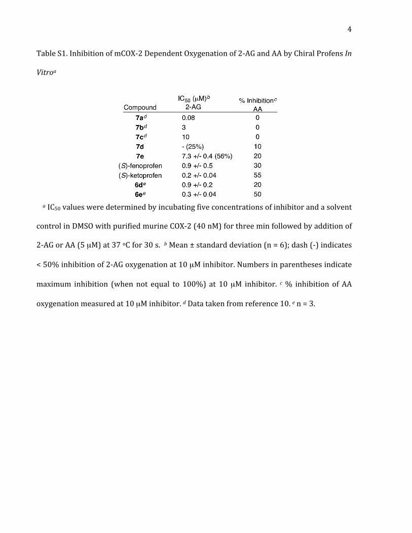
Table S1. Inhibition of mCOX-‐2 Dependent Oxygenation of 2-‐AG and AA by Chiral Profens In
Vitroa
a IC50 values were determined by incubating five concentrations of inhibitor and a solvent
control in DMSO with purified murine COX-‐2 (40 nM) for three min followed by addition of
2-‐AG or AA (5 µM) at 37 oC for 30 s. b Mean ± standard deviation (n = 6); dash (-‐) indicates
< 50% inhibition of 2-‐AG oxygenation at 10 µM inhibitor. Numbers in parentheses indicate
maximum inhibition (when not equal to 100%) at 10 µM inhibitor. c % inhibition of AA
oxygenation measured at 10 µM inhibitor. d Data taken from reference 10. e n = 3.
5
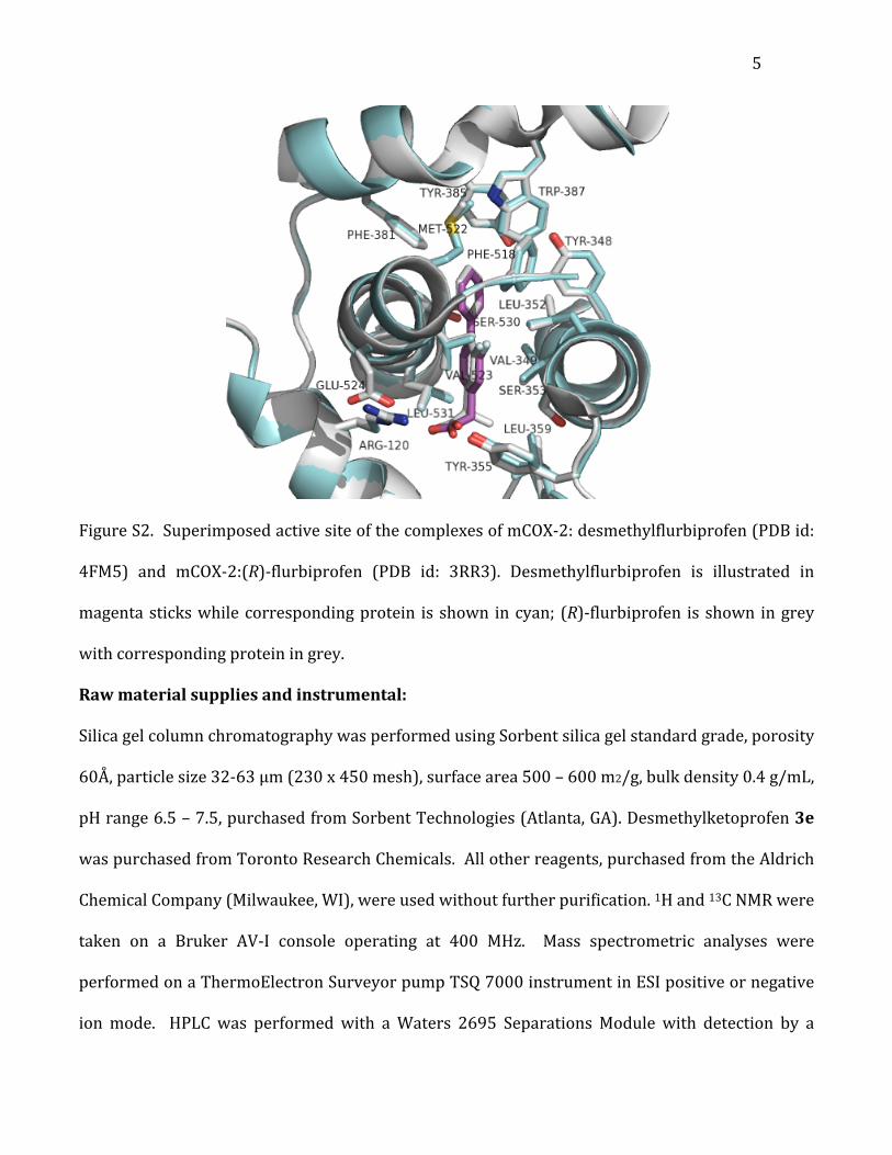
Figure S2. Superimposed active site of the complexes of mCOX-‐2: desmethylflurbiprofen (PDB id:
4FM5) and mCOX-‐2:(R)-‐flurbiprofen (PDB id: 3RR3). Desmethylflurbiprofen is illustrated in
magenta sticks while corresponding protein is shown in cyan; (R)-‐flurbiprofen is shown in grey
with corresponding protein in grey.
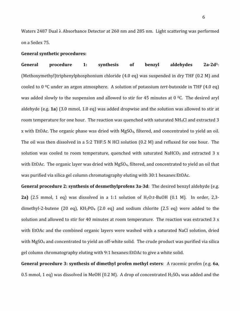
Raw material supplies and instrumental:
Silica gel column chromatography was performed using Sorbent silica gel standard grade, porosity
60Å, particle size 32-‐63 μm (230 x 450 mesh), surface area 500 – 600 m2/g, bulk density 0.4 g/mL,
pH range 6.5 – 7.5, purchased from Sorbent Technologies (Atlanta, GA). Desmethylketoprofen 3e
was purchased from Toronto Research Chemicals. All other reagents, purchased from the Aldrich
Chemical Company (Milwaukee, WI), were used without further purification. 1H and 13C NMR were
taken on a Bruker AV-‐I console operating at 400 MHz. Mass spectrometric analyses were
performed on a ThermoElectron Surveyor pump TSQ 7000 instrument in ESI positive or negative
ion mode. HPLC was performed with a Waters 2695 Separations Module with detection by a
6
Waters 2487 Dual λ Absorbance Detector at 260 nm and 285 nm. Light scattering was performed
on a Sedex 75.
General synthetic procedures:
General procedure 1: synthesis of benzyl aldehydes 2a-2d1:
(Methoxymethyl)triphenylphosphonium chloride (4.0 eq) was suspended in dry THF (0.2 M) and
cooled to 0 0C under an argon atmosphere. A solution of potassium tert-‐butoxide in THF (4.0 eq)
was added slowly to the suspension and allowed to stir for 45 minutes at 0 0C. The desired aryl
aldehyde (e.g. 1a) (3.0 mmol, 1.0 eq) was added dropwise and the solution was allowed to stir at
room temperature for one hour. The reaction was quenched with saturated NH4Cl and extracted 3
x with EtOAc. The organic phase was dried with MgSO4, filtered, and concentrated to yield an oil.
The oil was then dissolved in a 5:2 THF:5 N HCl solution (0.2 M) and refluxed for one hour. The
solution was cooled to room temperature, quenched with saturated NaHCO3 and extracted 3 x
with EtOAc. The organic layer was dried with MgSO4, filtered, and concentrated to yield an oil that
was purified via silica gel column chromatography eluting with 30:1 hexanes:EtOAc.
General procedure 2: synthesis of desmethylprofens 3a-3d: The desired benzyl aldehyde (e.g.
2a) (2.5 mmol, 1 eq) was dissolved in a 1:1 solution of H2O:t-‐BuOH (0.1 M). In order, 2,3-‐
dimethyl-‐2-‐butene (20 eq), KH2PO4 (2.0 eq) and sodium chlorite (2.5 eq) were added to the
solution and allowed to stir for 40 minutes at room temperature. The reaction was extracted 3 x
with EtOAc and the combined organic layers were washed with a saturated NaCl solution, dried
with MgSO4 and concentrated to yield an off-‐white solid. The crude product was purified via silica
gel column chromatography eluting with 9:1 hexanes:EtOAc to give a white solid.
General procedure 3: synthesis of dimethyl profen methyl esters: A racemic profen (e.g. 6a,
0.5 mmol, 1 eq) was dissolved in MeOH (0.2 M). A drop of concentrated H2SO4 was added and the
7
solution was refluxed for two hours. The reaction was quenched with sat. NaHCO3, extracted 3x
with EtOAc, dried with MgSO4 and concentrated to yield the methyl-‐ester protected racemic
profen as an oil. This oil was dissolved in dry THF (0.2 M) and cooled to -‐78 oC under argon. A
solution of LDA in THF (1.5 eq) was added slowly and allowed to stir at -‐78 oC for 30 minutes. The
solution was warmed to 0 oC and HMPA (1.4 eq) was added slowly and allowed to stir at 0 oC for
30 minutes. Finally, 1.9 eq. of MeI was added dropwise to the solution and allowed to stir for 30
minutes at 0 oC and then thirty minutes at room temperature. The reaction was quenched with
NH4Cl, extracted 3x with EtOAc, dried with MgSO4 and concentrated to yield an orange oil. The
crude product was purified via silica gel column chromatography eluting with 9:1 hexanes:EtOAc
to give an oil. Note: Compounds 3a-e can be used as the starting material with this procedure as
long as the equivalents of LDA, HMPA, and MeI are doubled from those described above.
General procedure 4: synthesis of cyclopropyl profen methyl esters: A desmethylprofen (e.g.
3a, 0.5 mmol, 1 eq) was dissolved in MeOH (0.2 M). A drop of concentrated H2SO4 was added and
the solution was refluxed for two hours. The reaction was quenched with sat. NaHCO3, extracted
3x with EtOAc, dried with MgSO4 and concentrated to yield the methyl-‐ester protected profen as
an oil. This oil was dissolved in dry THF (0.2 M) and cooled to -‐78 oC under argon. A solution of
LDA in THF (2.5 eq) was added slowly and allowed to stir at -‐78 oC for 30 minutes. The solution
was warmed to 0 oC and HMPA (2.0 eq) was added slowly and allowed to stir at 0 oC for 30
minutes. Finally, 1.5 eq. of 1,2-‐dibromoethane was added dropwise to the solution and allowed to
stir for 30 minutes at 0 oC and then thirty minutes at room temperature. The reaction was
quenched with NH4Cl, extracted 3x with EtOAc, dried with MgSO4 and concentrated to yield an
orange oil. The crude product was purified via silica gel column chromatography eluting with 9:1
hexanes:EtOAc to give an oil.
8
General procedure 5: synthesis of dimethyl (4a-e) and cyclopropyl (5a-e) profens: A
methyl-‐ester protected dimethyl profen or a methyl-‐ester protected cyclopropyl profen (0.2 mmol,
1.0 eq) was dissolved in dry THF (0.2 M). KOTMS (2.0 eq) was added to the reaction flask and
refluxed for two hours. The slurry was quenched at room temperature with NH4Cl, extracted 3 x
with EtOAc, dried with MgSO4 and concentrated to yield an off-‐white solid. The crude product was
purified via silica gel column chromatography eluting with 30:1 DCM:MeOH to give a dimethyl
profen (e.g. 4a) or a cyclopropyl profen (e.g. 5a), respectively, as a white solid.
Characterization of compounds:
2-(2-fluoro-[1,1'-biphenyl]-4-yl)acetaldehyde (2a): 2a was prepared via general procedure 1
as a clear oil (56% yield). 1H NMR (400 MHz, CDCl3) δ 9.82 (t, 1H), 7.60-‐7.58 (m, 2H), 7.40-‐7.52
(m, 4H), 7.10-‐7.08 (m, 2H), 3.77 (d, 2H). MS m/z (ESI): calc. for C14H11FO [M-‐H]-‐ 213.08, found
213.4.
2-(6-methoxynaphthalen-2-yl)acetaldehyde (2b): 2b was prepared via general procedure 1 as
a clear oil (48% yield). 1H NMR (400 MHz, CDCl3) δ 9.84 (t, 1H), 7.72-‐7.78 (m, 2H), 7.64 (s, 1H),
7.28-‐7.32 (m, 1H), 7.15-‐7.20 (m, 2H), 3.95 (s, 3H), 3.83 (d, 2H). MS m/z (ESI): calc. for C13H12O2
[M-‐H]-‐ 199.08, found 199.4.
2-(4-isobutylphenyl)acetaldehyde (2c): 2c was prepared via general procedure 1 as a clear oil
(77% yield). Note: intermediate and product are volatile when heated under reduced pressure. 1H
NMR (400 MHz, CDCl3) δ 9.76 (t, 1H), 7.16 (d, 2H), 7.10 (d, 2H), 3.65 (d, 2H), 2.47 (d, 2H), 1.85-‐
1.83 (m, 1H), 0.89 (d, 6H). MS m/z (ESI): calc. for C12H16O [M-‐H]-‐ 175.08, found 175.4.
2-(3-phenoxyphenyl)acetaldehyde (2d): 2d was prepared via general procedure 1 as a clear oil
(75% yield). 1H NMR (400 MHz, CDCl3) δ 9.75 (t, 1H), 7.33-‐7.31 (m, 3H), 7.13-‐7.11 (m, 1H), 6.88-‐
7.10-‐7.08 (m, 4H), 3.66 (d, 2H). MS m/z (ESI): calc. for C14H12O2 [M-‐H]-‐ 211.07, found 211.27.
9
2-(2-fluoro-[1,1'-biphenyl]-4-yl)acetic acid (3a): 3a was prepared via general procedure 2 as a
white solid (58% yield). 1H NMR (400 MHz, CDCl3) δ 7.55 (d, 2H), 7.50-‐7.35 (m, 4H), 7.20-‐7.10 (m,
2H), 3.70 (s, 2H). 13C NMR (400 MHz, CDCl3) δ 176.1, 160.8, 159.2, 135.6, 134.7 (d, JC-‐F = 150 Hz),
131.0 (d, JC-‐F = 20 Hz), 129.1, 128.6, 127.8, 125.5 (d, JC-‐F = 20 Hz), 117.3, 117.1, 40.4. HRMS m/z
(ESI): calc. for C14H11FO2 [2M+Na]-‐ 481.1233, found 481.1245.
2-(6-methoxynaphthalen-2-yl)acetic acid (3b): 3b was prepared via general procedure 2 as a
white solid (94% yield). 1H NMR (400 MHz, CD3OD) δ 7.75-‐7.65 (m, 3H), 7.38-‐7.32 (d, 1H), 7.30 (s,
1H), 7.11-‐7.08 (d, 1H), 3.9 (s, 3H), 3.72 (s, 2H). MS m/z (ESI): calc. for C13H12O3 [M-‐H]-‐ 215.08,
found 215.23.
2-(4-isobutylphenyl)acetic acid (3c): 3c was prepared via general procedure 2 as a white solid
(89% yield). 1H NMR (400 MHz, CDCl3) δ 7.20 (d, 2H), 7.15 (d, 2H), 3.64 (s, 2H), 2.5 (d, 2H), 1.93-‐
1.82 (m, 1H), 0.94-‐0.92 (d, 6H). MS m/z (ESI): calc. for C12H16O2 [M-‐H]-‐ 191.12, found 191.19.
2-(3-phenoxyphenyl)acetic acid (3d): 3d was prepared via general procedure 2 as a white solid
(99% yield). 1H NMR (400 MHz, CDCl3) δ 7.35-‐7.23 (m, 3H), 7.12-‐7.07 (m, 1H), 7.01-‐6.99 (m, 3H),
6.98-‐6.88 (m, 2H), 3.62 (s, 3H). MS m/z (ESI): calc. for C14H12O3 [M-‐H]-‐ 227.08, found 227.07.
Methyl-2-(2-fluoro-[1,1'-biphenyl]-4-yl)-2-methylpropanoate: The methyl-‐ester protected
form of 4a was prepared via general procedure 3 as an oil (57% yield). 1H NMR (400 MHz, CDCl3)
δ 7.54-‐7.50 (m, 2H), 7.47-‐7.30 (m, 4H), 7.17-‐7.10 (m, 2H), 3.70 (s, 3H), 1.61 (s, 6H). MS m/z (ESI):
calc. for C17H17FO2 [M+Na]+ 295.12, found 295.0.
Methyl-2-(6-methoxynaphthalen-2-yl)-2-methylpropanoate: The methyl-‐ester protected form
of 4b was prepared via general procedure 3 as an oil (58% yield). 1H NMR (400 MHz, CDCl3)
δ 7.75-‐7.68 (m, 3H), 7.44-‐7.40 (d, 1H), 7.17-‐7.11 (m, 2H), 3.92 (s, 3H), 3.66 (s, 3H), 1.67 (s, 6H). MS
m/z (ESI): calc. for C16H18O3 [M+Na]+ 281.13, found 281.07.
10
Methyl-2-(4-isobutylphenyl)-2-methylpropanoate: The methyl-‐ester protected form of 4c was
prepared via general procedure 3 as an oil (85% yield). 1H NMR (400 MHz, CDCl3) δ 7.25-‐7.20 (d,
2H), 7.10 (d, 2H), 3.68 (s, 3H), 2.47 (d, 2H), 1.90-‐1.80 (m, 1H), 1.60 (s, 6H), 0.93 (d, 6H). MS m/z
(ESI): calc. for C15H22O2 [M+Na]+ 257.16, found 257.07.
Methyl-2-methyl-2-(3-phenoxyphenyl)propanoate: The methyl-‐ester protected form of 4d
was prepared via general procedure 3 as an oil (48% yield). 1H NMR (400 MHz, CDCl3) δ 7.35-‐7.25
(m, 3H), 7.13-‐6.97 (m, 5H), 6.87-‐6.82 (m, 1H), 3.70 (s, 3H), 1.58 (s, 6H). MS m/z (ESI): calc. for
C17H18O3 [M+Na]+ 293.13, found 293.0.
Methyl-2-(3-benzoylphenyl)-2-methylpropanoate: The methyl-‐ester protected form of 4e was
prepared via general procedure 3 as an oil (64% yield). 1H NMR (400 MHz, CDCl3) δ 7.80 (m, 3H),
7.68-‐7.40 (m, 6H), 3.70 (s, 3H), 1.64 (s, 6H). MS m/z (ESI): calc. for C18H18O3 [M+Na]+ 305.13, found
305.20.
Methyl-1-(2-fluoro-[1,1'-biphenyl]-4-yl)cyclopropanecarboxylate: The methyl-‐ester
protected form of 5a was prepared via general procedure 4 as an oil (28% yield). 1H NMR (400
MHz, CDCl3) δ 7.57-‐7.49 (m, 2H), 7.45-‐7.33 (m, 4H), 7.20-‐7.12 (m, 2H), 3.66 (s, 3H), 1.66-‐1.62 (dd,
2H), 1.25-‐1.20 (dd, 2H). MS m/z (ESI): calc. for C17H15FO2 [M+Na]+ 293.11, found 293.10.
Methyl-1-(6-methoxynaphthalen-2-yl)cyclopropanecarboxylate: The methyl-‐ester protected
form of 5b was prepared via general procedure 4 as an oil (22% yield). 1H NMR (400 MHz, CDCl3)
δ 7.70-‐7.68 (m, 3H), 7.46-‐7.44 (m, 1H), 7.15-‐7.12 (m, 2H), 3.92 (s, 3H), 3.62 (s, 3H), 1.67-‐1.64 (dd,
2H), 1.28-‐1.24 (dd, 2H). MS m/z (ESI): calc. for C16H16O3 [M+Na]+ 279.11, found 279.09.
Methyl-1-(4-isobutylphenyl)cyclopropanecarboxylate: The methyl-‐ester protected form of 5c
was prepared via general procedure 4 as an oil. Note: the product could not be isolated as a pure
compound after column chromatography, but the impurities did not affect the next reaction (41%
11
crude yield). 1H NMR (400 MHz, CDCl3) δ 7.27-‐7.20 (d, 2H), 7.18-‐7.13 (d, 2H), 3.68 (s, 3H), 2.44-‐
2.40 (d, 2H), 1.88-‐1.78 (m, 1H), 1.60-‐1.55 (dd, 2H), 1.28-‐1.24 (dd, 2H). MS m/z (ESI): calc. for
C15H20O2 [M+Na]+ 255.15, found 255.12.
Methyl-1-(3-phenoxyphenyl)cyclopropanecarboxylate: The methyl-‐ester protected form of 5d
was prepared via general procedure 4 as an oil (17% yield). 1H NMR (400 MHz, CDCl3) δ 7.36-‐7.21
(m, 3H), 7.12-‐7.00 (m, 5H), 6.88-‐6.84 (m, 1H), 3.63 (s, 3H), 1.59-‐1.57 (dd, 2H), 1.19-‐1.16 (dd, 2H).
MS m/z (ESI): calc. for C17H16O3 [M+Na]+ 291.11, found 291.09.
Methyl-1-(3-benzoylphenyl)cyclopropanecarboxylate: The methyl-‐ester protected form of 5e
was prepared via general procedure 4 as an oil (18% yield). 1H NMR (400 MHz, CDCl3) δ 7.82-‐7.78
(m, 3H), 7.7-‐7.66 (m, 1H), 7.61-‐7.54 (m, 2H), 7.52-‐7.39 (m, 3H), 3.64 (s, 3H), 1.67-‐1.63 (d, 2H),
1.25-‐1.21 (dd, 2H). MS m/z (ESI): calc. for C17H16O3 [M+Na]+ 303.11, found 303.15.
2-(2-fluoro-[1,1'-biphenyl]-4-yl)-2-methylpropanoic acid (4a): 4a was prepared via general
procedure 5 as a white solid (84% yield). 1H NMR (400 MHz, CDCl3) δ 7.56-‐7.50 (m, 2H), 7.45-‐
7.37-‐7.35 (m, 4H), 7.17-‐7.08 (m, 2H), 1.65 (s, 6H). 13C NMR (400 MHz, CDCl3) δ 182.8, 161.9, 158.9,
146.1 (d, JC-‐F = 150 Hz), 135.6, 131.5 (d, JC-‐F = 20 Hz), 129.0, 128.5, 127.8, 122.0 (d, JC-‐F = 20 Hz),
114.3, 113.9, 46.0, 27.0. HRMS m/z (ESI): calc. for C16H15FO2 [2M+Na]-‐ 537.1859, found 537.1871.
2-(6-methoxynaphthalen-2-yl)-2-methylpropanoic acid (4b): 4b was prepared via general
procedure 5 as a white solid (92% yield). 1H NMR (400 MHz, CDCl3) δ 7.74-‐7.71 (m, 3H), 7.51-‐
7.49 (m, 1H), 7.17-‐7.12 (2H), 3.93 (s, 3H), 1.70 (s, 6H). MS m/z (ESI): calc. for C15H16O3 [M-‐H]-‐
243.11, found 243.31.
2-(4-isobutylphenyl)-2-methylpropanoic acid (4c): 4c was prepared via general procedure 5
as a white solid (92% yield). 1H NMR (400 MHz, CDCl3) δ 7.33-‐7.30 (d, 2H), 7.11-‐7.07 (d, 2H),
12
2.58-‐2.53 (d, 2H), 1.91-‐1.80 (m, 1H), 1.60 (s, 6H), 0.92-‐0.89 (d, 6H). MS m/z (ESI): calc. for
C14H20O2 [M-‐H]-‐ 219.15, found 219.13.
2-methyl-2-(3-phenoxyphenyl)propanoic acid (4d): 4d was prepared via general procedure 5
as a white solid (95% yield). 1H NMR (400 MHz, CDCl3) δ 7.36-‐7.24 (m, 3H), 7.13-‐6.98 (m, 5H),
6.85-‐6.80 (m, 1H), 1.58 (s, 6H). MS m/z (ESI): calc. for C16H16O3 [M-‐H]-‐ 255.11, found 255.07.
2-(3-benzoylphenyl)-2-methylpropanoic acid (4e): 4e was prepared via general procedure 5
as a white solid (89% yield). 1H NMR (400 MHz, CDCl3) δ 7.88-‐7.76 (m, 3H), 7.68-‐7.53 (m, 3H),
7.47-‐7.39 (m, 3H), 1.64 (s, 6H). MS m/z (ESI): calc. for C17H16O3 [M-‐H]-‐ 267.11, found 267.13.
1-(2-fluoro-[1,1'-biphenyl]-4-yl)cyclopropanecarboxylic acid (5a): 5a was prepared via
general procedure 5 as a white solid (77% yield). 1H NMR (400 MHz, CDCl3) δ 7.54-‐7.52 (m, 2H),
7.45-‐7.36 (m, 4H), 7.23-‐7.15 (m, 2H), 1.74-‐1.71 (dd, 2H), 1.34-‐1.31 (dd, 2H). 13C NMR (400 MHz,
CDCl3) δ 181.1, 160.9, 158.9, 140.0 (d, JC-‐F = 150 Hz), 135.5, 130.7 (d, JC-‐F = 20 Hz), 129.0, 128.5,
127.7, 126.4 (d, JC-‐F = 20 Hz), 118.4, 118.2, 28.0, 18.0. HRMS m/z (ESI): calc. for C16H13FO2 [M-‐H]-‐
255.0827, found 255.0835.
1-(6-methoxynaphthalen-2-yl)cyclopropanecarboxylic acid (5b): 5b was prepared via
general procedure 5 as a white solid (78% yield). 1H NMR (400 MHz, CDCl3) δ 7.70-‐7.68 (m, 3H),
7.49-‐7.47 (m, 1H), 7.14-‐7.11 (m, 2H), 3.91 (s, 3H), 1.74-‐1.71 (dd, 2H), 1.36-‐1.34 (dd, 2H). MS m/z
(ESI): calc. for C15H14O3 [M-‐H]-‐ 241.09, found 241.2.
1-(4-isobutylphenyl)cyclopropanecarboxylic acid (5c): 5c was prepared via general procedure
5 as a white solid. 1H NMR (400 MHz, CDCl3) δ 7.27-‐7.24 (d, 2H), 7.07-‐7.03 (d, 2H), 2.47-‐2.41 (d,
2H), 1.89-‐1.80 (m, 1H), 1.66-‐1.62 (dd, 2H), 1.25-‐1.21 (dd, 2H), 0.91-‐0.86 (d, 6H). MS m/z (ESI):
calc. for C14H18O2 [M-‐H]-‐ 217.13, found 217.30.
13
1-(3-phenoxyphenyl)cyclopropanecarboxylic acid (5d): 5d was prepared via general
procedure 5 as a white solid (59% yield). 1H NMR (400 MHz, CDCl3) δ 7.35-‐7.19 (m, 3H), 7.09-‐
6.96 (m, 5H), 6.87-‐6.83 (m, 1H), 1.70-‐1.67 (dd, 2H), 1.3-‐1.28 (dd, 2H). MS m/z (ESI): calc. for
C16H14O3 [M-‐H]-‐ 253.09, found 253.27.
1-(3-benzoylphenyl)cyclopropanecarboxylic acid (5e): 5e was prepared via general procedure
5 as a white solid (59% yield). 1H NMR (400 MHz, CDCl3) δ 7.83-‐7.81 (m, 3H), 7.73-‐7.68 (m, 1H),
7.61-‐7.56 (m, 2H), 7.49-‐7.39 (m, 3H), 1.74-‐1.71 (dd, 2H), 1.33-‐1.30 (dd, 2H). MS m/z (ESI): calc. for
C17H14O3 [M-‐H]-‐ 265.09, found 265.29.
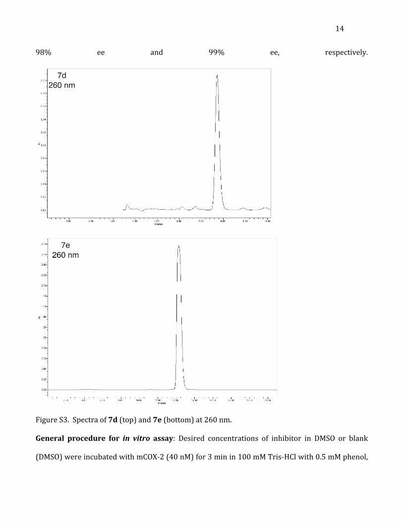
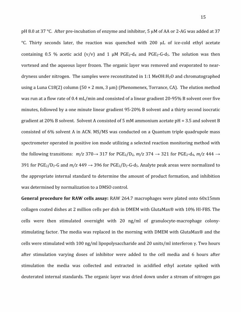
Separation of compounds 7d, 7e: Racemic profen (e.g. 6d) was dissolved in a 90:10 solution of
hexanes:IPA with 0.1% TFA for a final concentration of 10 mg/mL. An aliquot of 30 µL was
injected onto a Chiralcel® AD column using an isocratic gradient of 90:10 hexane:IPA with 0.1%
TFA. Compound 7d eluted at 7.7 minutes and compound 7e eluted at 14.2 minutes. Fractions
were collected manually. The identities of the enantiomers were identified after separation via
optical rotation.2, 3 Enantiomeric excess was determined by re-‐injecting the separated
enantiomers (e.g. 7d) and integrating each peak (Figure S3). Profens 7d and 7e were isolated at
14
98% ee and 99% ee, respectively.
Figure S3. Spectra of 7d (top) and 7e (bottom) at 260 nm.
General procedure for in vitro assay: Desired concentrations of inhibitor in DMSO or blank
(DMSO) were incubated with mCOX-‐2 (40 nM) for 3 min in 100 mM Tris-‐HCl with 0.5 mM phenol,
15
pH 8.0 at 37 °C. After pre-‐incubation of enzyme and inhibitor, 5 µM of AA or 2-‐AG was added at 37
°C. Thirty seconds later, the reaction was quenched with 200 µL of ice-‐cold ethyl acetate
containing 0.5 % acetic acid (v/v) and 1 µM PGE2-‐d4 and PGE2-‐G-‐d5. The solution was then
vortexed and the aqueous layer frozen. The organic layer was removed and evaporated to near-‐
dryness under nitrogen. The samples were reconstituted in 1:1 MeOH:H2O and chromatographed
using a Luna C18(2) column (50 × 2 mm, 3 μm) (Phenomenex, Torrance, CA). The elution method
was run at a flow rate of 0.4 mL/min and consisted of a linear gradient 20-‐95% B solvent over five
minutes, followed by a one minute linear gradient 95-‐20% B solvent and a thirty second isocratic
gradient at 20% B solvent. Solvent A consisted of 5 mM ammonium acetate pH = 3.5 and solvent B
consisted of 6% solvent A in ACN. MS/MS was conducted on a Quantum triple quadrupole mass
spectrometer operated in positive ion mode utilizing a selected reaction monitoring method with
the following transitions: m/z 370→ 317 for PGE2/D2, m/z 374 → 321 for PGE2-‐d4, m/z 444 →
391 for PGE2/D2-‐G and m/z 449 → 396 for PGE2/D2-‐G-‐d5. Analyte peak areas were normalized to
the appropriate internal standard to determine the amount of product formation, and inhibition
was determined by normalization to a DMSO control.
General procedure for RAW cells assay: RAW 264.7 macrophages were plated onto 60x15mm
collagen coated dishes at 2 million cells per dish in DMEM with GlutaMax with 10% HI-‐FBS. The
cells were then stimulated overnight with 20 ng/ml of granulocyte-‐macrophage colony-‐
stimulating factor. The media was replaced in the morning with DMEM with GlutaMax and the
cells were stimulated with 100 ng/ml lipopolysaccharide and 20 units/ml interferon γ. Two hours
after stimulation varying doses of inhibitor were added to the cell media and 6 hours after
stimulation the media was collected and extracted in acidified ethyl acetate spiked with
deuterated internal standards. The organic layer was dried down under a stream of nitrogen gas
16
and reconstituted in 1:1 water:methanol and analyzed by LC-‐MS/MS for prostaglandin levels using
the same instrumentation, column, solvents and conditions as described for the in vitro assay
above.
General procedure for in vivo mouse assay: The following animal protocol was approved by the
Vanderbilt University Institutional Animal Care and Use Committee, and all procedures were
performed in accordance with national guidelines and regulations. Male C57BL/6 mice were
dosed by intraperitoneal (i.p.) injection with 10 mg/kg of compound. For compound 3a at the
designated time points of one hr, two hr, and four hr, mice were euthanized. Separately, mice
were dosed with either 3a or 7a i.p. with 10 mg/kg at 0, 8, 24, 32, 48 and 56 hr. Blood was
collected from the mice at 72 hr. All blood samples were immediately collected into heparinized
syringes by cardiac puncture. The 16 hr time points for compounds 7a, 3a and (S)-‐flurbiprofen
were extracted from murine plasma as follows: 200 µL plasma was diluted 5:1 (v:v) with 1%
acetic acid (aqueous) then 100 µL acetonitrile and 1.2 mL hexane were added. The plasma
samples were mixed well and centrifuged to promote phase separation. The upper, organic layer
was removed and dried. The samples were reconstituted in 9:1 isopropyl alcohol:hexane (v:v)
immediately prior to analysis on an isocratic system where the mobile phase was 98:2 isopropyl
alcohol:hexane with 0.1% acetic acid. Chiral separation occurred on a Chiralcel OD column (25 x
0.46 cm) and fluorescence detection was employed (λex = 248 nm, λem = 312 nm).
The one, two and four hr timepoints for 3a were extracted from murine plasma as follows:
200 µL murine plasma was spiked with flurbiprofen (internal standard) and diluted with 1 mL 1%
acetic acid (aqueous) and loaded onto a pre-‐conditioned OASIS HLB solid phase extraction
column. The OASIS HLB cartridge had been conditioned with 3 mL methanol and 2 mL of 1%
acetic acid (aq). The loaded cartridge was washed with 3 mL of 1% acetic acid (aq) and 1.5 mL of
17
1% acetic acid (aq) with 20% methanol. The cartridge was eluted with 2 mL methanol and the
eluant was dried. The samples were reconstituted in 100 µL methanol and 50 µL water
immediately prior to analysis on a gradient of 30% B to 75% B over 11 min, where A = water and
B = 1:1 methanol:acetonitrile (v:v), each with 0.1% acetic acid. Separation occurred on a Zorbax
C18 column (15 x 0.21 cm) and fluorescence detection was employed (λex = 285 nm, λem = 340
nm).
General procedure for crystallization, X-ray data collection, structure determination and
refinement: Crystallization was performed as previously described with modest modification.4
Purified mCOX-‐2 (10 mg/mL) was reconstituted with a 2-‐fold molar excess of Fe3+-‐
protoprophyrin IX. After dialysis against 20 mM sodium phosphate buffer pH 6.7,100 mM NaCl,
0.01% NaN3, 0.6% (w/v) β-‐OG, β-‐OG concentration was adjusted to 1.2% and 10-‐fold molar excess
of chemical 3a was added prior to setup. Crystallization was performed using hanging drop vapor
diffusion method by combining 3.5 μL of the protein solution with 3.5 μL 50 mM EPPS pH 8.0,
80~120 mM MgCl2, 20~25% (v/v) PEG MME-‐550 equilibrating over reservoir solution containing
0.5 mL of 50 mM EPPS pH 8.0, 100~120 mM MgCl2, 20~25% (v/v) PEG MME-‐550 at 291 K.
Crystals were mounted after about 3 weeks growth and transferred to the stabilization solution
[50 mM EEPS, 28% (v/v) PEG MME 550, 100 mM MgCl2] for ~10 s and flash frozen for crystal
transportation.
Data sets were collected on an ADSC Quantum-‐315 CCD using the synchrotron radiation X-‐
ray source at a wavelength of 0.97929 Å with 100 K liquid nitrogen streaming at beamline 24-‐ID-‐E
of the Advance Photon Source at the Argonne National Laboratory. Diffraction data were collected
and processed with HKL 2000.5 Initial phases were determined by molecular replacement using a
search model (PDB 3NT1) with Phaser. Solution with four molecules in the asymmetric unit was
18
obtained. The models were improved with several rounds of model building in COOT6 and CCP4
suite.7 There were no significant structural differences evident among the monomers. Global non-‐
crystallographic symmetry was applied during the refinement. The ligand was built using SMILE
and waters were adding from COOT.6 The final model has Rwork and Rfree 0.240 and 0.289,
respectively. The potential of phase bias was excluded by simulated annealing using Phenix.8 The
values of the Ramachandran plot for the final refinement of the structure were obtained with
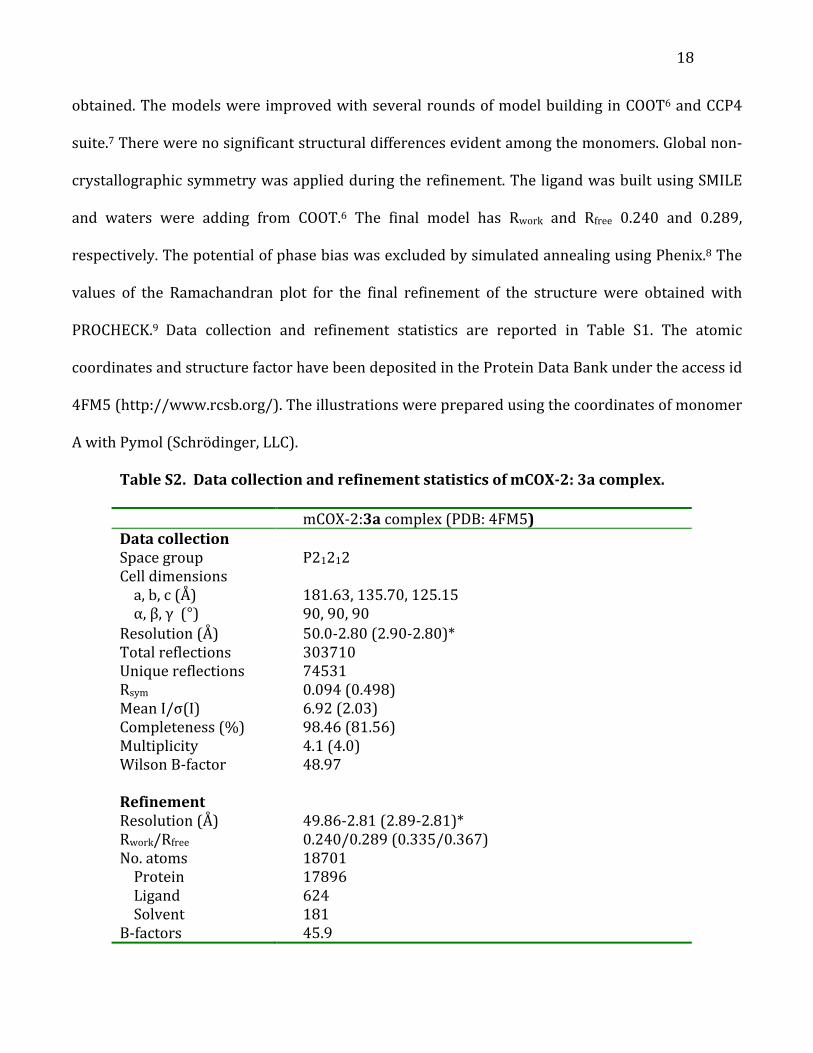
PROCHECK.9 Data collection and refinement statistics are reported in Table S1. The atomic
coordinates and structure factor have been deposited in the Protein Data Bank under the access id
4FM5 (http://www.rcsb.org/). The illustrations were prepared using the coordinates of monomer
A with Pymol (Schrödinger, LLC).
Table S2. Data collection and refinement statistics of mCOX-2: 3a complex. mCOX-‐2:3a complex (PDB: 4FM5) Data collection Space group P21212 Cell dimensions a, b, c (Å) 181.63, 135.70, 125.15 α, β, γ (°) 90, 90, 90 Resolution (Å) 50.0-‐2.80 (2.90-‐2.80)* Total reflections 303710 Unique reflections 74531 Rsym 0.094 (0.498) Mean I/σ(I) 6.92 (2.03) Completeness (%) 98.46 (81.56) Multiplicity 4.1 (4.0) Wilson B-‐factor 48.97 Refinement Resolution (Å) 49.86-‐2.81 (2.89-‐2.81)* Rwork/Rfree 0.240/0.289 (0.335/0.367) No. atoms 18701 Protein 17896 Ligand 624 Solvent 181 B-‐factors 45.9
19
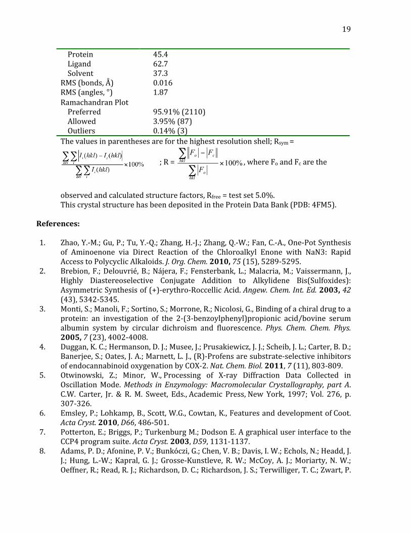
Protein 45.4 Ligand 62.7 Solvent 37.3 RMS (bonds, Å) 0.016 RMS (angles, °) 1.87 Ramachandran Plot Preferred 95.91% (2110) Allowed 3.95% (87) Outliers 0.14% (3) The values in parentheses are for the highest resolution shell; Rsym =
; R = , where Fo and Fc are the
observed and calculated structure factors, Rfree = test set 5.0%. This crystal structure has been deposited in the Protein Data Bank (PDB: 4FM5).
References:
1. Zhao, Y.-‐M.; Gu, P.; Tu, Y.-‐Q.; Zhang, H.-‐J.; Zhang, Q.-‐W.; Fan, C.-‐A., One-‐Pot Synthesis
of Aminoenone via Direct Reaction of the Chloroalkyl Enone with NaN3: Rapid Access to Polycyclic Alkaloids. J. Org. Chem. 2010, 75 (15), 5289-‐5295.
2. Brebion, F.; Delouvrié, B.; Nájera, F.; Fensterbank, L.; Malacria, M.; Vaissermann, J., Highly Diastereoselective Conjugate Addition to Alkylidene Bis(Sulfoxides): Asymmetric Synthesis of (+)-‐erythro-‐Roccellic Acid. Angew. Chem. Int. Ed. 2003, 42 (43), 5342-‐5345.
3. Monti, S.; Manoli, F.; Sortino, S.; Morrone, R.; Nicolosi, G., Binding of a chiral drug to a protein: an investigation of the 2-‐(3-‐benzoylphenyl)propionic acid/bovine serum albumin system by circular dichroism and fluorescence. Phys. Chem. Chem. Phys. 2005, 7 (23), 4002-‐4008.
4. Duggan, K. C.; Hermanson, D. J.; Musee, J.; Prusakiewicz, J. J.; Scheib, J. L.; Carter, B. D.; Banerjee, S.; Oates, J. A.; Marnett, L. J., (R)-‐Profens are substrate-‐selective inhibitors of endocannabinoid oxygenation by COX-‐2. Nat. Chem. Biol. 2011, 7 (11), 803-‐809.
5. Otwinowski, Z.; Minor, W., Processing of X-‐ray Diffraction Data Collected in Oscillation Mode. Methods in Enzymology: Macromolecular Crystallography, part A. C.W. Carter, Jr. & R. M. Sweet, Eds., Academic Press, New York, 1997; Vol. 276, p. 307-‐326.
6. Emsley, P.; Lohkamp, B., Scott, W.G., Cowtan, K., Features and development of Coot. Acta Cryst. 2010, D66, 486-‐501.
7. Potterton, E.; Briggs, P.; Turkenburg M.; Dodson E. A graphical user interface to the CCP4 program suite. Acta Cryst. 2003, D59, 1131-‐1137.
8. Adams, P. D.; Afonine, P. V.; Bunkóczi, G.; Chen, V. B.; Davis, I. W.; Echols, N.; Headd, J. J.; Hung, L.-‐W.; Kapral, G. J.; Grosse-‐Kunstleve, R. W.; McCoy, A. J.; Moriarty, N. W.; Oeffner, R.; Read, R. J.; Richardson, D. C.; Richardson, J. S.; Terwilliger, T. C.; Zwart, P.
20
H. PHENIX: a comprehensive Python-‐based system for macromolecular structure solution. Acta Cryst. 2010, D66, 213-‐221.
9. Laskowski, R. A.; MacArthur, M. W.; Moss, D. S.; Thornton J. M. PROCHECK: a program to check the stereochemical quality of protein structures, J. Appl. Cryst. 1993, 26, 283-‐291.
10. Duggan, K. C.; Hermanson, D. J.; Musee, J.; Prusakiewicz, J. J.; Scheib, J. L.; Carter, B. D.; Banerjee, S.; Oates, J. A.; Marnett, L. J., (R)-‐Profens are substrate-‐selective inhibitors of endocannabinoid oxygenation by COX-‐2. Nat. Chem. Biol. 2011, 7 (11), 803-‐809.