studies on the estimation of 3-methoxy-4-hydroxy-mandelic acid (vma) in human urine
TRANSCRIPT
ANALYTICAL BIOCHEMISTRY 7, &h!i-d94 (1964)
Studies on the Estimation of 3-Methoxy-4-hydroxy-
mandelic acid (VMA) in Human Urine
I-I. WEIL-MALHERBE
From the Clinical Neuropharmacology Research Center, National Institute of Mental Health, St. Elizabeths Hospital, Washington, D. C.
Rewired August 19, 1963
A method for the estimation of 3-methoxy-4-hydroxymandelic acid (VMA) in urine, based on its oxidation to vanillin, was introduced by Sandler and Ruthven (1, 2) : VMA was isolated from urine by ion exchange and ethyl acetate extraction and converted to vanillin by auto- claving in acid solution, in the presence of aluminum oxide as a catalyst. Vanillin was estimated calorimetrically by its reaction with indole. The method was later modified by Sunderman et al. (3), who used ferri- cyanide in acid solution at 37”C, and by Pisano et al. (4), who used sodium periodate in alkaline solution at 50°C as the oxidizing agent. While the color formation with indole was retained in the method of Sunderman et al. (3)) measurement of the light absorption of vanillin in the region of the 360-380 mp band was recommended by Sandler and Ruthven (2) and by Pisano et al. (4). The specificity of the method was improved by pretreatment of urine with Florisil (3) and by extraction of the vanillin formed with toluene either from acid solution (3) or, better, from neu- tral solution (4).
In this paper yet another method for the conversion of VMA to vanillin will be described. The oxidizing agent is cupric ion at a pH of about 10. It will be shown that the three methods of oxidation, by ferri- cyanide, periodate, or copper, give essentially identical results which compare well with the results obtained by the isotope dilution method of Weise et al. (5). It is confirmed that the absorption at 360 rnp is probably more specific than the color reaction with indole. While it is not claimed that the copper method offers any significant advantage over the other two methods, it presents some interesting features which arc thought to be worth recording.
EXPERIMENTAL
A&oxidation of VMA in the Presence of Catalytic Amounts of Cupric Ion and Other Heavy Metals
The present investigation arose from a study of the conversion of 3,4- 485

486 H. WEIL-MALHERBE
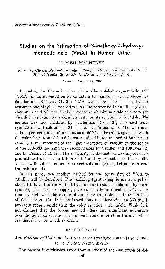
dihydroxymandelic acid to protoca techuic aldehyde. According to Miyake et al. (6), this reaction occurs when dihydroxymandelic acid is heated at 100°C in 10 M acetic acid. It has been found (8) that the reaction depends on the presence of catalytic amounts of cupric or other heavy metal ions and that its pH optimum is 5.5-6.0 at a temperature of 65”. The question arose whether a similar reaction would occur in the case of VMA. As is shown in Fig. 1, VMA is indeed converted to vanillin
.60
2 .=O 0 W
lo .40 l- a
c ii5 .30
El n
a 20 0 F
% .I0
IO 20 30 40 50 60 70 80
MINUTES
FIG. 1. Effect of pH on formation of vanillin from VMA in the presence of IO-‘M CuS04 at 100°C. Each sample contained 20 pg VMA in 2 ml buffer + 0.2 ml 0.025% CuSOa*5Hz0; 0.5M phosphate buffers were used for pH 6, 7, and 8 and a 0.5 M Na2HP04-NaC03 buffer for pH 9.
at 100°C in the presence of 1 X 10e4 U Cu ++. The reaction rate increases with rising pH and the end point is reached after about 10 min at pH 9. The end product appears to be stable under the conditions of the test even in alkaline medium.
Manganese was even more active than copper as a catalyst and several other heavy metals were shown to have catalytic activity while others were inactive or even inhibitory (Table 1).
Oxidation of VMA in Urine Extracts by Copper Ion
While VMA in pure solution was readily converted to vanillin under the conditions described, results were unsatisfactory when attempts were

ESTIMATION OF VMA IN URINE 487
TABLE 1 EFFECT OF IONS OF HEAVY METALS (AT IO+ M) ON OXIDATION OF VMA
Each sample contained 20 Mg VMA in 2 ml 0.5 M potassium phosphate pH 7.0. The solution of VMA in phosphate buffer was shaken for 10 min with about 0.03 vol of che- lating resin (Dowex A-l, 50-100 mesh, Na form) to remove traces of heavy metals. After heating at 100°C for 20 min, the solutions were extracted with 10 ml toluene, toluene was re-extracted with 0.5 N NH3--6y0 NaCl and optical density read at 360 rnlr.
IOn OD IOIl OD
MnZf 0.580 Fez+ 0.064 cue+ 0.437 Ax+ 0.057 HgP+ 0.312 Sn9+ 0.057 Ce2+ 0.263 Cd’+ 0.050 V6f 0.126 A13f 0.048 Fe’+ 0.125 Nie+ 0.048 Cot+ 0.111 Zns+ 0.048 Pb”+ 0.103 BiS+ 0.039 Cr’+ 0.066 No addition 0.066
made to carry out the reaction in the presence of material extracted from acidified urine by ethyl acetate. These difficulties were attributed to the presence of antioxidants, chelators, or both, and were overcome by increasing the concentration of copper ion, raising the pH to about 10, and extending the time of heating to 2 hr.
When an ethyl acetate extract of urine was re-extracted with 1 M Na,CO,, in the proportions described below, the pH of the resulting solu- tion was 10.0-10.1. The 1 M Na,C03 solution could be replaced by 0.5 M borate buffer pH 11.2, the pH of which also fell to 10.0 after equilibra- tion with an ethyl acetate extract of urine. The use of the Na,CO, solu- tion was preferred since the results seemed to be more consistent than when the borate buffer was used.
The end point of the reaction in urine extracts is reached after about 2 hr at 100°C although the increment due to added VMA ceased to increase after 1 hr (Fig. 2). The end product is stable under the condi- tions of the assay for at least 3 hr.
On the basis of these observations the following method was devised: Solutions: 1. Copper reagent: 2 gm CuSO,.5H,O, 10 gm sodium potas-
sium tartrate, and 10.6 gm Na,C03, anhydrous, dissolved to 100 ml. 2. Sodium carbonate, 1 M solution. 3. Sulfuric acid, 10N. 4. 50% (v/v) phosphoric acid: mix equal parts of 85% H,PO, and
water. 5. Ammonia, 0.5 N solution containing 6% (w/v) sodium chloride. 6. Standard solution of VMA (Calbiochem., Los Angeles) containing

488 H. WEIL-MALHERBE
1.00 2 .90
0 ,” .80
= .60
z” .50
: .40
60 120 180
MINUTES
FIG. 2. Time curve of vanillin formation from VMA; B, extract from 10 ml urine per sample; A, 10 gg VMA was added to each sample of the came urine extract aa used in curve B; A (A - B), Difference between curves A and B.
100 pg/ml in water saturated with 2-octanol. Store in dark bottle at refrigerator temperature. The standard is stable for several months.
Other Reagents: Florisil (Floridin Company, Tallahassee, Florida), ethyl acetate (reagent grade), toluene (reagent grade).
Procedure: A sampIe of 30 mI urine is acidified with 3 ml 10 N H,SO, and shaken for 10 min with 3 gm Florisil. The mixture is briefly cen- trifuged and the supernatant solution poured through a folded filter. A 22-ml aliquot of the filtrate (corresponding to 20 ml urine) is saturated with solid NaCl and extracted for 10 min first with 2 vol and again with 1 vol of ethyl acetate. The combined ethyl acetate extracts are re- extracted with 5 ml l.OM Na,CO,. The Na,C03 extract is separated and its volume measured: if necessary, it is made up to 4 ml with 1.0 M Na,CO,. It is divided into two equal aliquots, one to be oxidized with copper solution, the other to scrvc as a blank to correct for any pre- formed absorbing material which may be present.
The sample destined for oxidation is mixed with 2 ml of the copper reagent and placed for 2 hr in a boiling water bath. Reagent blank and standard solutions containing 5 and 20 pg V&IA are added to, 2 ml samples of 1 M Na,CO, and treated in the same way.
ESTIMMATION OF VMA IN URINE 489
The solutions are then cooled and brought to pH 7.0 by the addition of 0.26 ml 50% H3P04. The blank is diluted with 2 ml water and its pH adjusted to 7.0 by the addition of 0.13 ml 56% H,PO,. All samples are now saturated with solid NaCl and extracted with 5 vol toluene for 10 min. The toluene layer is separated and re-extracted with 3 m1 of the ammonia-NaC1 solution. The optical density is determined at 360 m,u; urine samples are read against. the urine blank, standards against. the reagent blank. If the urine sample should give a reading in excess of 0.900, it is diluted with ammonia solution as required. The result indi- cates the amount of VMA in 10 ml urine.
Comments: (1) According to Sunderman et al. (3), Florisil removes pigments and other interfering constituents from urine. This step was retained as a precaution after it had been confirmed that VML4 itself was not adsorbed by Florisil. Extraction of vanillin from neutral solution has been adopted from the procedure of Pisano et al. (4).
(,Z) Urine is extracted tIcice with ethyl acetate, A second extraction requires so little extra work and time that the more thorough separation and the saving of solvent (a total of 3 vol as against 4-5 vol used in the methods of Sunderman et al. (3) and Pisano et al. (4) were considered worthwhile.
(3) Ammonia solution was preferred to carbonate solution for the re-extraction of vanillin from toIuene because of troublesome bubble formation when the carb0nat.e solution was acidified prior to the addi- tion of the indole reagent. The addition of sodium rhloride facilitates phase separation.
(4) As will be shown below, the estimation of optical density is at least as specific as the color reaction with indole. Since it has the advan- tage of simplicity and since dilution is easy if the optical density is excessive, it is the preferred technique.
(5) The methods based on vanillin formation have been criticized (6) on the grounds that urine may contain preformed vanillin of dietary origin. This eventuality is easy to guard against by reading the optical density against an unoxidized blank [cf. refs. (3) and (4)]. ln fact, no significant absorption was observed when unoxidized urine blanks were read against the reagent blank. It is permissible, therefore, to omit the urine blank if insufficient urine is available. Significant, readings can be obtained with as little as 2 or 3 ml urine.
RESULTS
The optical density readings obtained with increasing volumes of urine lie on a straight line, up to an OD of about 0.9. Identical increments of OD are found when increasing amounts of VMA are added to a given volume of either urine or water and analyzed as described.

490 H. WEIL-MALHERBE
Specificity: The specificity of the conversion of VMA to vanillin has been studied by Sunderman et al. (3). They showed that there was no interference from a series of aromatic acids including mandelic acid, m-hydroxymandelic acid, and 3,4-dihydroxymandelic acid, The absence of interference from mandelic and 3,4-dihydroxymandelic acids is con- firmed by the data of Table 2. While mandelic acid is presumably
TABLE 2 INTERFERENCE IN ESTIMATION OF VMA BY COPPER METHOD
After measuring absorption at 360 mp the indole reaction was carried out with a por- tion of 2 ml of the solution, by adding 0.5 ml concentrated H2S04 at 0°C followed by 2 ml of a freshly prepared mixture of 1.5% ethanolic indole and 85% HzP04, 1:50.
Opt.ieal density
Addition
VMA, 20 rg
nn-Mandelic acid
2oag 100 Pg 300 Icg
on-p-Hydroxymandelic acid
20 I43
100 P&T 300 Peg
or&GDihydroxymandelic acid
20 Pi%
100 A% 300 Pcrg
n&3-Methoxy-Ghydroxyphenylglycol
20 Pg 100 fig
0 After addition of indole reagent.
360 m&t 405 mpa
0.545 0.320
0.030 0.005 0.030 0.006 0.032 0.005
0.060 0.095 0.210 0.560 0.410 0.850
0.040 0.035 0.040 0.030 0.028 0.005
0.272 0.160 1.36 0.780
resistant to oxidation under the conditions of the test, 3,4-dihydroxy- mandelic acid is oxidized very rapidly to protocatechuic aldehyde, which is decomposed under the conditions used. p-Hydroxymandelic acid is oxidized to p-hydroxybenzaldehyde. This interference can be reduced by the choice of the 360 mp wave length for the measurement of optical density as proposed by Pisano et al. [ (4) ; see also ref. (2)]. It amounts to about 10% of the reading given by an equal quantity of VMA, while the color formed in the indole reaction is 30% of that given by VMA.
As predicted by Sunderman et al. (3)) 3-methoxy-4-hydroxyphenyl-

ESTIMATION OF VMA IN URINE 491
glycol is oxidized to vanillin under the conditions of the VMA assay. The yield of vanillin is about 50% of t,hat, obtained from VMA. In prac- tice, however, the interference is minimal since only negligible amounts of methoxyhydroxyphenylglycol are extracted from strongly acid solu- tion by the proportions of ethyl acetate used for the extraction of VMA. In urine, the glycol mainly occurs in the conjugated form, which would be extract’ed with even greater difficulty.
Comparison of Ferricyanide, Periodate, and Copper Methods
For the purpose of comparing the different methods the procedures described by Sunderman et al. (3) and Pisano et al. (4) were slightly modified. The t,reatment of urine with Florisil and the initial extraction of VMA were performed on a sample of 50 ml urine as described under “Procedure.” Three 2-ml portions of carbonate extract, each correspond- ing to 8 ml urine, were used for oxidation by the three agents and a fourth served as unoxidized blank. The reagents used in the methods of Sunderman et al. (3) and Pisano et aL. (4) were suitably adapted to the scale of the experiment. The oxidized mixtures were adjusted to pH 7.0 and vanillin was extracted as described under “Procedure.” After determining the optical density at 360 mp the indole color was developed as described in Table 2. It should be noted that the concentration of indole was increased nearly threefold over that recommended by Sunder- man et al. (3) which, under our conditions, was not optimal for color formation.
A series of results is shown in Table 3. The table includes estimations on ten urine specimens that had previously been analyzed by the isotope dilution method of Weise et al. (5). The mean percentage differences shown at the bottom of Table 3 indicate that results obtained by absorp- tiometry at 360 rnp are identical, within the limits of error, for the periodate and the copper method. For the ferricyanide and copper methods, the difference, while statistically significant, is <1~)7~. The estimation by the indole color reaction usually gave higher values than absorptiometry at 360 rnp. This discrepancy is greatest after periodate oxidation and smallest aft,er copper oxidation. It may be inferred that material other than vanillin which reacts with indole is formed in small amounts, particularly when periodate is used as oxidizing agent,.
When the three methods based on vanillin formation are compared with the isotope dilution method of Weise et al. (5), it appears that,, in spite of considerable variation in individual experiments and conse- quently high standard errors, the differences of the results obtained by absorptiometry at 360 mp are not significant and are below 10yO in the case of the pcriodate and copper methods, whereas they are significantly

TABL
E 3
COM
PARI
SON
OF
THRE
E M
ETHO
DS
FOR
ESTI
MAT
ION
OF
VMA
BASE
D ON
IT
S
OXID
ATIO
N TO
VA
NILL
IN
Sam
ple
No.
Oxidi
zing
agen
t -
~___
Fe
rricya
nide
Perio
date
Copp
er ion
-.
360
mp
495
In@"
360m
r 49
5 m
p 36
0 m
p 49
5 m
p Me
thod
-- of
W&e
pg
VI
vlA/IO
m
l uri
ne
et al.
1 22
.6
21.8
20
.6
24.2
19
.4
21.9
-
2 25
.7
28.6
23
.6
31.8
21
.5
28.4
-
3 45
.6
50.6
41
.0
50.6
38
.6
45.2
-
4 28
.8
33.8
27
.6
34.7
27
.0
24.5
-
5 29
.8
34.7
30
.6
39.6
31
.6
35.8
-
6 30
.0
37.2
32
.2
40.6
30
.5
35.6
-
7 19
.2
21.4
15
.8
20.6
15
.0
18.7
10
.9
.x 8
15.1
15
.8
14.5
19
.3
12.5
13
.8
9.7
8 9
14.9
17
.6
15.0
18
.7
13.8
16
.5
11.4
,”
10
8.2
11.1
7.
9 10
.6
8.0
9.7
10.2
11
10
.2
13.8
8.
9 12
.8
9.2
10.9
8.
0 z
12
5.2
6.5
4.1
5.8
4.2
5.2
5.9
F
13
40.9
53
.8
40.6
51
.6
40.0
43
.0
50.4
9
14
7.7
9.6
7.9
11.1
8.
5 9.
9 6.
6 15
10
.0
13.4
9.
2 12
.7
9.2
10.9
8.
1 16
16
.7
20.7
18
.5
25.8
18
.7
22.7
18
.9
Ilean
pe
rcen
tage
di
ffere
nce
(&S.
E.M
.):
lhta
of
co
l. 8
$16.
8 +
10.3
3 +*
3.1
f 10
.22
+9.6
+
8.84
+4
7.8
f 11
.46
$7.3
*
7.53
j-2
6.7
I’C 9
.11
- =
100
Data
of
co
l. 6
+8.6
7 rf:
2.
97
- f2
.69
+ 1.
48
- -
- -
= 10
0 Da
ta
of
col.
7 -
+11.
81
f 3.
30
- +1
6.49
f
2.48
-
- -
= 10
0
a Af
ter
addi
tion
of
indo
le
reag
ent.

ESTIMATION OF VMA IN URIIGE 493
higher when the indole color reaction is used. Again, the excess is small- est after oxidation by copper ion.
DISCUSSION
In the series mandelic acid-VXIA-3,4-dihydroxymandeiic acid, the ease with which the side chain is oxidized increases in t.he same order. This clearly implicates the phenolic substituents although, at least in the case of VMA, they seem to emerge unchanged at the end of the reaction. The practical application of this rcact,ion is based on the great stability of vanillin, unusual for an aldehydc. As is well known (7), p-substitution with an hydroxyl group increases the stability of benzaldehyde. This effect is in contrast to the increased lability of the glycolate side chain caused by the same substitution.
The accuracy and specificity of the methods based on the conversion of VMA to vanillin appear to be adequate for most purposes and are clearly superior in simplicity and speed to paper chromatographic, elec- trophoretic, and isotope dilution methods. As for the comparison of the three met,hods studied, there is not much to choose among them. If speed is the main consideration, the periodate method of Pisano et al. (4) will be preferred.
SUMMARY
A method for the estimation of 3-methoxy-4-hydroxymandelic acid (VMA) in human urine, based on its oxidation to vanillin by copper ion at pH 10, is described. Pure solutions of VMA are subject to autoxi- dation in the presence of catalytic amounts of heavy metals, particularly manganese and copper, at pH values above 6 and at 100°C. Owing to the presence of interfering material in extracts from urine more drastic conditions are required, such as raising the pH to 10 and increasing the concentration of copper in the reaction mixture.
The method has been compared with other methods based on the same principle in which ferricyanide or periodate is used as the oxidizing agent. It has been shown that the results of the three methods, on the average, agree within 10% for a given urine specimen. Satisfactory agreement was also found with the results of an isotope dilution method.
The estimation of vanillin by absorptiometry at 360 m,,. seems to be slightly more specific than the color reaet,ion with indole.
ACKNOWLEDGMENTS
I am greatly indebted to Mrs. Viola Epps for technical assistance, to Mrs. Vir- ginia Weise for the gift of urine specimens previously analyzed by her method. and to Dr. Julius Axelrod for a specimen of 3-methoxy4-hydroxyphenylglycol.

494 H. WEIL-MALHERBE
NOTE ADDED IN PROOF
It has been noticed that homovanillic acid causes interference of about 10 percent in the copper method for VMA estimation.
REFERENCES
1. SANDLER, M., AND RUTHVEN, C. R. J., ikmxt 1959 II, 114, 1034. 2. SANDLER, M., AED RUTHVEN, C. R. J., B,iochem. J. 80, 78 (1961). 3. SUNDERMAN, F. W., JR., CLEVELAND, P. D., LAW, N. C., AND SUNDERMAN, F. W.,
Am. J. Chin. Path. 34, 293 (1960). 4. PISANO, J. J., CROUT, J. R., AND ABRAHAM, D., C&n. Chim. Acta 7, 285 (1962). 5. WEISE, V. K., MCDOXALD, R. K., AND LABROSE, E. H., Clin. Chim. Acta 6, 79
(1961). 6. MIYAKE, H., YOSHIDA, H., AND IMAIZUMI, R., Japan. J. Pharmacol. 12, 79 (1962). 7. HOUBEN-WEYL. “Methoden der organischen Chemie” (E. Miiller, ed.), 4th Ed.,
Band VII, Teii 1, p. 497. Thieme, Stuttgart, 1954. 8. WEIL-MALHERBE, H., to be submitted to Analytical Biochemistry.