studies on keratitis in reindeer : 3. experimentally induced keratitis in reindeer
TRANSCRIPT
Zbl. Vet. Med. A, 25, 110-128 (1978) @ 1978 Verlag Paul Parey, Berlin und Hamburg ISSN 0300-871 l/ASTM-Coden: ZVRAAX
From the Department of Anatomy and Histology College of Veterinary Medicine
Swedish University of Agricultural Sciences, Uppsala The Department of Radiobiology
National Defence Research Institute, Stockholm The Department of Bacteriology
National Veterinary Institute, Stockholm The Department of Applied Mathematics and Computations,
National Defence Research Institute, Stockholm The Department of Electric Power Systems Engineering Lightning Laboratory,
Royal Institute of Technology, Stockholm
Studies on Keratitis in Reindeer 3. Experimentally induced keratitis in reindeer
BY
C. REHBINDER, V. GLATTHARD, F. MOELL and A. OTTOSSON
W i t h 14 figures and 7 Tables
(Received for publication September 16, 1977)
Introduction The purpose of this investigation was to repeat on reindeer selected parts
of the experiments on mice to see whether the same factors were effective in reindeer.
Material and Methods Experimental animals
Twenty-seven one-year old reindeer, were used. Before the experiments all animals appeared healthy and were in a good nutritional state. The eyes showed no clinical signs of disease. Five days before the start of the experi- ments they were transferred to separate stalls, and each group was subjected to careful clinical and bacteriological control. During the experiment there was no contact between animals from different stalls.
Bacteriological control of experimental animals The bacterial flora of the conjunctival sacs was controlled according to
the methods described by REHBINDER and TSCHAPPAT (1974). The presence of a sparse bacterial growth of coagulase-negative staphylococci (35 eyes), a-
Studies on Keratitis in Reindeer / 3. 111
haemolytic streptococci (4 eyes) or a mixed flora of these bacteria ( 1 1 eyes) was considered to be normal. Samples from 4 eyes yielded no growth.
Bacteriu for inoculation experiments Bacteria, equipment and administration routines as for mice in paper 2.
Animals with normal flora were used as controls in each group.
Ultraviolet irradiation Equipment for irradiation experiments on reindeer according to paper 1
was used.
Dust Material and administration routines as for mice in paper 2.
Experimental procedure Seven different experiments were performed. In each experiment 6
I. Inoculation with different bacteria + controls. II. Inoculation with different bacteria + UV-irradiation 4 hrs; and
111. Inoculation with bacteria + dust; and dust alone, respectively. IV. Inoculation with different bacteria 4- dust + UV-irradiation 4 hrs;
and dust + UV-irradiation 4 hrs, respectively. V. Inoculation with different bacteria + erosion of the cornea; and
erosion of the cornea alone, respectively. VI. Inoculation with different bacteria + erosion of the cornea + UV-
irradiation 4 hrs; and erosion of the cornea + UV-irradiation 4 hrs, respec- tively.
VII. Inoculation with different bacteria + erosion of the cornea + dust + UV-irradiation 4 hrs; and erosion of the cornea + dust + UV-irradiation 4 hrs, respectively.
animals were used (cf. Tables). Alltogether 27 animals were used.
4 hrs UV-irradiation only, respectively.
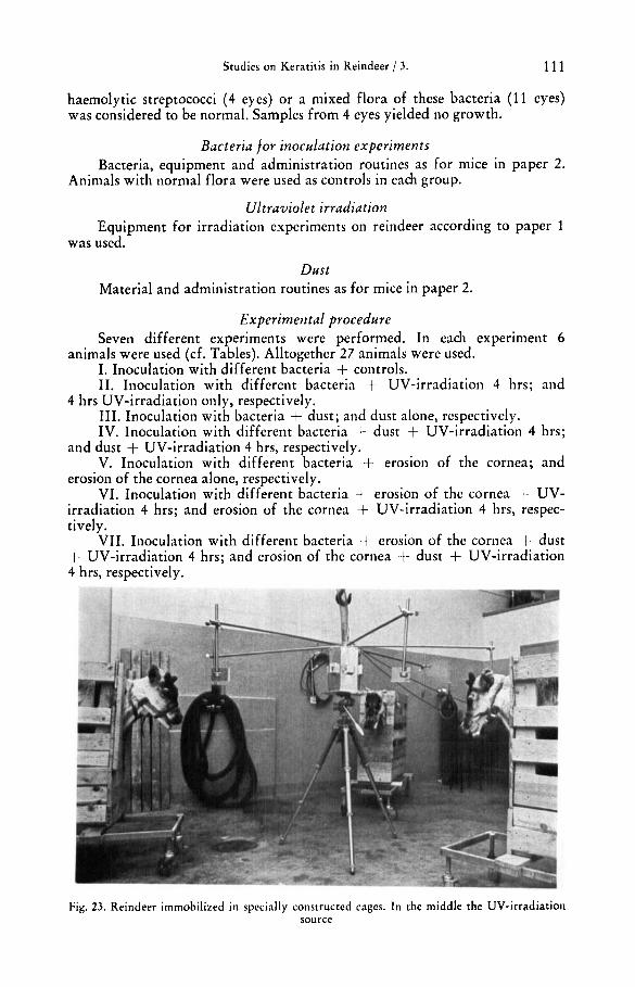
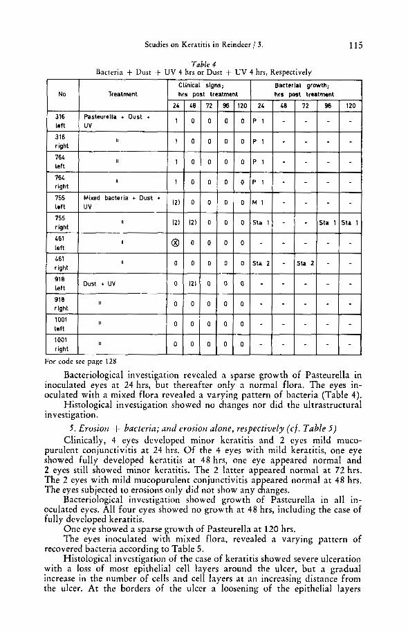
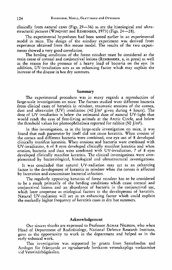
Fig. 23. Reindeer immobilized in specially constructed cages. In the middle the UV-irradiatioii source
112 REHBINDER, MOELL, GLATTHARD and OTTOSSON
Most animals were used in two related experiments and two animals were used in three experiments (cf. Tables 1-7). Experiment VI was performed first.
The bacteria were administered at the beginning of the experiment. The conjunctival flora was controlled before the experiment by checks every 24 hrs (5 days), and similarly after the experiment. Erosions of the cornea were produced at 0 hours as for mice in paper 2. The eyes of the animals were exposed to the different treatments, i. e., erosion, dust and bacteria immediate- ly prior to irradiation. UV-irradiation was administered with the equipment described in paper 1. Each animal was given a maximum dose of 40 J/m’. The animals were immobilized in a specially constructed cage (cf. Fig. 23). During irradiation the eyes of the animals were not touched and blinking was not prevented.
The eyes of the animals were examined at 24 hrs intervals with fluore- scein, a lens (4 X ) and a lamp. The character of the eye secretions was defined as for mice in paper 2. Photophobia was considered to be present if the animals pinched up their eyelids when in light. Cases of keratitis were categorized in 2 groups, I ) a minor greyish swelling or an initial diffuse opacity of the cornea and 2) a well-defined large swelling or diffuse opacity involving most of the cornea.
Animals with fully developed keratitis on one eye, disregarding the status of the other eye, were submitted to ordinary slaughter. The material for
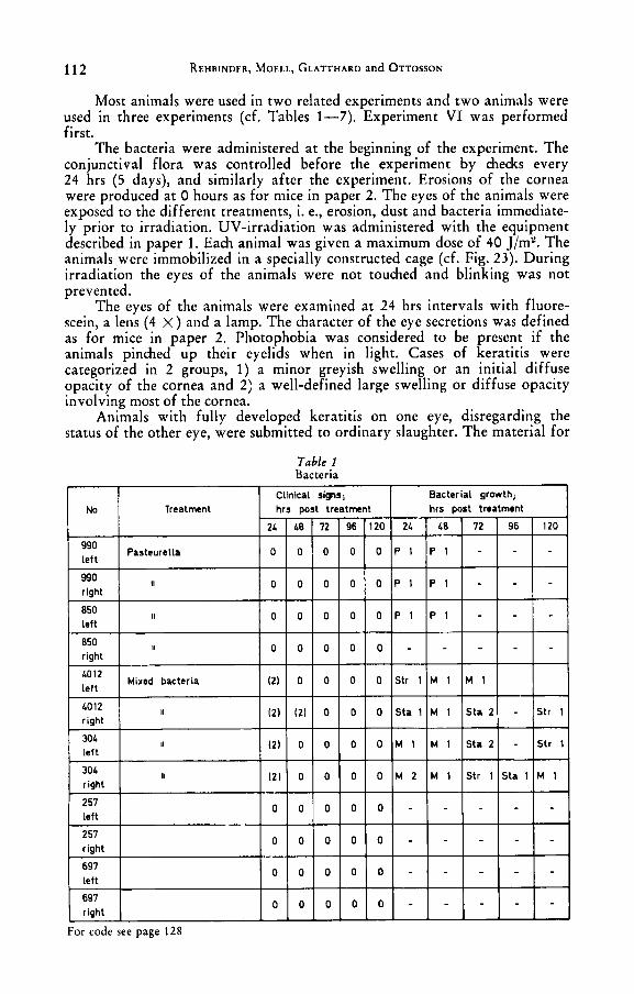
Table 1 Bacteria

Studies on Keratitis in Reindeer / 3. 113
histology and electron microscopy was obtained and handled as described for mice in paper 2 with the exception that the corneae were removed directly and put into glutaraldehyde and not the whole eye as for mice.
Results 1. Bacteria (cf . Table 1 )
Four eyes inoculated with a mixed flora of staphylococci and streptococci developed clinically a mild mucopurulent conjunctivitis at 24 hrs, which had disappeared at 48 hrs in 3 eyes and at 72 hrs in the remaining eye. Bacterio- logical investigation showed no growth of Pasteurella after 48 hrs, but a sparse growth of staphylococci and streptococci remained established in the conjunc- tival sac without resulting in clinical signs of conjunctivitis.
2. Bacteria + UV-irradiation 4 hrs and UV-irradiation 4 hrs alone, respectively ( c f . Table 2 )
Pasteurella did not cause any clinical alterations. In the eyes inoculated with staphylococci and streptococci all 4 eyes developed mucopurulent con- junctivitis according to Table 2.
Bacteriological investigation showed no growth of the inoculated bacteria in 6 of 8 eyes at 24 hrs. The eyes then revealed a varying pattern of recovered bacteria according to Table 2.
Histological investigation revealed no changes, nor did the ultrastructural
114 REHBINDER, MOELL, GLATTHARD and OTTOSSON
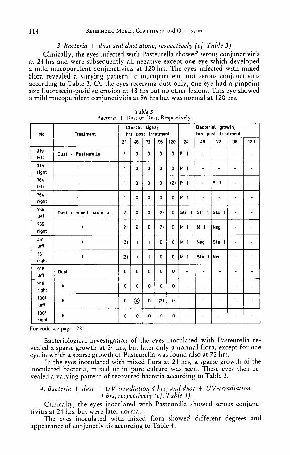
3. Bacteria + dust and dust alone, respectively ( c f . Table 3 ) Clinically, the eyes infected with Pasteurella showed serous conjunctivitis
at 24 hrs and were subsequently all negative except one eye which developed a mild mucopurulent conjunctivitis at 120 hrs. The eyes infected with mixed flora revealed a varying pattern of mucopurulent and serous conjunctivitis according to Table 3. Of the eyes receiving dust only, one eye had a pinpoint size fluorescein-positive erosion a t 48 hrs but no other lesions. This eye showed a mild mucopurulent conjunctivitis at 96 hrs but was normal at 120 hrs.
Table 3 Bacteria + Dust or Dust, Respectively
Bacteriological investigation of the eyes inoculated with Pasteurella re- vealed a sparse growth at 24 hrs, but later only a normal flora, except for one eye in which a sparse growth of Pasteurella was found also at 72 hrs.
In the eyes inoculated with mixed flora at 24 hrs, a sparse growth of the inoculated bacteria, mixed or in pure culture was seen. These eyes then re- vealed a varying pattern of recovered bacteria according to Table 3.
4 . Bacteria -t dust t UV-irradiation 4 hrs; and dust + UV-irradiation 4 hrs, respectively ( c f . Table 4 )
Clinically, the eyes inoculated with Pasteurella showed serous conjunc-
The eyes inoculated with mixed flora showed different degrees and tivitis at 24 hrs, but were later normal.
appearance of conjunctivitis according to Table 4.
Studies on Keratitis in Reindeer / 3. 115
Bacteriological investigation revealed a sparse growth of Pasteurella in inoculated eyes at 24 hrs, but thereafter only a normal flora. The eyes in- oculated with a mixed flora revealed a varying pattern of bacteria (Table 4).
Histological investigation showed no changes nor did the ultrastructural investigation.
f i . Erosion -+ bacteria; and erosion alone, respectively (c f . Table 5 ) Clinically, 4 eyes developed minor keratitis and 2 eyes mild muco-
purulent conjunctivitis a t 24 hrs. Of the 4 eyes with mild keratitis, one eye showed fully developed keratitis at 48 hrs, one eye appeared normal and 2 eyes still showed minor keratitis. The 2 latter appeared normal a t 72 hrs. The 2 eyes with mild mucopurulent conjunctivitis appeared normal at 48 hrs. The eyes subjected to erosions only did not show any changes.
Bacteriological investigation showed growth of Pasteurella in all in- oculated eyes. All four eyes showed no growth at 48 hrs, including the case of fully developed keratitis.
One eye showed a sparse growth of Pasteurella at 120 hrs. The eyes inoculated with mixed flora, revealed a varying pattern of
recovered bacteria according to Table 5. Histological investigation of the case of keratitis showed severe ulceration
with a loss of most epithelial cell layers around the ulcer, but a gradual increase in the number of cells and cell layers a t an increasing distance from the ulcer. At the borders of the ulcer a loosening of the epithelial layers
116 REHBINDER, MOELL, GLATTHARD and OTTOSSON
formed a shallow pocket. A general intra- and intercellular oedema was present in all epithelial cell layers. The tissue underlying the ulcer was pro- truding and undulating. It was heavily infiltrated with neutrophils and had many capillaries. However, most of the severe changes in the stroma were concentrated beneath the ulcer. The endothelium was slightly swollen. The remaining cases showed no changes in the epithelium. In two corneae a few capillaries were present in the anterior part ( ' I s ) of the stroma.
Ultrastructural investigation of clinically normal corneae showed changes and confirmed the histological picture of keratitis, showing ulceration and acute inflammatory changes. A rominent feature was a marked oedema
by long cellular processes connected with desmosomes, some of the processes being broken in or near the desmosomes. Around the ulcer most of the surface cells were lost and olyhedral cells were found outermost. Some cells were
appeared normal but most were pycnotic o r showed other degenerative changes. The stroma showed an intense oedema and around the ulcer the orderly structure was more of less completely lost, with only a few collagen bundles in a flocculent ground substance with many neutrophils and mono- cytes.
The endothelium showed intracellular oedema of the same type as the epithelial cells and in addition many pinocytic vesicles.
in and between the epithelial cells. T K e intercellular spaces were often bridged
very pale and swol P en while others were dark and shrunken. Many nuclei
Studies on Keratitis in Reindeer / 3. 117
6. Erosion + bacteria + UV-irradiation 4 hrs; and erosion + UV-irradiation 4 hrs, respectively ( c f . Table 6 )
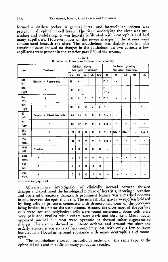
Clinically, 4 eyes had a fully developed keratitis at 24 hrs. Of these one animal was affected bilaterally. Both unilateral cases showed mucopurulent
Fig. 24. Ulcerating keratitis. Note the bulging of the scrorna and the loss of epithelium. Erosion + bacteria (Pasteurella) and UV 4 hours. Embedded in Epon, toluidine blue ( x 150)
118 REHBINDER, MOELL, GLATTHARD and OTTOSSON
conjunctivitis of the other eye. One animal showed bilateral mucopurulent conjunctivitis and photophobia a t 24 hrs and fully developed bilateral kera- titis a t 48 hrs.
The eyes subject to erosion and UV-irradiation did not show any signs. Bacteriological investigation of the eyes inoculated with Pasteurella
showed no growth in one case of keratitis and one case of mucopurulent conjunctivitis and photophobia, one eye with keratitis and one with muco- purulent conjunctivitis and photophobia showed sparse growth. The two eyes which at 48 hrs had developed keratitis revealed no growth of the inoculated bacteria.
Of the eves inoculated with mixed flora. 3 of 4 showed a sparse growth of inoculated'bacteria at 24 hrs, while the remaining eycs showed a mGderate growth of staphylococci and a sparse growth of streptococci.
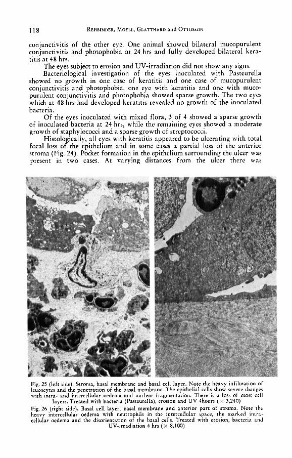
Histologically, all eyes with keratitis appeared to be ulcerating with total focal loss of the epithelium and in some cases a partial loss of the anterior stroma (Fig. 24). Pocket formation in the epithelium surrounding the ulcer was present in two cases. At varying distances from the ulcer there was
Fig. 25 (left side). Stroma, basal membrane and basal cell layer. Note the heavy infiltration of leucocytes and the penetration of the basal membrane. The epithelial cells show severe change\ with intra- and intercellular oedema and nuclear fragmentation. There is a loss of most cell
layers. Treated with bacteria (Pasteurella), erosion and UV 4hours ( x 3,240) Fig. 26 (right side). Basal cell layer, basal membrane and anterior par t of stroma. Note thc heavy intercellular oedema with neutrophils in the intercellular space, the marked intra- cellular oedema and the disorientation of the basal cells. Treated with erosion, bacteria and
UV-irradiation 4 hrs ( x 8,100)
Studies on Keratitis in Reindeer / 3. 119
a gradual increase in the number of cell layers. In many cases neutrophils were present in the epithelium. A regular finding was also an inter- and intra- cellular oedema.
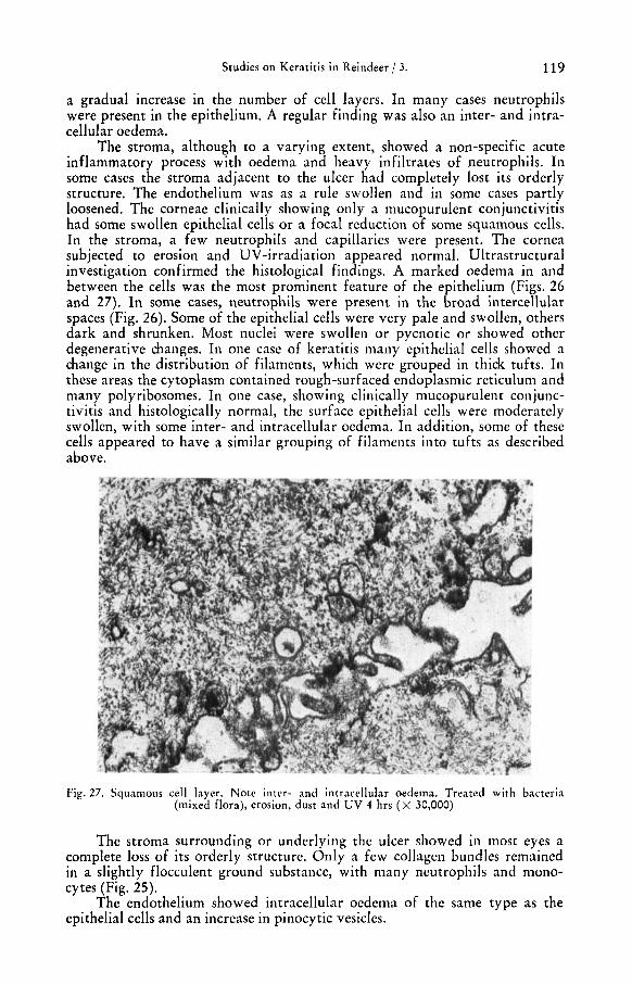
The stroma, although to a varying extent, showed a non-specific acute inflammatory process with oedema and heavy infiltrates of neutrophils. In some cases the stroma adjacent to the ulcer had completely lost its orderly structure. The endothelium was as a rule swollen and in some cases partly loosened. The corneae clinically showing only a mucopurulent conjunctivitis had some swollen epithelial cells or a focal reduction of some squamous cells. In the stroma, a few neutrophils and capillaries were present. The cornea subjected to erosion and UV-irradiation appeared normal. Ultrastructural investigation confirmed the histological findings. A marked oedema in and between the cells was the most prominent feature of the epithelium (Figs. 26 and 27). In some cases, neutrophils were present in the broad intercellular spaces (F,ig. 26). Some of the epithelial cells were very pale and swollen, others dark and shrunken. Most nuclei were swollen or pycnotic or showed other degenerative changes. In one case of keratitis many epithelial cells showed a change in the distribution of filaments, which were grouped in thick tufts. In these areas the cytoplasm contained rough-surfaced endoplasmic reticulum and many polyribosomes. In one case, showing clinically mucopurulent conjunc- tivitis and histologically normal, the surface epithelial cells were moderately swollen, with some inter- and intracellular oedema. In addition, some of these cells appeared to have a similar grouping of filaments into tuf t s as described above.
Fig. 27. Squamous cell layer. Note inter- and intracellular oedema. Treated with bacteria (mixed flora), erosion, dust and UV 4 hrs ( X 30,000)
The stroma surrounding or underlying the ulcer showed in most eyes a complete loss of its orderly structure. Only a few collagen bundles remained in a slightly flocculent ground substance, with many neutrophils and mono- cytes (Fig. 25).
The endothelium showed intracellular oedema of the same type as the epithelial cells and an increase in pinocytic vesicles.
120 REHBINDER, MOELL, GLATTHARD and OTTOSSON
In cases without clinical and histological changes, no changes were found on ultrastructural investigation.
7. Erosion + dust + bacteria + UV-irridiation 4 hrs; or erosion + dust + UV-irradiation 4 hrs, respectively (Table 7)
Clinically, all inoculated aiiinials had developed a minor keratitis a t 24 hrs. At 48 hrs, 7 of 8 eyes had a fully developed keratitis, while the re- maining eye still showed a minor keratitis. Bacteriological investigation re-
Table 7 Bacteria + Erosion + Dust + UV 4 hrs or Erosion, Dust, UV 4 hrs, Respectively
Treatment I No I Erosion, dust, I Pasteurella + UV I “;t t-q-77- right
right
right
left
Clinical signs; Bacterial growth; hrs Dost treatment hrs post treatment
405 right
99L left
994 right
left
997 rlght
II
Erosion, dust i UV
W
991 I1
ll - For code see page 128
vealed a sparse growth of inoculated bacteria in all eyes. At 48 hrs, 3 of the eyes inoculated with Pasteurella still showed a sparse growth, while the re- maining eye had a moderate to rich growth. Of the eyes inoculated with mixed flora there was at 48 hrs a sparse to moderate growth of staphylococci in 2 eyes, a sparse growth of staphylococci in one eye and the remaining eye showed no growth of the inoculated bacteria. The eyes which had not been inoculated showed no changes.
Histological investigation revealed ulceration of the epithelium in one eye with a marked reduction of the surface epithelium. The other eyes showed a marked general inter- and intracellular oedema of all cell layers with a marked general reduction in the number of cell layers. Neutrophils were present in the epithelium to a varying extent in 4 cases and were absent in 3. In 4 cases the changes were focal in the centre of the cornea while in the other four they seemed to involve the whole cornea. In all cases, the stroma,
Studies on Keratitis in Reindeer / 3. 121
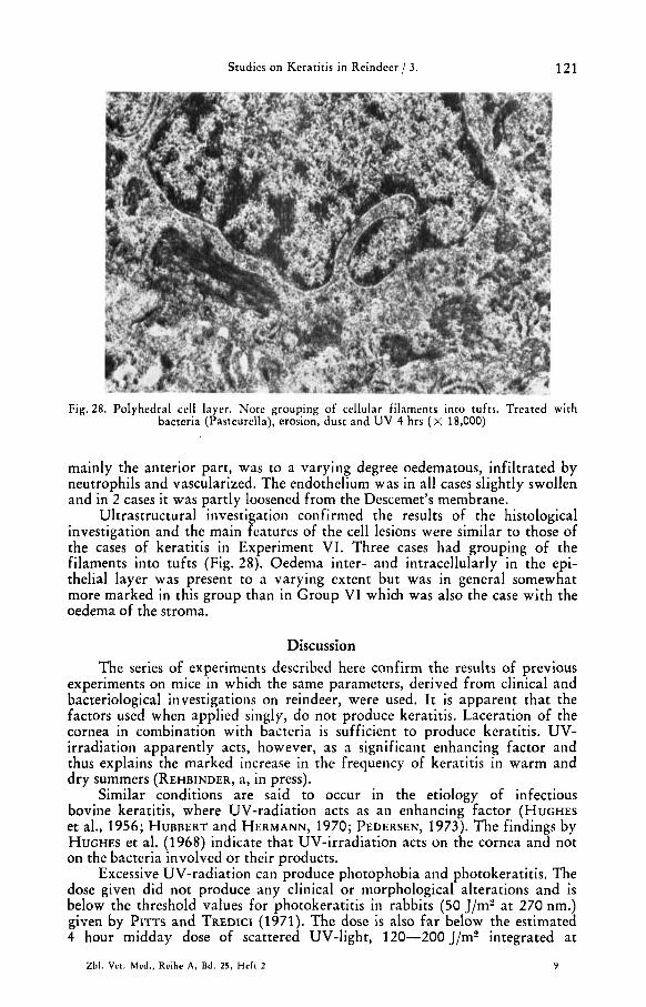
Fig. 28. Polyhedral cell layer. Note grouping of cellular filaments into tufts. Treated with bacteria (Pasreurella), erosion, dust and UV 4 hrs ( x 18,000)
mainly the anterior part, was to a varying degree oedematous, infiltrated by neutrophils and vascularized. The endothelium was in all cases slightly swollen and in 2 cases it was partly loosened from the Descemet's membrane.
Ultrastructural investigation confirmed the results of the histological investigation and the main features of the cell lesions were similar to those of the cases of keratitis in Experiment VI. Three cases had grouping of the filaments into tufts (Fig. 28). Oedema inter- and intracellularly in the epi- thelial layer was present to a varying extent but was in general somewhat more marked in this group than in Group VI which was also the case with the oedema of the stroma.
Discussion The series of experiments described here confirm the results of previous
experiments on mice in which the same parameters, derived from clinical and bacteriological investigations on reindeer, were used. I t is apparent that the factors used when applied singly, do not produce keratitis. Laceration of the cornea in combination with bacteria is sufficient to produce keratitis. UV- irradiation apparently acts, however, as a significant enhancing factor and thus explains the marked increase in the frequency of keratitis in warm and dry summers (REHBINDER, a, in press).
Similar conditions are said to occur in the etiology of infectious bovine keratitis, where UV-radiation acts as an enhancing factor (HUGHES et al., 1956; HUBBERT and HERMANN, 1970; PEDERSEN, 1973). The findings by HUGHES et al. (1968) indicate that UV-irradiation acts on the cornea and not on the bacteria involved or their products.
Excessive UV-radiation can produce photophobia and photokeratitis. The dose given did not produce any clinical or morphological alterations and is below the threshold values for photokeratitis in rabbits (50 J/m' at 270 nm.) given by PITTS and TREDICI (1971). The dose is also far below the estimated 4 hour midday dose of scattered UV-light, 120-200 J/me integrated at
Zbl. Vet. Med., Reihe A, Bd. 25, H e f t 2 9
122 REHBINUER, MOELL, GLATTHARD and OTTOSSON

Studies on Keratitis in Reindeer / 3. 123
270 nm., under natural conditions at 66.5' North and with average cloudiness (cf. paper 1).
Laceration of the cornea is essential if the bacteria are to produce kerati- tis. The circumstances during which forest reindeer are herded apparently give rise to numerous corneal and conjunctival lacerations. These lacerations in naturally occurring cases as well as in these experiments are of different magnitudes and depths, thus providing somewhat different conditions in each case. This may contribute to the examples of healing seen both experimentally and in natural cases (REHBINDER, a, in press; REHBINDER, 1977; REHBINDER and GLATTHARD, 1977).
The bacteria used were obtained from clinical cases of keratitis in rein- deer. In addition, what was considered the normal flora was also included in the experiment (coagulase negative staphylococci, a-haemolytic streptococci and a mixed flora of these bacteria). A similar flora is normally found in other animals and man (CASON and WINKLER, 1954; BISTNER et al., 1969; OJO et al., 1972; FAHMY et al., 1974; REHBINDER and TSCHAPPAT, 1974).
Normal flora has been connected with clinical cases of keratitis in rein- deer (REHBINDER and GLATTHARD, 1977). In these experiments it was not possible to produce a single case of keratitis with the normal flora as the sole etiological agent. In several cases of experimental keratitis it was found that the primary etiological agent e. g. Pasteurella, was not recovered but that instead a growth of what in these experiments has been considered as the normal flora was present. The well established normal flora is probably not affected to the same extent by the local defence reaction as are the inoculated bacteria. It seems probable that the same circumstances prevail among the freely living forest reindeer and so the more potentially pathogenic bacteria may play a more important role than the results from examinations of field cases by REHBINDER and GLATTHARD (1977) indicate. The effect of dust is probably mostly on the conjunctiva, making it more suitable for the inocula- ted bacteria. In field cases, however, most foreign bodies are much larger (REHBINDER, a, in press), and must be considered as one of the factors that also produce corneal laceration.
It is possible that the faster development of keratitis in Expt. VI com- pared to Expt. VII was due to stress. The animals were all in their first experiment, prior to which they had been exposed to severe transportation stress. The animals of Expt. VII in contrast had been handled during a much longer period and several had been used in one or two previous experiments. Most of these animals had become very tame and submitted to the treatment readily.
The histological and ultrastructural investigations revealed a non-specific purulent keratitis, in many cases ulcerating. The only somewhat specific feature was a grouping of cytoplasmic filaments into tufts. This was seen in 5 of 15 experimentally induced cases of keratitis in reindeer (Fig. 28) and also in experimentally produced keratitis in mice. The same observation was made on spontaneous cases of keratitis in reindeer by WINQVIST and REH- BINDER (1973). These observations are discussed in paper 2 in connection with the findings in mice and thereby suggested not to be pathognomonic of UV induced cases of keratitis. Also discussed in paper 2 is the significance of corneal lacerations.
ossible to produce keratitis experimentally in reindeer with
tions. The experimentally produced cases of keratitis are indistinguishable
It has been factors derived P rom clinical, epizootiological and bacteriological investiga-
9.
124 REHBINDER, MOELL, GLATTHARD and OTTOSSON
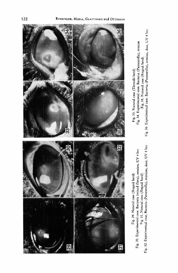
clinically from natural cases (Figs. 29-36) as are the histological and ultra- structural pictures (WINQVIST and REHBINDER, 1973) (Figs. 24-28).
The experimental hypotheses had been tested earlier in an experimental model in mice. The design of the reindeer experiment was derived from experience obtained from this mouse model. The results of the two experi- ments showed a very good correlation.
The herding conditions of the forest reindeer must be considered as the main cause of corneal and conjunctival lesions (REHBINDER, a, in press) as well as the reason for the presence of a heavy load of bacteria on the eye. In addition, UV-irradiation acts as an enhancing factor which may explain the increase of the disease in hot dry summers.
Summary The experimental procedure was in many regards a reproduction of
large-scale investigations on mice. The factors studied were different bacteria from clinical cases of keratitis in reindeer, traumatic erosions of the cornea, dust and ultraviolet (UV) irradiation (40 J/m2 given during 4 hours). The dose of UV irradiation is below the estimated dose of natural UV-light that would reach the eyes of free-living animals at the Arctic Circle, and below the threshold values for photoophthalmia reported for rabbits (50 J/m2).
In this investigation, as in the large-scale investigation on mice, it was found that each parameter by itself did not cause keratitis. When erosion of the cornea and different bacteria were combined, one eye out of 8 developed clinically manifest keratitis. When erosions and bacteria were combined with UV-irradiation, 6 of 8 eyes developed clinically manifest keratitis and when erosion, bacteria and dust were combined with UV-irradiation, 7 of 8 eyes developed clinically manifest keratitis. The clinical investigations were com- plemented by bacteriological, histological and ultrastructural investigations.
It was concluded that natural UV-radiation may act as an enhancing factor in the development of keratitis in reindeer when the cornea is affected by laceration and concomitant bacterial infection.
The regularly appearing keratitis of forest reindeer has to be considered to be a result primarily of the herding conditions which cause corneal and conjunctival lesions and an abundance of bacteria in the conjunctival sac, which later cooperate as etiological factors in the development of keratitis. Natural UV-radiation will act as an enhancing factor which could explain the markedly higher frequency of keratitis cases in dry hot summers.
Acknowledgement Our sincere thanks are expressed to Professor AGNAR NILSSON, who when
Head of Department of Radiobiology, National Defence Research Institute, gave us the opportunity to work in the department and helped us in the early technical work.
This investigation was supported by grants from Samefonden and Anslaget for framjande av ograduerade forskares vetenskapliga verksamhet vid Veterinarhogskolan.

Studies on Keratitis in Reindeer / 3. 125
Zusammenfassung Untersuchungen iiber die Keratitis beim Rentier
3. Teil: Experimentell induzierte Keratitis beim Rentier Die Methodik glich in mancher Hinsicht derjenigen, welche bei den
Mauseexperimenten zur Anwendung kam (vgl. 2. Teil dieser Arbeit). Als Aus- loserfaktoren wurden untersucht : 1. verschiedene Bakterienarten, welche bei klinischen Fallen von Rentierkeratitiden isoliert wurden, 2. traumatische Ero- sionen der Kornea, 3. Staubapplikation, 4. UV-Bestrahlung (40 J/m2 wahrend 4 Stunden). Die UV-Dosis war geringer als die geschatzte Dosis der naturlichen UV-Strahlung, welche die Augen frei lebender Tiere im Gebiet des nordlichen Polarkreises erreicht, sie lag auch unter dem Schwellenwert fur die Auslosung einer Bestrahlungs-Ophthalmie bei Kaninchen (50 J/m2).
In Analogie zu den im groi3en Maastab durchgefuhrten Untersuchungen an Mausen zeigten auch diese Versuche, dai3 eine Noxe allein keine Keratitis erzeugt. Wurden Korneaerosionen mit Bakterieninokulationen kombiniert, so trat in einem von acht Augen eine klinisch manifeste Keratitis auf. Wurden Korneaerosionen mit Bakterieninokulationen kombiniert und zusatzlih eine UV-Bestrahlung durchgefuhrt, so entwickelten sich bei 6 von 8 Augen klinisch manifeste Keratitiden. Wurden Korneaerosionen mit Bakterieninokulationen und Staubapplikationen kombiniert und zusatzlich eine UV-Bestrahlung durchgefuhrt, so entwickelten sich bei 7 von 8 Augen Keratitiden. Die klini- schen Untersuhungen wurden durch bakteriologische, histologische und elek- tronenoptische erganzt.
Folgerungen: UV-Bestrahlung verstarkt die Keratitisentwicklung beim Rentier, wenn ein Trauma vorliegt, und die Kornea bakteriell infiziert wird.
Die regelmaaig auftretenden Keratitiden beim Rentier sind die Folge der Verhaltnisse in der Herde, welche zu Verletzungen der Kornea und Konjunk- tiva sowie einer reichlichen Bakterienbesiedlung des Konjunktivalsackes Anlai3 geben. Diese bilden gemeinsam die atiologischen Faktoren der Keratitis. Die natiirliche UV-Bestrahlung ist ein Verstarkungsfaktor, welcher das haufigere Auftreten von Keratitiden in trockenen und heii3en Sommern erklaren durfte.
Resume Recherches sur la KCratite chez le renne
Troisikme partie: KCratite experimentale induite chez le renne La mkthode resemble en beaucoup de points A celle utilisCe dans
I’expkrimentation sur les souris (voir deuxieme partie). Les facteurs favorisants examinks furent les suivants: 1. Diffkrentes esphces bactkriennes isolkes lors de cas cliniques de Kkratite chez le renne. 2. Erosions traumatiques de la cornke. 3. Application de poussiilre 4. Rayonnement UV (40 J/m2 durant 4 heures). La dose d’UV fut plus faible que celle du rayonnement U V nature1 qui atteint les animaux sauvages des rkgions du cercle polaire arctique et se situe au- dessous des valeurs nkcessaires pour dkclencher une ophtalmie A rayonnement chez le lapin (50 J/mz).
En analogie avec I’expkrimentation de masse sur les souris, ces recherches ont Cgalement montrk qu’un seul facteur n’induit pas une Kkratite. Des krosions de la cornke avec inoculations de bactkries ont montrk une Kkratite clinique manifeste chez un oeil sur huit. Des krosions de la cornke combinkes A des inoculations bactkriennes avec un rayonnement aux UV ont dkveloppk des Kkratites cliniques manifestes chez six yeux sur huit. Sept yeux sur huit ont developpk une Kkratite lors d’krosions de la cornke combinkes avec des inoculations bactkriennes, des applications de poussiere et un rayonnement
126 REHBINDER, MOELL, GLATTHARD and OTTOSSON
aux UV. Les examens cliniques furent complktks par des analyses bactkrio- logiques, histologiques et klectro-optiques.
Conclusions: Un rayonnement aux UV augmente le dkveloppement de KCratite chez le renne s'il y a traumatisme et infection bactkrienne de la cornke.
Les Kkratites frkquentes du renne sont la conskquence de conditions dans le troupeau qui occasionnent des blessures de la cornke et de la conjonctive ainsi qu'une r i h e colonisation bactkrienne du sac conjonctival. Ces facteurs forment ensemble des facteurs Ctiologiques de la Kkratite. Le rayonnement nature1 des UV est un facteur supplkmentaire qui devrait expliquer l'appari- tion plus frkquente des Kkratites durant les CtCs chauds et secs.
Resumen Estudios sobre la queratitis en el reno
3. Queratitis experimental inducida en el reno La tkcnica se asemejaba mucho en varios aspectos a la empleada en 10s
experimentos con ratones (vkase la parte 2" de este trabajo). Como factores desencadenantes se examinaron: I . varias especies bacterianas, aisladas en casos clinicos de queratitis de renos, 2. erosiones traumiticas en la cbrnea, 3. aplicaci6n de polvo, 4. radiaci6n UV (40 J/m2 durante 4 horas). La dosis UV era menor que la dosis de radiacibn UV natural calculada que alcanza 10s ojos de animales de vida libre en la regibn del circulo polar Artico y tambiin menor que 10s valores dinteles necesarios para desencadenar una oftalmia por radiacibn en conejos (50 J/m').
En analogia con 10s experimentos realizados a escala grande en ratones, estos ensayos tambikn evidenciaron que un solo factor no induce ninguna queratitis. A1 combinar erosiones en la cbrnea con inoculaciones de bacterias apareci6 una queratitis clinica manifiesta en uno de cada o h 0 ojos. Cuando se combinaron erosiones de c6rnea con inoculaciones de bacterias y se realizb una irradiacibn UV aditiva se desarrollaron queratitis clinicas manifiestas en 6 de 8 ojos. Si se combinaban erosiones de c6rnea con inoculaciones de bacterias y aplicaciones de polvo y se llevb a cab0 una radiacibn UV adicional se desa- rrollaron queratitis en 7 de 8 ojos. Los exAmenes clinicos fueron completados por otros bacteriol6gicos, histopatolbgicos y electrbnicobpticos.
Conclusiones: la irradiacibn UV refuerza el desarrollo de queratitis en el reno cuando la cbrnea se hallaba afectada por traumatismo e infeccibn bac- teriana.
Las queratitis que aparecen con regularidad en el reno son consecuencia de las circunstancias en la tropa, las cuales motivan lesiones en la c6rnea y con- juntiva asi como una cuantiosa colonizacibn bacteriana del sac0 conjuntival. Las mismas forman, en conjunto, 10s factores etiolbgicos de la queratitis. La irradiacibn UV natural es un factor de refuerzo que podria explicar la apari- cibn mis frecuente de queratitis durante 10s veranos secos y calurosos.
References BASS, A. N., 1948: Short wave cut-off filters for the ultraviolet radiation. J. Optical SOC.
BERGMAN, A., 1912: Smittosam hornhinneinflaniniation, keratitis infectiosa, hos ren. (Con-
BISTNER, S. J., S. R. ROBERTS, and R. P. ANDERSON, 1969: Conjunctival bacteria. Mod. Vet.
CASON, L. M. A., and C. H. WINKLER, 1954: Bacteriology of the eye. 1 . Normal flora. Arch.
COGAN, D. G., and V. E. KINSEY, 1946: Action spectrum of keratitis produced by ultrn-
Am. 38, 977-979.
tagious keratitis in reindeer). Skand. Vet.-T. 2 , 145-154, 177-207.
Pract. 50, 45-47.
Ophtal. 5 1 , 196-199.
violet radiation. Arch. Ophthal. 35, 670-677.
Studies on Keratitis in Reindeer / 3. 127
COGAN, D. G., and V. E. KINSEY, 1956: Ophthalmic ultraviolet action spectra. Amer. J.
DAVE, J. V., and P. M. F U R U K A W A , 1966: Scattered radiation in the ozone absorption bands
DUKE-ELDER, W. S., 1926: Pathological action of light upon the eyes. Lancet, I ,
DUKE-ELDER, W. S., 1954: Textbook of Ophthalmology. St. Louis, C. V. Mosby CO. 6 ,
DUKE-ELDER, W. S., 1972: System of Ophthalmology. vol. XIV. Henry Kimpton, London. EHLERS, N., 1965: The precorncal film. Bioniicroscopical, histological and chemical in-
vestigations. Thesis, Acta ophthal. Copenhagen, Suppl. 8, pp. 136. ENEQVIST, N., 1956: Renens sjukdomar (Diseases of reindeer). Lantbrukets djurbok, Stock-
holm, 143-150. FAHMY, J. A., S. M B L L E R , and M. WEISS BENTZON, 1974: Bacterial flora of the normal
conjunctiva 1. Topographical distribution. Acta ophthal. 52, 786-800. FRIEDENWALD, J., and W. BUSCHKI., 1944: The influence of some experimental variables 011
the epithelial movements in the healing of corneal wounds. J. Cell and Comp. Physiol.
FUKUYAMA, K., W. L. EPSTEIN, and J. H. EPSTEIN, 1967: Effect of ultraviolet light on K N A
GRAWITZ, P. B., 1958 : Weitere Beitrage zur Physiologie der Keratitis (Further contributions
HADWEN, S., and L. J. PALMER, 1922: Reindeer in Alaska. U. S. Department of Agriculture,
Hagner Universal Photometer, 1974: Instruction manual. B. Hagner AB, Solna, Sweden. HAMERSKI, W., 1971 : Badaniia doiwiadczalne nad zawartokia kwasbw nukleinowych w
rogbwcc poddanej dziat aniu promieni pozafioletowych. (Experimental investigation on the content of nucleinic acids in the cornea subjected to the influence of ultraviolet rays.) Klin. Oczna. 4 1 , 639-642.
HEMMINGSEN, E. A., and E. L. DOUGLAS, 1970: Ultraviolet radiation thresholds for corned injury in antarctic and temperate-zone animals. Comp. Biochem. Physiol. 32,
HOGAN, M. J., and L. E. ZIMMERMAN, 1962: Ophthalmic Pathology. An atlas and textbook. W. B. Saunders Co. 2nd Ed.
HUBEERT, W. T., and G. J. HERMANN, 1970: A winter epizootic of infectious bovine kerato- conjunctivitis. J. Amer. vet. mcd. Ass. 157, 452-454.
HUGHES, D. E., and G . W. PUGH, 1970: A five year study of infectious bovine kerato- conjunctivitis in a beef herd. J. Amer. vet. med. Ass. 157, 443-451.
HUGHES, D. E., G. W. PUGH, and T. J. MCDONALD, 1965: Ultraviolet radiation and Moraxe!l.i bovis in the etiology of bovine infectious keratoconjunctivitis. Amer. J. vet. Res. 26,
HUGHES, D. E., G . W. PUGH Jr.* and T. J. MCDONALI), 1968: Experimental bovine keraco- conjunctivitis caused by sunlamp irradiation and Moraxella bovis infection: Deter- mination of optimal irradiation. Amer. J. vet. Res. 29, 821-827.
KOLLER, L. R., 1952: Ultraviolet Radiation. John Wiley & Sons, N e w York. KUMMENEJE, K. L., 1976: Isolation of Neisscria ovis and a Colesiota conjunctivae-like
organism from cases of kerato-conjunctivitis i n reindeer in northern Norway. Acta vet. scand. 17, 107-108.
LATESSA, A. J., and M. H. Ross, 1964: Electron microscope studies of nonpenetrating corneal wounds in the early stages of healing. Exp. Eye Res. 3, 298-303.
LUDWIGH, E., and V. E. KINSEY, 1946: Effect of long ultraviolet radiation on the human eye. Science. 104, 246-247.
MEYER, A. E. H., and E. C. SEITZ, 1942: Ultraviolette Strahlen (Ultraviolet radiation). Wal- ter de Gruyter &i Co., Berlin.
NIX, T. E., R. E. NORDQUIST, and M. A. EVERETT, 1965: Ultrastructural changes induced by ultraviolet light in human epidermis: Granular and transitional cell layers. J. Ultra- structure Kes. 12, 547-573.
NORDQVIST, M., 1960: Renens sjukdomar. Kort oversikt. (Diseases of the reindeer. Short survey). Lappvasendet - Renforskningen. Smiskrif t 4.
OJO, M. O., T. T. ISOUN, and D. H. HILL, 1972: Avian kerato-conjunctivitis in Nigeria. Trop. Anim. Hlth. Prod. 4 , 156-159.
PAINTER, R. B., 1970: The action of ultraviolet light on mammalian cells. Photophys. 5 ,
PEDERSEN, K. B., 1973: Infectious kerato-conjunctivitis in cattle. Thesis. Royal Veterinary
Ophthal. 4 1 , 969-975.
a t selected levels of terrestrial rayleigh atmosphere. Meteor. Mon. 7, 1-353.
1137-1140.
pp. 1196.
23,95-107.
and protein synthesis on differentiated epidermal cells. Nature. 216, 1031-1032.
on the physiology of keratitis). Alb. v. G r a d e s Arch. Ophthal. 159, 459-485.
Bulletin No. 1089, Washington.
593-600.
1331-1338.
169-189.
and Agricultural University. Copenhagen, pp. 156.

128 REHBINDER, MOELL, GLATTHARD and OTTOSSON
PITTS, D. G., J. E. PRINCE, W. J. BUTCHER, K. R. KAY, R. W. BOWMAN, H. W. CASEY, D. G. RICHEY, L. H. MORI, J. E. STRONG, and T. J. TREDICI, 1969: The effects of ultraviolet radiation on the eye. U. S. A. F. School of Aerospace Medicine. Aerospace medical division (A. F. S. C.) Brook Air Force Base, Texas. 1-120.
PITTS, D. G., and T. J. TREDICI, 1971: The effects of ultraviolet on the eye. Amer. Industr.
PULHORN, G., und H.-J. THIEL, 1974: Ultrastrukturelle Untersuchungen zur Regeneration der Basalmembran des Hornhautsepithels. (Electron microscope investigation in basal mem- brane regeneration of epithelium.) Alb. v. Graefes Arch. 0 hthal. 189, 21-32.
in the eye of reindeer and their relation to keratitis in this animal. Acta. vet. scand. I f , 338-339.
KEHBINDER, C., 1977: Keratitis in reindeer. Relation to the presence of 1st instar larvae of the nostril fly (Cephenomyia trompe L) in the conjunctival sac and to natural ultra- violet radiation. Acta vet. scand. 18, 75-85.
Hyg. ASS. J. 32, 235-246.
REHBINDER, C., 1970: Observation on 1st instar larvae of nostri P fly (Cephenomyia trompe L)
REHBINDER, C.: Clinical and epizootiological studies on keratitis in reindeer (a, in press). REHBINDER, C.: Fine structure of the mouse cornea (b, in press). REHBINDER, C., and V. TSCHAPPAT, 1974: Pasteurella pneuniotropica, isoliert von der Kon-
junktivalschleimhaut gesunder Laboratoriumsmiiuse. (Pasteurella pnrimotropica isolated from the conjunctival sac of healthy laboratory mice.) 2. Versuchstierk. 16, 359-365.
REHBINDER, C., and V. GLATTHAKD, 1977: Keratitis in reindeer. Relation to bacterial infec- tions. Acta vet. s c a d 18, 54-64.
ROBINSON, N., 1966: Solar Radiation. Elsevier Publ. Co., Amsterdam. SAUNDERS, L. Z., 1971: Pathology of the eye of domestic animals. Verlag Paul Parey, Berlin. SCHOLANDER, P. F., L. IRVING, E. A. HEMMINGSEN, and E. BRADSTREET, 1969: Ultraviolet
absorption in the cornea of arctic and alpine animals. I n : F. URBACH (ed.): The Biologic Effects of Ultraviolet Radiation, Pergamon Press, N e w York, 469-471.
Schott und Genossen, 1970, Mainz, Germany. Filter catalogue. SCHULTZE, R., and K. GRAw, 1969: Consideration of sky ultraviolet radiation in the measure-
ment of solar ultraviolet radiation. I n : 1:. URBACH (ed.): The Biologic Effects of Ultraviolet Radiation, Pcrgamon Press, N e w York, 359-373.
SCOTTO, J., T. R. FEARS, and G. B. GORI, 1976: Ultraviolet exposure patterns. Environmental Research, 12, 228-237.
VERHOEFF, F. H., L. BELL, and C. B. WALKER, 1916: The pathological effects of radiant energy on the eye. Proc. Anier. Acad. Arts. Sci. I f , 629-818.
WIER, K. A., K. FUKUYAMA, and \V. L. ErsrEiN, 1971: Nuclear changes during ultraviolet light induced depression of ribonucleic acid and protein synthesis in human epidermis. Lab. Invest. 21, 451-456.
WINQVIST, G., and C. K E I ~ B I N L I K R , 1973: Fine structure of the reindeer cornea in normal conditions and in keratitis. Acta vet. scand. 14, 292-300.
YOUNG, C., and A. H I L L , 1974: Conjunctivitis in a colony of rats. Lab. anim. 8, 301-304. ZIGMAN, S., J. GROFF, T. YUOLO, and T. VAUGHAN, 1975: The response of mouse ocular
tissues to continuous near-UV light exposure. Invest. Ophthalm. 14, 710-713.
Authors' addresses: CLAES REHBINDER, Swedish University of Agricultural Sciences, College of veterinary Medicine, Department of Pathology, Clinical Centre, Ultuna, S-750 07 Uppsala 7, Sweden. FREDRIK MOELL, National Defence Research Institute, P. 0. Box 416, $172 04 Sundbyberg, Sweckii. VERENA GLATTHARD, National Veterinary Institute, Depart- ment of Bacteriology, FACK, s-104 05 Stockholm 50, Sweden. ALLAN OTTOSSON, Royal Institute of Technology, Department of Electric Power Systems Engineering Lightning Laboratory, FACK, S-I 00 44 Stockholm, Sweden. PER LINDIJERG, National Defence Research Institute, FACK, S-104 50 Stockholm, Sweden.
Code for Tables 1-7
Treatment Past, = Pasteurella rnultocida Mixed bacteria = Streptococci + Staphylococci Clinical signs; hrs post treatment 0 = no reaction (1) = minor serous conjunctivitis 1 = serous conjunctivitis (2) = minor mucopurulent conjunctivitis 2 = mucopurulent conjunctivitis (3) = minor mucopurulent conjunctivitis and photophobia 3 = Mucopurulent conjunctivitis and photophobia (k) = keratitis
K = keratitis (X) = minor erosion Bacteria given; hrs post treutment Neg. = no growth
~ = normal flora P = Pasteurella multocida M = Mixed bacteria Str = Streptococci Sta = Staphylococci 1 = slight growth 2 = moderate rowth 3 = rich growt i