structure two pectate lyase genes erwinia chrysanthemi ec16
TRANSCRIPT
Vol. 168, No. 2JOURNAL OF BACTERIOLOGY, Nov. 1986, P. 595-6060021-9193/86/110595-12$02.00/0Copyright © 1986, American Society for Microbiology
Structure of Two Pectate Lyase Genes from Erwinia chrysanthemiEC16 and Their High-Level Expression in Escherichia coli
N. T. KEEN* AND S. TAMAKIDepartment of Plant Pathology, University of California, Riverside, California 92521
Received 12 May 1986/Accepted 8 July 1986
The pelB and pelE genes from Erwinia chrysanthemi EC16, which encode different pectate lyase enzymes,were sequenced and expressed at a high level in Escherichia coli. The genes possessed little similarity to eachother in 5' signal regions, signal peptide sequences, coding sequences, or 3' noncoding regions. Both genescontained their own promoters as well as sequences 3' to the coding regions with considerable secondarystructure which may function as rho-independent transcriptional termination signals. High-level expressionplasmids were constructed with both genes, which led to 20% or more of E. coli cellular protein. The pectatelyases were secreted efficiently to the periplasm and, to ra lesser extent, the culture medium. The matureproteins in E. coli periplasmic fractions were obtained in milligram amounts and high purity with asingle-column affinity purification method. E. coli cells which produced high amounts of the pelE proteinmacerated potato tuber tissue as efficiently as E. chrysanthemi EC16 cells but cells producing high amounts ofthe pelB protein were less effective. Thus, the pelE gene product is an important pathogenicity factor whichsolely enables E. coli to cause a soft-rot disease on potato tuber tissue under laboratory conditions.
We cloned genes coding for two different pectate lyase(EC 4.2.2.2) enzymes from the phytopathogenic bacteriumErwinia chrysanthemi EC16 (13) and observed their expres-sion in Escherichia coli. Pectate lyases have previously beenshown to account largely or entirely for the maceration orsoft rotting of plant tissue caused by Erwinia spp. (4).Confirming this, E. coli cells containing the cloned pectatelyase genes macerated plant tissue, albeit less efficiently thanE. chrysanthemi (13). Several groups subsequently clonedsimilar genes from other strains of E. chrysanthemi (5, 14,28, 34) and the related bacterium Erwinia carotovora (18, 29,40). The genes that we cloned did not cross-hybridize (13),but coded for enzymes with similar physical properties(molecular weights of ca. 40,000 and isoelectric points of 8.8and 9.8) which both catalyzed the random eliminative cleav-age of sodium polypectate. These enzymes were efficientlysecreted to the periplasm and, to a lesser extent, the culturemedium of E. coli. For reasons discussed below, the clonedDNA fragments appear to contain pelB and pelE, describedby others, and the mature proteins which they encode arePLb and PLe, respectively. Plasmids containing our pelgenes were named pPL in the previous paper (13), but thisdesignation was found to be already entered in the PlasmidReference Center (17). Accordingly, our pel gene plasmidconstructs have been renamed pPEL, a designation that wehave registered in the Plasmid Reference Center (17).The cloned pelB and pelE genes were both regulated by
catabolite repression in E. coli (13) but were not induced bysodium polypectate, as occurs in E. chrysanthemi (4). Due totheir pathogenic importance in diseases caused by Erwiniaspp. and to their regulation properties, we elected to furtherstudy the cloned pel genes coding for the pectate lyaseenzymes. Since multigene families coding for functionallysimilar proteins are uncommon in procaryotes, it was also ofinterest to compare the structures of the two genes. Thispaper presents the further subcloning and sequencing ofthese genes, their high-level expression in E. coli, and the
* Corresponding author.
demonstration that their high-level expression enables E.coli cells to efficiently macerate plant tissue.
MATERIALS AND METHODS
Purification and assay of pectate lyases. E. coli cells (usu-ally HB101 or JA-221; Table 1) containing various expres-sion plasmids were grown into the stationary phase for ca. 16h at 28°C, usually in 15 ml of L broth with 50 jig of ampicillinper ml in 50-ml shaken DeLong flasks. These bacteria werealso grown in the same way on M9 or M9CA medium (19)with glucose at 2 mg ml-1, thiamine hydrochloride at 2 ,ugml- 1, and proline, tryptophan, and le4cine all at 20 p,g ml-.Cultures received isopropyl-p-p-thiogalactopyranoside(IPTG) at 1 mM either when initiated or after attaining anabsorbance at 600 nm of 0.5 to 0.7. The resultant cells (ca.0.12 g per flask) were harvested by centrifugation, washedwith 0.2 M Tris hydrochloride (pH 8.0), and induced to formspheroplasts by the method of Witholt et al. (37), resulting ina final periplasmic fraction of 16 ml from each culture. Theperiplasmic fractions, culture fluids, and cellular fractionswere assayed as described previously (13).For purification of the pectate lyases, periplasmic frac-
tions were dialyzed extensively against 5 mM Tris hydro-chloride (pH 8.0), containing 0.1 mM CaCl2 at 4°C andcentrifuged at 20,000 x g for 10 min to remove traces ofinsoluble material. Pectate lyase activity was easily purifiedto near homogeneity by a modification of techniques basedon the affinity of pectate lyases for agarose gel matrices (7).The dialyzed periplasmic fraction resulting from eight cul-ture flasks (- 120 ml) was pumped through a 1.5 by 7.0-cmcolumn of Bio-Rad A-1.5m or Bio-Rad CM Bio-Gel at ca. 3ml min- at roomn temperature. The columns were washedwith 0.1 mM CaCl24 mM Tris hydrochloride (pH 8.0) andthen eluted with a linear gradient composed of 60 ml of 5 mMTris hydrochloride (pH 8.0) and 60 ml of 0.2 M NaCl in thesame buffer. In later experiments the columns were bulkeluted with 0.2 M NaCl in the Tris hydrochloride buffer torelease the enzymes. Fractions (3 ml) were collected and
595
Dow
nloa
ded
from
http
s://j
ourn
als.
asm
.org
/jour
nal/j
b on
10
Nov
embe
r 20
21 b
y 12
3.24
0.23
5.68
.
596 KEEN AND TAMAKI
TABLE 1. Plasmids and bacterial strains employed
Plasmid or strain Genotype or Description Source or Reference
E. coliHB101
JA-221
RB791C600706M5219
F- hsdS20 (hsdR hsdM) recA13 ara-14 proA2 lacYl galK2 rpsL20(StrW) xyl-S mtl-1 supE44 X-
hsdR AtrpE5 leuB6 lacY recA thi F'(lacIq lacZ+ lacY+ lacA +proA+ proB+)
W3110 lacIqL8F- thr-I leuB6 thi-) supE44 lacY1 flzuA2J X-F- proA thr leu argH his lac phoSt rpsL lky-207Stri lacZ(Am) trp(Am) (A bio252 cI857 AHi)
E. chrysanthemi EC16
(19)
(20)
(1)(19)(16)(27)
E. Chatterjee
PlasmidspINI11113 A-1 andA-2pCQV2pKC30pPLc2820pUC8pUC18 and pUC19pJRD158pINK-1pPEL3pPEL34
pPEL342pPEL343pPEL344
pPEL7pPEL74
pPEL742pPEL7421pPEL7422pPEL743pPEL743JpPEL746pPEL711pPEL712pPEL747pPEL748pPEL749
(20)
Deletion of PstI site from pPLc2819
Expression vector for transcriptional fusionsCosmid clone encoding PLb and PLc; formerly pPL36.6-kb PstI fragment from pPEL3 coding for PLb only; formerlypPL34
2.0-kb KpnI-ScaI fragment from pPEL34 cloned in pUC182.0-kb SstI-HindIII fragment from pPEL342 cloned in pINK-11.3-kb DraI-HindIII fragment from pPEL342 cloned into theSmaI-HindIII sites of pINK-1
Cosmid clone encoding PLa and PLe; formerly pPL78.2-kb PstI fragment from pPEL7 encoding for PLa and PLe;
formerly pPL741.75-kb EcoRI fragment from pPEL74 encoding PLe only1.2-kb EcoRI-SalI fragment from pPEL742, cloned in pUC81.2-kb EcoRI-SalI fragment cloned in pUC192.0-kb HindIII-SalI fragment from pPEL74 cloned in pUC192.0-kb HindIII-SalI fragment from pPEL74 cloned in PJRD1581.2-kb EcoRI-HindIII fragment from pPEL7421 cloned in pINK-1pPEL7421 with 92-base-pair internal BclI fragment deletedpPEL7421 with 361-base-pair internal EcoRV fragment deletedPectate lyase-negative translational fusion of pelEMutant pectate lyase-positive translational fusion of pelEPectate lyase-positive translational fusion of pelE
(26)(31)(27; Remaut, unpublished data)(38)(38)(8)This paper(13)(13)
This paperThis paperThis paper
(13)(13)
(13)This paperThis paperThis paperThis paperThis paperThis paperThis paperFig. 4, this paperFig. 4, this paperFig. 4, this paper
assayed for pectate lyase activity (13) and absorbance at 280nm. Peak fractions were pooled, dialyzed against distilledwater at 4°C, and lyophilized. The white, fluffy enzymeswere stored dry at - 20°C. Care was taken to use dialysistubing with 6,000- to 8,000-molecular-weight cutoff in theabove steps, since tubing with a 12,000- to 14,000-molecular-weight cutoff resulted in significant loss of the 9.8 pectatelyase.
Electrophoresis of whole cell proteins. Cells of E. coliHB101 containing desired plasmids were grown for variousperiods at 28°C on 15 ml of L broth containing 50 tag ofampicillin per ml; some cultures received 1 mM IPTG atvarious times. The cultures were centrifuged, and the result-ant cells (ca. 0.10 to 0.15 g, fresh weight) were washed oncewith 10 ml of 0.01 M Tris hydrochloride (pH 7.5). The pelletswere then suspended in 0.25 ml of water, an equal volume of2.5 x Laemmli sample solution (15) was added, and thesamples were boiled for 5 min. Samples were then electro-phoresed directly on 10% sodium dodecyl sulfate(SDS)-polyacrylamide gels (1 mm thick) with a Laemmlibuffer system (15). Protein standards (Sigma Chemical Co.)and purified pectate lyases were prepared and electropho-
resed similarly. The gels were run at 160 V for 4 h at roomtemperature and directly stained with Coomassie blue R250and destained, both as described previously (13).
Plant tissue maceration tests. Cells of E. chrysanthemiEC16 or log-phase E. coli cells carrying desired plasmidswere grown on LB medium, and various cell concentrationswere placed into wells on potato tuber cylinders. Thecylinders (1.4-cm diameter) were cut from store-boughtRusset Burbank potato tubers with a no. 7 cork borer andsliced to 7-mm lengths. A well 3 mm deep and 6 mm widewas made at one end of each cylinder with a no. 2 cork borersuch that 0.1 ml of cell suspension could be added. Thecylinders were then incubated on moistened filter paper inpetri plates at 28°C and observed for maceration at varioustime intervals. Maceration severity was rated on a 0 to 4scale by probing cylinders with a spatula to assess the degreeof softening and tissue disintegration.DNA manipulations. Subcloning and plasmid construc-
tions were generally done by ligating desired DNA fragmentsrecovered from low-melting-point agarose gels by themethod of Crouse et al. (6), except that gelatin was added to100 ,ug ml- 1. End-filling reactions used T4 DNA polymerase
J. BACTERIOL.
Dow
nloa
ded
from
http
s://j
ourn
als.
asm
.org
/jour
nal/j
b on
10
Nov
embe
r 20
21 b
y 12
3.24
0.23
5.68
.
STRUCTURE OF PECTATE LYASE GENES 597
Xba 1, Bam HI, Sma 1, Kpn 1,Sst l,EcoRI, HindfI, Bam HI
Pstln~~~~~Kp I
/z Rp Kpn I
| ~PINK-1
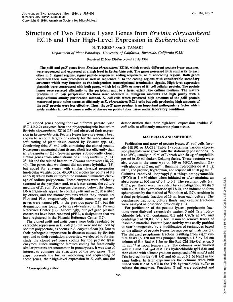
FIG. 1. Restriction map of pINK-1, constructed from PINIII"3A-1 (20), showing polylinker used for insertion of E. chrysanthemipel genes bearing their own translation signals.
(6). Constructs were checked by mini-boil plasmid extrac-tions (6, 13) followed by restriction with the appropriateenzymes. The junction sequences of plasmids producingfusion proteins were confirmed by DNA sequencing asdescribed below. Plasmid constructs were transformed intofrozen, competent cells of E. coli by the method of Morrison(22). Other cloning details were as described previously (13,19).N-terminal amino acid sequencing. The purified mature
PLb or PLe pectate lyases (50 pmol) were dissolved in 0.5%SDS, and microsequencing was performed with an AppliedBiosystems 470A vapor-phase microsequencer. Fifteenamino acid residues were determined for each pectate lyase.DNA sequencing. A series of BAL 31 deletions were
generated from either end of the fragments and sequenced bythe dideoxy chain termination method of Dean et al. (9). Alldata were confirmed by sequencing both strands. Twononcoding regions, both ca. 20 base pairs in length, associ-ated with the insert DNA of pPEL342 could not be deter-mined accurately by this method and were therefore se-quenced by the Maxam and Gilbert method (21). Sequencedata were analyzed by the computer programs of Pustell andKafatos (25) and the BIONET system supplied throughIntelligenetics Corp., Palo Alto, Calif.
RESULTSConstruction of pINK-1. To test the expression of the
subcloned pectate lyase genes in E. coli with E.chrysanthemi translation signals, a plasmid was constructedfrom the lac-regulated expression vector pINIII"13 A-1 (20).Plasmid DNA was restricted at the XbaI and EcoRI sites,and the large fragment was ligated with pUC19 DNA cutwith the same enzymes. The resulting construct containedmost of the pUC19 polylinker region downstream from thetriple lac UV5 promoters of pINIII and was called pINK-1(Fig. 1). It permitted use of the strong regulated promoterswith E. chrysanthemi genes carrying their own translationalsignals and allowed the insertion of DNA fragments withvarious restriction termini. The other advantages of thepINIII vectors, such as transcriptional termination se-quences 3' to the inserted coding region and presence in thevector of the lacIq gene, were retained.
Expression of the peiB gene in E. coli. The 6.6-kilobase (kb)PstI fragment from pPEL34 (13) coding for PLb was further
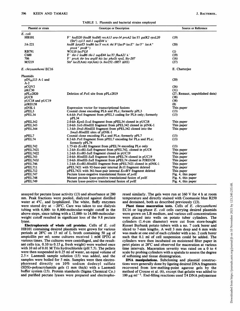
subcloned. Preliminary experiments indicated that the cod-ing region resided on the left-hand side of the fragment, sincea KpnI-EcoRI fragment cloned in pUC18 retained the activ-ity, but a BstEII-PstI fragment cloned in pJRD158 did not. A2.0-kb KpnI-ScaI fragment cloned in pUC18 conferred pec-tate lyase activity in E. coli, but a 1.3-kb KpnI-EcoRVfragment was inactive. Accordingly, the KpnI-ScaI fragmentcloned in pUC18 was retained and designated pPEL342 (Fig.2). This fragment was further mapped, and the additionalrestriction sites are shown in Fig. 2.
Pectate lyase production was high in E. coli cells carryingpPEL342 with or without IPTG (Table 2). However, a smallincrease with IPTG was consistently observed withpPEL342 but not the reverse insert orientation in pUC19(data not shown). The same was true when the 2.0-kbKpnI-ScaI fragment from pPEL342 was inserted into pINK-1 to give pPEL343. These suggestions for the direction oftranscription were later confirmed by DNA sequence data.pPEL344, a pINK-1 construct lacking the E. chrysanthemipromoter regions (see Table 1 and Fig. 2 and 9) led tosomewhat lower constitutive pectate lyase production,which was increased ca. threefold when cells were inducedwith IPTG (Table 2). Unlike the prior constructs, IPTGsupplied at culture initiation severely inhibited the growth ofcells carrying pPEL344.
High-level expression of the pelE gene. The 1.75-kb EcoRIinsert in pPEL742 (13) was further restricted, giving a 1.2-kbEcoRI-SalI fragment that retained PLe activity when clonedin pUC8 to yield pPEL7421 (Table 2). Expression of the1.2-kb insert in pPEL7421 was found to be orientationdependent, since little pectate lyase activity was detectedwhen the 1.2-kb EcoRI-SalI fragment was reversed inpUC19 to yield pPEL7422 (Table 2). To retain the E.chrysanthemi transcription signals of the pI 9.8 pectatelyase, a 2.0-kb HindIII-SalI fragment from pPEL74 wascloned in pUC19 to yield pPEL743. The same fragment wasalso cloned in pJRD158, and the resultant plasmid was calledpPEL743J. Significantly, pPEL743 gave considerably lesspectate lyase activity in HB101 than pPEL7421, and only asmall increase in activity was observed when cells harboringpPEL743 were supplied with IPTG (Table 2). Somewhatmore pectate lyase was produced by HB101 cells carryingpPEL743J, but this was still below the induced level forpPEL7421 and may be attributable to the reported high copynumber of the vector, pJRD158 (8). These results raise thepossibility that a regulatory DNA element in the 5' noncod-ing region of the pelE gene may be repressing expression inE. coli. In contrast, pPEL7421 lacks most of the 5' DNA andled to an unexpectedly higher uninduced pectate lyase level(Table 2).
High-level expression of the pelE gene was initially at-
0
c a 0 - 0a , -0 C)v 4 a en WI II
w(Ico
0C.)C,)
II
.I I
0 0.5 1.0 1.5 2.0
FIG. 2. Restriction map of a ca. 2.0-kb KpnI-ScaI fragmentisolated from pPEL34 which conferred production of PLb in E. coliHB101. The fragment was cloned into the KpnI and HincII sites ofpUC18, and the resultant construct was called pPEL342. Map unitsare in kilobase pairs. Sequence data subsequently showed thepresence of two closely spaced AvaI-XhoI sites rather than one site.
VOL. 168, 1986
Dow
nloa
ded
from
http
s://j
ourn
als.
asm
.org
/jour
nal/j
b on
10
Nov
embe
r 20
21 b
y 12
3.24
0.23
5.68
.
598 KEEN AND TAMAKI
TABLE 2. Production of pectate lyase by E. coli cells containing various expression plasmidsPectate lyase U per g of cells (% of total activity)c
Plasmida E. coi strain IFTGb Periplasmic Intracellular Extracellular Totalfraction fraction fraction activity
pPEL342 HB101 No 4,500pPEL342 HB101 Yes 6,000pPEL343 HB101 No 7,500 (93) 200 (3) 350 (4) 8,050pPEL343 HB101 Yes 8,060 (93) 160 (2) 490 (5) 8,710pPEL344 HB101 No 3,100 30pPEL344 HB101 Yes, 7 h 8,900 55pPEL7421 HB101 No 310 (84) 20 (5) 40 (11) 370pPEL7421 HB101 Yes 1,750 (93) 80 (4) 50 (3) 1,880pPEL7422 HB101 Yes 55 (92) <2 5 (8) 60pPEL711 HB101 Yes <0.2 <1 <0.2 <1pPEL712 HB101 Yes <0.2 <1 <0.2 <1pPEL743 HB101 No 90 (88) 5 (5) 7 (7) 102pPEL743 HB101 Yes 130 (86) 10 (7) 11 (7) 151pPEL743J HB101 No 520pPEL746 RB791 No 1,650 (88) 90 (5) 30 (7) 1,870pPEL746 RB791 Yes 4,360 (95) 170 (4) 60 (1) 4,590pPEL746 JA-221 No 4,830pPEL746 JA-221 Yes 8,750pPEL746 HB101 No 1,700 (90) 135 (7) 55 (3) 1,890pPEL746 HB101 Yes 6,400 (94) 220 (3) 200 (3) 6,820pPEL746 706 No 146 (32) 50 (11) 258 (57) 454pPEL746 706 Yes 245 (7) 85 (3) 2,890 (90) 3,220pPEL747 HB101 Yes <0.2 <1 <0.2 <1pPEL748 HB101 No 10,500 (91) 1,030 (9) 55 (<1) 11,585pPEL748 HB101 Yes, 7 h 12,000 (89) 450 (3) 1,100 (8) 13,550pPEL748 JA-221 No 3,280 (44) 600 (8) 3,630 (48) 7,510pPEL748 JA-221 Yes, 5 h 6,780 (51) 720 (6) 5,590 (43) 13,090pPEL749 HB101 No 63pPEL749 HB101 Yes 325 (86) 20 (5) 35 (9) 380
a pPEL342, 343, and 344 all contain the pelB gene; all other plasmids contain pelE.b Unless otherwise noted, 1 mM IPTG was added at culture initiation; since IPTG severely inhibited the growth of cells containing pPEL344 or pPEL748, it
was added after the noted culture times when cell density was between 0.5 and 0.7 at 600 nm; cultures were then harvested 5 or 7 h later. The fully induced levelof total pectate lyase activity produced by E. chrysanthemi EC16 was about 500 U per g of cells (13).
c All cultures harvested after growth at 28°C on 15 ml of medium; growth was for 14 to 18 h in all cases except pPEL344 or pPEL748, where growth was forca. 12 h; cell fractions were prepared and assayed as described in Materials and Methods.
tempted with several vectors carrying temperature-regulatedphage lambda promoters (Table 1), but these constructsresulted in lower pectate lyase activity in E. coli at 37 and42°C (100 to 200 U per g of cells) than at 32°C (200 to 600 Ug 1). To test whether the temperature dependence of en-zyme production resulted from the use of lambda promotervectors or was peculiar to the particular gene, the lac-regulated pPEL746 was examined. At lower temperatures,cells with pPEL746 produced substantially more pectatelyase in the periplasmic fraction and responded to inductionby IPTG (Table 3). At 32 and 37°C, however, a largereduction in PLe activity was observed, and production wasnot increased by IPTG induction. Production of PLb di-rected by pPEL343 was also less at 37°C than at 28°C, butthe decrease was much less than for PLe, and IPTG in-creased production of PLb at all temperatures (Table 3). Thedecreased expression of pelE at 37°C therefore appeared toexplain the poor performance of constructs utilizing lambdapromoters. Further expression experiments were accord-ingly conducted with lac promoter vectors in cells grown at280C.
Strain HB101 containing pPEL746 produced considerablyhigher PLe levels at 28°C than the comparable construct inpUC8, pPEL7421 (Table 2). Most of the pectate lyaseactivity encoded by pPEL746 occurred in the periplasmicfraction, even when IPTG was supplied. Strain JA-221containing pPEL746 produced higher levels of pectate lyasethan HB101, RB791, or 706. However, the lac repressor-
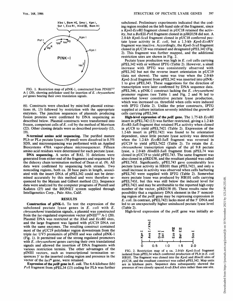
overproducing JA-221 and RB791 strains did not yield lowerlevels of pectate lyase in the absence of IPTG than HB101.The periplasmic leaky strain, 706, resulted in extensivesecretion of pectate lyase to the culture medium (Table 2) atall stages of growth. However, total activity was lower thanwith the other tested strains. The increased enzyme produc-tion conferred by pPEL746 in the presence ofIPTG (Table 2,Fig. 3) was expected since the previous results (13) sug-gested that the EcoRI subcloning site was within or 3' to theE. chrysanthemi promoter or the catabolite activator pro-tein-binding site associated with the coding region of pelE.The constitutive level of pectate lyase production directedby pPEL746 in the absence of IPTG was unexpectedly high,however, even in strains RB791 and JA-221, which overpro-duce lac repressor (Table 2). Electrophoresis of whole E.coli cells bearing pPEL746 (Fig. 3) confirmed the highconstitutive and induced expression of PLe but did notreveal a detectable preprotein band at -2 kilodaltons (kDa)above the secreted protein.
Various plasmid constructs were made to test expressionof the pelE gene under the control of vector transcription andtranslation signals. An in-frame fusion with pINIII A-1,pPEL749 (Fig. 4 and 5), produced pectate lyase activity in E.coli as predicted, but levels were relatively low with orwithout IPTG in all cell fractions (Table 2). The reason forthis relatively low expression is not known, although severalamino acid changes at the N terminus of the pectate lyasepreprotein occurred relative to the native preprotein (Fig. 5).
J. BACTERIOL.
Dow
nloa
ded
from
http
s://j
ourn
als.
asm
.org
/jour
nal/j
b on
10
Nov
embe
r 20
21 b
y 12
3.24
0.23
5.68
.

STRUCTURE OF PECTATE LYASE GENES 599
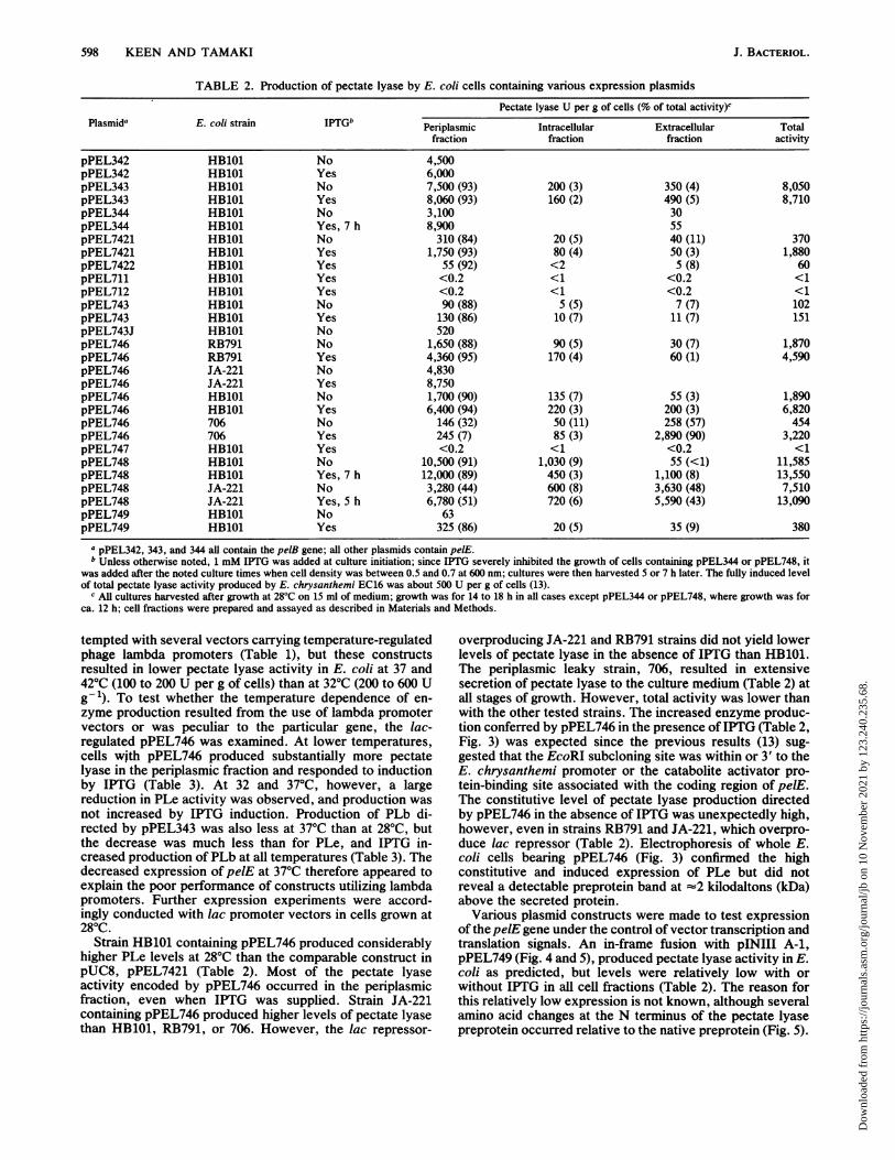
A control pectate lyase-negative construct of the pelE codingsequence was made by fusion with pINIII A-2 (Fig. 4). Ofthe resulting HB101 transformants, 47 were pectate lyasenegative on the pectate plate assay (13) as expected; one ofthese was retained and called pPEL747. One transformant,however, was strongly pectate lyase positive and was alsofound to lack a reconstituted EcoRI site as seen in pPEL747(Fig. 5). The pectate lyase-positive construct was calledpPEL748. Sequencing confirmed that plasmids pPEL747 andpPEL749 had the predicted junction sequences (Fig. 5), butpPEL748 was found to be a mutant plasmid in which a5'-terminal cytosine residue had been lost from the MluI 5'overhang during the T4 polymerase end fill. This accountedfor the loss of the EcoRI site and for the regained pectatelyase activity, since the one-base deletion threw the pectatelyase coding region into frame with the translational startcodon of the vector (Fig. 5). Unexpectedly, pPEL748 led tohigher pectate lyase production in E. coli than any othertested construct (Table 2, Fig. 6); most of the activity wassecreted into the periplasm, even in the presence of IPTG.However, pectate lyase production was also extensive in theabsence of IPTG, and cells exhibited severe growth inhibi-tion and premature lysis when IPTG was supplied at theinitiation of culture growth. Although 95% or more of thepectate lyase activity was secreted into the periplasm andculture medium from IPTG-induced cells with pPEL748(Table 2), electrophoresis of crude lysates from induced cellsdisclosed a putative preprotein band ca. 2 kDa above thesecreted PLe protein (Fig. 6). Scanning of the lanes in Fig. 6indicated that PLe and its preprotein constituted ca. 25%of the total cellular protein in induced cells (data notshown).Maceration of potato tissue. As observed previously (13),
E. coli cells containing only the plasmid vector pINIII113 A-2did not macerate potato tuber cylinders at any tested con-centration. Of the pel constructs, those expressing PLe(pPEL746 and pPEL748) were all more active than pPEL343or pPEL344, which encode PLb (data not shown). ConstructpPL748 in strains JA-221 and HB101 gave the highestmacerating activity, which was comparable to or greaterthan that caused by E. chrysanthemi EC16. The periplasmicleaky strain 706 caused less maceration with plasmidspPEL746 and pPEL748 than strains JA-221 and HB101,despite its more efficient secretion of pectate lyase to theculture medium (Table 2). This may have been due to poorerviability of this strain carrying the high-expression pectatelyase constructs, since it transformed at 1% or less thefrequency of the other E. coli strains. A detailed study of the
TABLE 3. Production of pectate lyase by E. coli HB101 cellscontaining pPEL746 or pPEL343 and grown at various
temperaturesa
Pectate lyase activity (U per g ofPlasmid IPTG fresh cells) at:
250C 280C 32°C 370C
pPEL746 No 1,680 1,600 300 100pPEL746 Yes 5,550 6,100 1,300 50pPEL343 No 5,210 1,760pPEL343 Yes 6,150 2,150
a Cultures were grown for 24 h, ca. 14 h of which was stationary phase;IPTG at 1 mM was supplied at the initiation of culture growth on 15 ml of LBplus 50 ,ug of amplicillin per ml; flasks were shaken at ca. 100 reciprocal cyclesper min; growth was similar in all cases, between 0.11 and 0.14 g (freshweight) cells. Data are for periplasmic fractions only, but other fractionsshowed relative percentage values similar to those in Table 2.
29- mm
1 2 3 4 5 6 7. . -, . .. .......
205--
116- -977- _
66- *
45-4--,.
0. 4*
FIG. 3. SDS-gel electrophoresis of HB101 cells containingpINK-1, pPEL343, or pPEL746 grown with or without IPTG addedat the start of culturing. Cultures were grown for 16 h at 28°C and,after washing, were treated with sample buffer as described inMaterials and Methods, and 5 ,ul was applied to each lane. Lanes: 1,standards; 2, pINK-1 plus IPTG; 3, pPEL746 without IPTG; 4,pPEL746 with IPTG; 5, pPEL343 without IPTG; 6, pPEL343 withIPTG; 7, size standards. Arrows denote mature PLe (upper) andPLb (lower). Sizes of standard proteins in kilodaltons are noted onthe left.
pathogenic properties of E. coli cells carrying the pel expres-sion plasmids will be published elsewhere (Payne, Collmer,and Keen, manuscript in preparation).
Purification of the pectate lyases. HB101 cells harboringpPEL748 or pPEL344 grown at 28°C in the absence of IPTGfor ca. 12 h into the stationary phase were a good source ofPLe or PLb, respectively, for purification. Cells were har-vested, the periplasmic fraction was prepared, and either theaffinity or ion-exchange chromatographic technique de-scribed in Materials and Methods was used to purify theenzymes. Single symmetrical and coincident peaks of pec-tate lyase activity and material absorbing at 280 nm elutedfrom the CM Bio-Gel or Bio-Gel A-1.5m columns with anNaCl gradient (Fig. 7). Application of more than 3 to 5 mg ofthe enzymes overloaded the A-1.5m column, but as much as10 mg adsorbed to the CM Bio-Gel column. Otherwise, thetwo columns performed similarly. Because no significantcontaminating proteins were detected during the gradient,the columns were, after washing, eluted batchwise with 0.2M NaCl in the Tris hydrochloride buffer to preparativelypurify the pectate lyases. The suggestion of high purity notedin the elution profile from the columns (Fig. 7) was confirmedwhen the purified pectate lyase preparations were electro-phoresed on SDS-polyacrylamide gels and when N-terminalamino acid analyses were performed. For instance, a singlepolypeptide which comprised 98% or more of the total wasobserved on SDS gels at the same position (estimatedmolecular mass, 43 kDa) as the PLe protein present in E. colicells containing high-level expression plasmids (lane 6, Fig.6).DNA sequencing of the pelE gene. The data summarized in
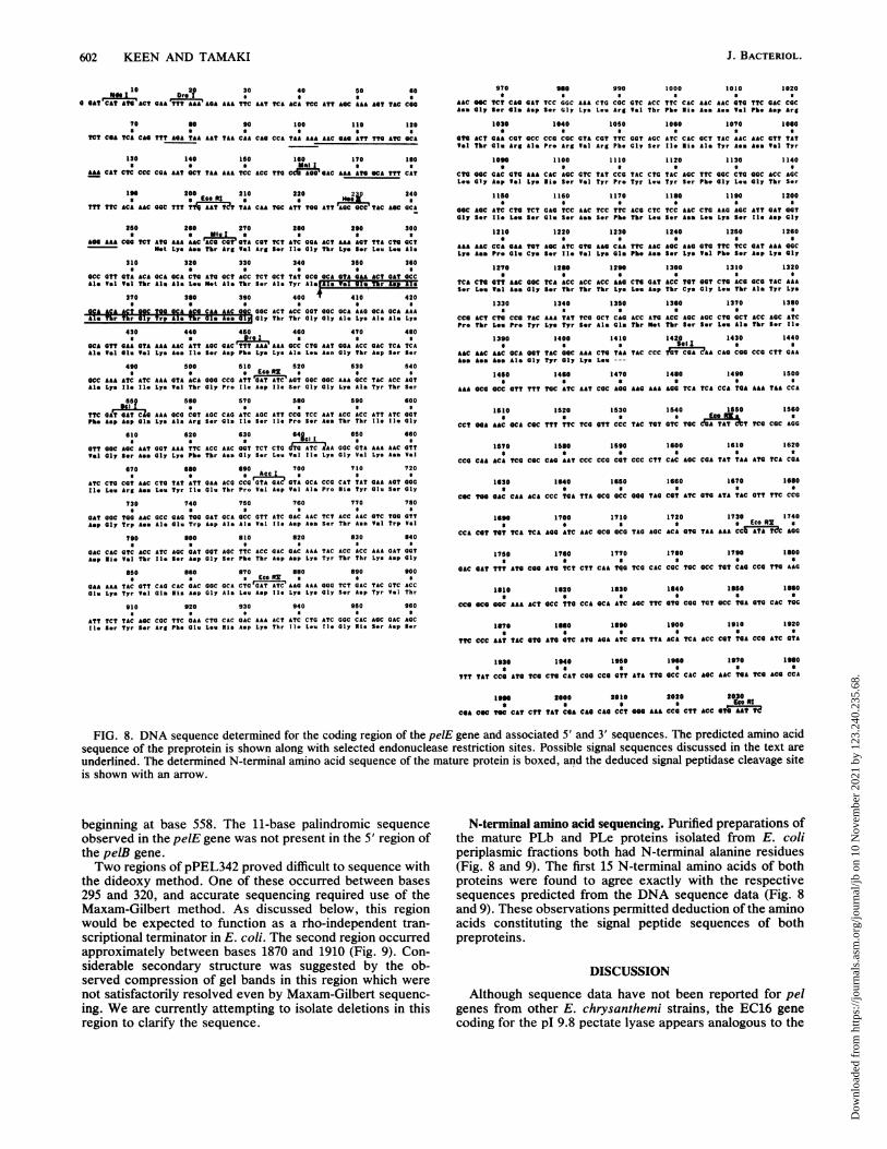
Fig. 8 revealed a single long open reading frame occurringbetween the EcoRI and Sall sites of plasmid pPEL7421, aspredicted from expression data. This is believed to encodethe PLe preprotein. Confirming this suspicion, pPEL711 andpPEL712, lacking internal BclI and EcoRV fragments, re-spectively, were negative for pectate lyase (Table 2) aspredicted. A purine-rich Shine-Delgarno sequence with an
VOL. 168, 1986
Dow
nloa
ded
from
http
s://j
ourn
als.
asm
.org
/jour
nal/j
b on
10
Nov
embe
r 20
21 b
y 12
3.24
0.23
5.68
.
600 KEEN AND TAMAKI
Eco RIRI,Hindm Mlu I
Eco RI1
Eco R3
t PEL 722
II, PstI,Hindm
RI, Hind m
RESTRICT WITH Eco RI,END-FILL AND RESTRICTWITH Hind m
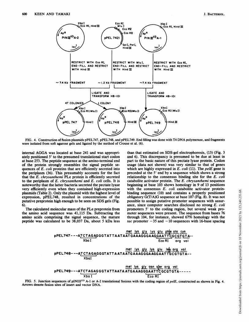
FIG. 4. Construction offusion plasmids pPEL747, pPEL748, and pPEL749. End filling was done with T4 DNA polymerase, and fragmentswere isolated from soft agarose gels and ligated by the method of Crouse et al. (6).
internal AGGA was located at base 241 and was appropri-ately positioned 5' to the presumed translational start codonat base 253. The peptide sequence at the amino-terminal endof the protein strongly resembles the signal peptide se-quences of E. coli proteins that are efficiently secreted intothe periplasm (36). This presumably accounts for the factthat the E. chrysanthemi PLe protein is efficiently secretedto the periplasm of E. chrysanthemi and E. coli cells. It isnoteworthy that the latter bacteria secreted the pectate lyasevery efficiently even when they contained high-expressionplasmids (Table 2). Only the plasmid with the highest level ofexpression, pPEL748, resulted in concentrations of theputative preprotein high enough to be seen on SDS gels (Fig.6).The calculated molecular mass of the PLe preprotein from
the amino acid sequence was 41,115 Da. Subtracting theamino acids comprising the signal sequence, the maturepeptide was calculated to be 38,037 Da, about 5 kDa less
than that estimated on SDS-gel electrophoresis, (13) (Fig. 3and 6). This discrepancy is presumed to be due at least inpart to the basic nature of this pectate lyase protein. Codonusage (data not shown) was very similar to that of geneswhich are highly expressed in E. coli (11). The pelE gene ispreceded at the 5' end by a sequence which shows a strongrelationship to the consensus binding site for the E. colicatabolite activator protein. The E. chrysanthemi sequencebeginning at base 103 shows homology in 9 of 13 positionswith the consensus E. coli catabolite activator protein-binding sequence (10) and contains a properly positionedobligatory G(T/A)G sequence at base 107 (Fig. 8). It was notpossible to assign putative promoter sequences with assur-ance, since computer searches disclosed no strong E. colipromoters 5' to the coding region, but several weak pro-moter sequences were present. The sequence from bases 76through 104, for instance, showed 67% homology with thetac promoter - 35 and -10 sequences with 16-base spacing
rret IysglyIysqlu pha alo cyspPEL747 - - -ACTAGAGGGTATTAATAAT<iAAAGGGAAG,GAATT'CGCGTGTA--
Xbo I Eco RI arg vol
met I g I lu lu org volpPEL748- --AITCTAGA,GGGTATTAATAATGAAAGGGAAGGAAT CGTGTA--
XbaI
met Iy g asn sler org volpPEL749---ATCTAGAGGGTATTAATAATGAAAGGGAATTCGCGTGTA-----
Xbo I Eco RIFIG. 5. Junction sequences of pINIII113 A-1 or A-2 translational fusions with the coding region of pelE, constructed as shown in Fig. 4.
Arrows denote fusion sites of insert and vector DNA.
J. BACTERIOL.
Dow
nloa
ded
from
http
s://j
ourn
als.
asm
.org
/jour
nal/j
b on
10
Nov
embe
r 20
21 b
y 12
3.24
0.23
5.68
.
STRUCTURE OF PECTATE LYASE GENES 601
1 2 3 4 5 6 7
** -205
-' --- 116- 97
-66'"t
__ ._ ._~
a; -It-45
..do A'__ ___do_m _-0 -_
- 29
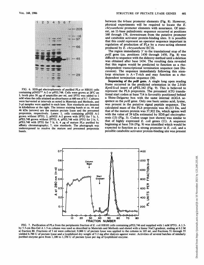
FIG. 6. SDS-gel electrophoresis of purified PLe or HB101 cellscontaining pINIII'13 A-2 or pPEL748. Cells were grown at 28°C onL broth plus 50 ,g of ampicillin per ml, and IPTG was added to 1mM when the cells attained an absorbance at 600 nm of 0.7. Cultureswere harvested at intervals as noted in Materials and Methods, and3-IlI samples were applied to each lane. Size standards are denotedin kilodaltons at the right. The intense staining bands at ca. 44 and46 kDa (arrows) are the mature pectate lyase and the presumedpreprotein, respectively. Lanes: 1, cells containing pINIII A-2grown without IPTG; 2, pINIII A-2 grown with IPTG for 7 h; 3,pPEL748 grown without IPTG; 4, pPEL748 with IPTG for 2 h; 5,pPEL748 with IPTG for 7 h; 6, 4 jg of mature PLe purified byaffinity chromatography; 7, size standards. The photograph wasunderexposed to resolve the mature and presumed preproteinbands.
between the 6-base promoter elements (Fig. 8). However,physical experiments will be required to locate the E.chrysanthemi promoter elements with assurance. Of inter-est, an 11-base palindromic sequence occurred at positions168 through 178, downstream from the putative promoterand catabolite activator protein-binding sites. It is possiblethat this could represent an operator sequence important inregulation of production of PLe by a trans-acting elementproduced by E. chrysanthemi EC16.The region immediately 3' to the translational stop of the
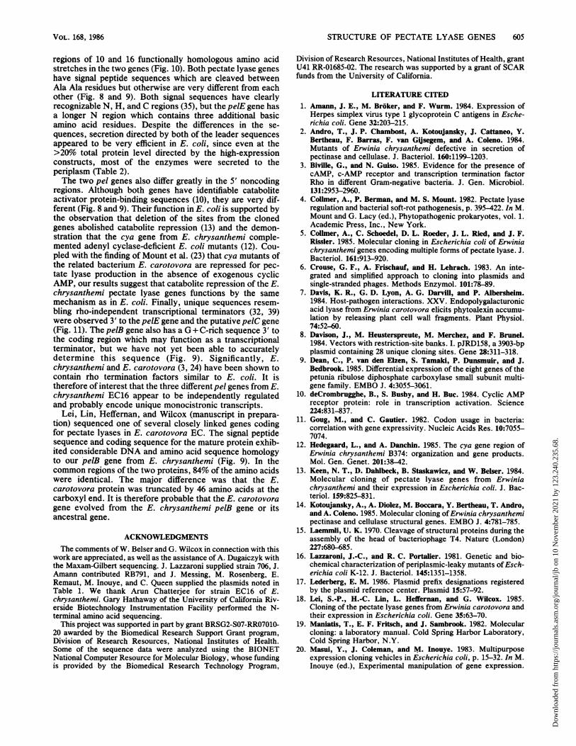
pelE gene (ca. positions 1430 through 1450, Fig. 8) wasdifficult to sequence with the dideoxy method until a deletionwas obtained after base 1434. The resulting data revealedthat this region would be predicted to function as a rho-independent transcriptional termination sequence (see Dis-cussion). The sequence immediately following this stem-loop structure is A + T-rich and may function as a rho-dependent termination sequence (30).
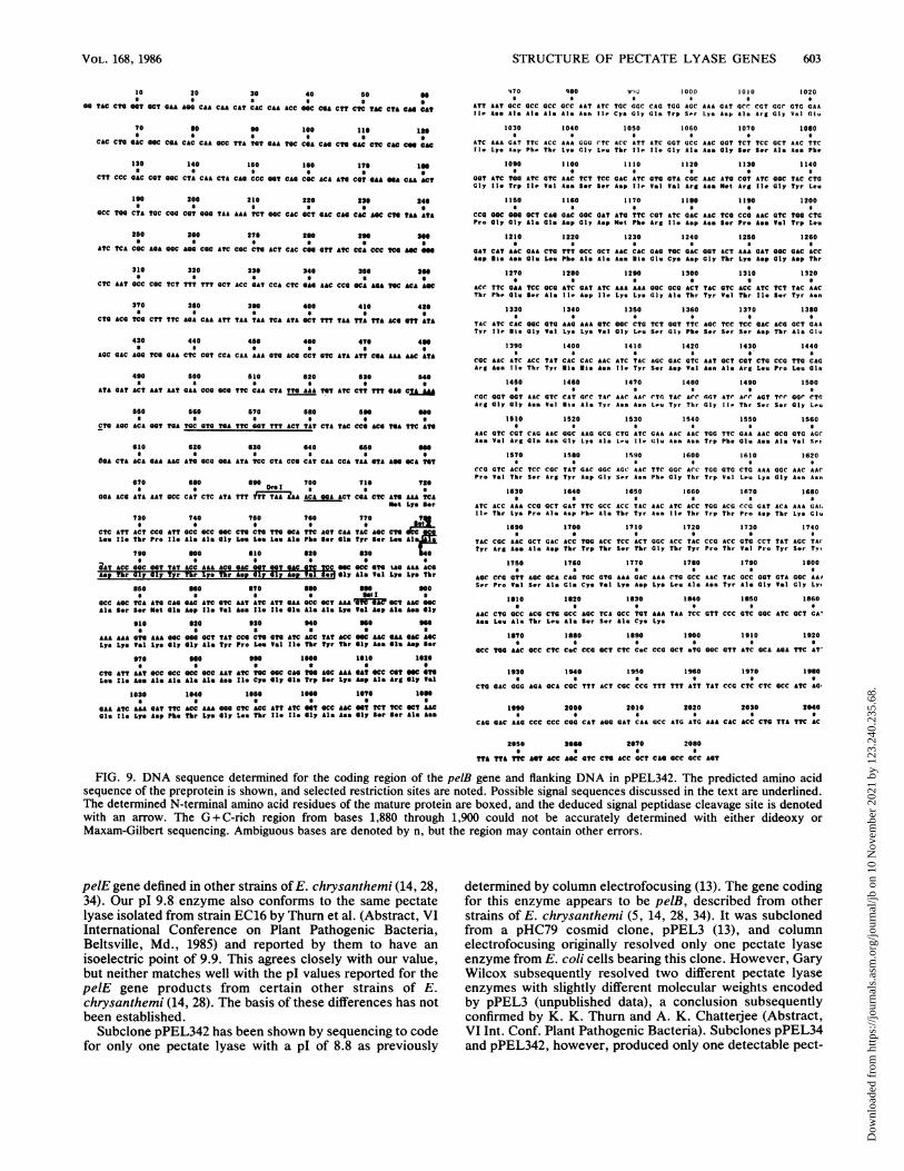
Sequencing of the pelB geile. A single long open readingframe occurred in the predicted orientation in the 2.0-kbKpnI/ScaI insert of pPEL342 (Fig. 9). This is believed torepresent the PLb preprotein. The presumed ATG transla-tional start codon at base 714 is favorably positioned behinda Shine-Delgarno box with the same internal AGGA se-quence as the pelE gene. Only one basic amino acid, lysine,was present in the putative signal peptide sequence. Thecalculated mass of the PLb preprotein was 40,213 Da, andthat of the mature protein was 37,922 Da, which agrees wellwith the value of 39 kDa estimated by SDS-gel electropho-resis (13) (Fig. 3). Codon usage (not shown) was similar tothat of highly expressed E. coli genes (11). A sequencebeginning at base 516 (Fig. 9) was identified which would beexpected to function as a strong promoter in E. coli, and apossible catabolite activator protein-binding site was present
CY
a
1400
1200 j
1000 6
0
800 LA
w600 a-
U)
400 Z
200C
10 20 30 40 50 60 70 80FRACTION NUMBER
FIG. 7. Purification of PLe from the periplasmic fraction of E. coli HB101 cells containing pPEL746 and supplied with 1 mM IPTG. A 1.5-by 5.5-cm Bio-Gel A 1.5-m column was used as described in Materials and Methods and eluted with a linear NaCl gradient, ending at 0.2 Mat fraction 80. Fractions of 3 ml were collected; 9,000 U of pectate lyase was applied to the column in 105 ml, and fractiotis 51 through 55yielded 6,700 U of pectate lyase and a lyophilized dry weight of 5.2 mg after dialysis against water. Activities of several batches of similarlypurified enzyme gave from 1,100 to 1,350 U of pectate lyase per mg of lyophilized enzyme.
VOL. 168, 1986
14 -A""
..._".
."Im, 99M.Aw :NOW
;., im .11M.---W ...*,
Dow
nloa
ded
from
http
s://j
ourn
als.
asm
.org
/jour
nal/j
b on
10
Nov
embe
r 20
21 b
y 12
3.24
0.23
5.68
.
602 KEEN AND TAMAKI
10 20 20 40 s0 60
OAT CAT AftOACTCAL 'W.AA'AOL ALL TTC ALT TCA ACA TCC ATT AGC :AA ACT TAC CCC
70 60 90 100 110 120
TCT COA TCO CAC TTT AGO TAA ALT TAA CAL CAO CCA TAL ALA AAC GAG AT? TTC ATC OCC
130 140 100 160 170 160
* .M.^1 t
LA CAT CTC CCC CA ALT OCCT TAA ALA TCC ACC TTC CcdAG'ACAC ALA AT oCC TTT CAT
194 200 210 220 220 240* E£o * Ho ,
TTT TTC ACA AAC "C TTT TIq ALT Tdt TAA CAL TOC ATT TOO ATT LC 0cc TAC ACC QC
260 260 270 280 290 300
GOC ALA COO TCT ATO ALA LAC'ACG COT'GTA COT TCT ATC aOa ACT ALL LOT TTA cTo OCT
Not Lys Ass Thr Arg Vol Arg $or I1l Oly Thr Lys $or Lou Los Als
310 320 330 340 260 360
GCC CTT GTA ACA GCC GCC CTG ATG OCT ACc TCT OCt TAT GCG GCC GTA CA ACT OAT Cc
Ale Vsl Val Thr Ale Als Lsu Not Als Tbr Ser Als Tyr AlaAlaaUuTras m270 280 390 400 410 420
GCS AO ACT $GC CA ACG CAL AACGCC GGC ACT ACC OCT aC GCC AOA GCC GCC ALAAla Tbr t r ly Trp AleTs r Os Ass 01710ly Tbr Thr 0ly Cly Als Lys Als Als Lys
420 440 450 460 470 460* * ~~~ ~~~OroI * *
GCA OTT CAL GA ALA AAC AT? AGC GAC T?T ALA' AA aCC CTG ALT CaL ACC CAC TCA TC
Als Vsl GIs Vsl Lys Ass Ile S-r Asp Phs Lys Lys Als Lou Ass Oly Thr Asp Ser $or
490 S00 510 520 530 540~~~~~*£co Nml
CCC ALL ATC ATC ALA OTA :CA COO CCC ATT'?A TCACT CCC GC ALA GCC TAC ACC ACAla Lys II- Ile Lys Val Tbr Gly Pro Ile Asp Ile Sr Gly Gly Lys Ale Tyr Thr S-r
559 560 570 S60 590 600
TTC OAT GAT CLO ALA aCa COT AOC CAC ATC AOC ATT CCC TCC AAT ACC LCC ATT ATC COTPh. Asp Asp Gls Lys Ala Arg gar Ols Ile Sor 11. Pro Sor Ass Thr Thr lII II. Cly
610 620 630 141 650 660
OTT GGC Ac ALT GOT ALA TTC ACC AAC COT TCT CTO dTG ATC CAasC OTA ALL AAC OaTVal Cly Sor Ass Cly Lys Pbo Tbr Ass Cly ger Lou Val Ile Lys 0ly Vol Lys Ass Vel
670 660 690 700 710 720e * * W ~~~~~AccI
ATC CT COT AAC CTG TAT ATT CAL ACG CCO at CAC'ATAc CCC CAT TAT CAL ALT CCGII* Lou Arg Ass Lou Tyr 11- Clu Tbr Pro Vol Asp Val Ale Pro Nie Tyr Olu S.r 0ly
730 740 750 760 770 780
CAT GCC TOO aACCCC GAG TOO CAT CCL GcC OTT ATC CAC LAC TCT ACC :AC GTC TOO OTTAsp 0ly Trp Ass Ala Glu Trp Asp Ale Ale Val 11e Asp Ass S-r Thr Ans Vol Trp Vol
790 g00 610 820 830 840
CAC CAC CTC ACC ATC AOC CAT OT AOC 4TC ACC CAC GAC ALL TAC ACC :CC ALA CAT OCTAsp Nis Vsl Tbr I1. gar Asp Cly s-r Phe Tbr Asp Asp Lys Tyr Tbr Thr Lye Asp 0ly
850 660 870 So0 690 900* $ EeocooRR
CAL ALA TAC CTT CAG CAC :AC CCC aCC CTG1CAT ATC' LA ALA OGG TCT CAC TAC GTC LCcalu Lys Tyr Vel Ole 31i Asp 0ly Ale Lov Asp 11- Lye Lye Cly S-r Asp Tyr Val Thr
910 920 930 940 950 960* *
ATT TCT TAC AGC CCC TTC CAL CTO CAC GAC ALA ACT LT: CTG ATC 0CC CAC AOCcAC LACIle S-r Tyr S-r Arg Pbs Clu Lou Nis Asp Lye Thr Lou lb Gly01is S-r Asp Bor
970 980 990 1000 1010 1020
AAC TCT CAC GAT TCC GGC AAA CYG COC GTC ACC TTC CAC AAC AAC GTG TTC cAC C¢C£s6 Gly S-r CIs Asp S-r Gly Lys Leou Arg Sal Tbr Pb. Nis Ass Aso Val Ph. Asp Are
1030 1040 1050 1060 1070 1080
OG AC? CGA CGT 0CC CCG CGC GTA CGT TTC CCT ACC ATC CAC GCt TAC AAC AAC CTT TATVal Tbr CGl Arg A1l Pro Arg V1a Arg Ph. Cly Ser II1 Rio Ala Tyr Ass Ass T.1 Tyr
1090 1100 1110 1120 1130 1140
Cta GCC CAC G? AAA CAC AC CtC TAT CCC TAC CTG TAC AGC TTC cGC CTO GC ACC AGC
Lss Cly Asp Val Lys Nis Ssr Val Tyr Pro Tyr Lou Tyr sr Pbh 0ly Lsu 0ly Tbr s.r
1150 1160 1170 1180 1190 1200
GCC LGC ATC CTc TCT GAO TCC AAC TCC TTC ACc CTC TCC AAC CTc AG AcC ATT CGT OCT
01y S-r IIs Lss Sr Cl. Ssr Ass Ser Pbs Tbr Lou S-r Asa Lou Lys S-r II* Asp Cly
1210 1220 1230 1240 1260 1260
AAA LAC CCL G&A ?t LGC LTC GTG LAO CAL TTC AAC AGC AAG aa ttC tCC OAT ALA GGCLys Ass Pro Ols Cys s-r Ile Val Lys GIs Phb Ass Bsr Lys Val Pbs Ser Asp Lys Gly
1270 1260 1290 1300 1310 1320
TCA CC GTT AAC COC TCA ACC ACC ACC LAA CtC CAT LCc TGT 0T0 C 6ACG GCC TAC AAA
S-r Lss Tel Ass Cly Csr Tbr Tbr Tbr Lys Lss Asp Thr Cys Cly Lsu Tbr Al. Tyr Lys
1330 1340 1350 1360 1370 1380
CCG ACT CtG CCC TAC ALA TAT ?CC OCT CAC ACC AtO ACC AOC AGC CTG OCT ACC AGC ATCPro Tbr Pro Tyr Lys Tyr S-r Ala Gls Tbr M.t Tbr Osr OSr Lss Ale Tbr Sor 11e
1390 1400 1410 1420 1430 1440
AAC AAC AAC GCC OCT TAC CGC ALL CTC T:A TAC CCC faT CCL C'eA CAG COG CCC CT? CAL
Ass Ass Ass Als 0ly Tyr Cly Lys Las ---
1450 1460 1470 1480 1490 1500
ALA GCG GCC CTT TT? TGC ATC ALT CCC AGG AOA ALL ACc TCA TCA CCL TGA ALA TAA CCA
IS10 1620 1530 1540 1550 1560e s Eeo OX a
CCT oa AAC GCC CGC TTT ??C TCO OTT CCC TAC TCT CTC TGC CcA TAT CT TCG CCC ACc
1570 1580 1990 1600 1610 1620
CCC CAL ACA TCC CCC CLC ALT CCC CCC CCT CCC CTT CAC AOC CCa TAT ?AA a" TCA CCA
1630 1640 1650 1660 1670 1680
CCC TO CAC CAL ACA CCC TGA ?TA GCa CCC TAG CCT A?C GTG ATA TAC 0?T TTC CCC
1690 1700 1710 1720 1730 1740s 0 * EcoRo
CCL CCT TOT ?CA TCA ACO AcC AAC GCC aCa TAG AOC ACA GTG TAA AL CCd ATA TC' Ac
1750 1760 1770 1760 1790 1800
GAC CAT TTT A" COO ATG TCT CTT CAL TQG TCC CAC CCC TOC OCC TOT CAO CCG TO AAG
1610 1620 1830 1840 1860 1660
CCI CC GGC ALA ACt GCC TTG CCL GCC ATC AGC TTC GTO CGC TGT GCC TGA GTO CAC TOO
1670 1660 1690 1900 1910 1920
TYC CCC ALT TAC A"O OTC ATG AOL ATC OTA TTA ACA TCA ACC COT TGA CCG ATC OTA
1930 1940 1960 1960 1970 1980
TTT TAT CCC ATO TCO CTO CAT COO CcO OTT TTO QCC CAC AOC AAC TOA TCG CO CCL
190 2000 2010 2020 2030S ECO Ott
COIL COO TOO OAT C?? TAT COA CAO CAC COT ALA CC: CT? ACC ITOl TO
FIG. 8. DNA sequence determined for the coding region of the pelE gene and associated 5' and 3' sequences. The predicted amino acidsequence of the preprotein is shown along with selected endonuclease restriction sites. Possible signal sequences discussed in the text are
underlined. The determined N-terminal amino acid sequence of the mature protein is boxed, and the deduced signal peptidase cleavage siteis shown with an arrow.
beginning at base 558. The 11-base palindromic sequence
observed in the pelE gene was not present in the 5' region ofthe pelB gene.Two regions of pPEL342 proved difficult to sequence with
the dideoxy method. One of these occurred between bases295 and 320, and accurate sequencing required use of theMaxam-Gilbert method. As discussed below, this regionwould be expected to function as a rho-independent tran-scriptional terminator in E. coli. The second region occurredapproximately between bases 1870 and 1910 (Fig. 9). Con-siderable secondary structure was suggested by the ob-served compression of gel bands in this region which were
not satisfactorily resolved even by Maxam-Gilbert sequenc-
ing. We are currently attempting to isolate deletions in thisregion to clarify the sequence.
N-terminal amino acid sequencing. Purified preparations ofthe mature PLb and PLe proteins isolated from E. coliperiplasmic fractions both had N-terminal alanine residues(Fig. 8 and 9). The first 15 N-terminal amino acids of bothproteins were found to agree exactly with the respectivesequences predicted from the DNA sequence data (Fig. 8and 9). These observations permitted deduction of the aminoacids constituting the signal peptide sequences of bothpreproteins.
DISCUSSION
Although sequence data have not been reported for pelgenes from other E. chrysanthemi strains, the EC16 gene
coding for the pl 9.8 pectate lyase appears analogous to the
J. BACTERIOL.
Dow
nloa
ded
from
http
s://j
ourn
als.
asm
.org
/jour
nal/j
b on
10
Nov
embe
r 20
21 b
y 12
3.24
0.23
5.68
.
STRUCTURE OF PECTATE LYASE GENES 603
10 20 30 40 50 60
GO ?AC C?G GO? OCT GAA AGO CAA CAA CAT CAC CAA ACC 0C CGA C?? CTC t?A C?A CM C:A70 s0 90 100 11 1is
CAC CTG GAC NC CGA CAC CAA GCC TTA T0? GAA TGC CCA COG CN SAC C?C COe CO :AC130 140 160 10 170 IN
CTT CCC GAC CGT GGC CTA CAA CTA CG CCC N? CAG CCC ACA ATG CGT GAA NA CA ACt
190 200 210 220 230 240GCC TGG CTA TGC CGO COT GG ?AA AAA ?C?T NC CAC CT GAC CA CMC ^C ?tAA TA?
280 260 270 280 290 UO 10 a 0S
AtC TCA CGC AGA GCC AN CGC A?C CGC CTG AC? CAC COO Ctt A?C CCA CCC C ANe
310 320 330 340 360 3NC?C AAT GCC CGC TCT TTT TTT GCT ACC GAT CCA C?C GAO AAC CCG GCA AGONC ACA AC
370 320 390 400 410 420
CTG ACG TCG CTT ?TC AGA CAA A?TT AA ?AA ?CA A? N? T?? ?AA ?TA ??A AC CTT tA?
430 440 450 460 4?0 4NAGC GAC AGO TCG GAO C?C CcT CCA CAA AAA GTG ACG CCT GTC ATA A?? CCA AAA AAC TA?
490 500 510 020 030 040
ATA GAT A:? AA? AT GA: CCG GCG TTcAA CTA TTG AL TO ATC CT TTT ^S C,L;550 560 570 580 S90 "0
CTG AGC ACA GT GA TGc aTG TGA TT GOaTT ACTcTA CTA TAC CC: ACG TA TT ATO
o0 620 630 640 O 0C?O AC ? G NCGO?O ? OG C ?Aa?A CC C A?CA
OGA C?A ACA OAA AAC AN OCO "A ATA NC C?A CCC CA? CAA CCA ?AA OA AO NA NTT
670 680 690 700 710 7200 Drol *OA ACO AA AAT 0CC CA? CTC ATA TTT fT ?AA A'AAA ALC NA ACT COA C?C AN AA NA
Net Lye 0er
730 740 700 760 770 T
CTC ATT AC? CCG A?? 0CC Cc NC CTO CN ??CNA T? AG? CAA TAC AC CNcLou I1e Thr Pro Ile Ala Ale Oly Lou Lou Lou Ala Pbh Oer OIs Tyr Our Leo Al
790 000 010 020 030 4
ATACC 0CC 00? ?A? A.CC AAA AceOGAC N?1 N? GAC ON NgC NC NCC ON %AG AAA ACOAsp Tlr Gly Gly Tyr Tbr Lys r As alyly pa* e Cly 1 V1 Ly Ly Tbr
050 go0 *70 000 goo 900s s * 10 SolI s
CCC AC CA A?G CA GAC AN OC AA? A?C AT? GAA CCC OCT AAA %IWIU CCT AAC NacAle Our Our Net Cls Asp Ile al Ass I1e I1e Olu Ale Ale Lys V1a Asp Ale Asa 01y
910 920 930 940 90 0oAAA AAA ON AAA NC COO CT ?A? CCC Cn ON AC ACC ?AT CC C AAC AA SAC ANcLys Lye Va1 Lys Oly ly Ale Tyr Pro Lea Va1 Ila ?hr Tyr Thr Sly Ass Ols AsptO r
970 9OC 990 160o 1010 1020
CN A?? ? NCCoCCNcCcCC AAT ANC NeC CM No ANC MA GA? NCC CC? NCLee II Ass Ale Ale Ale Ala Ass II Cy Oly oIs trp *or Lye Asp Al Avrg sly Pal
1020 1040 1000 lo0 1070 16o0OAA A?C AAA GA? ?TC ACC AAA CC CC ACC ATT ANC "T *CC AACA O? N? CC CT AM0lu I1. Lys Asp Pb. Thr Lye Oly Lee Thr Ila II- Oly Ale Ass Cly Our Ser Ale Ass
470 9809so 1000 1010 1020
ATT AAT 0CC aCC 0CC aCC OAT ATC TCC CGC CAG TGG AGC AAA GAT GCC CGT GGC GT7 GAAfie Aee Ale Ale Ale Ala Asn fie Cys Gly Gin Trp Srr Lys Asp Ale Arg Gly Val Glu
1030 1040 1050 1060 1070 1010
ATC AAA GAT TtC ACC AAA GGG CTC ACC ATT AtC GGT GCC AAC OGT TCT TCC OCT AAC TTCII Lye Asp Ph. Thr Lye Gly L-e thr fie 11e ly Ale Ass Gly SOr gor Ale Asn Phe
1090 1100 1110 1120 1130 1140
CCt ATC TOO ATC OTC AAC TC? TCC CAC ATC 070 C?A CaC AAC ATC CCT ATC GOC ?AC CtcCly 11e Trp fir VPl Asn S-r Ser Asp 11PVsl Vsl Org Ass Mlt Arg lie Cly Tyr Leu
1160 1160 1170 1180 1190 1200
CCC 0CC 0CC OCT CAG CAC CCcA?T ATO TTC cOT ATc CAC AAC ?TC CCC AAC OTC TOO CTGPro 0ly Cly Ale Cl. Asp Cly Asp Ml Phb. Arg 11- Asp Ass O,r Pro Ass VPl Trp Lee
1210 1220 1230 1240 1250 1260
GAT CAT AAC CAA CTO TT? CCC OCT AAC CAC GAO TGC GAC GOT ACT AAA GAT GOC GAC ACCAep lie Ass Olu Lee Ph. Ale Ale Ass lie Glu Cys Asp Cly T?r Lys Asp Cly Asp thr
1270 1260 1290 1300 1310 1320
ACC TTC CAA ?CC CC A?C CA?T ATC AAA AAA CCC aCC AC? TAC OTC ACc ATC ?TC TAC AACTbr Ph. Olu $er Ale Ile Asp 11- Lys Ly Gly Ale Thr Tyr Vsl Tbr Ile S-r Tyr Aen
1330 1340 1350 1360 1370 1380
TAC ATC CAC GGC 0T7 AAG AAA OTC 0aC CTC TCT CCT TTC AC TCC TCC CAC ACG GCT GAAtyr fIr Nis Cly VPl Lye Lys Val Cly Leu Ser Gly Pbe Ser Ier Ser Asp Tbr Ale Clu
1390 1400 1410 1420 1430 1440
CaC AAC ATC ACC ?TA CAC CAC AAC ATC TAC AGC CAC CTC AA? OCT CCT CTO CCC 7T7 CAGArg Ass 11- Thr Tyr Nie Nie Ass Ile Tyr Ser Asp Vel Aes Ale Arg Lee Pro Leu Oln
1450 1460 1470 1480 1490 1500
CCC CC? 00? AAC CtC CAT GCC tAC AAC AAC CT7 TAC ACCr nGT ATC A3 ACT 7Te Ce 3TcArg Cly Cly Ass Val Sis Ale Tyr Ass Asn Leu Tyr Thr Gly lir Thr Scr Ser Cly Leu
1510 1520 1530 1540 1550 1560
AAC OTC CCT CAC AAC CCC AAC CGC CTG ATC GAA AAC AAC TCG TTC CAA AAC OCO 070 AGCAss Vel Arg Oln Ass Gly Lys Ale L-u 11. filu Asn Aen trp Pbe Olu Ass Ale Vel Ser
1670 1500 1590 1600 1610 1620
CCC ctC ACC tCC CGC tAt GAC acC AGC AAC ttC CCC ACC TGG GtG CtG AAA acC AAC AACPro VPl tbr S-r Arg tyr Asp Gly Ser Ans Phe Cly Thr Trp Val Lou Lye Cly Asn Aesn
1630 1640 1650 1660 1670 1680
ATC ACC AAA CCC OCt CAT TTC CCC ACC TAC AAC AtC ACC TCC ACG CC GA? ACA AAA GAl.1Ir tbr Lys Pro Ale Asp Pbh Ale Tbr Tyr Aen Ile Thr Trp Thr Pro Asp Tbr Lye Glu
1690 1700 1710 1720 1730 1740
TAC CCC AAC OCT CAC ACC T70 ACC TCC ACT acC ACC TAC CCC ACC CTC CCT tAT AGC TA(tyr Arg Ass Ale Asp thr trp tbr S-r thr Gly thr tyr Pro Thr Vel Pro Tyr S-r tv,
1760 1760 1770 1780 1790 1800* 5 0 * s e
AGC CCC CTT AOC OCA COA TcC 7G0 AAA CAC AAA CTG CCC AAC TAC CCC GCT CTA CGC AA#Ser Pro VPl Ser Ale Gle Cys Val Lye Asp Lye Lee Al Aesn Tyr Ale Gly Psi Gly Ly.
1610 1820 1630 1840 1o50 1860
AAC CTO CCC ACG CTG CCC AC TCA aCC T7T AAA TAA TCC CTT CCC OTC CCC ATC OCT GA'Aes Lee Ale Tbr Lou Ale Bar Ser Ale Cys Lys
1870 100 1890 1900 1910 1920
0CC TCC AAC GCC C?C CeC CCC OCT CTC CC CCC OCC eTC CCC 077 ATC OCA AGA TTC At'
1930 1940 1950 1960 1970 1980* e s e o s
CTO AC GCCC AGA OCA CCC TT7 ACT CCC CCC TTT 7TT ATT TAT CCC CTC CtC aCC ATC AG
1990 2000 2010 2020 2030 2040
CAG GAC AAG CCC CCC CCC CAT AGG CAT CAA cCC ATG ATc AAA CAC ACC CTO TTA TTC AC
2050 20" 2070 20806 0 0
A ??A ?N Ac? 0CANe ONCNCACC CTC CAC 0CCCCCAC?T
FIG. 9. DNA sequence determined for the coding region of the pelB gene and flanking DNA in pPEL342. The predicted amino acidsequence of the preprotein is shown, and selected restriction sites are noted. Possible signal sequences discussed in the text are underlined.The determined N-terminal amino acid residues of the mature protein are boxed, and the deduced signal peptidase cleavage site is denotedwith an arrow. The G+C-rich region from bases 1,880 through 1,900 could not be accurately determined with either dideoxy orMaxam-Gilbert sequencing. Ambiguous bases are denoted by n, but the region may contain other errors.
pelE gene defined in other strains of E. chrysanthemi (14, 28,34). Our pI 9.8 enzyme also conforms to the same pectatelyase isolated from strain EC16 by Thurn et al. (Abstract, VIInternational Conference on Plant Pathogenic Bacteria,Beltsville, Md., 1985) and reported by them to have anisoelectric point of 9.9. This agrees closely with our value,but neither matches well with the pl values reported for thepelE gene products from certain other strains of E.chrysanthemi (14, 28). The basis of these differences has notbeen established.
Subclone pPEL342 has been shown by sequencing to codefor only one pectate lyase with a pl of 8.8 as previously
determined by column electrofocusing (13). The gene codingfor this enzyme appears to be peiB, described from otherstrains of E. chrysanthemi (5, 14, 28, 34). It was subclonedfrom a pHC79 cosmid clone, pPEL3 (13), and columnelectrofocusing originally resolved only one pectate lyaseenzyme from E. coli cells bearing this clone. However, GaryWilcox subsequently resolved two different pectate lyaseenzymes with slightly different molecular weights encodedby pPEL3 (unpublished data), a conclusion subsequentlyconfirmed by K. K. Thurn and A. K. Chatterjee (Abstract,VI Int. Conf. Plant Pathogenic Bacteria). Subclones pPEL34and pPEL342, however, produced only one detectable pect-
VOL. 168, 1986
Dow
nloa
ded
from
http
s://j
ourn
als.
asm
.org
/jour
nal/j
b on
10
Nov
embe
r 20
21 b
y 12
3.24
0.23
5.68
.
604 KEEN AND TAMAKI
860 GAC GGC GCA CTG GAT ATC AAG AAA GGG TCT GAC TAC GTC ACC ATT TCT TAC 910 EAsp Gly Ala Leu Asp Ile Lys Lys Gly Ser Asp Tyr Vol Thr Ile Ser Tyr
I I II II 1I II II II 11 II 11 II II IIGlu Ser Alo Ile ASD Ile Lvs Lvs Gly Ala Thr Tyr Vol Thr Ile Ser Tyr
1272 GAA TCC GCG ATC GAT ATC AAA AAA GGC GCG ACT TAC GTC ACC ATC TCT TAC 1322
1052 GGT AGC ATC CAC GCT TAC AAC AAC GTT TAT 1081Gly Ser lie His Ala Tyr Asn Asn Vol Tyr 251E
11 1I II 1I II II 1IGly Asp Vol His Ala Tyr Asn Asn Lou Tyr
1452 GGT AAC GTC CAT GCC TAC AAC AAC CTG TAC 1481 E1
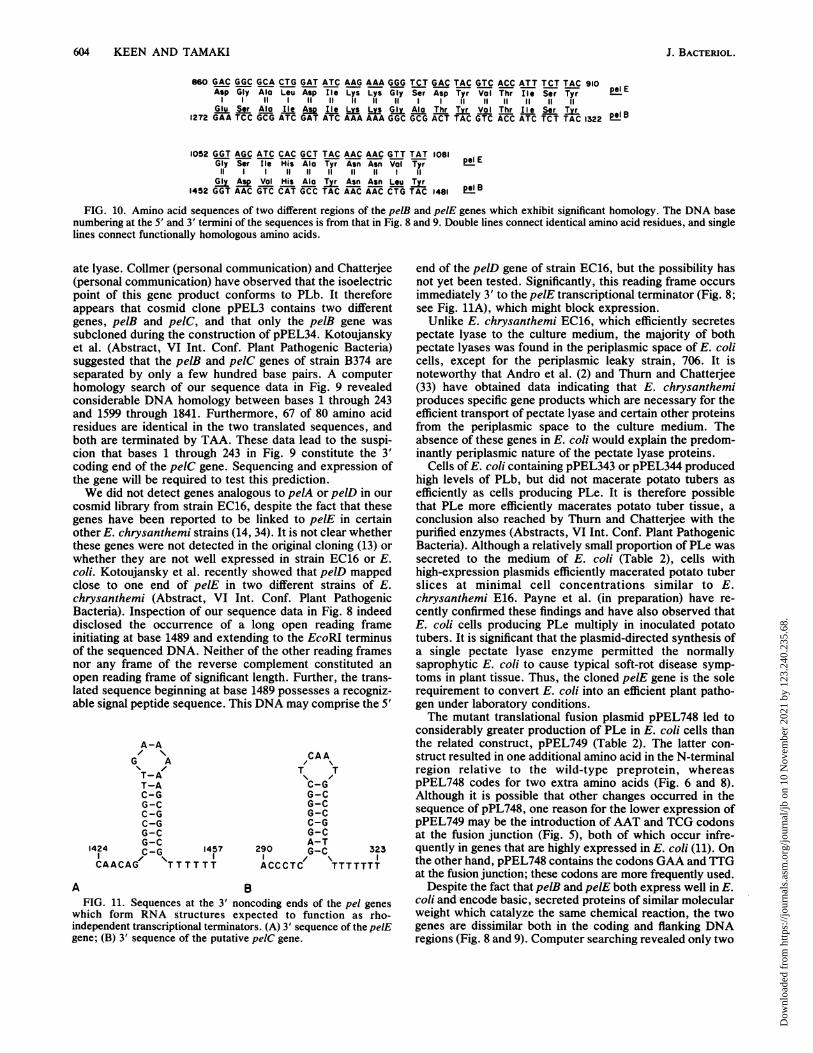
FIG. 10. Amino acid sequences of two different regions of the pelB and pelE genes which exhibit significant homology. The DNA basenumbering at the 5' and 3' termini of the sequences is from that in Fig. 8 and 9. Double lines connect identical amino acid residues, and singlelines connect functionally homologous amino acids.
ate lyase. Collmer (personal communication) and Chatterjee(personal communication) have observed that the isoelectricpoint of this gene product conforms to PLb. It thereforeappears that cosmid clone pPEL3 contains two differentgenes, pelB and pe1C, and that only the pelB gene wassubcloned during the construction of pPEL34. Kotoujanskyet al. (Abstract, VI Int. Conf. Plant Pathogenic Bacteria)suggested that the pelB and pelC genes of strain B374 areseparated by only a few hundred base pairs. A computerhomology search of our sequence data in Fig. 9 revealedconsiderable DNA homology between bases 1 through 243and 1599 through 1841. Furthermore, 67 of 80 amino acidresidues are identical in the two translated sequences, andboth are terminated by TAA. These data lead to the suspi-cion that bases 1 through 243 in Fig. 9 constitute the 3'coding end of the pelC gene. Sequencing and expression ofthe gene will be required to test this prediction.We did not detect genes analogous to pelA or pelD in our
cosmid library from strain EC16, despite the fact that thesegenes have been reported to be linked to pelE in certainother E. chrysanthemi strains (14, 34). It is not clear whetherthese genes were not detected in the original cloning (13) orwhether they are not well expressed in strain EC16 or E.coli. Kotoujansky et al. recently showed that pelD mappedclose to one end of pelE in two different strains of E.chrysanthemi (Abstract, VI Int. Conf. Plant PathogenicBacteria). Inspection of our sequence data in Fig. 8 indeeddisclosed the occurrence of a long open reading frameinitiating at base 1489 and extending to the EcoRI terminusof the sequenced DNA. Neither of the other reading framesnor any frame of the reverse complement constituted anopen reading frame of significant length. Further, the trans-lated sequence beginning at base 1489 possesses a recogniz-able signal peptide sequence. This DNA may comprise the 5'
A-A
G AT-AT-AC-GG-CC-GC-GG-C
1424 G_C 1457CAACAG TTTTTTTT
CAAT T\ ,C-GG-CG-CG-CC-GG-CA-T
290 G-C 323I I/\ACCCTC TTTTTTT
BFIG. 11. Sequences at the 3' noncoding ends of the pel genes
which form RNA structures expected to function as rho-independent transcriptional terminators. (A) 3' sequence of the pelEgene; (B) 3' sequence of the putative pelC gene.
end of the pelD gene of strain EC16, but the possibility hasnot yet been tested. Significantly, this reading frame occursimmediately 3' to the pelE transcriptional terminator (Fig. 8;see Fig. 11A), which might block expression.
Unlike E. chrysanthemi EC16, which efficiently secretespectate lyase to the culture medium, the majority of bothpectate lyases was found in the periplasmic space of E. colicells, except for the periplasmic leaky strain, 706. It isnoteworthy that Andro et al. (2) and Thurn and Chatterjee(33) have obtained data indicating that E. chrysanthemiproduces specific gene products which are necessary for theefficient transport of pectate lyase and certain other proteinsfrom the periplasmic space to the culture medium. Theabsence of these genes in E. coli would explain the predom-inantly periplasmic nature of the pectate lyase proteins.
Cells of E. coli containing pPEL343 or pPEL344 producedhigh levels of PLb, but did not macerate potato tubers asefficiently as cells producing PLe. It is therefore possiblethat PLe more efficiently macerates potato tuber tissue, aconclusion also reached by Thurn and Chatterjee with thepurified enzymes (Abstracts, VI Int. Conf. Plant PathogenicBacteria). Although a relatively small proportion of PLe wassecreted to the medium of E. coli (Table 2), cells withhigh-expression plasmids efficiently macerated potato tuberslices at minimal cell concentrations similar to E.chrysanthemi E16. Payne et al. (in preparation) have re-cently confirmed these findings and have also observed thatE. coli cells producing PLe multiply in inoculated potatotubers. It is significant that the plasmid-directed synthesis ofa single pectate lyase enzyme permitted the normallysaprophytic E. coli to cause typical soft-rot disease symp-toms in plant tissue. Thus, the cloned pelE gene is the solerequirement to convert E. coli into an efficient plant patho-gen under laboratory conditions.The mutant translational fusion plasmid pPEL748 led to
considerably greater production of PLe in E. coli cells thanthe related construct, pPEL749 (Table 2). The latter con-struct resulted in one additional amino acid in the N-terminalregion relative to the wild-type preprotein, whereaspPEL748 codes for two extra amino acids (Fig. 6 and 8).Although it is possible that other changes occurred in thesequence of pPL748, one reason for the lower expression ofpPEL749 may be the introduction of AAT and TCG codonsat the fusion junction (Fig. 5), both of which occur infre-quently in genes that are highly expressed in E. coli (11). Onthe other hand, pPEL748 contains the codons GAA and TTGat the fusion junction; these codons are more frequently used.
Despite the fact that pelB and pelE both express well in E.coli and encode basic, secreted proteins of similar molecularweight which catalyze the same chemical reaction, the twogenes are dissimilar both in the coding and flanking DNAregions (Fig. 8 and 9). Computer searching revealed only two
A
J. BACTERIOL.
Dow
nloa
ded
from
http
s://j
ourn
als.
asm
.org
/jour
nal/j
b on
10
Nov
embe
r 20
21 b
y 12
3.24
0.23
5.68
.
STRUCTURE OF PECTATE LYASE GENES 605
regions of 10 and 16 functionally homologous amino acidstretches in the two genes (Fig. 10). Both pectate lyase geneshave signal peptide sequences which are cleaved betweenAla Ala residues but otherwise are very different from eachother (Fig. 8 and 9). Both signal sequences have clearlyrecognizable N, H, and C regions (35), but the pelE gene hasa longer N region which contains three additional basicamino acid residues. Despite the differences in the se-quences, secretion directed by both of the leader sequencesappeared to be very efficient in E. coli, since even at the>20%o total protein level directed by the high-expressionconstructs, most of the enzymes were secreted to theperiplasm (Table 2).The two pel genes also differ greatly in the 5' noncoding
regions. Although both genes have identifiable cataboliteactivator protein-binding sequences (10), they are very dif-ferent (Fig. 8 and 9). Their function in E. coli is supported bythe observation that deletion of the sites from the clonedgenes abolished catabolite repression (13) and the demon-stration that the cya gene from E. chrysanthemi comple-mented adenyl cyclase-deficient E. coli mutants (12). Cou-pled with the finding of Mount et al. (23) that cya mutants ofthe related bacterium E. carotovora are repressed for pec-tate lyase production in the absence of exogenous cyclicAMP, our results suggest that catabolite repression of the E.chrysanthemi pectate lyase genes functions by the samemechanism as in E. coli. Finally, unique sequences resem-bling rho-independent transcriptional terminators (32, 39)were observed 3' to the pelE gene and the putative pelC gene(Fig. 11). The pelB gene also has a G + C-rich sequence 3' tothe coding region which may function as a transcriptionalterminator, but we have not yet been able to accuratelydetermine this sequence (Fig. 9). Significantly, E.chrysanthemi and E. carotovora (3, 24) have been shown tocontain rho termination factors similar to E. coli. It istherefore of interest that the three different pel genes from E.chrysanthemi EC16 appear to be independently regulatedand probably encode unique monocistronic transcripts.
Lei, Lin, Heffernan, and Wilcox (manuscript in prepara-tion) sequenced one of several closely linked genes codingfor pectate lyases in E. carotovora EC. The signal peptidesequence and coding sequence for the mature protein exhib-ited considerable DNA and amino acid sequence homologyto our pelB gene from E. chrysanthemi (Fig. 9). In thecommon regions of the two proteins, 84% of the amino acidswere identical. The major difference was that the E.carotovora protein was truncated by 46 amino acids at thecarboxyl end. It is therefore probable that the E. carotovoragene evolved from the E. chrysanthemi pelB gene or itsancestral gene.
ACKNOWLEDGMENTSThe comments of W. Belser and G. Wilcox in connection with this
work are appreciated, as well as the assistance of A. Dugaiczyk withthe Maxam-Gilbert sequencing. J. Lazzaroni supplied strain 706, J.Amann contributed RB791, and J. Messing, M. Rosenberg, E.Remaut, M. Inouye, and C. Queen supplied the plasmids noted inTable 1. We thank Arun Chatterjee for strain EC16 of E.chrysanthemi. Gary Hathaway of the University of California Riv-erside Biotechnology Instrumentation Facility performed the N-terminal amino acid sequencing.
This project was supported in part by grant BRSG2-S07-RRO7010-20 awarded by the Biomedical Research Support Grant program,Division of Research Resources, National Institutes of Health.Some of the sequence data were analyzed using the BIONETNational Computer Resource for Molecular Biology, whose fundingis provided by the Biomedical Research Technology Program,
Division of Research Resources, National Institutes of Health, grantU41 RR-01685-02. The research was supported by a grant of SCARfunds from the University of California.
LITERATURE CITED1. Amann, J. E., M. Broker, and F. Wurm. 1984. Expression of
Herpes simplex virus type 1 glycoprotein C antigens in Esche-richia coli. Gene 32:203-215.
2. Andro, T., J. P. Chambost, A. Kotoujansky, J. Cattaneo, Y.Bertheau, F. Barras, F. van G"segem, and A. Coleno. 1984.Mutants of Erwinia chrysanthemi defective in secretion ofpectinase and cellulase. J. Bacteriol. 160:1199-1203.
3. Biville, G., and N. Guiso. 1985. Evidence for the presence ofcAMP, c-AMP receptor and transcription termination factorRho in different Gram-negative bacteria. J. Gen. Microbiol.131:2953-2960.
4. CoUmer, A., P. Berman, and M. S. Mount. 1982. Pectate lyaseregulation and bacterial soft-rot pathogenesis, p. 395-422. In M.Mount and G. Lacy (ed.), Phytopathogenic prokaryotes, vol. 1.Academic Press, Inc., New York.
5. Collmer, A., C. Schoedel, D. L. Roeder, J. L. Ried, and J. F.Rissler. 1985. Molecular cloning in Escherichia coli of Erwiniachrysanthemi genes encoding multiple forms of pectate lyase. J.Bacteriol. 161:913-920.
6. Crouse, G. F., A. Frischauf, and H. Lehrach. 1983. An inte-grated and simplified approach to cloning into plasmids andsingle-stranded phages. Methods Enzymol. 101:78-89.
7. Davis, K. R., G. D. Lyon, A. G. Darvill, and P. Albersheim.1984. Host-pathogen interactions. XXV. Endopolygalacturonicacid lyase from Erwinia carotovora elicits phytoalexin accumu-lation by releasing plant cell wall fragments. Plant Physiol.74:52-60.
8. Davison, J., M. Heusterspreute, M. Merchez, and F. Brunel.1984. Vectors with restriction-site banks. I. pJRD158, a 3903-bpplasmid containing 28 unique cloning sites. Gene 28:311-318.
9. Dean, C., P. van den Elzen, S. Tamaki, P. Dunsmuir, and J.Bedbrook. 1985. Differential expression of the eight genes of thepetunia ribulose diphosphate carboxylase small subunit multi-gene family. EMBO J. 4:3055-3061.
10. deCrombrugghe, B., S. Busby, and H. Buc. 1984. Cyclic AMPreceptor protein: role in transcription activation. Science224:831-837.
11. Goug, M., and C. Gautier. 1982. Codon usage in bacteria:correlation with gene expressivity. Nucleic Acids Res. 10:7055-7074.
12. Hedegaard, L., and A. Danchin. 1985. The cya gene region ofErwinia chrysanthemi B374: organization and gene products.Mol. Gen. Genet. 201:38-42.
13. Keen, N. T., D. Dahibeck, B. Staskawicz, and W. Belser. 1984.Molecular cloning of pectate lyase genes from Erwiniachrysanthemi and their expression in Escherichia coli. J. Bac-teriol. 159:825-831.
14. Kotoujansky, A., A. Diolez, M. Boccara, Y. Bertheau, T. Andro,and A. Coleno. 1985. Molecular cloning ofErwinia chrysanthemipectinase and cellulase structural genes. EMBO J. 4:781-785.
15. Laemmli, U. K. 1970. Cleavage of structural proteins during theassembly of the head of bacteriophage T4. Nature (London)227:680-685.
16. Lazzaroni, J.-C., and R. C. Portalier. 1981. Genetic and bio-chemical characterization of periplasmic-leaky mutants ofEsch-erichia coli K-12. J. Bacteriol. 145:1351-1358.
17. Lederberg, E. M. 1986. Plasmid prefix designations registeredby the plasmid reference center. Plasmid 15:57-92.
18. Lei, S.-P., H.-C. Lin, L. Heffernan, and G. Wilcox. 1985.Cloning of the pectate lyase genes from Erwinia carotovora andtheir expression in Escherichia coli. Gene 35:63-70.
19. Maniatis, T., E. F. Fritsch, and J. Sambrook. 1982. Molecularcloning: a laboratory manual. Cold Spring Harbor Laboratory,Cold Spring Harbor, N.Y.
20. Masui, Y., J. Coleman, and M. Inouye. 1983. Multipurposeexpression cloning vehicles in Escherichia coli, p. 15-32. In M.Inouye (ed.), Experimental manipulation of gene expression.
VOL. 168, 1986
Dow
nloa
ded
from
http
s://j
ourn
als.
asm
.org
/jour
nal/j
b on
10
Nov
embe
r 20
21 b
y 12
3.24
0.23
5.68
.
606 KEEN AND TAMAKI
Academic Press, Inc., New York.21. Maxatn, A. M., and W. Gilbert. 1980. Sequencing end-labeled
DNA with base-specific chemical cleavages. Methods Enzymol.65:499-560.
22. Morrison, D. A. 1977. Transformation in Escherichia coli: cryo-genic preservation of competent cells. J. Bacteriol. 132:349-351.
23. Mount, M. S., P. M. Berman, R. P. Mortlock, and J. P.Hubbard. 1979. Regulation of endopolygalacturonate trans-eliminase in an adenosine 3',5'-cyclic monophosphate deficientmutant of Erwinia carotovora. Phytopathology 69:117-120.
24. Nwanko, D. O., and S. K. Guterman. 1985. Purification ofRNApolymerase and transcription-termination factor Rho fromErwinia carotovora. Eur. J. Biochem. 146:383-389.
25. Pustell, J., and F. C. Kafatos. 1984. A convenient and adaptablepackage of computer programs for DNA and protein sequencemanagement, analysis and homology determination. NucleicAcids Res. 12:643-655.
26. Queen, C. 1983. A vector that uses phage signals for efficientsynthesis of proteins in Escherichia coli. J. Mol. Appl. Genet.2:1-10.
27. Remaut, E,, H. Tsao, and W. Fiers. 1983. Improved plasmidvectors with a thermoinducible expression and temperature-regulated runaway replication. Gene 22:103-113.
28. Reverchon, S., N. Hugouvieux-Cotte-Pattat, and R. Robert-Baudouy. 1985. Cloning of genes encoding pectolytic enzymesfrom a genomic library of the phytopathogenic bacterium,Erwinia chrysanthemi. Gene 35:121-130.
29. Roberts, D. P., P. M. Berman, C. Alien, V. K. Stromberg, G. H.Lacy, and M. S. Mount. 1986. Erwinia carotovora: molecularcloning of a 3.4 kilobase DNA fragment mediating production ofpectate lyases. Can. J. Plant Pathol. 8:17-27.
30. Rosenberg, M., and D. Court. Regulatory sequences involved inthe promotion and termination of RNA transcription. Annu.Rev. Genet. 13:319-353.
31. Rosenberg, M., Y.-S. Ho, and A. Shatzman. 1983. The use of
pKC30 and its derivatives for controlled expression of genes.Methods Enzymol. 101:123-138.
32. Rosenberg, M., and U. Schmeissner. 1982. Regulation of geneexpression by transcription termination and RNA processing, p.1-16. In M. Grunberg-Manago and B. Saler (ed.), Interaction oftranslational and transcriptional controls in the regulation ofgene expression. Elsevier Science Publishing, Inc., NewYork.
33. Thurn, K. K., and A. K. Chatterjee. 1985. Single-site chromo-somal TnS insertions affect the export of pectolytic andcellulolytic enzymes in Erwinia chrysanthemi EC16. Appl.Environ. Microbiol. 50:894-898.
34. Vani GUsegem, F., A. Toussaint, and E. Schoonejans. 1985. Invivo cloning of the pectate lyase and cellulase genes of Erwiniachrysanthemi. EMBO J. 4:787-792.
35. Von HeijUne, G. 1985. Signal sequences. The limits of variation.J. Mol. Biol. 184:99-105.
36. Watson, M. E. E. 1984. Compilation of published signal se-quences. Nucleic Acids Res. 12:5145-5164.
37. Witholt, B., M. Boekhout, M. Brock, J. Kingma, H. vanHeerikhuizen, and L. deLej. 1976. An efficient and reproducibleprocedure for the formation of spheroplasts from variouslygrown Escherichia coli. Anal. Biochem. 74:160-170.
38. Yanisch-Perron, C., J. Veira, and J. Messing. 1985. ImprovedM13 phage cloning vectors and host strains: nucleotide se-quences of the M13mpl8 and pUC19 vectors. Gene 33:103-119.
39. Yanofsky, C. 1982. Attenuation in the control of tryptophanoperon expression, p. 17-24. In M. Grunberg-Manago and B.Safer (ed.), Interaction of translational and transcriptional con-trols in the regulation of gene expression. Elsevier SciencePublishing, Inc., New York.
40. Zink, R. T., and A. K. Chatterjee. 1985. Cloning and expressionin Escherichia coli of pectinase genes of Erwinia carotovorasubsp. carotovora. Appl. Environ. Microbiol. 49:714-717.
J. BACTERIOL.
Dow
nloa
ded
from
http
s://j
ourn
als.
asm
.org
/jour
nal/j
b on
10
Nov
embe
r 20
21 b
y 12
3.24
0.23
5.68
.