structure of active dimeric human telomerase
DESCRIPTION
Structure of Active Dimeric Human Telomerase Telomerase, MoleculerTRANSCRIPT
Structure of Active, Dimeric Human Telomerase
Anselm Sauerwald1,2,*, Sara Sandin1,3,*, Gaël Cristofari2,†, Sjors H.W. Scheres1, JoachimLingner2, and Daniela Rhodes1,3
1Medical Research Council Laboratory of Molecular Biology, Hills Road, Cambridge CB2 0QH,United Kingdom. 2Swiss Institute for Experimental Cancer Research (ISREC), School of LifeSciences, Frontiers in Genetics National Center of Competence in Research, EcolePolytechnique Fédérale de Lausanne (EPFL), 1015 Lausanne, Switzerland. 3School of BiologicalSciences, Nanyang Technological University, 60 Nanyang Drive, SBS-01s-45, Singapore 637551
AbstractTelomerase contains a large RNA subunit TER and a protein catalytic subunit TERT. Whethertelomerase functions as monomer or dimer has been a matter of debate. Here we reportbiochemical and labeling data that show that in vivo assembled human telomerase contains twoTERT subunits and binds two telomeric DNA substrates. Importantly, catalytic activity requiresboth TERT active sites to be functional, demonstrating that human telomerase functions as adimer. We also present the three-dimensional structure of active, full-length human telomerasedimer, determined by single-particle electron microscopy in negative stain. Telomerase has abilobal architecture, with the two monomers linked by a flexible interface. The monomerreconstruction at 23Å resolution, and fitting of the atomic structure of the beetle TERT subunitreveals the spatial relationship between RNA and protein subunits, providing insights into thetelomerase architecture.
IntroductionTelomeres, the protein/DNA complexes that cap the ends of eukaryotic chromosomes areessential for genomic stability and cell viability1. In eukaryotes, telomere length ismaintained by the telomerase enzyme, a specialized reverse transcriptase capable of de novoDNA synthesis. Telomere maintenance in the majority of cancer cells involves the activationof telomerase2. Originally discovered by Greider and Blackburn in 1985 in the ciliate T.thermophila3, the telomerase enzyme consists of an RNA subunit TER that contains aninternal template for telomeric DNA-repeat synthesis4 and a protein subunit TERT thatcontains a reverse transcriptase domain that catalyzes nucleotide addition5,6. The telomeraseis a processive enzyme: In the first step of G-overhang synthesis, the 3′ end of the telomericprimer, the G-overhang, is positioned in the active site of the telomerase forming base pairswith the alignment region of the RNA template. In a second elongation step, nucleotideaddition takes place to synthetize the telomeric DNA repeat, and in a third step thetelomerase translocates to restart telomere repeat synthesis7. To accomplish all these
Correspondence and requests for materials should be addressed to: [email protected] and [email protected].*These authors contributed equally to this work.†Present address: University of Nice - Sophia-Antipolis, 06107 Nice Cedex 2, France.
Author contributions A.S. designed and carried out all the biochemical work and contributed to image processing. S.S. collected EMdata and solved the structures. G.C. developed the super-telomerase cells. S.H.W.S designed and contributed to the structurerefinement. J.L. and D.R. designed and supervised the project. All authors contributed to the writing of the paper.
Author Information The authors declare no competing financial interests.
Europe PMC Funders GroupAuthor ManuscriptNat Struct Mol Biol. Author manuscript; available in PMC 2013 October 01.
Published in final edited form as:Nat Struct Mol Biol. 2013 April ; 20(4): 454–460. doi:10.1038/nsmb.2530.
Europe PM
C Funders A
uthor Manuscripts
Europe PM
C Funders A
uthor Manuscripts
catalytic steps, the telomerase enzyme would likely have to undergo substantialconformational changes.
Together TER and TERT form a tight complex that is sufficient for telomeric DNA repeatsynthesis in vitro8, whilst accessory proteins involved in assembly and localization havebeen found associated with the telomerase holoenzyme isolated from cells9-12. Whetherhuman telomerase functions as a monomer13 or a dimer10,14-17 is a matter of debate10,18. Invitro reconstitution experiments demonstrated that telomerase contains two telomerase RNAmoieties14. However, it was suggested that in vitro reconstitution may lead to formation ofnon-native multi-subunit complexes and that in vivo assembled human telomerase wasmonomeric13.
A full understanding of telomerase function requires information on the three-dimensionalstructure of the telomerase. This has been hindered by the difficulty in obtaining sufficientquantities of purified, active and full-length telomerase complex. Therefore, in contrast toother important enzymes, such as the ribosome and RNA/DNA polymerases, knowledge ofthe telomerase structure has been limited to subdomains of the TER and TERTsubunits19-21. Amino acid sequence analysis and mutational studies have established that theTERT subunit contains three domains: an N-terminal G-overhang binding (TEN) domain,the RNA binding domain (TRBD) and a C-terminal domain consisting of the reversetranscriptase (RT) domain (Fig.1a)7,20.The most comprehensive structural information onthe TERT subunit comes from the crystal structure of the beetle T. castaneum TERT incomplex with a short DNA/RNA helix22,23 confirming a structural conservation with otherRTs such as HIV RT24 and revealing the relative location of the TRBD with respect to theRT domains within TERT25. This beetle TERT subunit lacks the TEN domain present at theN-terminus of most TERTs, including hTERT, but the structure of this domain is knownfrom the crystal structure of the isolated TEN domain of the ciliate T. thermophila26.
Although the length of the RNA subunit is less well conserved than the protein subunit(from 147 nucleotides in protozoa to almost 2000 nucleotides in budding yeasts),phylogenetic and functional studies have revealed that all TERs contain two conservedstructural elements: the catalytically essential pseudoknot/template core domain and a stemterminus element called CR4/CR5 in vertebrates27, both of which have been shown tointeract with TERT28,29. Structural information on the TER subunit is restricted to isolatedsegments of the RNA, including the functionally important pseudoknot21. Despite thestructural information on TER and TERT sub-domains, little is known about the overallstructure of human telomerase and how the various domains contribute to the architecture offull-length telomerase to form a functional enzyme. Here we present the first three-dimensional structural analysis of active, full-length human telomerase by single particleelectron microscopy which reveals a dimeric bilobal structure, as well as experimentalevidence that the two TERT subunits in the telomerase dimer cooperate in catalytic activity.
ResultsIn vivo assembled human telomerase is a dimer
To obtain sufficient quantities of purified, active human telomerase for structural analyses,we made use of HEK293T ‘super-telomerase’ cells which over-express hTER and hTERTfrom transiently transfected plasmid constructs30, and have 200-fold higher telomeraseactivity than untransfected HEK293T cells31. The purification exploits the protein-A tagcloned at the N-terminus of hTERT, followed by affinity chromatography and sucrosegradient fractionation. To facilitate the tracking of the telomerase in the sucrose gradient, thetelomerase was incubated with a [32P] 5′ labeled G-overhang oligonucleotide (5′-(TTAGGG)2-3′) prior to the sedimentation. Figure 1b shows a well resolved peak that
Sauerwald et al. Page 2
Nat Struct Mol Biol. Author manuscript; available in PMC 2013 October 01.
Europe PM
C Funders A
uthor Manuscripts
Europe PM
C Funders A
uthor Manuscripts
sediments in the same position as the 670 kDa thyroglobulin size marker analysed in aparallel sucrose gradient (fig. S1c). Furthermore, tracking of the telomerase using a directtelomerase activity assay 10 shows that the peak of activity coincides with the position of the670 kDa peak in the sucrose gradient (Fig. 1b. and 1c). The purified telomerase sample wasfurther analysed for protein content by SDS-PAGE, which shows a band with the MW ofhTERT (fig. S1a). Incubation of purified telomerase with 5′ [32P] labeled G-overhangoligonucleotide (5′-(TTAGGG)3-3′) followed by native PAGE shows that the telomerasecomplex migrates as a very tight band, and hence is of homogeneous MW (fig. S1b). Thecomposition of the purified telomerase sample was analysed by mass spectroscopy. Thisanalysis (table S1) reveals that the purified telomerase contains the hTERT subunits and twoaccessory proteins, Nop10 and dyskerin32. In conclusion, our data show that the purifiedhuman telomerase is active and that both the G-overhang bound and unbound telomerasecomplexes have a molecular weight consistent with that of a dimer consisting of two hTERT(127 kDa) and two hTER (153 kDa) subunits, as well as the two accessory proteins Nop10(7.7 kDa) and dyskerin (58 kDA). This observation is in agreement with findings from aprevious purification of the human telomerase complex from untransfected HEK239Tcells10.
Figure 1D shows an electron micrograph of purified, in vivo assembled human telomerasebound to the G-overhang oligonucleotide 5′-(TTAGGG)2-3′. Inspection of the micrographshows side views of many particles with a bilobal structure. Two- and three-dimensionalstructural information was obtained by single particle classification (Fig. 1e) and electrontomography (Fig. 1f) respectively (For image processing procedures, see SupplementaryMaterial). These analyses reveal that the telomerase enzyme has an elongated structure, 280Å in length and 125 Å in width, consisting of two spherical lobes connected by a region ofthin density. The size of the telomerase particle is consistent with the estimated molecularweight (670 kDa) and the distribution of density into two lobes of similar size suggests thepresence of a homodimeric complex. To establish whether the telomerase complex can bindone or two telomeres, a purified telomerase preparation was incubated with a G-overhangoligonucleotide 5′-(TTAGGG)2-3′ labeled at its 5′ end with biotin and 5nm colloidal goldcoated with monovalent streptavidin33. Colloidal gold can clearly be recognized as 5nmblack dots bound to telomerase dimers (Fig. 1g, encircled), whereas excess monodispersegold have a thin streptavidin coat (Fig. 1g, arrows). No particles resembling telomerasecould be detected in a control sample containing streptavidin gold (Fig. 1h). Out of 450telomerase dimers analyzed, 13.5% had no gold bound, 50% had one gold particle boundand 36.5% had two gold particles bound. This analysis suggests that one dimeric telomeraseparticle can bind up to two G-overhang substrates. This in turn is consistent with thepresence of two RNA templates, and hence two TER subunits per telomerase complex.
Telomerase is a functional dimerTo address the question of whether active human telomerase contains one or more hTERTsubunits, the telomerase enzyme was assembled in vivo from differently tagged TERTsubunits. Expression plasmids of WT-hTERT containing either a N-terminal 13×Myc or anN-terminal ZZ(=2-protein A-tags)_(TEVsite)_3×Flag tag were generated and the twoplasmids were co-expressed at a ratio of 1:1 or 1:6 together with hTER in HEK293T cells asdescribed above. The differential tagging not only permits affinity purification, but results inTERT subunits of different MWs, which permits unambiguous interpretation of thecomposition of the in vivo assembled telomerase. Firstly, western blot analysis of celllysates using an affinity purified polyclonal antibody against the C-terminus of hTERT,shows that the differently tagged TERT subunits are expressed in approximately the ratiosexpected from the input plasmid concentrations (Fig. 2a, Lanes 3 and 5 and fig.S2a-b).Secondly, the composition of telomerase complexes was investigated by binding ZZ-tagged
Sauerwald et al. Page 3
Nat Struct Mol Biol. Author manuscript; available in PMC 2013 October 01.
Europe PM
C Funders A
uthor Manuscripts
Europe PM
C Funders A
uthor Manuscripts
hTERT to IgG sepharose followed by TEV protease cleavage of the TEV site present in theZZ-tagged hTERT subunit only. Western blot analysis of the released immunoaffinitypurified complexes shows that 13×Myc_hTERT co-purifies with ZZ-tagged hTERT (Fig.2a, Lanes 7 and 9), whilst in the absence of co-expressed ZZ-tagged hTERT,13×Myc_hTERT does not bind to IgG sepharose (fig. S2 c-d, Lane 6). Furthermore, theintensity of the bands in the western blot (Fig. 2a, Lanes 7 and 9) is consistent with TEVcleavage releasing 3×Flag_hTERT homodimers and 3×Flag_hTERT/13×Myc hTERTheterodimers in the appropriate concentrations (as determined by the expression plasmidratios) (Fig. 2a). Thus, the 13×Myc_hTERT subunit must have been in complex with theZZ-tagged hTERT subunit to co-purify in the immunoaffinity purification. This resultprovides strong evidence that two TERT subunits are present per human telomerasecomplex and is consistent with both the G-overhang binding data (Fig. 1g) and themolecular weight estimation of purified telomerase (Fig. 1b).
We next asked whether the two TERT subunits in a telomerase complex functionindependently of each other, or whether they cooperate for catalytic activity. For thisanalysis we generated a catalytically dead active site mutant (V709A, D710I) 34: (DN)-ZZ_(TEVsite)_3×Flag_hTERT (fig. S2a-b, Lane 4). This mutant, or the corresponding wildtype (WT)-ZZ_(TEVsite)_3×Flag_hTERT were co-expressed with (WT)-13×Myc_hTERTat plasmid ratios of 1:1 or 6:1. The western blot analysis (Fig. 2a, Lanes 2 to 5) shows thatthe WT/DN TERT subunits are expressed in the expected ratios. To test whether TERTsubunits function independently or cooperate, telomerase complexes were affinity purifiedvia the ZZ-tag and complexes released by TEV cleavage as described above (Fig. 2a, Lanes6 to 9). Thus, with the (DN) ZZ-tagged hTERT mutant, the complexes released contain WThTERT only as DN/WT heterodimer, but not as WT/WT homodimers (Fig. 2a, Lanes 6 and8). Significantly, quantification of telomerase catalytic activity, by both the telospot anddirect telomerase activity assays14 (Fig. 2b-c, Lanes 6 and 8), reveal that the telomerasecomplexes containing a mixture of catalytically inactive and catalytically active hTERT aretotally catalytically inactive for both repeat addition processivity and nucleotide additionprocessivity (Fig. 2c, Lanes 6 and 8) (see fig. S2 c-d, Lanes 2 to 5, 7 and 8 forquantification). In contrast, the WT/WT TERT containing complexes are active as expected(Fig. 2b-c, Lanes 7 and 9). Therefore, the presence of a catalytically inactive TERT subunitin the telomerase complex has a dominant-negative impact on catalytic activity. Theseresults establish that the human telomerase not only is structurally a dimer, but thattelomerase activity requires the cooperation between the two TERT subunits in a dimer.
3-D reconstruction of the telomerase dimerFor single particle EM analysis of the telomerase dimer bound to the G-overhangoligonucleotide 5′-(TTAGGG)2-3′, the sucrose gradient fractionation described above (Fig.1b) was carried out in the presence of 0 to 0.1% glutaraldehyde (GraFix)35. This gentlecross-linking method has previously been demonstrated to improve structural homogeneity,particularly for low-abundance complexes that may otherwise dissociate during thepreparation of EM grids. Importantly, parallel sucrose gradient fractionations of purifiedtelomerase samples in the presence or absence of glutaraldehyde, show the samesedimentation profile (Fig. 1b). This indicates that the cross-linking did not effect thestructural composition and that the cross-linked telomerase sample represents the activeconformation of the enzyme (Fig. 1c). After the sucrose gradient fractionation, theconcentration of the purified telomerase was very low and this precluded 3D structuredetermination by cryo EM. After concentrating the sample onto a freshly prepared carboncoated grid (Supplementary Material), we could detect 30-100 particles/image plate whichwas sufficient for single particle analysis by negative stain.
Sauerwald et al. Page 4
Nat Struct Mol Biol. Author manuscript; available in PMC 2013 October 01.
Europe PM
C Funders A
uthor Manuscripts
Europe PM
C Funders A
uthor Manuscripts
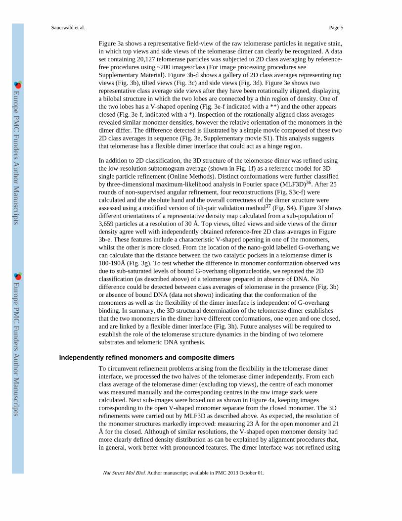
Figure 3a shows a representative field-view of the raw telomerase particles in negative stain,in which top views and side views of the telomerase dimer can clearly be recognized. A dataset containing 20,127 telomerase particles was subjected to 2D class averaging by reference-free procedures using ~200 images/class (For image processing procedures seeSupplementary Material). Figure 3b-d shows a gallery of 2D class averages representing topviews (Fig. 3b), tilted views (Fig. 3c) and side views (Fig. 3d). Figure 3e shows tworepresentative class average side views after they have been rotationally aligned, displayinga bilobal structure in which the two lobes are connected by a thin region of density. One ofthe two lobes has a V-shaped opening (Fig. 3e-f indicated with a **) and the other appearsclosed (Fig. 3e-f, indicated with a *). Inspection of the rotationally aligned class averagesrevealed similar monomer densities, however the relative orientation of the monomers in thedimer differ. The difference detected is illustrated by a simple movie composed of these two2D class averages in sequence (Fig. 3e, Supplementary movie S1). This analysis suggeststhat telomerase has a flexible dimer interface that could act as a hinge region.
In addition to 2D classification, the 3D structure of the telomerase dimer was refined usingthe low-resolution subtomogram average (shown in Fig. 1f) as a reference model for 3Dsingle particle refinement (Online Methods). Distinct conformations were further classifiedby three-dimensional maximum-likelihood analysis in Fourier space (MLF3D)36. After 25rounds of non-supervised angular refinement, four reconstructions (Fig. S3c-f) werecalculated and the absolute hand and the overall correctness of the dimer structure wereassessed using a modified version of tilt-pair validation method37 (Fig. S4). Figure 3f showsdifferent orientations of a representative density map calculated from a sub-population of3,659 particles at a resolution of 30 Å. Top views, tilted views and side views of the dimerdensity agree well with independently obtained reference-free 2D class averages in Figure3b-e. These features include a characteristic V-shaped opening in one of the monomers,whilst the other is more closed. From the location of the nano-gold labelled G-overhang wecan calculate that the distance between the two catalytic pockets in a telomerase dimer is180-190Å (Fig. 3g). To test whether the difference in monomer conformation observed wasdue to sub-saturated levels of bound G-overhang oligonucleotide, we repeated the 2Dclassification (as described above) of a telomerase prepared in absence of DNA. Nodifference could be detected between class averages of telomerase in the presence (Fig. 3b)or absence of bound DNA (data not shown) indicating that the conformation of themonomers as well as the flexibility of the dimer interface is independent of G-overhangbinding. In summary, the 3D structural determination of the telomerase dimer establishesthat the two monomers in the dimer have different conformations, one open and one closed,and are linked by a flexible dimer interface (Fig. 3h). Future analyses will be required toestablish the role of the telomerase structure dynamics in the binding of two telomeresubstrates and telomeric DNA synthesis.
Independently refined monomers and composite dimersTo circumvent refinement problems arising from the flexibility in the telomerase dimerinterface, we processed the two halves of the telomerase dimer independently. From eachclass average of the telomerase dimer (excluding top views), the centre of each monomerwas measured manually and the corresponding centres in the raw image stack werecalculated. Next sub-images were boxed out as shown in Figure 4a, keeping imagescorresponding to the open V-shaped monomer separate from the closed monomer. The 3Drefinements were carried out by MLF3D as described above. As expected, the resolution ofthe monomer structures markedly improved: measuring 23 Å for the open monomer and 21Å for the closed. Although of similar resolutions, the V-shaped open monomer density hadmore clearly defined density distribution as can be explained by alignment procedures that,in general, work better with pronounced features. The dimer interface was not refined using
Sauerwald et al. Page 5
Nat Struct Mol Biol. Author manuscript; available in PMC 2013 October 01.
Europe PM
C Funders A
uthor Manuscripts
Europe PM
C Funders A
uthor Manuscripts
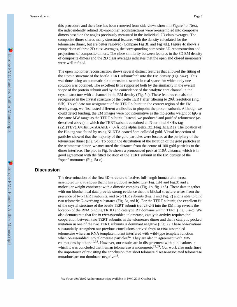
this procedure and therefore has been removed from side views shown in Figure 4b. Next,the independently refined 3D-monomer reconstructions were re-assembled into compositedimers based on the angles previously measured in the individual 2D class averages. Thecomposite dimer shares many structural features with the density calculated for thetelomerase dimer, but are better resolved (Compare Fig 3f. and Fig 4d.). Figure 4c shows acomparison of three 2D class averages, the corresponding composite 3D-reconstruction andprojections of composite dimers. The close similarity between features in the 3D EM densityof composite dimers and the 2D class averages indicates that the open and closed monomerswere well refined.
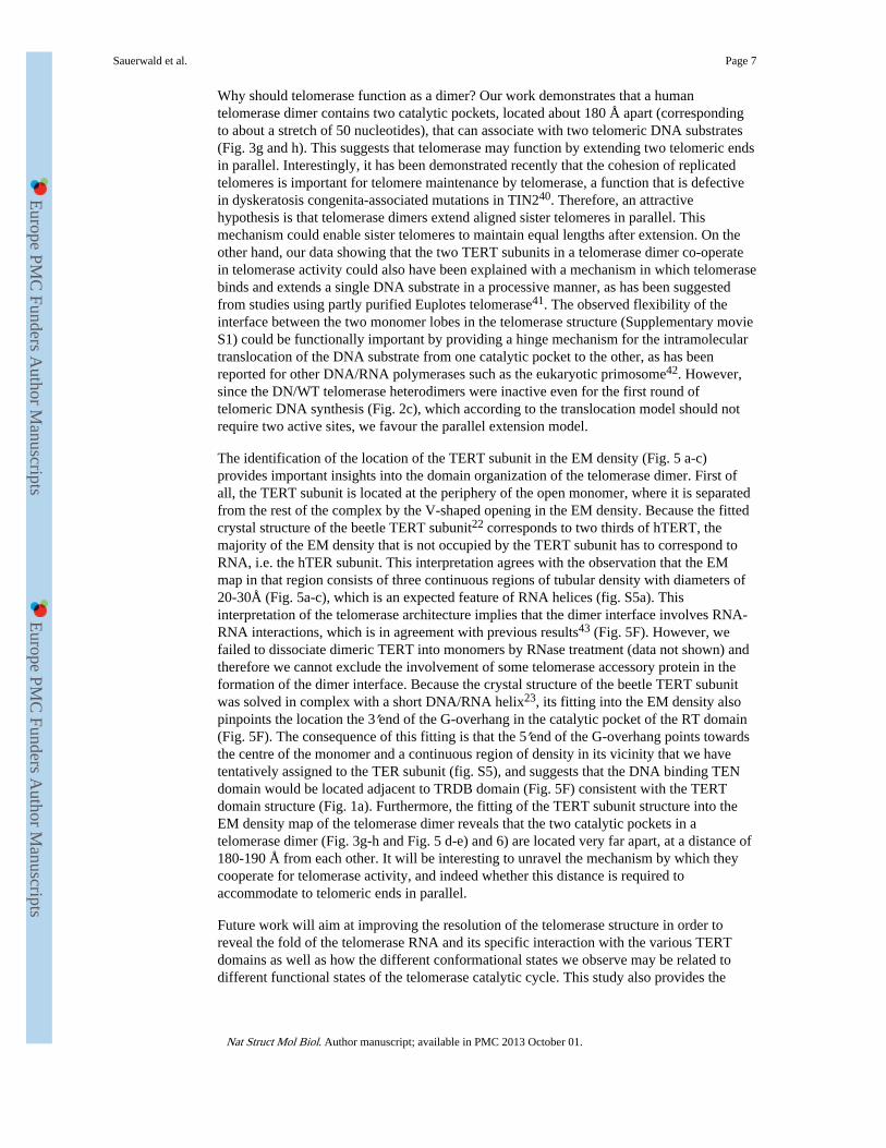
The open monomer reconstruction shows several distinct features that allowed the fitting ofthe atomic structure of the beetle TERT subunit22,25 into the EM density (Fig. 5a-c). Thiswas done using an automatic six dimensional search in real space, for which only onesolution was obtained. The excellent fit is supported both by the similarity in the overallshape of the protein subunit and by the coincidence of the catalytic core channel in thecrystal structure with a channel in the EM density (Fig. 5c). These features can also berecognized in the crystal structure of the beetle TERT after filtering to 20Å resolution (Fig.S5b). To validate our assignment of the TERT subunit to the correct region of the EMdensity map, we first tested different antibodies to pinpoint the protein subunit. Although wecould detect binding, the EM images were not informative as the molecular weight of IgG isthe same MW range as the TERT subunit. Instead, we produced and purified telomerase (asdescribed above) in which the TERT subunit contained an N-terminal 6×His-tag(ZZ_(TEV)_6×His_5x(AAAKE) ~37Å long alpha Helix_3x_Flag_hTERT). The location ofthe His-tag was found by using Ni-NTA coated 5nm colloidal gold. Visual inspection ofparticles showed that the majority of the gold particles were located at the periphery of thetelomerase dimer (Fig. 5d). To obtain the distribution of the location of the gold particles inthe telomerase dimer, we measured the distance from the centre of 100 gold particles to thedimer interface. The plot in Fig. 5e shows a pronounced peak at 110Å distance, which is ingood agreement with the fitted location of the TERT subunit in the EM density of the“open” monomer (Fig. 5a-c).
DiscussionThe determination of the first 3D-structure of active, full-length human telomeraseassembled in vivo shows that it has a bilobal architecture (Fig. 1d-f and Fig.3) and amolecular weight consistent with a dimeric complex (Fig. 1b, fig. 1aS). These data togetherwith our biochemical data provide strong evidence that the bilobal structure arises from thepresence of two TERT subunits, and two TER subunits (Fig. 1 and Fig. 2) and is able to bindtwo telomeric G-overhang substrates (Fig. 3g and h). For the TERT subunit, the excellent fitof the crystal structure of the beetle TERT subunit (ref 23-24) into the EM map reveals thelocation of the RNA binding TRBD and catalytic RT domains within TERT (Fig. 5 a-c). Wealso demonstrate that for in vivo assembled telomerase, catalytic activity requires thecooperation between two TERT subunits in the telomerase dimer and that a catalytic pockedmutation in one of the two TERT subunits is dominant negative (Fig. 2). These observationssubstantially strengthen our previous conclusions derived from in vitro assembledtelomerase where an RNA template mutant interfered with wild-type template functionwhen co-assembled into telomerase particles14. They are also in agreement with MWestimations by others10,38. However, our results are in disagreement with publications inwhich it was concluded that human telomerase is monomeric13,39. Our work also underlinesthe importance of revisiting the conclusion that short telomere disease-associated telomerasemutations are not dominant-negative13.
Sauerwald et al. Page 6
Nat Struct Mol Biol. Author manuscript; available in PMC 2013 October 01.
Europe PM
C Funders A
uthor Manuscripts
Europe PM
C Funders A
uthor Manuscripts
Why should telomerase function as a dimer? Our work demonstrates that a humantelomerase dimer contains two catalytic pockets, located about 180 Å apart (correspondingto about a stretch of 50 nucleotides), that can associate with two telomeric DNA substrates(Fig. 3g and h). This suggests that telomerase may function by extending two telomeric endsin parallel. Interestingly, it has been demonstrated recently that the cohesion of replicatedtelomeres is important for telomere maintenance by telomerase, a function that is defectivein dyskeratosis congenita-associated mutations in TIN240. Therefore, an attractivehypothesis is that telomerase dimers extend aligned sister telomeres in parallel. Thismechanism could enable sister telomeres to maintain equal lengths after extension. On theother hand, our data showing that the two TERT subunits in a telomerase dimer co-operatein telomerase activity could also have been explained with a mechanism in which telomerasebinds and extends a single DNA substrate in a processive manner, as has been suggestedfrom studies using partly purified Euplotes telomerase41. The observed flexibility of theinterface between the two monomer lobes in the telomerase structure (Supplementary movieS1) could be functionally important by providing a hinge mechanism for the intramoleculartranslocation of the DNA substrate from one catalytic pocket to the other, as has beenreported for other DNA/RNA polymerases such as the eukaryotic primosome42. However,since the DN/WT telomerase heterodimers were inactive even for the first round oftelomeric DNA synthesis (Fig. 2c), which according to the translocation model should notrequire two active sites, we favour the parallel extension model.
The identification of the location of the TERT subunit in the EM density (Fig. 5 a-c)provides important insights into the domain organization of the telomerase dimer. First ofall, the TERT subunit is located at the periphery of the open monomer, where it is separatedfrom the rest of the complex by the V-shaped opening in the EM density. Because the fittedcrystal structure of the beetle TERT subunit22 corresponds to two thirds of hTERT, themajority of the EM density that is not occupied by the TERT subunit has to correspond toRNA, i.e. the hTER subunit. This interpretation agrees with the observation that the EMmap in that region consists of three continuous regions of tubular density with diameters of20-30Å (Fig. 5a-c), which is an expected feature of RNA helices (fig. S5a). Thisinterpretation of the telomerase architecture implies that the dimer interface involves RNA-RNA interactions, which is in agreement with previous results43 (Fig. 5F). However, wefailed to dissociate dimeric TERT into monomers by RNase treatment (data not shown) andtherefore we cannot exclude the involvement of some telomerase accessory protein in theformation of the dimer interface. Because the crystal structure of the beetle TERT subunitwas solved in complex with a short DNA/RNA helix23, its fitting into the EM density alsopinpoints the location the 3′end of the G-overhang in the catalytic pocket of the RT domain(Fig. 5F). The consequence of this fitting is that the 5′end of the G-overhang points towardsthe centre of the monomer and a continuous region of density in its vicinity that we havetentatively assigned to the TER subunit (fig. S5), and suggests that the DNA binding TENdomain would be located adjacent to TRDB domain (Fig. 5F) consistent with the TERTdomain structure (Fig. 1a). Furthermore, the fitting of the TERT subunit structure into theEM density map of the telomerase dimer reveals that the two catalytic pockets in atelomerase dimer (Fig. 3g-h and Fig. 5 d-e) and 6) are located very far apart, at a distance of180-190 Å from each other. It will be interesting to unravel the mechanism by which theycooperate for telomerase activity, and indeed whether this distance is required toaccommodate to telomeric ends in parallel.
Future work will aim at improving the resolution of the telomerase structure in order toreveal the fold of the telomerase RNA and its specific interaction with the various TERTdomains as well as how the different conformational states we observe may be related todifferent functional states of the telomerase catalytic cycle. This study also provides the
Sauerwald et al. Page 7
Nat Struct Mol Biol. Author manuscript; available in PMC 2013 October 01.
Europe PM
C Funders A
uthor Manuscripts
Europe PM
C Funders A
uthor Manuscripts
starting point for investigating the structural basis of telomerase dimerization and itssignificance for catalysis and function.
Online MethodsTelomerase expression and purification
The telomerase complex was over-expressed using HEK293T cells1 transiently transfectedwith hTERT- and hTER-expressing plasmid DNA (pVan145 [pCDNA6-ZZ-(TEV)-3×Flag-hTERT(WT)], [pCDNA6-ZZ-(TEV)-3×Flag- hTERT(DN)], or [pCDNA6-13×Myc-hTERT(WT)], and pBS-U1-hTR) as described2 and whole-cell lysates (WCL) wereprepared as described3. For telomerase affinity purification, the WCL was adjusted to aprotein concentration of 4 mg/ml with buffer A (20 mM HEPES- KOH pH 7.9, 2 mMMgCl2, 300 mM KCl, 10 % glycerol (v/v), 1 mM DTT, 1 mM EDTA, 0.1 % Triton X-100(v/v), 1 mM PMSF) and clarified by filtration to produce the Input. To 20 ml Input 1 mlbuffer A equilibrated IgG –Sepharose 6 Fast Flow (GE Healthcare #17-0969-01) was addedand rotated for 3 hrs at 4°C and washed extensively with ice-cold buffer A. Telomerase wasreleased from 1ml IgG –Sepharose by overnight incubation with 5 ml 0.05 mg/ml TEVprotease (>90% efficient) in buffer A at 4°C. In a second affinity purification step, thesoluble telomerase containing fraction was applied to a 1 ml HiTrap Heparin HP column(GE Healthcare #17-0406-01) previously equilibrated with buffer A and washed with 10column volumes of buffer A to remove unbound proteins. Bound proteins were eluted usinga gradient of 0.3 – 1 M KCl in buffer A. Fractions containing telomerase were identifiedusing a direct telomerase activity assay (Fig. 1c) as described3 and active fractions werepooled, concentrated, and dialyzed against buffer B (20 mM HEPES-KOH pH 7.9, 2 mMMgCl2, 150 mM KCl, 10 % glycerol (v/v), 1 mM DTT, 1 mM EDTA, 1 mM PMSF).Telomerase yields were quantified by Northern blot analysis using hTR run-off transcriptsas standards and the purity was assessed by SDS-PAGE.
Telomerase sucrose gradient sedimentation in combination with fixation (GraFix)For EM analysis the telomerase complex was further purified using GraFix4. Briefly,solutions of buffer B containing 5 % and 30 % Sucrose were prepared in Falcon tubes. 0.1%glutaraldehyde (EM grade 25%, Science Services GmbH, Munich, Germany) was added tothe high-density solution only. 4 ml gradients for centrifugation using a Beckman SW60rotor were prepared using a gradient former (Gradient Master 107, BioComp Instruments,Canada). Freshly prepared gradients were kept at 4°C for one hr before sample loading.Subsequently 200 μl, containing 10-30 pmol of purified telomerase bound to 5′-[32P]-(TTAGGG)2-3′ was loaded on top of the gradient and run for 20 hr at max 215,000 × g and4°C. Sucrose gradients were run in parallel without fixation reagent to assess whether thepresence of glutaraldehyde caused any differences in particle sedimentation. 200 μl fractionswere collected manually from top to bottom (Fig. 1b). The glutaraldehyde was inactivatedafter fractionation by adding glycine to a final concentration of 80 mM. For EM analysis, thesucrose from the GraFix fractions was removed by applying the sample to a buffer exchangespin column (Zeba spin columns, Pierce, Rockford, IL, USA) previously equilibrated withbuffer C (20 mM Tris – HCl pH 7.6, 150 mM KCl, 1 mM MgCl2).
SDS-PAGE and E MSA analysisFor SDS-PAGE analysis, 10 μl of extract were directly boiled for 5 min in Laemmli samplebuffer and fractionated on a 4–20 % SDS–PAGE gradient gel. Proteins were silver stained.For the EMSA analysis, a two fold dilution series of IgG-Sepharose/Heparin purifiedtelomerase fraction was incubated with 250 nM 32P-(TTAGGG)3 telomeric DNA in thepresence of 5 μM CTAGACCTGTCATCA competitor DNA and 0.1 mg/ml acetylated BSAin heparin binding buffer (20 mM HEPES-KOH pH7.9, 300 mM KCl, 2 mM MgCl2, 1 mM
Sauerwald et al. Page 8
Nat Struct Mol Biol. Author manuscript; available in PMC 2013 October 01.
Europe PM
C Funders A
uthor Manuscripts
Europe PM
C Funders A
uthor Manuscripts
EDTA pH8, 1 mM DTT, 10 % glycerol). Reactions were incubated at RT for 5 min beforeelectrophoresis on a 6.7% acrylamide native gel (80:1 acrylamide:bis acrylamide, 5%glycerol, 0.5× Tris borate-EDTA) run at 200 V for 15 h at 4°C and fixed in MeOH/Acetate.As control, oligo DNA was incubated without telomerase. The gel was exposed to X-rayfilm.
Nano-liquid chromatography tandem mass spectrometry (nanoLC-MS/MS)Analysis of the protein content of the purified telomerase samples was carried out by adding20 μl of a trypsin solution (10 ng/μl in 10 mM Tris/2 mM CaCl2, pH 8.2) to a 200-μl aliquot(estimated at 0.5 pmol) of the purified telomerase or control samples. Microwave (CEMDiscover, CEM Corp., USA) assisted trypsin digestion was performed at 60°C at 5 W for 30min. After digestion the sample was concentrated to approximately 50 μl in a vacuumcentrifuge (Savant Instruments Inc., USA), diluted with 50 μl 0.1% formic acid and 4 μl wasloaded onto a nano-UPLC system (nanoAcquity UPLC system, Waters, USA) through atrap-column (Symmetry, C18, 180 μm i.d. x 20 mm length, Waters, USA). Peptides wereseparated on a C18-column (BEH300 C18, 75 μm i.d. x 150 mm length, Waters, USA) at250 nl/min using a linear gradient from 1 to 60% solvent B (solvent A: 0.1% formic acid inwater, solvent B: 0.1% formic acid in acetonitrile) in 60 min. Mass spectra were recorded ona Synapt G2 HDMS (Waters, USA). Information-dependent data acquisition was carried outusing a 0.5-second survey scan from which the five most abundant doubly and triplycharged peptides were selected for product ion scans. The resulting data were searchedagainst the SwissProt database using the ProteinLynx Global Server (Waters, USA) searchengine (table S1).
Direct Telomerase Activity AssaysConventional telomerase assays were carried out for 45 min at 30°C in 20-μl reactionscontaining 1 μl of cell extract, 50mM Tris– HCl (pH 8.0), 50mM KCl, 1mM spermidine,1mM β-mercaptoethanol, 1mM MgCl2, 0.5mM dATP, 0.5mM dTTP, 2 mM dGTP, 20 μCiof [α-32P]dGTP (3000 Ci/mmol) and 1 μM of telomeric primer 5′-Biotin(T2AG3)-3′.Reactions were stopped by the addition of 5mM EDTA and 1% SDS. Biotinylated telomericprimers were recovered with Dynabeads® M-280 Streptavidin (Invitrogen) following theinstructions provided by the manufacturer. Beads were re-suspended in 98% formamidecontaining 10mM EDTA and 0.005% xylene cyanol, heated to 95 °C for 5 min, andanalyzed on a 12% polyacrylamide-urea sequencing gels.
Western analysisFor Western analysis, 4 μl of extract were boiled for 5 min in Laemmli sample buffer andfractionated on 4–15 % SDS–PAGE gradient gels. Immunoblots with standard protocols,using an affinity purified rabbit polyclonal antibody against hTERT (1:10000, R484) 5. Assecondary IRDye 800CW Goat anti-Rabbit IgG (Li Cor) was used (1:7000). The Li-CorInfrared fluorescence detection of IRDye infrared dye substrate and imaging system wasused to detect bound antibodies.
Telospot assayTelospot telomerase activity assays were performed as previously described6. Briefly,telomerase activity reactions were carried out for 45 min at 30 °C in 20 μl volumescontaining 1 μl of cell extracts, 50mM Tris– HCl (pH 8.0), 50mM KCl, 1mM spermidine,1mM β-mercaptoethanol, 1mM MgCl2, 33 μM dATP, 33 μM dTTP, 33 μM dGTP, and 0.25mM of telomeric primer (T2AG3)3. Reactions were stopped by the addition of 2 μl of 0.25MEDTA / 0.05% bromophenol blue solution. 0.5 μl of the reaction mix was spotted inquadruplicate on a GeneScreen Plus charged nylon membrane (PerkinElmer). DNA samples
Sauerwald et al. Page 9
Nat Struct Mol Biol. Author manuscript; available in PMC 2013 October 01.
Europe PM
C Funders A
uthor Manuscripts
Europe PM
C Funders A
uthor Manuscripts
were UV crosslinked to the membrane with the auto-crosslink function of a Stratalinker(Stratagene). Without prior denaturation membranes were blocked for 1 hr at 60 °C withchurch buffer. After overnight hybridization at 60 °C with a randomly labeled probe derivedfrom a 600 bp TTAGGG-repeat containing DNA fragment, membranes were washed twicefor 15 min with 2× SSC buffer at room temperature and twice for 30 min with 2× SSC, 0.1% SDS at 65 °C and exposed to a phosphoimager screen. Spot intensities were quantifiedusing 2D-densitometry and the Aida software (Raytest).
EM sample preparation and imagingContinuous carbon-coated grids were freshly prepared and glow-discharged before use. 13μl of telomerase sample (8-10 nM) were deposited on the grid for 15-30 minutes, blottedwith filter paper and negatively stained with 2 drops of 1-2% (w/v) uranyl acetate solution.A homogenous sample preparation for EM was obtained using these conditions although theparticle concentration was low (30-100 particles/image). Single particle EM data werecollected on a FEI CM12 transmission electron microscope, operated at 120 keV and liquidnitrogen temperature. The nominal magnification was 42,000× (calibrated magnification,42,550×). Micrographs were recorded on Kodak SO-163 film with an electron dose of 10-12e/Å2 and a defocus of 1.0–1.5 μm. The films were developed in Kodak developer at fullstrength for 12 min and digitized with a Zeiss SCAI scanner using a step size of 7 μm. Themicrographs were compressed x4 giving a final object pixel size of 6.6Å/pixel. Figures 1dand colloidal gold data were recorded on a 2k TVIPS CCD camera with an object pixel sizeof 3.25 Å. Single axis tilt series were recorded with SerialEM7 on a FEI Tecnai G2 Polaramicroscope at 300keV and 3.5 μm underfocus. The specimen was tilted ±60° or ±65° andimages were recorded every second degree on a 2k TVIPS CCD detector. The electron doseper image was 10 e/Å2 and the pixel size was 5.65 Å at the specimen level.
Particle selection and 2D class averagingA data set of 26,361 telomerase particles bound to oligonucleotide 5′-(TTAGGG)2- 3′ wereselected manually with Ximdisp8 using a box size of 78×78 pixels. The image stack wasnormalised and sorted by statistics in Xmipp9, and 20,127 images were selected for furtheranalysis. A maximum likelihood target function in Fourier space (MLF2D10) was used formulti-reference alignment and 2D classification.
Initial low-resolution 3D reconstruction by electron tomographyElectron tomography was used to calculate a low-resolution reference model. Single axis tiltseries were compressed x2 giving a pixel size of 11.3 Å at the specimen level. Themicrographs were coarsely aligned by cross-correlation and reconstructed by filtered back-projection in IMOD11. An iterative refinement procedure was used to refine the alignmentparameters and the reconstruction in EMTIAR12. 50 evenly stained sub-tomogramscorresponding to individual telomerase particles were manually selected in Bsoft13 andextracted using a box size of 40×40×40 voxels. An average 3D reconstruction wascalculated by missing-wedge weighted, reference-free alignment of sub-tomograms inXmipp14. 13 sub-tomograms with the highest cross correlation to the average (Fig. 1f andfig. S3a) were included in the final sub-tomogram average.
Map refinement by single particle electron microscopyInitial single-particle refinement was performed in EMAN215 using the sub-tomogramaverage (fig. S3a) as a starting reference. The reference model was initially filtered to 80 Åresolution and refined (fig. S3b) by 20 rounds of multi-reference alignment, classification bymultivariate statistical analysis and angular assignment by projection matching. Classaverages with 6 degree angular spacing were calculated using 6 averaging iterations. The 3D
Sauerwald et al. Page 10
Nat Struct Mol Biol. Author manuscript; available in PMC 2013 October 01.
Europe PM
C Funders A
uthor Manuscripts
Europe PM
C Funders A
uthor Manuscripts
structure was calculated at 35 Å resolution by direct Fourier inversion. To analyse structuralvariability in the telomerase dimer we used 3D-classification by maximum-likelihood inFourier space (MLF3D10). After 25 rounds of non-supervised angular refinement againstfive reference models, four homogenous subsets of images were selected for furtherrefinement (a fifth class was interpreted as an accumulation of particles of low quality). Theselected classes contained 2608, 2631, 3659 and 5519 particles respectively. Subsequentrefinement in EMAN2 (as described above) yielded reconstructions with resolutions in therange of 30-40 Å (fig. S3c-f).
Map validation and absolute hand determinationThe absolute hand and the overall correctness of the dimer structure in Figure 3f wereassessed using the tilt-pair validation method16. From one of the tilt series that was used tocalculate electron tomograms for our reference-free initial model calculation, we selectedthe individual projections of one of the dimer particles (selection of other particles gavesimilar results). These single-particle projections were then aligned independently againstreference projections of the dimer structure. Analysis of the relative tilt angles between theseimages (fig. S4) validated the structure, while comparison with an identical analysis for a tiltseries that was collected on the same microscope on a sample of known handedness (helicalassemblies of acetyl choline receptors) served to determine its absolute hand.
Independent monomer refinement and reconstruction of composite dimersFrom 2D class averages of telomerase dimer side-views, we calculated the centre of eachmonomer in the raw image stack; 9,380 sub-images were selected of the open monomer andthe closed monomer (4,690 each), and extracted using a box size of 34×34 pixels. Thesesub-images were used for independent monomer refinement, using a two-reference modelprocedure, in Xmipp (Fig. 4-5). To re-combine the monomers into composite dimers,reference-free 2D class averages (Fig. 4c top panel), were used to determine the orientationof the open and closed monomers (calculated as the average orientation of all sub-imagesthat were included in the 2D class average). This analysis provided a reconstruction of therelative orientation of the independely refined monomers (Fig. 4c middle panel). Since theanalysis was carried out using 2D class averages of side views, the Z-height of themonomers were adjusted by hand and compared to the previously refined intact dimer (Fig.3f). Visual inspection shows that projections of the assembled composite dimer (Fig. 4c,lower panel) agreed well with the 2D class averages (Fig. 4c top panel).
Colloidal gold labeling of telomerase domainsTwo types of 5 nm colloidal gold, coated with either streptavidin (prepared as described 17)or Ni-NTA (Nanoprobes, Inc Yaphank, NY, USA), was used to identify the presence andlocation of the telomeric G-overhang and the TERT subunit respectively. The bindingintegrity of the monovalent streptavidin gold complex to 5′-[32P]/biotin 5′-[32P]-(TTAGGG)2-3′ -3′ was analysed by EMSA. After GraFix purification of the telomerase (asdescribed above), streptavidin coated gold was added in a stochiometric ratio of 2:1 totelomerase bound to 5′- Biotin dT-(TTAGGG)2- 3′ and incubated for 30 minutes on icebefore preparing EM grids (Fig. 1g). To map the location of the TERT subunit, telomeraseN-terminal 6×His-tag (ZZ_(TEV)_6×His_5x(AAAKE) ~37Å long alphahelix_3x_Flag_hTERT) was incubated with Ni-NTA coated gold for 3 hours at roomtemperature (Fig. 5d). We found more telomerase particles in complex with streptavidincoated gold (87%) compared to Ni-NTA coated gold (16%), which is likely to reflect thehigh affinity of streptavidin-biotin (not shown).
Sauerwald et al. Page 11
Nat Struct Mol Biol. Author manuscript; available in PMC 2013 October 01.
Europe PM
C Funders A
uthor Manuscripts
Europe PM
C Funders A
uthor Manuscripts
Model fitting and density visualizationThe crystal structure of the TERT protein subunit (pdb code: 3KYL) was docked into theEM density of the monomeric subunit using automatic docking procedures in Chimera18.This program was also used for 3D density visualization and generating a 23 Å density mapof the TERT subunit (Fig. S5b).
Supplementary MaterialRefer to Web version on PubMed Central for supplementary material.
AcknowledgmentsWe are indebted to Patrick Reichenbach, Benoit Zuber, Steven J. Ludtke, Richard Henderson, Jude Short, JakeGrimmett, Shaoxia Chen and Alix Christen for help and advice. We thank Sarah Thurnheer and David Hacker(EPFL-PECF) for telomerase production and Peter Hunziker from the Functional Genomics Center Zurich forprotein identification. We thank the Human Frontier Science Program for funding through a grant (numberRGP0032/2005-C) awarded to J.L. and D.R. and a post-doctoral fellowship to A.S.. We thank the EuropeanMolecular Biology Organization for post-doctorial fellowships to A.S and S.S; the UK Medical Research Councilfor a career-development fellowship to S.S.. Work in J.L.’s laboratory is supported by the Swiss National ScienceFoundation and a European Research Council advanced investigator grant (grant agreement number 232812). Workin DR’s and S.H.W.S.’s laboratory is supported by the UK Medical Research Council (MC_UP_A025_1013).
References1. de Lange T. How telomeres solve the end-protection problem. Science. 2009; 326:948–952.
[PubMed: 19965504]
2. Harley CB. Telomerase and cancer therapeutics. Nat Rev Cancer. 2008; 8:167–179. [PubMed:18256617]
3. Greider CW, Blackburn EH. A telomeric sequence in the RNA of Tetrahymena telomerase requiredfor telomere repeat synthesis. Nature. 1989; 337:331–337. [PubMed: 2463488]
4. Yu GL, Bradley JD, Attardi LD, Blackburn EH. In vivo alteration of telomere sequences andsenescence caused by mutated Tetrahymena telomerase RNAs. Nature. 1990; 344:126–132. doi:10.1038/344126a0. [PubMed: 1689810]
5. Cech T. Beginning to understand the end of the chromosome. Cell. 2004; 116:273–279. [PubMed:14744437]
6. Lingner J, et al. Reverse transcriptase motifs in the catalytic subunit of telomerase. Science. 1997;276:561–567. [PubMed: 9110970]
7. Blackburn EH, Collins K. Telomerase: an RNP enzyme synthesizes DNA. Cold Spring HarbPerspect Biol. 2011; 3
8. Weinrich S, et al. Reconstitution of human telomerase with the template RNA component hTR andthe catalytic protein subunit hTRT. Nat Genet. 1997; 17:498–502. doi:10.1038/ng1297-4980.[PubMed: 9398860]
9. Autexier C, Lue N. The structure and function of telomerase reverse transcriptase. Annu RevBiochem. 2006; 75:493–517. [PubMed: 16756500]
10. Cohen S, et al. Protein composition of catalytically active human telomerase from immortal cells.Science. 2007; 315:1850–1853. [PubMed: 17395830]
11. Egan E, Collins K. Specificity and stoichiometry of subunit interactions in the human telomeraseholoenzyme assembled in vivo. Mol Cell Biol. 2010; 30:2775–2786. [PubMed: 20351177]
12. Venteicher A, et al. A human telomerase holoenzyme protein required for Cajal body localizationand telomere synthesis. Science. 2009; 323:644–648. doi:10.1126/science.11653570. [PubMed:19179534]
13. Errington TM, Fu D, Wong JM, Collins K. Disease-associated human telomerase RNA variantsshow loss of function for telomere synthesis without dominant-negative interference. Mol CellBiol. 2008; 28:6510–6520. [PubMed: 18710936]
Sauerwald et al. Page 12
Nat Struct Mol Biol. Author manuscript; available in PMC 2013 October 01.
Europe PM
C Funders A
uthor Manuscripts
Europe PM
C Funders A
uthor Manuscripts

14. Wenz C, et al. Human telomerase contains two cooperating telomerase RNA molecules. EMBO J.2001; 20:3526–3534. doi:10.1093/emboj/20.13.35260. [PubMed: 11432839]
15. Arai K, et al. Two independent regions of human telomerase reverse transcriptase are important forits oligomerization and telomerase activity. J Biol Chem. 2002; 277:8538–8544. Epub 2001 Dec85180. [PubMed: 11751869]
16. Beattie TL, Zhou W, Robinson MO, Harrington L. Functional multimerization of the humantelomerase reverse transcriptase. Mol Cell Biol. 2001; 21:6151–6160. [PubMed: 11509658]
17. Moriarty TJ, Huard S, Dupuis S, Autexier C. Functional multimerization of human telomeraserequires an RNA interaction domain in the N terminus of the catalytic subunit. Mol Cell Biol.2002; 22:1253–1265. [PubMed: 11809815]
18. Gardano L, Holland L, Oulton R, Le Bihan T, Harrington L. Native gel electrophoresis of humantelomerase distinguishes active complexes with or without dyskerin. Nucleic Acids Res. 2011;19:19.
19. Sekaran V, Soares J, Jarstfer M. Structures of telomerase subunits provide functional insights.Biochim Biophys Acta. 2010; 1804:1190–1201. doi:10.1016/j.bbapap.2009.07.0190. [PubMed:19665593]
20. Wyatt H, West S, Beattie T. InTERTpreting telomerase structure and function. Nucleic Acids Res.2010 doi:10.1093/nar/gkq370.
21. Zhang Q, Kim NK, Feigon J. Architecture of human telomerase RNA. Proc Natl Acad Sci U S A.2011; 108:20325–20332. Epub 22011 Aug 203150. [PubMed: 21844345]
22. Mitchell M, Gillis A, Futahashi M, Fujiwara H, Skordalakes E. Structural basis for telomerasecatalytic subunit TERT binding to RNA template and telomeric DNA. Nat Struct Mol Biol. 2010;17:513–518. doi:10.1038/nsmb.17770. [PubMed: 20357774]
23. Schuller AP, Harkisheimer MJ, Skordalakes E. In vitro reconstitution of the active T. castaneumtelomerase. J Vis Exp. 2011:e2799. doi: 2710.3791/27990. [PubMed: 21775967]
24. Kohlstaedt L, Wang J, Friedman J, Rice P, Steitz T. Crystal structure at 3.5 A resolution of HIV-1reverse transcriptase complexed with an inhibitor. Science. 1992; 256:1783–1790. [PubMed:1377403]
25. Gillis A, Schuller A, Skordalakes E. Structure of the Tribolium castaneum telomerase catalyticsubunit TERT. Nature. 2008; 455:633–637. [PubMed: 18758444]
26. Jacobs S, Podell E, Cech T. Crystal structure of the essential N-terminal domain of telomerasereverse transcriptase. Nat Struct Mol Biol. 2006; 13:218–225. doi:10.1038/nsmb10540. [PubMed:16462747]
27. Theimer C, Feigon J. Structure and function of telomerase RNA. Curr Opin Struct Biol. 2006;16:307–318. doi:10.1016/j.sbi.2006.05.0050. [PubMed: 16713250]
28. Mitchell J, Collins K. Human telomerase activation requires two independent interactions betweentelomerase RNA and telomerase reverse transcriptase. Mol Cell. 2000; 6:361–371. [PubMed:10983983]
29. Kelleher C, Teixeira M, Forstemann K, Lingner J. Telomerase: biochemical considerations forenzyme and substrate. Trends Biochem Sci. 2002; 27:572–579. [PubMed: 12417133]
30. Cristofari G, et al. Low- to high-throughput analysis of telomerase modulators with Telospot. NatMethods. 2007; 4:851–853. [PubMed: 17893679]
31. Cristofari G, Lingner J. Telomere length homeostasis requires that telomerase levels are limiting.EMBO J. 2006; 25:565–574. [PubMed: 16424902]
32. Mitchell J, Wood E, Collins K. A telomerase component is defective in the human diseasedyskeratosis congenita. Nature. 1999; 402:551–555. doi:10.1038/9901410. [PubMed: 10591218]
33. Howarth M, et al. A monovalent streptavidin with a single femtomolar biotin binding site. NatMethods. 2006; 3:267–273. [PubMed: 16554831]
34. Hahn WC, et al. Inhibition of telomerase limits the growth of human cancer cells. Nat Med. 1999;5:1164–1170. [PubMed: 10502820]
35. Kastner B, et al. GraFix: sample preparation for single-particle electron cryomicroscopy. NatMethods. 2008; 5:53–55. [PubMed: 18157137]
Sauerwald et al. Page 13
Nat Struct Mol Biol. Author manuscript; available in PMC 2013 October 01.
Europe PM
C Funders A
uthor Manuscripts
Europe PM
C Funders A
uthor Manuscripts
36. Scheres SH, et al. Modeling experimental image formation for likelihood-based classification ofelectron microscopy data. Structure. 2007; 15:1167–1177. [PubMed: 17937907]
37. Rosenthal PB, Henderson R. Optimal determination of particle orientation, absolute hand, andcontrast loss in single-particle electron cryomicroscopy. J Mol Biol. 2003; 333:721–745.[PubMed: 14568533]
38. Gardano L, Holland L, Oulton R, Le Bihan T, Harrington L. Native gel electrophoresis of humantelomerase distinguishes active complexes with or without dyskerin. Nucleic Acids Res. 2012;40:e36. Epub 2011 Dec 20190. [PubMed: 22187156]
39. Alves D, et al. Single-molecule analysis of human telomerase monomer. Nat Chem Biol. 2008;4:287–289. [PubMed: 18391947]
40. Canudas S, et al. A role for heterochromatin protein 1gamma at human telomeres. Genes Dev.2011; 25:1807–1819. Epub 2011 Aug 18240. [PubMed: 21865325]
41. Fouche N, Moon IK, Keppler BR, Griffith JD, Jarstfer MB. Electron microscopic visualization oftelomerase from Euplotes aediculatus bound to a model telomere DNA. Biochemistry. 2006;45:9624–9631. [PubMed: 16878997]
42. Nunez-Ramirez R, et al. Flexible tethering of primase and DNA Pol alpha in the eukaryoticprimosome. Nucleic Acids Res. 2011; 39:8187–8199. Epub 2011 Jun 81280. [PubMed: 21715379]
43. Ren X, et al. Identification of a new RNA.RNA interaction site for human telomerase RNA (hTR):structural implications for hTR accumulation and a dyskeratosis congenita point mutation. NucleicAcids Res. 2003; 31:6509–6515. [PubMed: 14602909]
References1. Graham F, Smiley J, Russell W, Nairn R. Characteristics of a human cell line transformed by DNA
from human adenovirus type 5. J Gen Virol. 1977; 36:59–74. [PubMed: 886304]
2. Cristofari G, et al. Human telomerase RNA accumulation in Cajal bodies facilitates telomeraserecruitment to telomeres and telomere elongation. Mol Cell. 2007; 27:882–889. [PubMed:17889662]
3. Cohen S, et al. Protein composition of catalytically active human telomerase from immortal cells.Science. 2007; 315:1850–1853. [PubMed: 17395830]
4. Kastner B, et al. GraFix: sample preparation for single-particle electron cryomicroscopy. NatMethods. 2008; 5:53–55. [PubMed: 18157137]
5. Wenz C, et al. Human telomerase contains two cooperating telomerase RNA molecules. EMBO J.2001; 20:3526–3534. doi:10.1093/emboj/20.13.3526. [PubMed: 11432839]
6. Cristofari G, et al. Low- to high-throughput analysis of telomerase modulators with Telospot. NatMethods. 2007; 4:851–853. [PubMed: 17893679]
7. Mastronarde DN. Automated electron microscope tomography using robust prediction of specimenmovements. J Struct Biol. 2005; 152:36–51. doi:10.1016/j.jsb.2005.07.007. [PubMed: 16182563]
8. Smith JM. Ximdisp--A visualization tool to aid structure determination from electron microscopeimages. J Struct Biol. 1999; 125:223–228. doi:10.1006/jsbi.1998.4073. [PubMed: 10222278]
9. Sorzano CO, et al. XMIPP: a new generation of an open-source image processing package forelectron microscopy. J Struct Biol. 2004; 148:194–204. doi:10.1016/j.jsb.2004.06.006. [PubMed:15477099]
10. Scheres SH, et al. Modeling experimental image formation for likelihood-based classification ofelectron microscopy data. Structure. 2007; 15:1167–1177. [PubMed: 17937907]
11. Kremer JR, Mastronarde DN, McIntosh JR. Computer visualization of three-dimensional imagedata using IMOD. J Struct Biol. 1996; 116:71–76. [PubMed: 8742726]
12. Chen H, Hughes DD, Chan TA, Sedat JW, Agard DA. IVE (Image Visualization Environment): asoftware platform for all three-dimensional microscopy applications. J Struct Biol. 1996; 116:56–60. [PubMed: 8742723]
13. Heymann JB, Belnap DM. Bsoft: image processing and molecular modeling for electronmicroscopy. J Struct Biol. 2007; 157:3–18. [PubMed: 17011211]
Sauerwald et al. Page 14
Nat Struct Mol Biol. Author manuscript; available in PMC 2013 October 01.
Europe PM
C Funders A
uthor Manuscripts
Europe PM
C Funders A
uthor Manuscripts
14. Scheres SH, Melero R, Valle M, Carazo JM. Averaging of electron subtomograms and randomconical tilt reconstructions through likelihood optimization. Structure. 2009; 17:1563–1572.[PubMed: 20004160]
15. Tang G, et al. EMAN2: an extensible image processing suite for electron microscopy. J StructBiol. 2007; 157:38–46. doi:10.1016/j.jsb.2006.05.009. [PubMed: 16859925]
16. Rosenthal PB, Henderson R. Optimal determination of particle orientation, absolute hand, andcontrast loss in single-particle electron cryomicroscopy. J Mol Biol. 2003; 333:721–745.[PubMed: 14568533]
17. Beesley, JE. Colloidal gold : a new perspective for cytochemical marking. Oxford University Press[for] Royal Microscopical Society; 1989. p. 17Vol. Microscopy handbooks
18. Pettersen EF, et al. UCSF Chimera--a visualization system for exploratory research and analysis. JComput Chem. 2004; 25:1605–1612. doi:10.1002/jcc.20084. [PubMed: 15264254]
Sauerwald et al. Page 15
Nat Struct Mol Biol. Author manuscript; available in PMC 2013 October 01.
Europe PM
C Funders A
uthor Manuscripts
Europe PM
C Funders A
uthor Manuscripts

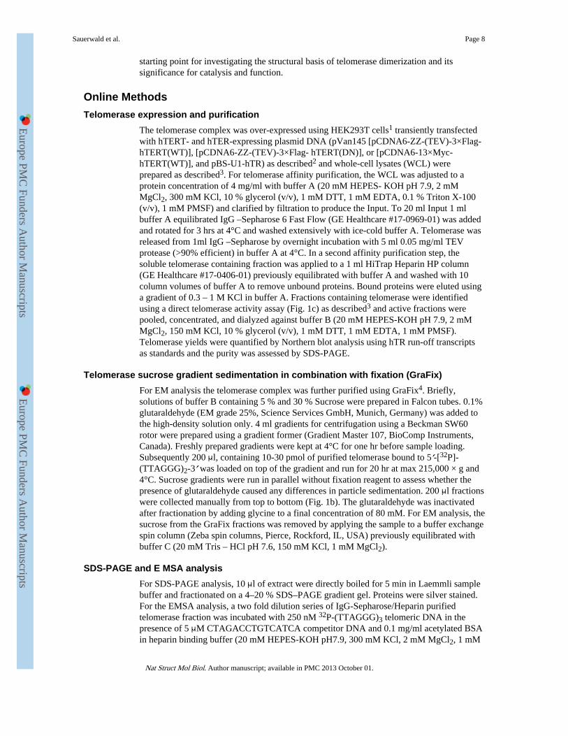
Figure 1. Human telomerase is a dimera, Schematic of hTERT domain arrangement: The essential N-terminal domain involved inDNA binding (TEN), the RNA binding domain (TRBD), the reverse transcriptase domain(RT) that contains the catalytic site and the C-terminal extension (CTE). b, Elution profile ofthe in vivo assembled human telomerase in complex with the G-overhang oligonucleotide5′-[32P]/biotin dT](TTAGGGT)2-3′ fractionated on a sucrose gradient. c, Telomeraseactivity profile of a telomerase sample fractionated as in (b), but in the absence of G-overhang. G-overhang bound (b) and unbound (c) telomerase complexes migrate in the sameposition on the sucrose gradient. d, Electron micrograph of negatively stained telomerasebound to the G-overhang. Top views are indicated by black circles and side views by whitecircles. e, Example of reference-free 2D class averages. f, 3D structure obtained bysubtomogram averaging. g and h, Electron micrographs of telomerase bound by 5 nmcolloidal gold coated with streptavidin (g) and 5 nm colloidal gold coated with streptavidinin the absence of telomerase (h). Telomerase dimers in complex with one gold particle arecircled in red and in complex with two gold particles circled in yellow. The arrows indicatedcolloidal gold particles that are not bound to telomerase. The scale bar in images (d), (g)and (h) is 50 nm and in images (e) and (f) is 5nm.
Sauerwald et al. Page 16
Nat Struct Mol Biol. Author manuscript; available in PMC 2013 October 01.
Europe PM
C Funders A
uthor Manuscripts
Europe PM
C Funders A
uthor Manuscripts
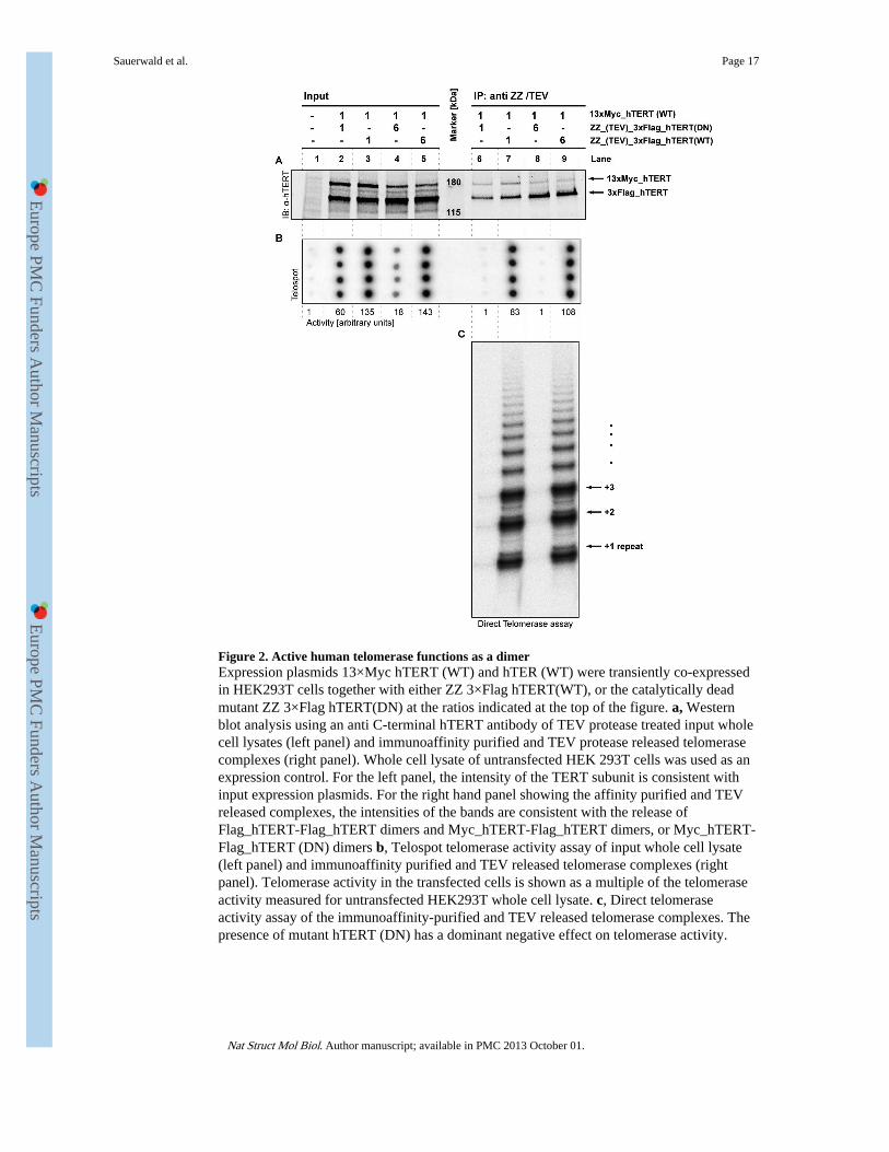
Figure 2. Active human telomerase functions as a dimerExpression plasmids 13×Myc hTERT (WT) and hTER (WT) were transiently co-expressedin HEK293T cells together with either ZZ 3×Flag hTERT(WT), or the catalytically deadmutant ZZ 3×Flag hTERT(DN) at the ratios indicated at the top of the figure. a, Westernblot analysis using an anti C-terminal hTERT antibody of TEV protease treated input wholecell lysates (left panel) and immunoaffinity purified and TEV protease released telomerasecomplexes (right panel). Whole cell lysate of untransfected HEK 293T cells was used as anexpression control. For the left panel, the intensity of the TERT subunit is consistent withinput expression plasmids. For the right hand panel showing the affinity purified and TEVreleased complexes, the intensities of the bands are consistent with the release ofFlag_hTERT-Flag_hTERT dimers and Myc_hTERT-Flag_hTERT dimers, or Myc_hTERT-Flag_hTERT (DN) dimers b, Telospot telomerase activity assay of input whole cell lysate(left panel) and immunoaffinity purified and TEV released telomerase complexes (rightpanel). Telomerase activity in the transfected cells is shown as a multiple of the telomeraseactivity measured for untransfected HEK293T whole cell lysate. c, Direct telomeraseactivity assay of the immunoaffinity-purified and TEV released telomerase complexes. Thepresence of mutant hTERT (DN) has a dominant negative effect on telomerase activity.
Sauerwald et al. Page 17
Nat Struct Mol Biol. Author manuscript; available in PMC 2013 October 01.
Europe PM
C Funders A
uthor Manuscripts
Europe PM
C Funders A
uthor Manuscripts
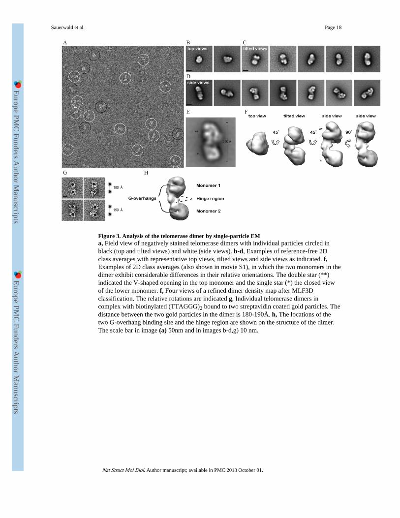
Figure 3. Analysis of the telomerase dimer by single-particle EMa, Field view of negatively stained telomerase dimers with individual particles circled inblack (top and tilted views) and white (side views). b-d, Examples of reference-free 2Dclass averages with representative top views, tilted views and side views as indicated. f,Examples of 2D class averages (also shown in movie S1), in which the two monomers in thedimer exhibit considerable differences in their relative orientations. The double star (**)indicated the V-shaped opening in the top monomer and the single star (*) the closed viewof the lower monomer. f, Four views of a refined dimer density map after MLF3Dclassification. The relative rotations are indicated g, Individual telomerase dimers incomplex with biotinylated (TTAGGG)2 bound to two streptavidin coated gold particles. Thedistance between the two gold particles in the dimer is 180-190Å. h, The locations of thetwo G-overhang binding site and the hinge region are shown on the structure of the dimer.The scale bar in image (a) 50nm and in images b-d,g) 10 nm.
Sauerwald et al. Page 18
Nat Struct Mol Biol. Author manuscript; available in PMC 2013 October 01.
Europe PM
C Funders A
uthor Manuscripts
Europe PM
C Funders A
uthor Manuscripts
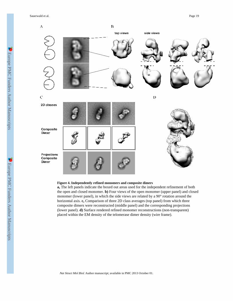
Figure 4. Independently refined monomers and composite dimersa, The left panels indicate the boxed out areas used for the independent refinement of boththe open and closed monomer. b) Four views of the open monomer (upper panel) and closedmonomer (lower panel), in which the side views are related by a 90° rotation around thehorizontal axis. c, Comparison of three 2D class averages (top panel) from which threecomposite dimers were reconstructed (middle panel) and the corresponding projections(lower panel). d) Surface rendered refined monomer reconstructions (non-transparent)placed within the EM density of the telomerase dimer density (wire frame).
Sauerwald et al. Page 19
Nat Struct Mol Biol. Author manuscript; available in PMC 2013 October 01.
Europe PM
C Funders A
uthor Manuscripts
Europe PM
C Funders A
uthor Manuscripts
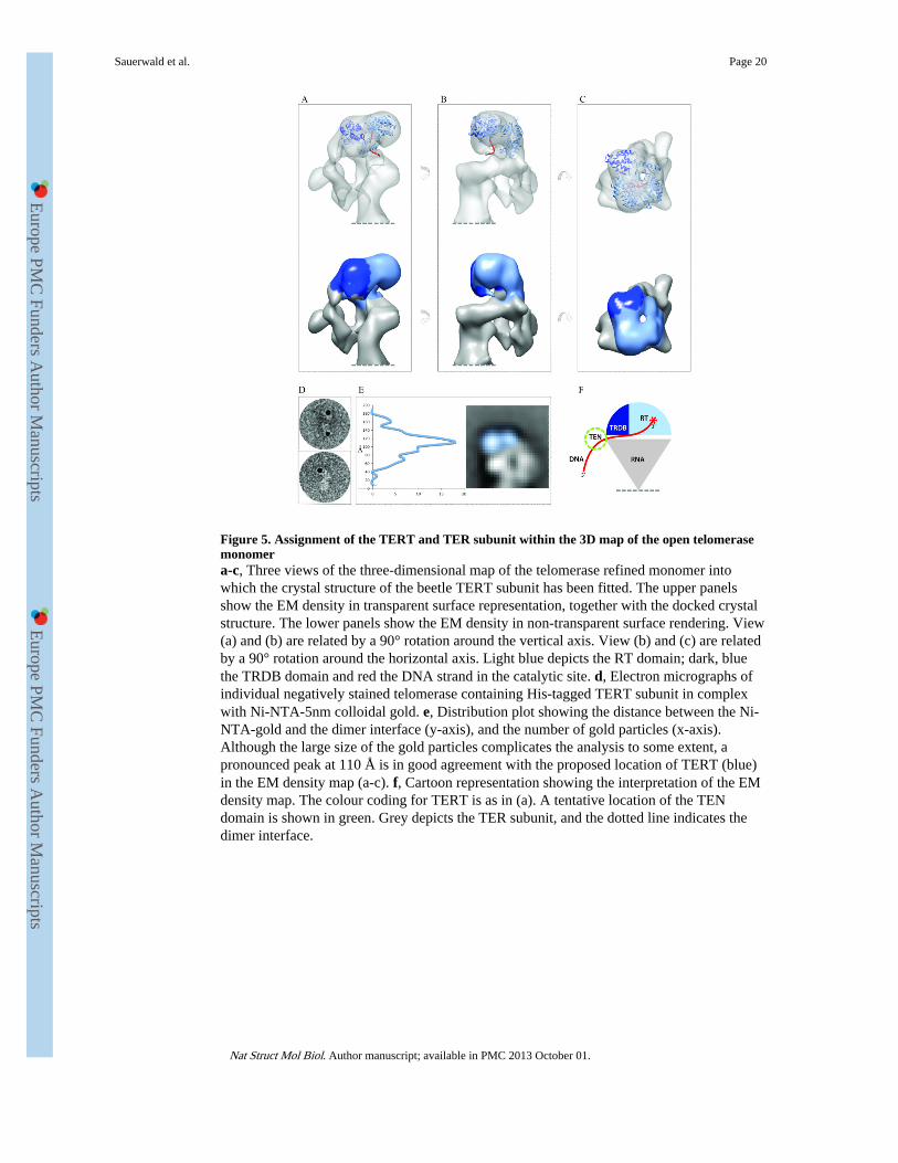
Figure 5. Assignment of the TERT and TER subunit within the 3D map of the open telomerasemonomera-c, Three views of the three-dimensional map of the telomerase refined monomer intowhich the crystal structure of the beetle TERT subunit has been fitted. The upper panelsshow the EM density in transparent surface representation, together with the docked crystalstructure. The lower panels show the EM density in non-transparent surface rendering. View(a) and (b) are related by a 90° rotation around the vertical axis. View (b) and (c) are relatedby a 90° rotation around the horizontal axis. Light blue depicts the RT domain; dark, bluethe TRDB domain and red the DNA strand in the catalytic site. d, Electron micrographs ofindividual negatively stained telomerase containing His-tagged TERT subunit in complexwith Ni-NTA-5nm colloidal gold. e, Distribution plot showing the distance between the Ni-NTA-gold and the dimer interface (y-axis), and the number of gold particles (x-axis).Although the large size of the gold particles complicates the analysis to some extent, apronounced peak at 110 Å is in good agreement with the proposed location of TERT (blue)in the EM density map (a-c). f, Cartoon representation showing the interpretation of the EMdensity map. The colour coding for TERT is as in (a). A tentative location of the TENdomain is shown in green. Grey depicts the TER subunit, and the dotted line indicates thedimer interface.
Sauerwald et al. Page 20
Nat Struct Mol Biol. Author manuscript; available in PMC 2013 October 01.
Europe PM
C Funders A
uthor Manuscripts
Europe PM
C Funders A
uthor Manuscripts