structure and receptor binding specificity of
TRANSCRIPT
Structure and Receptor Binding Specificity ofHemagglutinin H13 from Avian Influenza A VirusH13N6Xishan Lu, China Agricultural UniversityJianxun Qi, Chinese Academy of SciencesYi Shi, Chinese Academy of SciencesMing Wang, China Agricultural UniversityDavid Smith, Emory UniversityJamie Heimburg-Molinaro, Emory UniversityYanfang Zhang, Chinese Academy of SciencesJames C. Paulson, Scripps Research InstituteHaixia Xiao, Chinese Academy of SciencesGeorge F. Gao, China Agricultural University
Journal Title: Journal of VirologyVolume: Volume 87, Number 16Publisher: American Society for Microbiology | 2013-08-01, Pages 9077-9085Type of Work: Article | Final Publisher PDFPublisher DOI: 10.1128/JVI.00235-13Permanent URL: https://pid.emory.edu/ark:/25593/s75rt
Final published version: http://dx.doi.org/10.1128/JVI.00235-13
Copyright information:© 2013, American Society for Microbiology.
Accessed December 2, 2021 1:47 AM EST

Structure and Receptor Binding Specificity of Hemagglutinin H13from Avian Influenza A Virus H13N6
Xishan Lu,a,b Jianxun Qi,b Yi Shi,b,f Ming Wang,a David F. Smith,c Jamie Heimburg-Molinaro,c Yanfang Zhang,e James C. Paulson,d
Haixia Xiao,e George F. Gaoa,b,e,f,g
College of Veterinary Medicine, China Agricultural University, Beijing, Chinaa; CAS Key Laboratory of Pathogenic Microbiology and Immunology, Institute of Microbiology,Chinese Academy of Sciences, Beijing, Chinab; Department of Biochemistry, and the Glycomics Center, Emory University School of Medicine, O. Wayne Rollins ResearchCenter, Atlanta, Georgia, USAc; Department of Cell and Molecular Biology, The Scripps Research Institute, La Jolla, California, USAd; Laboratory of Protein Engineering andVaccines, Tianjin Institute of Industrial Biotechnology, Chinese Academy of Sciences, Tianjin, Chinae; Research Network of Immunity and Health, Beijing Institutes of LifeScience, Chinese Academy of Sciences, Beijing, Chinaf; Institute for Viral Disease Control and Prevention, Chinese Center for Disease Control and Prevention, Beijing,Chinag
Interspecies transmission (host switching/jumping) of influenza viruses is a key scientific question that must be addressed. Inaddition to the vigorous research on highly pathogenic avian influenza viruses (HPAIVs), studies of the mechanism of interspe-cies transmission of low-pathogenic avian influenza viruses (LPAIVs) could also provide insights into host tropism and viru-lence evolution. Influenza A viruses harboring hemagglutinin (HA) H13 (e.g., H13N6) are LPAIVs. In this study, soluble H13 HAglycoprotein was purified, and its receptor binding activity was characterized. The results revealed that H13 exclusively binds theavian �2-3-linked sialic acid receptor; no binding to the mammalian �2-6-linked sialic acid receptor was detected. Furthermore,the molecular basis of the H13 receptor binding specificity was revealed by comparative analysis of the crystal structures of bothreceptor-bound H13 and H5 HAs, which might be contributed by the hydrophobic residue V186. Work with an H13N186 mu-tant confirmed the importance of V186 in the receptor binding specificity of H13 HA, which shows that the mutant protein re-duced the binding of an avian receptor analog but increased the binding of a human receptor analog. Detailed structural analysisalso demonstrated that the conserved binding sites of the recently well-studied broadly neutralizing human monoclonal anti-bodies targeting the HA2 domain are found in H13. Our results expand our understanding of virulence evolution, receptor bind-ing preference, and species tropism of the LPAIVs and HPAIVs.
There are three types of influenza virus: A, B, and C. Influenza Aviruses account for all known major epidemics and pandem-
ics, though some mild epidemics of influenza B virus have beenrecorded (1–4). Influenza A viruses are classified into subtypesaccording to their two surface glycoproteins, hemagglutinin (HA)and neuraminidase (NA) (5, 6). For HA, which functions in sialicacid receptor binding and membrane fusion during virus infec-tion, 16 functional antigenic subtypes (H1 to H16) and one batHA homolog (H17) have been reported (7–10). For NA, the re-ceptor-destroying enzyme that removes sialic acid from the virusand cellular glycoproteins to release newly made virus from theinfected cells, there are 9 functional antigenic subtypes (N1 to N9)and one bat NA homolog (N10) (9–11). Influenza A virus infectsa broad spectrum of species, including many animals, humans,and birds (5). Interspecies transmission (host switching/jumping)is a major virulence factor for influenza viruses. In general, humanviruses preferentially interact with an N-acetyl sialic acid attachedto galactose with an �2-6 linkage, whereas avian viruses mostlybind to N-acetyl sialic acid attached to galactose with an �2-3linkage (12). This specificity corresponds, to a degree, with theavailability and density of such glycans at the principle sites ofinfection in the host. For example, �2-3-linked glycans predomi-nate in the intestinal tracts of ducks, where viral replication gen-erally occurs during natural infection (13, 14), while in humans,�-2-6-linked glycans predominate in the upper respiratory tract(15–17).
The 17 HA subtypes are divided into two groups based onphylogenic analysis. Group 1 can be further divided into threeclades (containing H8, H9, and H12; H1, H2, H5, H6 and H17;
and H11, H13, and H16), and group 2 includes two clades (con-taining H3, H4, and H14 and H7, H10, and H15) (18, 19). Sincethe first HA structure was solved in 1981 (20), nine of the 17known HAs (H1, H2, H3, H5, H7, H9, H14, H16, and H17) havebeen crystallized (19, 21–28), and structural correlates of thesesubtypes have been made from comparisons of the three-di-mensional structures of representative HAs from differentclades (9, 19).
To date, only three HA subtypes have adapted to cause pan-demics in humans: H1N1 in 1918 and most recently 2009, H2N2in 1957, and H3N2 in 1968 (18). Other subtypes (e.g., H5N1,H6N1, H7N2, H7N7, and H9N2) have caused epidemics in do-mestic poultry in certain areas of the world (29). Moreover, someviruses from poultry involving subtypes H5, H7, and H9 haveresulted in sporadic infections with high fatality in humans, buttheir low transmissibility among humans has prevented any newpandemics or epidemics (30–32). However, two recent papers re-port that only a few mutations in the HA protein give the H5N1viruses the ability to spread through the air between ferrets (33,
Received 24 January 2013 Accepted 31 May 2013
Published ahead of print 12 June 2013
Address correspondence to George F. Gao, [email protected].
Supplemental material for this article may be found at http://dx.doi.org/10.1128/JVI.00235-13.
Copyright © 2013, American Society for Microbiology. All Rights Reserved.
doi:10.1128/JVI.00235-13
August 2013 Volume 87 Number 16 Journal of Virology p. 9077–9085 jvi.asm.org 9077

34). Evidence was also provided that the wild H5N1 virus couldpotentially evolve to spark a pandemic on its own (33, 34).
Generally speaking, many scientists focus on the highly patho-genic H5N1 virus, and less attention is paid to other avian HAsubtypes (e.g., H13 and H16), which are commonly seen in somegulls and shorebirds (35). However, these avian subtypes still havethe potential to cross species barriers to infect humans and thusare a major concern for public health. Investigation of the receptorbinding properties of these avian subtypes could contribute to theunderstanding of host range switching during virus transmission.
The H13 subtype virus was first isolated in 1977 from gulls(H13N6) in the United States (36) and has subsequently beendetected primarily in gulls and shorebirds as a virus with lowpathogenicity (37). To better understand the receptor bindingspecificity of this subtype, we performed a series of receptor bind-ing experiments, including surface plasmon resonance (SPR)analysis and glycan microarray analysis, and further determinedthe three-dimensional atomic structure of H13 and that of itscomplexes with an avian receptor analog (LSTa). We demon-strated here that H13 specifically binds the avian receptor analogbut not the human receptor. Comparative analysis of the crystalstructures of both receptor-bound H13 and H5 HAs revealed thatthis specificity might be contributed by the hydrophobic residueV186. We generated the H13N186 mutant and found that thismutant protein reduced the binding of the avian receptor analogbut increased the binding of the human receptor analog. Theseresults provide the structural basis for the receptor binding spec-ificity of H13 and important insight into the interaction of H13with avian hosts.
MATERIALS AND METHODSH13 cloning, expression, and purification. Highly stable and pure H13protein was prepared using previously established methods (38), withslight modifications, using the Bac-to-Bac baculovirus expression system(Invitrogen). The cDNA corresponding to residues 11 to 329 (HA1) and 1to 176 (HA2) of the ectodomain of HA from the A/gull/Maryland/704/1977 (H13N6) virus was cloned into the baculovirus transfer vector pFast-Bac1 (Invitrogen), with a GP67 signal peptide at the N terminus, a throm-bin cleavage site, a trimerizing sequence, and a His6 tag at the C terminus.The recombinant baculovirus was prepared based on the manufacturer’sprotocol (Invitrogen). H13 and H13N186 proteins were obtained frominfected Hi5 insect cells using previously reported purification methods(38).
SPR analysis. Ion-exchange chromatography-purified H13 andH13N186 proteins were subjected to thrombin digestion (3 U/mg proteinovernight at 4°C) and purified by gel filtration chromatography usingstandard phosphate-buffered saline (PBS) with 0.005% Tween 20 (PBST)as the running buffer. The affinity and kinetics of the binding of solubleHAs to receptor analogs were measured at 25°C on a BIAcore 3000 instru-ment using streptavidin chips (SA chips; Biacore) by SPR. Two biotinyl-ated receptor analogs, the �-2-6 glycans {6=S-Di-LN [Neu5Aca2-6(Galb1-4GlcNAcb1-3)2b-SpNH-LC-LC-biotin]} and the �-2-3 glycans{3=S-di-LN [Neu5Aca2-3(Galb1-4GlcNAcb1-3)2b-SpNH-LC-LC-bio-tin]} were kindly provided by the Consortium for Functional Glycomics.The �-2-6 glycans and �-2-3 glycans were immobilized on a CM5 chipwith 500 response units. H13 HA protein (20, 10, 5, 2.5, 1.25, 0.63, 0.31, or0 �M) was allowed to flow through the chip, and the response units wererecorded. In a comparable assay, H13 and H13N186 proteins were ele-vated to a higher concentration (100 �M). The data were analyzed usingBIAevaluation software and fitted to a 1:1 binding model using HA mono-mer for calculating molecular weight.
Glycan microarray analysis. Glycan microarray analysis was per-formed using 20 mM Tris-HCl (pH 8.0) and 150 mM NaCl as the running
buffer. Glycan microarray printing and recombinant HA analyses havebeen described previously (27). The analyses were performed by applica-tion of the protein to the array at 200 �g/ml and detection in the secondstep with an anti-His antibody labeled with Alexa 488. Version 5.0 of theprinted array consists of 611 glycans in replicates of six. The highest andlowest points from each set of six replicates were removed, so the averageis of four values rather than six.
Crystallization, data collection, and structure determination. Crys-tallization conditions were screened using the hanging-drop vapor diffu-sion method with commercial kits (Hampton Research). H13 crystalswere obtained with a reservoir solution (0.2 ml) of 0.2 M L-proline, 0.1 MHEPES (pH 7.2), 10% polyethylene glycol (PEG) 3350, and 0.01 M so-dium bromide at 20°C. For receptor analog complexes, crystals weresoaked in a reservoir solution containing 8 mM LSTa or LSTc for 4 h.X-ray diffraction data were collected at 100 K at beamline NE3A of thePhoton Factory, Tsukuba, Japan. X-ray diffraction data for the complexstructure of H13 HA with LSTa (through crystal soaking) were collected at100 K at Shanghai Synchrotron Radiation Facility (SSRF) beamlineBL17U. These data were processed and scaled using the HKL-2000 pro-gram (39). Data collection and processing statistics are summarized inTable 1. The structures were solved by the molecular replacement(MR) method using Phaser (40) from the CCP4 program suite (41),with the structure of H16HA (Protein Data Bank [PDB] identifier4F23) as the search model. Model building and refinement were per-formed using the COOT (42) and REFMAC5 (43) programs, respec-tively. The stereochemical quality of the final models was assessed withthe program PROCHECK (44).
Protein structure accession numbers. The crystal structures were de-posited in the Protein Data Bank (PDB) under accession numbers 4KPQand 4KPS.
TABLE 1 Data collection and refinement statistics
Parameter
Valuea for:
H13 H13-LSTa
Data collection statisticsSpace group P1 P1Cell dimensions
a, b, c (Å) 75.11, 77.06, 76.47 75.64, 76.80, 76.38�, �, � (°) 85.50, 82.55, 87.37 85.60, 82.67, 87.52
Resolution (Å) 50–2.50 (2.59–2.50) 50–2.60 (2.69–2.60)Rmerge 0.086 (0.588) 0.106 (0.586)I/�I 15.0 (2.1) 11.4 (3.5)Completeness (%) 98.0 (97.6) 95.7 (97.8)Redundancy 3.7 (3.5) 3.0 (3.2)
Refinement statisticsResolution (Å) 42.4–2.50 42.5–2.60No. of reflections 52,627 47,912Rwork/Rfree 0.2238/0.2569 0.2161/0.2548No. of atoms
Protein 11,615 11,615Ligand/ion 0 78Water 226 103
B factorsProtein 71.08 76.44Ligand/ion 123.80Water 61.82 87.08
RMSDBond length (Å) 0.003 0.005Bond angle (°) 0.736 0.896
PDB code 4KPQ 4KPSa Values in parentheses are for highest-resolution shell.
Lu et al.
9078 jvi.asm.org Journal of Virology

RESULTSPreferential avian �2-3-linked receptor binding activity. The se-quence encoding the ectodomain of the H13 hemagglutinin frominfluenza A virus A/gull/Maryland/704/1977 (H13N6) was clonedinto the pFastbac1 vector and expressed using a baculovirus ex-pression system as previously described (38). Next, SPR technol-ogy and glycan microarrays were used to investigate the receptorbinding properties of the H13 protein. SPR experiments revealedthat H13 binds exclusively to the canonical avian �2-3-linked re-ceptor and not to the canonical human �2-6-linked receptor (Fig.1A and B). The large-scale glycan microarray analysis with 611different glycans, including natural sialosides (�2-3-linkage, �2-6-linkage, �2,8-linkage, and mixed linkage) and other glycans thatmay be relevant to influenza virus biology, revealed that the H13protein binds to only a few �2-3-linked glycans, with differentrelative fluorescence units (RFU) (Fig. 1C; see Table S1 in thesupplemental material). The structural formulas of the top repre-sentative five glycans are shown in Fig. 1C. Although H13 shares a
general preference for sialic acid with an �2-3 linkage to Gal-2,alternative linkages from GlcNAc-3 to Gal-4 and subsequent gly-can linkages could affect its binding activity.
Overall H13 structural features. The H13 structure was solvedby molecular replacement (MR) at a resolution of 2.5 Å usingH16HA (PDB identifier 4F23) (19) as a search model (Table 1),and the sequence identity between H13 and H16 is 80.04%. Al-though the H13 protein was expressed and purified in HA0 form,the solved H13 structure exhibits as a cleaved HA1/HA2 form.Only one molecule is observed in the asymmetrical unit. The crys-tal structure displays a classical homotrimer oligomerization asseen in other HA subtypes (Fig. 2). Seven asparagine-linked gly-cosylation sites are predicted in the H13 HA monomer, but inter-pretable carbohydrate electron density was observed at only onesite, N169. Regarding this site, only one N-acetylglucosamine(GlcNAc) could be interpreted.
The phylogenetic tree in Fig. 2A shows that H13 indeed belongsto group 1 HAs, indicating that the H13 structure should resemble
FIG 1 Receptor binding of the H13 protein. (A and B) SPR diagrams of H13 protein binding to the �2-3-linked and �2-6-linked sialic acid receptors. The H13protein displayed exclusive binding to the �2-3-linked receptor. (C) Glycan microarray analysis of the H13 protein. Binding to different types of glycans on thearray is highlighted, where blue represents Neu5Gc, cyan represents �2,8 ligands, magenta represents �2-6 ligands, green represents �2-3 ligands, and yellowrepresents other glycans. The H13 protein displayed a good avidity exclusively to �2-3 ligands. The structure formulas of the top representative are shown. Errorbars represent the standard deviation of the mean value. Percent coefficient of variation (%CV) � 100 � standard deviation/mean. A %CV of less than 50%indicates data reliability.
Influenza Virus H13 Structure and Receptor Binding
August 2013 Volume 87 Number 16 jvi.asm.org 9079

the structures of group 1 HAs rather than those of group 2. Thesuperimposition of other HA structures onto the H13 monomerby means of their HA2 domains (root mean square deviations[RMSD] are given in Table 2) demonstrated that H13 is mostclosely related to the 1957 Singapore H2 subtype (RMSD �0.634), whereas the avian H14 subtype is the most divergent(RMSD � 1.164). Based on their HA1 domains, H13 is mostclosely related to the avian H16 subtype (RMSD � 0.480), whereasthe 1968 Hong Kong H3 subtype is the most divergent (RMSD �2.391). Concerning the receptor binding region (R region) andvestigial esterase region (E region), H13 is still most closely relatedto the avian H16 subtype (RMSD � 0.454 and 0.359, respectively).
Previously solved HA structures demonstrate that there aregroup-specific features at sites where extensive conformationalchanges occur for HA activation, including the conformation ofthe interhelix loop and the rigid-body orientation of the globulardomain. Here, we studied only whether the rule of the rigid-bodyorientation of the globular domain is applicable to recently solvedH13 and H16 structures or not. Superimposition with other
solved HA structures by means of the long central �-helices ofHA2 revealed that the rigid-body orientations of globular do-mains fall into three clusters: group 1, including H1, H2, H5, andH9; group 2, including H3, H7, and H14; and “cluster 3,” consist-ing of H16 and H13. Cluster 3 (H13 and H16) is located betweenthe group 1 HAs and group 2 HAs. These differences may result indifferent mechanisms of HA activation, which should be the sub-ject of a future study.
Structural basis for receptor binding specificity. The crystalsof H13 were soaked with the pentasaccharides LSTc and LSTa,which represent �2-6- and �2-3-linked glycan analogs of humanand avian receptors, respectively. However, only LSTa successfullysoaked into the crystal to form a complex, which is consistent withthe SPR experiments (i.e., only LSTa binds H13). The structuralformula of LSTa is NeuAc�2-3Gal�1-3GlcNAc�1-3Gal�1-4Glc.In the H13/LSTa complex structure, strong electron density forthe glycan ligand is observed in the receptor binding sites of two ofthe three monomers in the asymmetric unit. It is possible that thecrystal packing limits the glycan receptor analog binding in the
FIG 2 Phylogenetic tree and overall structure. (A) Phylogenetic tree showing that H13 belongs to group 1, in the same clade as H16. (B) Overall structure of H13and comparison of the globular domain of the H13 monomer with those of other solved HA subtype structures (H1, light blue; H2, cyan; H3, limon; H5, lightorange; H7, yellow; H9, pink; H14, pale yellow; H13, green; and H16, red). H13 displays a typical homotrimer structure. The 190 helix (residues 188 to 195) inthe receptor binding domain was used as a representative of the globular subdomain of each HA subtype. The rigid-body orientation of the globular domain inthe H13 structure is similar to that in the H16 subtype, by means of superimposition through the long helix of HA2. They are located between other group 1 HAsand group 2 HAs.
Lu et al.
9080 jvi.asm.org Journal of Virology
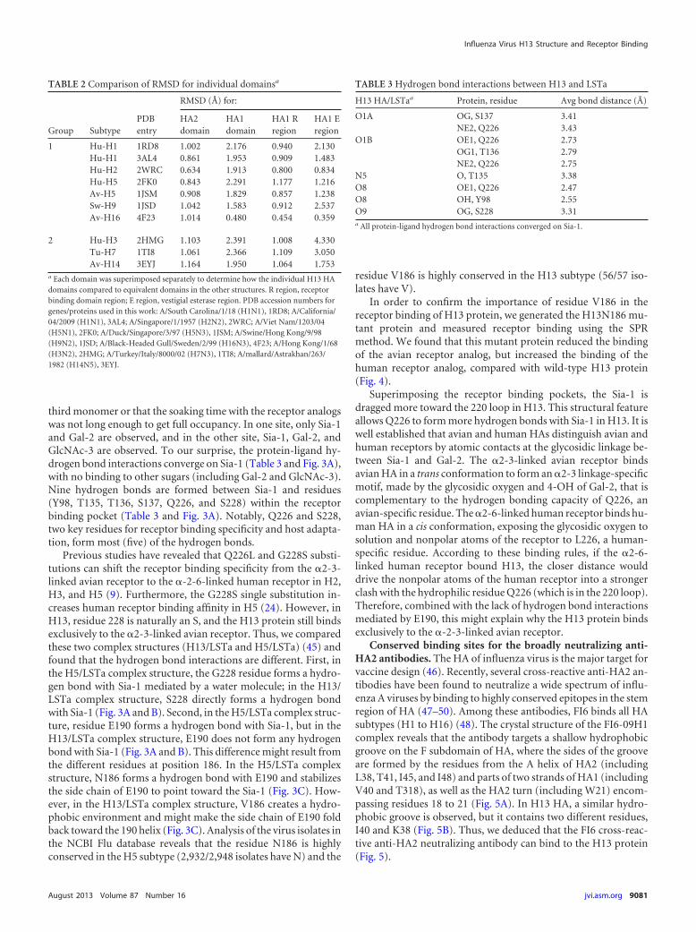
third monomer or that the soaking time with the receptor analogswas not long enough to get full occupancy. In one site, only Sia-1and Gal-2 are observed, and in the other site, Sia-1, Gal-2, andGlcNAc-3 are observed. To our surprise, the protein-ligand hy-drogen bond interactions converge on Sia-1 (Table 3 and Fig. 3A),with no binding to other sugars (including Gal-2 and GlcNAc-3).Nine hydrogen bonds are formed between Sia-1 and residues(Y98, T135, T136, S137, Q226, and S228) within the receptorbinding pocket (Table 3 and Fig. 3A). Notably, Q226 and S228,two key residues for receptor binding specificity and host adapta-tion, form most (five) of the hydrogen bonds.
Previous studies have revealed that Q226L and G228S substi-tutions can shift the receptor binding specificity from the �2-3-linked avian receptor to the �-2-6-linked human receptor in H2,H3, and H5 (9). Furthermore, the G228S single substitution in-creases human receptor binding affinity in H5 (24). However, inH13, residue 228 is naturally an S, and the H13 protein still bindsexclusively to the �2-3-linked avian receptor. Thus, we comparedthese two complex structures (H13/LSTa and H5/LSTa) (45) andfound that the hydrogen bond interactions are different. First, inthe H5/LSTa complex structure, the G228 residue forms a hydro-gen bond with Sia-1 mediated by a water molecule; in the H13/LSTa complex structure, S228 directly forms a hydrogen bondwith Sia-1 (Fig. 3A and B). Second, in the H5/LSTa complex struc-ture, residue E190 forms a hydrogen bond with Sia-1, but in theH13/LSTa complex structure, E190 does not form any hydrogenbond with Sia-1 (Fig. 3A and B). This difference might result fromthe different residues at position 186. In the H5/LSTa complexstructure, N186 forms a hydrogen bond with E190 and stabilizesthe side chain of E190 to point toward the Sia-1 (Fig. 3C). How-ever, in the H13/LSTa complex structure, V186 creates a hydro-phobic environment and might make the side chain of E190 foldback toward the 190 helix (Fig. 3C). Analysis of the virus isolates inthe NCBI Flu database reveals that the residue N186 is highlyconserved in the H5 subtype (2,932/2,948 isolates have N) and the
residue V186 is highly conserved in the H13 subtype (56/57 iso-lates have V).
In order to confirm the importance of residue V186 in thereceptor binding of H13 protein, we generated the H13N186 mu-tant protein and measured receptor binding using the SPRmethod. We found that this mutant protein reduced the bindingof the avian receptor analog, but increased the binding of thehuman receptor analog, compared with wild-type H13 protein(Fig. 4).
Superimposing the receptor binding pockets, the Sia-1 isdragged more toward the 220 loop in H13. This structural featureallows Q226 to form more hydrogen bonds with Sia-1 in H13. It iswell established that avian and human HAs distinguish avian andhuman receptors by atomic contacts at the glycosidic linkage be-tween Sia-1 and Gal-2. The �2-3-linked avian receptor bindsavian HA in a trans conformation to form an �2-3 linkage-specificmotif, made by the glycosidic oxygen and 4-OH of Gal-2, that iscomplementary to the hydrogen bonding capacity of Q226, anavian-specific residue. The �2-6-linked human receptor binds hu-man HA in a cis conformation, exposing the glycosidic oxygen tosolution and nonpolar atoms of the receptor to L226, a human-specific residue. According to these binding rules, if the �2-6-linked human receptor bound H13, the closer distance woulddrive the nonpolar atoms of the human receptor into a strongerclash with the hydrophilic residue Q226 (which is in the 220 loop).Therefore, combined with the lack of hydrogen bond interactionsmediated by E190, this might explain why the H13 protein bindsexclusively to the �-2-3-linked avian receptor.
Conserved binding sites for the broadly neutralizing anti-HA2 antibodies. The HA of influenza virus is the major target forvaccine design (46). Recently, several cross-reactive anti-HA2 an-tibodies have been found to neutralize a wide spectrum of influ-enza A viruses by binding to highly conserved epitopes in the stemregion of HA (47–50). Among these antibodies, FI6 binds all HAsubtypes (H1 to H16) (48). The crystal structure of the FI6-09H1complex reveals that the antibody targets a shallow hydrophobicgroove on the F subdomain of HA, where the sides of the grooveare formed by the residues from the A helix of HA2 (includingL38, T41, I45, and I48) and parts of two strands of HA1 (includingV40 and T318), as well as the HA2 turn (including W21) encom-passing residues 18 to 21 (Fig. 5A). In H13 HA, a similar hydro-phobic groove is observed, but it contains two different residues,I40 and K38 (Fig. 5B). Thus, we deduced that the FI6 cross-reac-tive anti-HA2 neutralizing antibody can bind to the H13 protein(Fig. 5).
TABLE 3 Hydrogen bond interactions between H13 and LSTa
H13 HA/LSTaa Protein, residue Avg bond distance (Å)
O1A OG, S137 3.41NE2, Q226 3.43
O1B OE1, Q226 2.73OG1, T136 2.79NE2, Q226 2.75
N5 O, T135 3.38O8 OE1, Q226 2.47O8 OH, Y98 2.55O9 OG, S228 3.31a All protein-ligand hydrogen bond interactions converged on Sia-1.
TABLE 2 Comparison of RMSD for individual domainsa
Group SubtypePDBentry
RMSD (Å) for:
HA2domain
HA1domain
HA1 Rregion
HA1 Eregion
1 Hu-H1 1RD8 1.002 2.176 0.940 2.130Hu-H1 3AL4 0.861 1.953 0.909 1.483Hu-H2 2WRC 0.634 1.913 0.800 0.834Hu-H5 2FK0 0.843 2.291 1.177 1.216Av-H5 1JSM 0.908 1.829 0.857 1.238Sw-H9 1JSD 1.042 1.583 0.912 2.537Av-H16 4F23 1.014 0.480 0.454 0.359
2 Hu-H3 2HMG 1.103 2.391 1.008 4.330Tu-H7 1TI8 1.061 2.366 1.109 3.050Av-H14 3EYJ 1.164 1.950 1.064 1.753
a Each domain was superimposed separately to determine how the individual H13 HAdomains compared to equivalent domains in the other structures. R region, receptorbinding domain region; E region, vestigial esterase region. PDB accession numbers forgenes/proteins used in this work: A/South Carolina/1/18 (H1N1), 1RD8; A/California/04/2009 (H1N1), 3AL4; A/Singapore/1/1957 (H2N2), 2WRC; A/Viet Nam/1203/04(H5N1), 2FK0; A/Duck/Singapore/3/97 (H5N3), 1JSM; A/Swine/Hong Kong/9/98(H9N2), 1JSD; A/Black-Headed Gull/Sweden/2/99 (H16N3), 4F23; A/Hong Kong/1/68(H3N2), 2HMG; A/Turkey/Italy/8000/02 (H7N3), 1TI8; A/mallard/Astrakhan/263/1982 (H14N5), 3EYJ.
Influenza Virus H13 Structure and Receptor Binding
August 2013 Volume 87 Number 16 jvi.asm.org 9081

DISCUSSION
An increasing number of cases of human infections by the H5N1highly pathogenic avian influenza virus (HPAIV) underscores thereal public health concern about potential pandemics or endemicscaused by influenza viruses with an avian origin (51). Recent work(33, 34) on H5N1 virus adaptation to horizontal transmission inferrets has again alerted the public and the scientific community tothe possibility of human infection by avian-origin influenza virus.Studies on the virulence evolution and/or interspecies transmis-sion of avian viruses cannot be suspended under such circum-stances. The H13 subtype was originally isolated from seagulls in1977 (36) and causes illness in ferrets upon direct inoculation(36). Thus, detailed studies of H13 receptor binding and structureare highly relevant to avian-to-mammalian transmission of low-pathogenic avian influenza virus (LPAIV).
In this study, we solved the H13 crystal structure and char-acterized its receptor binding, revealing that H13 has a typicalHA structure and is cleaved into HA1 and HA2 in the crystalstructure. Our results demonstrated that the avian H13 dis-plays exclusive binding to the avian receptor (i.e., the �2-3-linked sialic acid receptor), whereas other avian HAs (e.g., H1,H2, H3, and H5) both bind the avian receptor and possess weakbinding affinity for the human receptor (i.e., the �2-6-linkedsialic acid receptor). Notably, the H13 HA bound well to linear-sequence fragments (structures 2 and 4 in Fig. 1C) and the same
sequences on selected O-linked glycans but bound with re-duced avidity to glycans containing the same terminal se-quences attached to N-linked glycan cores, consistent with pre-vious analysis on a more limited set of glycans (52). Structuralanalysis revealed that H13 has a typical �2-3-linked sialic acidreceptor binding groove, as confirmed by a receptor-H13 com-plex structure. The hydrogen bond interactions between thesialyl glycan receptor and the receptor binding site of H13 con-verge on the sialic acid (Sia-1), with no binding to the glycans.Furthermore, the residue E190 does not form hydrogen bondswith Sia-1 in the H13/LSTa complex structure. This lack ofhydrogen bond interactions results from the fold-back confor-mation of E190, which might be generated by the nearby hy-drophobic residue V186. However, in other avian HAs (H5 andH2 subtypes), the residue at position 186 is usually an aspara-gine (N) (308/308 virus isolates of H2 in the NCBI Flu databasehave N). Previous studies show that the residues E190 andN186 contribute to the binding of the human receptor via awater-mediated hydrogen bond network in other HA subtypes(26). Thus, the hydrophobic residue V186 precludes the bind-ing of H13 to the human receptor, which is also confirmed byour work with the H13N186 mutant. In terms of interspeciestransmission, a Q226L substitution in the receptor binding sitemight help H13 to obtain human �2-6-linked sialic acid recep-tor binding, as the same Q226L substitution helps H5 to obtain
FIG 3 Interaction of H13 protein with an avian receptor analog and comparison with the H5/avian receptor analog complex. The three secondary structureelements of the binding site (the 130 loop, 190 helix, and 220 loop) are labeled in ribbon representation together with selected residues in stick representation.Hydrogen bonds are shown as dashed lines. (A) H13 protein with the avian receptor analog LSTa (�2-3) pentasaccharide bound, colored in green. Theinteraction with the receptor binding site converges on Sia-1. (B) H5 protein with the avian receptor analog LSTa pentasaccharide bound (PDB accession number1JSN), colored in cyan. Both Sia-1 and Gal-2 are involved in the interaction with the receptor binding site. (C and D) Comparison of the receptor binding sitesof the H13/LSTa and H5/LSTa complexes. The hydrophobic residue V186 creates a hydrophobic environment to force the side chain of E190 to adopt a fold-backconformation in H13. The equivalent residue N186 stabilizes the side chain of E190 to point to the receptor binding site in H5. The glycan ligand is closer to the220 loop in the H13/LSTa complex than in the H5/LSTa complex.
Lu et al.
9082 jvi.asm.org Journal of Virology

human receptor binding (34). Therefore, evidence of this mu-tation should be closely monitored in the future for influenzavirus surveillance.
Detailed analysis of the HA structure also revealed that thebinding sites of the recently well-studied HA2-targeting neutral-izing monoclonal antibodies are highly conserved. Therefore,these human monoclonal antibodies should neutralize viruses en-coding H13, but they need to be (minimally) tested in an animalmodel. Future work should also focus on the structures and recep-tor binding activities of other LPAIV HAs.
ACKNOWLEDGMENTS
This work was supported by grants from the Ministry of Science andTechnology of China (project 973, grant no. 2011CB504703; http://www.most.gov.cn/eng/). We acknowledge the Consortium for Functional Gly-comics for glycan array analysis and synthetic glycans used in bindingassays (NIH grant GM62116). G.F.G. is a leading principal investigator ofthe Innovative Research Group of the National Natural Science Founda-tion of China (grant no. 81021003; http://www.nsfc.gov.cn/Portal0/default106.htm).
FIG 5 The conserved hydrophobic groove in the H13 protein reveals thestructural basis of binding by the broadly neutralizing antibody FI6. Surfacerepresentations of the F subdomains of 09H1 HA (A) and H13 HA (B) withselected side chains that contribute to the conserved hydrophobic groove areshown. The approximate boundaries of the hydrophobic grooves are indicatedby the black lines. Although the residues contributing to the hydrophobicgroove are moderately different between 09H1 and H13, similar hydrophobicgrooves guarantee the binding potential by the FI6 antibody.
FIG 4 Receptor binding of the H13N186 mutant protein. (A and B) SPR diagrams of H13 protein binding to the �2-3-linked and �2-6-linked sialic acidreceptors with a series of higher concentrations. The H13 protein still displayed exclusive binding to the �2-3-linked receptor. (C and D) SPR diagrams ofH13N186 protein binding to the �2-3-linked and �2-6-linked sialic acid receptors. The H13N186 protein reduced the binding to the �2-3-linked receptor butincreased the binding to �2-6-linked receptor.
Influenza Virus H13 Structure and Receptor Binding
August 2013 Volume 87 Number 16 jvi.asm.org 9083

The funders had no role in study design, data collection and analysis,decision to publish, or preparation of the manuscript.
We thank the staff at the Shanghai Synchrotron Radiation Facility andthe staff at the Photon Factory (Tsukuba, Japan) for assistance.
G.F.G. supervised and oversaw the project; G.F.G., X.L., and Y.S. con-ceived the research; X.L. performed the protein expression, purification,and crystallization; Y.Z. prepared the insect cells; J.Q. performed thestructure determination; D.F.S., and J.H.-M. performed the glycan arrayexperiments; J.C.P. provided advice about receptor binding experiments;G.F.G., Y.S., X.L., H.X., and M.W. performed the data analysis; Y.S., X.L.,and G.F.G. wrote the manuscript; all authors reviewed and edited themanuscript.
We declare that we have no conflict of interest.
REFERENCES1. Frank AL, Taber LH, Glezen WP, Geyer EA, McIlwain S, Paredes A.
1983. Influenza B virus infections in the community and the family. Theepidemics of 1976-1977 and 1979-1980 in Houston, Texas. Am. J. Epide-miol. 118:313–325.
2. Gao GF, Sun Y. 2010. It is not just AIV: from avian to swine-origininfluenza virus. Sci. China Life Sci. 53:151–153.
3. Guan Y, Vijaykrishna D, Bahl J, Zhu H, Wang J, Smith GJ. 2010. Theemergence of pandemic influenza viruses. Protein Cell 1:9 –13.
4. Neumann G, Noda T, Kawaoka Y. 2009. Emergence and pandemicpotential of swine-origin H1N1 influenza virus. Nature 459:931–939.
5. Webster RG, Bean WJ, Gorman OT, Chambers TM, Kawaoka Y. 1992.Evolution and ecology of influenza A viruses. Microbiol. Rev. 56:152–179.
6. WHO. 1980. A revision of the system of nomenclature for influenza vi-ruses: a WHO memorandum. Bull. World Health Organ. 58:585–591.
7. Air GM. 1981. Sequence relationships among the hemagglutinin genes of12 subtypes of influenza A virus. Proc. Natl. Acad. Sci. U. S. A. 78:7639 –7643.
8. Nobusawa E, Aoyama T, Kato H, Suzuki Y, Tateno Y, Nakajima K.1991. Comparison of complete amino acid sequences and receptor-binding properties among 13 serotypes of hemagglutinins of influenza Aviruses. Virology 182:475– 485.
9. Gamblin SJ, Skehel JJ. 2010. Influenza hemagglutinin and neuraminidasemembrane glycoproteins. J. Biol. Chem. 285:28403–28409.
10. Tong S, Li Y, Rivailler P, Conrardy C, Castillo DA, Chen LM, RecuencoS, Ellison JA, Davis CT, York IA, Turmelle AS, Moran D, Rogers S, ShiM, Tao Y, Weil MR, Tang K, Rowe LA, Sammons S, Xu X, Frace M,Lindblade KA, Cox NJ, Anderson LJ, Rupprecht CE, Donis RO. 2012.A distinct lineage of influenza A virus from bats. Proc. Natl. Acad. Sci.U. S. A. 109:4269 – 4274.
11. Russell RJ, Haire LF, Stevens DJ, Collins PJ, Lin YP, Blackburn GM,Hay AJ, Gamblin SJ, Skehel JJ. 2006. The structure of H5N1 avianinfluenza neuraminidase suggests new opportunities for drug design. Na-ture 443:45– 49.
12. Rogers GN, Pritchett TJ, Lane JL, Paulson JC. 1983. Differential sensi-tivity of human, avian, and equine influenza A viruses to a glycoproteininhibitor of infection: selection of receptor specific variants. Virology 131:394 – 408.
13. Webster RG, Yakhno M, Hinshaw VS, Bean WJ, Murti KG. 1978.Intestinal influenza: replication and characterization of influenza virusesin ducks. Virology 84:268 –278.
14. Naeve CW, Hinshaw VS, Webster RG. 1984. Mutations in the hemag-glutinin receptor-binding site can change the biological properties of aninfluenza virus. J. Virol. 51:567–569.
15. Baum LG, Paulson JC. 1990. Sialyloligosaccharides of the respiratoryepithelium in the selection of human influenza virus receptor specificity.Acta Histochem. Suppl. 40:35–38.
16. Couceiro JN, Paulson JC, Baum LG. 1993. Influenza virus strains selec-tively recognize sialyloligosaccharides on human respiratory epithelium;the role of the host cell in selection of hemagglutinin receptor specificity.Virus Res. 29:155–165.
17. Matrosovich MN, Matrosovich TY, Gray T, Roberts NA, Klenk HD.2004. Human and avian influenza viruses target different cell types incultures of human airway epithelium. Proc. Natl. Acad. Sci. U. S. A. 101:4620 – 4624.
18. Medina RA, Garcia-Sastre A. 2011. Influenza A viruses: new researchdevelopments. Nat. Rev. Microbiol. 9:590 – 603.
19. Lu X, Shi Y, Gao F, Xiao H, Wang M, Qi J, Gao GF. 2012. Insights intoavian influenza virus pathogenicity: the hemagglutinin precursor HA0 ofsubtype H16 has an alpha-helix structure in its cleavage site with ineffi-cient HA1/HA2 cleavage. J. Virol. 86:12861–12870.
20. Wilson IA, Skehel JJ, Wiley DC. 1981. Structure of the haemagglutininmembrane glycoprotein of influenza virus at 3 A resolution. Nature 289:366 –373.
21. Ha Y, Stevens DJ, Skehel JJ, Wiley DC. 2002. H5 avian and H9 swineinfluenza virus haemagglutinin structures: possible origin of influenzasubtypes. EMBO J. 21:865– 875.
22. Russell RJ, Gamblin SJ, Haire LF, Stevens DJ, Xiao B, Ha Y, Skehel JJ.2004. H1 and H7 influenza haemagglutinin structures extend a structuralclassification of haemagglutinin subtypes. Virology 325:287–296.
23. Stevens J, Corper AL, Basler CF, Taubenberger JK, Palese P, Wilson IA.2004. Structure of the uncleaved human H1 hemagglutinin from the ex-tinct 1918 influenza virus. Science 303:1866 –1870.
24. Stevens J, Blixt O, Tumpey TM, Taubenberger JK, Paulson JC, WilsonIA. 2006. Structure and receptor specificity of the hemagglutinin from anH5N1 influenza virus. Science 312:404 – 410.
25. Russell RJ, Kerry PS, Stevens DJ, Steinhauer DA, Martin SR, GamblinSJ, Skehel JJ. 2008. Structure of influenza hemagglutinin in complex withan inhibitor of membrane fusion. Proc. Natl. Acad. Sci. U. S. A. 105:17736 –17741.
26. Liu J, Stevens DJ, Haire LF, Walker PA, Coombs PJ, Russell RJ,Gamblin SJ, Skehel JJ. 2009. Structures of receptor complexes formed byhemagglutinins from the Asian influenza pandemic of 1957. Proc. Natl.Acad. Sci. U. S. A. 106:17175–17180.
27. Sun X, Shi Y, Lu X, He J, Gao F, Yan J, Qi J, Gao GF. 2013. Bat-derivedinfluenza hemagglutinin h17 does not bind canonical avian or humanreceptors and most likely uses a unique entry mechanism. Cell Rep.3:769 –778.
28. Zhu X, Yu W, McBride R, Li Y, Chen LM, Donis RO, Tong S, PaulsonJC, Wilson IA. 2013. Hemagglutinin homologue from H17N10 bat influ-enza virus exhibits divergent receptor-binding and pH-dependent fusionactivities. Proc. Natl. Acad. Sci. U. S. A. 110:1458 –1463.
29. Brown IH. 2010. Summary of avian influenza activity in Europe, Asia, andAfrica, 2006-2009. Avian Dis. 54:187–193.
30. de Jong JC, Rimmelzwaan GF, Bartelds AI, Wilbrink B, Fouchier RA,Osterhaus AD. 2003. The 2002/2003 influenza season in the Netherlandsand the vaccine composition for the 2003/2004 season. Ned. Tijdschr.Geneeskd. 147:1971–1975.
31. Peiris M, Yuen KY, Leung CW, Chan KH, Ip PL, Lai RW, Orr WK,Shortridge KF. 1999. Human infection with influenza H9N2. Lancet 354:916 –917.
32. Subbarao K, Klimov A, Katz J, Regnery H, Lim W, Hall H, Perdue M,Swayne D, Bender C, Huang J, Hemphill M, Rowe T, Shaw M, Xu X,Fukuda K, Cox N. 1998. Characterization of an avian influenza A (H5N1)virus isolated from a child with a fatal respiratory illness. Science 279:393–396.
33. Herfst S, Schrauwen EJ, Linster M, Chutinimitkul S, de Wit E, MunsterVJ, Sorrell EM, Bestebroer TM, Burke DF, Smith DJ, Rimmelzwaan GF,Osterhaus AD, Fouchier RA. 2012. Airborne transmission of influenzaA/H5N1 virus between ferrets. Science 336:1534 –1541.
34. Imai M, Watanabe T, Hatta M, Das SC, Ozawa M, Shinya K, Zhong G,Hanson A, Katsura H, Watanabe S, Li C, Kawakami E, Yamada S, KisoM, Suzuki Y, Maher EA, Neumann G, Kawaoka Y. 2012. Experimentaladaptation of an influenza H5 HA confers respiratory droplet transmis-sion to a reassortant H5 HA/H1N1 virus in ferrets. Nature 486:420 – 428.
35. Velarde R, Calvin SE, Ojkic D, Barker IK, Nagy E. 2010. Avian influenzavirus H13 circulating in ring-billed gulls (Larus delawarensis) in southernOntario, Canada. Avian Dis. 54:411– 419.
36. Hinshaw VS, Air GM, Gibbs AJ, Graves L, Prescott B, Karunakaran D.1982. Antigenic and genetic characterization of a novel hemagglutininsubtype of influenza A viruses from gulls. J. Virol. 42:865– 872.
37. Hofle U, Van de Bildt MW, Leijten LM, Van Amerongen G, VerhagenJH, Fouchier RA, Osterhaus AD, Kuiken T. 2012. Tissue tropism andpathology of natural influenza virus infection in black-headed gulls(Chroicocephalus ridibundus). Avian Pathol. 41:547–553.
38. Zhang W, Qi J, Shi Y, Li Q, Gao F, Sun Y, Lu X, Lu Q, Vavricka CJ, LiuD, Yan J, Gao GF. 2010. Crystal structure of the swine-origin A (H1N1)-2009 influenza A virus hemagglutinin (HA) reveals similar antigenicity tothat of the 1918 pandemic virus. Protein Cell 1:459 – 467.
39. Vaguine AA, Richelle J, Wodak SJ. 1999. SFCHECK: a unified set of
Lu et al.
9084 jvi.asm.org Journal of Virology

procedures for evaluating the quality of macromolecular structure-factordata and their agreement with the atomic model. Acta Crystallogr. D Biol.Crystallogr. 55:191–205.
40. Read RJ. 2001. Pushing the boundaries of molecular replacement withmaximum likelihood. Acta Crystallogr. D Biol. Crystallogr. 57:1373–1382.
41. Collaborative Computational Project. 1994. The CCP4 suite: programsfor protein crystallography. Acta Crystallogr. D Biol. Crystallogr. 50:760 –763.
42. Emsley P, Cowtan K. 2004. Coot: model-building tools for moleculargraphics. Acta Crystallogr. D Biol. Crystallogr. 60:2126 –2132.
43. Murshudov GN, Vagin AA, Dodson EJ. 1997. Refinement of macromo-lecular structures by the maximum-likelihood method. Acta Crystallogr.D Biol. Crystallogr. 53:240 –255.
44. Morris AL, MacArthur MW, Hutchinson EG, Thornton JM. 1992.Stereochemical quality of protein structure coordinates. Proteins 12:345–364.
45. Ha Y, Stevens DJ, Skehel JJ, Wiley DC. 2001. X-ray structures of H5avian and H9 swine influenza virus hemagglutinins bound to avian andhuman receptor analogs. Proc. Natl. Acad. Sci. U. S. A. 98:11181–11186.
46. Xuan C, Shi Y, Qi J, Zhang W, Xiao H, Gao GF. 2011. Structuralvaccinology: structure-based design of influenza A virus hemagglutininsubtype-specific subunit vaccines. Protein Cell 2:997–1005.
47. Ekiert DC, Friesen RH, Bhabha G, Kwaks T, Jongeneelen M, Yu W,Ophorst C, Cox F, Korse HJ, Brandenburg B, Vogels R, Brakenhoff JP,Kompier R, Koldijk MH, Cornelissen LA, Poon LL, Peiris M, Koudstaal
W, Wilson IA, Goudsmit J. 2011. A highly conserved neutralizing epitopeon group 2 influenza A viruses. Science 333:843– 850.
48. Corti D, Voss J, Gamblin SJ, Codoni G, Macagno A, Jarrossay D,Vachieri SG, Pinna D, Minola A, Vanzetta F, Silacci C, Fernandez-Rodriguez BM, Agatic G, Bianchi S, Giacchetto-Sasselli I, Calder L,Sallusto F, Collins P, Haire LF, Temperton N, Langedijk JP, Skehel JJ,Lanzavecchia A. 2011. A neutralizing antibody selected from plasma cellsthat binds to group 1 and group 2 influenza A hemagglutinins. Science333:850 – 856.
49. Ekiert DC, Bhabha G, Elsliger MA, Friesen RH, Jongeneelen M, Thro-sby M, Goudsmit J, Wilson IA. 2009. Antibody recognition of a highlyconserved influenza virus epitope. Science 324:246 –251.
50. Sui J, Hwang WC, Perez S, Wei G, Aird D, Chen LM, Santelli E, Stec B,Cadwell G, Ali M, Wan H, Murakami A, Yammanuru A, Han T, CoxNJ, Bankston LA, Donis RO, Liddington RC, Marasco WA. 2009.Structural and functional bases for broad-spectrum neutralization ofavian and human influenza A viruses. Nat. Struct. Mol. Biol. 16:265–273.
51. Neumann G, Macken CA, Karasin AI, Fouchier RA, Kawaoka Y. 2012.Egyptian H5N1 influenza viruses-cause for concern? PLoS Pathog.8:e1002932. doi:10.1371/journal.ppat.1002932.
52. Nycholat CM, McBride R, Ekiert DC, Xu R, Rangarajan J, Peng W, RaziN, Gilbert M, Wakarchuk W, Wilson IA, Paulson JC. 2012. Recognitionof sialylated poly-N-acetyllactosamine chains on N- and O-linked glycansby human and avian influenza A virus hemagglutinins. Angewandte Che-mie. 51:4860 – 4863.
Influenza Virus H13 Structure and Receptor Binding
August 2013 Volume 87 Number 16 jvi.asm.org 9085