structure and reaction mechanism of basil eugenol...
TRANSCRIPT

Structure and Reaction Mechanism of Basil EugenolSynthaseGordon V. Louie1, Thomas J. Baiga1, Marianne E. Bowman1, Takao Koeduka2, John H. Taylor1, Snejina M. Spassova1, Eran Pichersky2, Joseph P.Noel1*
1 Howard Hughes Medical Institute, Jack H. Skirball Center for Chemical Biology and Proteomics, The Salk Institute for Biological Studies, La Jolla,California, United States of America, 2 Department of Molecular, Cellular and Developmental Biology, University of Michigan, Ann Arbor, Michigan,United States of America
Phenylpropenes, a large group of plant volatile compounds that serve in multiple roles in defense and pollinator attraction,contain a propenyl side chain. Eugenol synthase (EGS) catalyzes the reductive displacement of acetate from the propenyl sidechain of the substrate coniferyl acetate to produce the allyl-phenylpropene eugenol. We report here the structuredetermination of EGS from basil (Ocimum basilicum) by protein x-ray crystallography. EGS is structurally related to the short-chain dehydrogenase/reductases (SDRs), and in particular, enzymes in the isoflavone-reductase-like subfamily. The structure ofa ternary complex of EGS bound to the cofactor NADP(H) and a mixed competitive inhibitor EMDF ((7S,8S)-ethyl (7,8-methylene)-dihydroferulate) provides a detailed view of the binding interactions within the EGS active site and a starting pointfor mutagenic examination of the unusual reductive mechanism of EGS. The key interactions between EMDF and the EGS-holoenzyme include stacking of the phenyl ring of EMDF against the cofactor’s nicotinamide ring and a water-mediatedhydrogen-bonding interaction between the EMDF 4-hydroxy group and the side-chain amino moiety of a conserved lysineresidue, Lys132. The C4 carbon of nicotinamide resides immediately adjacent to the site of hydride addition, the C7 carbon ofcinnamyl acetate substrates. The inhibitor-bound EGS structure suggests a two-step reaction mechanism involving theformation of a quinone-methide prior to reduction. The formation of this intermediate is promoted by a hydrogen-bondingnetwork that favors deprotonation of the substrate’s 4-hydroxyl group and disfavors binding of the acetate moiety, akin toa push-pull catalytic mechanism. Notably, the catalytic involvement in EGS of the conserved Lys132 in preparing the phenolicsubstrate for quinone methide formation through the proton-relay network appears to be an adaptation of the analogous rolein hydrogen bonding played by the equivalent lysine residue in other enzymes of the SDR family.
Citation: Louie GV, Baiga TJ, Bowman ME, Koeduka T, Taylor JH, et al (2007) Structure and Reaction Mechanism of Basil Eugenol Synthase. PLoSONE 2(10): e993. doi:10.1371/journal.pone.0000993
INTRODUCTIONThe phenylpropenes are a diverse group of plant secondary
metabolites characterized by a phenyl ring bearing a propenyl side
chain (Figure 1A). A variety of phenylpropenes occur in
angiosperms, whereas a more limited subset of these compounds
exist in gymnosperms. In plants, the phenylpropenes function in
defense and interspecies communication. Because some phenyl-
propenes are toxic to animals and microorganisms, these
compounds are typically produced and stored in plant vegetative
tissues to act as deterrents against herbivores and microbial
pathogens [1]. Moreover, some volatile phenylpropenes are
emitted by flowering plants and serve as attractants for insect
pollinators [2]. Historically, humans have exploited both the
aromatic and toxic properties of the phenylpropenes in perfumes,
flavorings, preservatives, and general antiseptics.
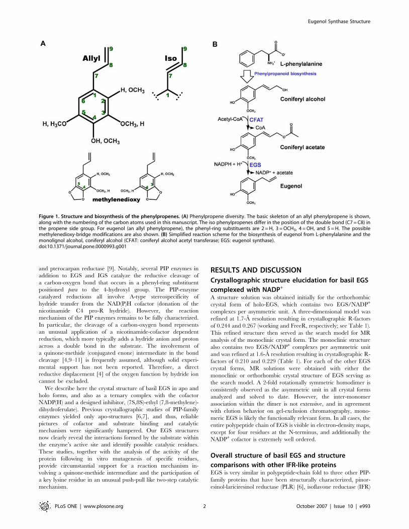
The phenylpropenes are derived from coumaryl, coniferyl, and
sinapyl alcohol, which are also intermediates in the lignin and
lignan biosynthetic pathways. As precursors for phenylpropene
production, the monolignol alcohols undergo first acetylation of
the C9 hydroxyl group [3] and then reductive cleavage of the
acetate moiety to yield the propenyl side group [4,5]. This
reduction reaction is catalyzed by enzymes that produce an allyl
propene (with the double bond between C9 and C8) or an ‘‘iso’’
propene (with the double bond between C8 and C7). An example
of the former is basil (Ocimum basilicum) eugenol synthase (EGS),
which converts coniferyl acetate to eugenol, and an example of the
latter is petunia (Petunia hybrida) isoeugenol synthase (IGS), which
converts coniferyl acetate to isoeugenol (Figure 1). Further
modifications required for formation of the known natural
phenylpropenes include additional hydroxylation of the benzene
ring, methylation of any of the hydroxyl groups on the benzene
ring, and formation of a methylenedioxy bridge (Figure 1). Some
of these modifications may occur before the formation of the
propene moiety, but a free hydroxyl group at the para position
appears to be a requirement for the reduction reaction [4]. The
biosynthetic routes to the phenylpropenes generate considerable
chemical diversity.
The basil EGS and petunia IGS are closely related to a number
of other NADPH-dependent enzymes that act on phenylpropa-
noid-derived substrates. These enzymes constitute the PIP family,
named after the three initially identified members, pinoresinol-
lariciresinol reductase (PLR) [6], isoflavone reductase (IFR) [7],
and phenylcoumaran benzylic ether reductase (PCBER) [6].
Other enzymes in this family are leucocyanidin reductase [8],
Academic Editor: Martin Egli, Vanderbilt University, United States of America
Received June 26, 2007; Accepted September 13, 2007; Published October 3,2007
Copyright: � 2007 Louie et al. This is an open-access article distributed underthe terms of the Creative Commons Attribution License, which permitsunrestricted use, distribution, and reproduction in any medium, provided theoriginal author and source are credited.
Funding: This work was supported by National Science Foundation grants0331353, 0312466 and 0718152 to EP and 0236027 and 0718064 to JNP. JPN is aninvestigator of the Howard Hughes Medical Institute.
Competing Interests: The authors have declared that no competing interestsexist.
* To whom correspondence should be addressed. E-mail: [email protected]
PLoS ONE | www.plosone.org 1 October 2007 | Issue 10 | e993
and pterocarpan reductase [9]. Notably, several PIP enzymes in
addition to EGS and IGS catalyze the reductive cleavage of
a carbon-oxygen bond that occurs in a phenyl-ring substituent
positioned para to the 4-hydroxyl group. The PIP-enzyme
catalyzed reductions all involve A-type stereospecificity of
hydride transfer from the NAD(P)H cofactor (donation of the
nicotinamide C4 pro-R hydride). However, the reaction
mechanism of the PIP enzymes remains to be fully characterized.
In particular, the cleavage of a carbon-oxygen bond represents
an unusual application of a nicotinamide-cofactor dependent
reduction, which more typically adds a hydride anion and proton
across a double bond in the substrate. The involvement of
a quinone-methide (conjugated enone) intermediate in the bond
cleavage [4,9–11] is frequently assumed, although solid experi-
mental support has not been reported. Therefore, a direct
reductive displacement [4] of the oxygen function by hydride ion
cannot be excluded.
We describe here the crystal structure of basil EGS in apo and
holo forms, and also as a ternary complex with the cofactor
NADP(H) and a designed inhibitor, (7S,8S)-ethyl (7,8-methylene)-
dihydroferulate). Previous crystallographic studies of PIP-family
enzymes yielded only apo-structures [6,7], and thus, reliable
pictures of cofactor and substrate binding and catalytic
mechanism were significantly hampered. Our EGS structures
now clearly reveal the interactions formed by the substrate within
the enzyme’s active site and identify possible catalytic residues.
These studies, together with the analysis of the activity of the
protein following in vitro mutagenesis of specific residues,
provide circumstantial support for a reaction mechanism in-
volving a quinone-methide intermediate and the participation of
a key lysine residue in an unusual push-pull like two-step catalytic
mechanism.
RESULTS AND DISCUSSION
Crystallographic structure elucidation for basil EGS
complexed with NADP+
A structure solution was obtained initially for the orthorhombic
crystal form of holo-EGS, which contains two EGS/NADP+
complexes per asymmetric unit. A three-dimensional model was
refined at 1.7-A resolution resulting in crystallographic R-factors
of 0.244 and 0.267 (working and FreeR, respectively; see Table 1).
This refined structure then served as the search model for MR
analysis of the monoclinic crystal form. The monoclinic structure
also contains two EGS/NADP+ complexes per asymmetric unit
and was refined at 1.6-A resolution resulting in crystallographic R-
factors of 0.210 and 0.229 (Table 1). For each of the other EGS
crystal forms, MR solutions were obtained with either the
monoclinic or orthorhombic crystal structure of EGS serving as
the search model. A 2-fold rotationally symmetric homodimer is
consistently observed as the asymmetric unit in all crystal forms
analyzed and solved to date. However, the inter-monomer
association within the dimer is not extensive, and in agreement
with elution behavior on gel-exclusion chromatography, mono-
meric EGS is likely the functionally relevant form. In all cases, the
entire polypeptide chain of EGS is visible in electron-density maps,
except for four residues at the N-terminus, and additionally the
NADP+ cofactor is extremely well ordered.
Overall structure of basil EGS and structure
comparisons with other IFR-like proteinsEGS is very similar in polypeptide-chain fold to three other PIP-
family proteins that have been structurally characterized, pinor-
esinol-lariciresinol reductase (PLR) [6], isoflavone reductase (IFR)
Figure 1. Structure and biosynthesis of the phenylpropenes. (A) Phenylpropene diversity. The basic skeleton of an allyl phenylpropene is shown,along with the numbering of the carbon atoms used in this manuscript. The iso phenylpropenes differ in the position of the double bond (C7 = C8) inthe propene side group. For eugenol (an allyl phenylpropene), the phenyl-ring substituents are 2 = H, 3 = OCH3, 4 = OH, and 5 = H. The possiblemethylenedioxy-bridge modifications are also shown. (B) Simplified reaction scheme for the biosynthesis of eugenol from L-phenylalanine and themonolignol alcohol, coniferyl alcohol (CFAT: coniferyl alcohol acetyl transferase; EGS: eugenol synthase).doi:10.1371/journal.pone.0000993.g001
Eugenol Synthase Structure
PLoS ONE | www.plosone.org 2 October 2007 | Issue 10 | e993

[7], and PCBER [6]. Structurally, the PIP-family proteins
organize around an N-terminal, Rossman-fold domain, containing
a core, six-stranded parallel b-sheet flanked on each face by an a-
helical layer (Figure 2A). One edge of the core b-sheet provides the
extended binding surface for the NADP+ cofactor (as discussed
further below). The C-terminal polypeptide-chain segment of the
PIP-family proteins forms a predominantly a-helical domain, and
this C-terminal segment also contributes three additional b-strands
Table 1. Summary of data collection and refinement statistics for EGS structures. . . . . . . . . . . . . . . . . . . . . . . . . . . . . . . . . . . . . . . . . . . . . . . . . . . . . . . . . . . . . . . . . . . . . . . . . . . . . . . . . . . . . . . . . . . . . . . . . . . . . . . . . . . . . . . . . . . . . . . . . . . . . . . . . . . . . . . . . . . . . . . . . .
EGS-NADP+
monoclinicEGS-NADP+
orthorhombic EGS-NADPH Apo-EGS{ EGS-NADP+-EMDFEGS(Lys132Gln)-EMDF
Space group P21 P212121 P21 P212121 P21 P21
Unit-cell parametersa (A) 53.8 79.3 54.3 79.4 54.0 53.8
b (A) 85.9 85.9 86.0 86.3 85.4 86.2
c (A) 76.2 99.2 76.4 98.2 76.9 76.8
b (u) 107.3 90 107.7 90 107.5 107.6
Monomers per asymmetric unit 2 2 2 2 2 2
Resolution range* (A) 43-1.60 (1.78-1.60) 100-1.72 (1.84-1.72) 34.-1.60 (1.69-1.60) 43-1.80 (1.88-1.80) 34-1.60 (1.69-1.60) 44-1.80 (1.90-1.80)
Number of reflections measured 316974 492798 268704 443874 354814 167942
Merging R-factor* 0.083 (0.676) 0.110 (0.663) 0.105 (0.286) 0.086 (0.477) 0.129 (0.443) 0.109 (0.341)
Mean (I/s I)* 9.9 (2.4) 10.0(2.9) 8.9 (1.9) 23.0 (2.5) 8.3 (1.6) 7.9 (1.6)
Completeness* 0. 972 (0.959) 0. 972 (0.961) 0.912 (0.592) 0.956 (0.732) 0.975 (0.950) 0.921 (0.826)
Redundancy* 3.74 (3.74) 6.73 (5.07) 3.3 (2.2) 7.4 (5.1) 4.1 (2.4) 2.9 (2.2)
Number of reflections used 84786 73101 80436 60262 85858 57422
R-factor* 0.205 (0.322) 0.229 (0.361) 0.201 (0.277) 0.224 (0.327) 0.259 (0.318) 0.248 (0.312)
Free R-factor* 0.226 (0.329) 0.256 (0.411) 0.221 (0.306) 0.242 (0.341) 0.286 (0.328) 0.280 (0.361)
Number of amino-acid residues 620 620 620 620 620 620
Number of water molecules 472 547 674 388 374 350
Residues with most favorableconformation (%)
93.0 93.2 93.2 92.1 93.6 92.7
PDB entry 2QW8 2QX7 2R6J 2QYS 2QZZ 2R2G
Merging R-factor =ghkl gi|Ii(hkl)–<I(hkl)>|/ghkl gi Ii(hkl)*Values in parentheses describe the highest resolution shell.{In the apo-EGS crystal structure, a small amount of nicotinamide cofactor is likely present (less than 20% occupancy, as judged from residual electron density at theexpected NADP(H) binding site).
doi:10.1371/journal.pone.0000993.t001....
....
....
....
....
....
....
....
....
....
....
....
....
....
....
....
....
....
....
....
....
....
....
....
....
..
Figure 2. Structure of EGS and comparison with other PIP-family enzymes. (A) Orthogonal views of EGS/NADP+ monomer. The polypeptide chainof EGS is represented as a ribbon, with coloring varying from blue for the N-terminus to red for the C-terminus. The atoms of the NADP+ cofactor aredrawn as balls and sticks, and are colored coded according to element (carbon: gray; nitrogen: blue; oxygen: red; phosphorus: orange). (B)Superposition of the polypeptide-chain backbones of EGS and other PIP-family enzymes (color coding as shown in inset). The NADP+ cofactor of EGSis also shown (the structures of the other PIP-family enzymes were determined in the absence of cofactor).doi:10.1371/journal.pone.0000993.g002
Eugenol Synthase Structure
PLoS ONE | www.plosone.org 3 October 2007 | Issue 10 | e993
to the Rossman-fold domain. The C-terminal domain is presumed
(see below) to function in substrate binding. Indeed, this domain
together with the last a-helix of the Rossman-fold domain
surround a cavity located immediately adjacent to the nicotin-
amide ring of the NADP+ cofactor. Within the IFR-like PIP
family, the substrate-binding domains (residues 154–314 in EGS)
appear more structurally divergent than the nicotinamide-cofactor
binding domains (residues 1–153 in EGS). For example, from
comparisons of polypeptide-chain backbones (Figure 2B), EGS
differs from PCBER by 1.40 A (rmsd) overall, but by only 0.83 A
for the Rossman-fold domain alone; and similarly, EGS differs
from IFR by 1.63 A overall and 1.09 A for the Rossman-fold
domain alone. Curiously, PCBER, PLR, and IFR-like EGS-all
form 2-fold rotationally symmetric homodimers, but the various
homodimeric associations are distinct in each case. An additional
structural element unique to EGS (absent in the other PIP-family
proteins) is a proline-rich extension at the C-terminus. This tail
segment passes across the mouth of the active-site region, and the
side chain of the C-terminal phenylalanine residue participates
directly in forming the substrate-binding pocket. The positioning
of the tail segment in EGS precludes the formation of the
homodimeric associations observed in PCBER and IFR.
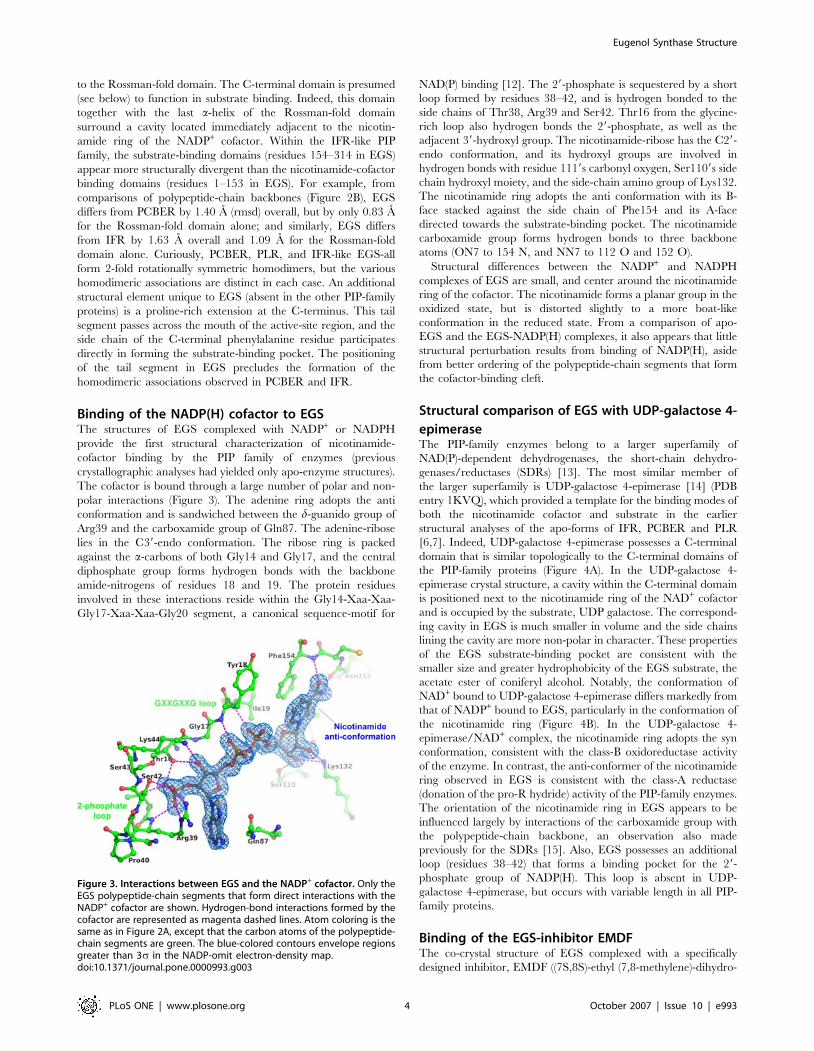
Binding of the NADP(H) cofactor to EGSThe structures of EGS complexed with NADP+ or NADPH
provide the first structural characterization of nicotinamide-
cofactor binding by the PIP family of enzymes (previous
crystallographic analyses had yielded only apo-enzyme structures).
The cofactor is bound through a large number of polar and non-
polar interactions (Figure 3). The adenine ring adopts the anti
conformation and is sandwiched between the d-guanido group of
Arg39 and the carboxamide group of Gln87. The adenine-ribose
lies in the C39-endo conformation. The ribose ring is packed
against the a-carbons of both Gly14 and Gly17, and the central
diphosphate group forms hydrogen bonds with the backbone
amide-nitrogens of residues 18 and 19. The protein residues
involved in these interactions reside within the Gly14-Xaa-Xaa-
Gly17-Xaa-Xaa-Gly20 segment, a canonical sequence-motif for
NAD(P) binding [12]. The 29-phosphate is sequestered by a short
loop formed by residues 38–42, and is hydrogen bonded to the
side chains of Thr38, Arg39 and Ser42. Thr16 from the glycine-
rich loop also hydrogen bonds the 29-phosphate, as well as the
adjacent 39-hydroxyl group. The nicotinamide-ribose has the C29-
endo conformation, and its hydroxyl groups are involved in
hydrogen bonds with residue 1119s carbonyl oxygen, Ser1109s side
chain hydroxyl moiety, and the side-chain amino group of Lys132.
The nicotinamide ring adopts the anti conformation with its B-
face stacked against the side chain of Phe154 and its A-face
directed towards the substrate-binding pocket. The nicotinamide
carboxamide group forms hydrogen bonds to three backbone
atoms (ON7 to 154 N, and NN7 to 112 O and 152 O).
Structural differences between the NADP+ and NADPH
complexes of EGS are small, and center around the nicotinamide
ring of the cofactor. The nicotinamide forms a planar group in the
oxidized state, but is distorted slightly to a more boat-like
conformation in the reduced state. From a comparison of apo-
EGS and the EGS-NADP(H) complexes, it also appears that little
structural perturbation results from binding of NADP(H), aside
from better ordering of the polypeptide-chain segments that form
the cofactor-binding cleft.
Structural comparison of EGS with UDP-galactose 4-
epimeraseThe PIP-family enzymes belong to a larger superfamily of
NAD(P)-dependent dehydrogenases, the short-chain dehydro-
genases/reductases (SDRs) [13]. The most similar member of
the larger superfamily is UDP-galactose 4-epimerase [14] (PDB
entry 1KVQ), which provided a template for the binding modes of
both the nicotinamide cofactor and substrate in the earlier
structural analyses of the apo-forms of IFR, PCBER and PLR
[6,7]. Indeed, UDP-galactose 4-epimerase possesses a C-terminal
domain that is similar topologically to the C-terminal domains of
the PIP-family proteins (Figure 4A). In the UDP-galactose 4-
epimerase crystal structure, a cavity within the C-terminal domain
is positioned next to the nicotinamide ring of the NAD+ cofactor
and is occupied by the substrate, UDP galactose. The correspond-
ing cavity in EGS is much smaller in volume and the side chains
lining the cavity are more non-polar in character. These properties
of the EGS substrate-binding pocket are consistent with the
smaller size and greater hydrophobicity of the EGS substrate, the
acetate ester of coniferyl alcohol. Notably, the conformation of
NAD+ bound to UDP-galactose 4-epimerase differs markedly from
that of NADP+ bound to EGS, particularly in the conformation of
the nicotinamide ring (Figure 4B). In the UDP-galactose 4-
epimerase/NAD+ complex, the nicotinamide ring adopts the syn
conformation, consistent with the class-B oxidoreductase activity
of the enzyme. In contrast, the anti-conformer of the nicotinamide
ring observed in EGS is consistent with the class-A reductase
(donation of the pro-R hydride) activity of the PIP-family enzymes.
The orientation of the nicotinamide ring in EGS appears to be
influenced largely by interactions of the carboxamide group with
the polypeptide-chain backbone, an observation also made
previously for the SDRs [15]. Also, EGS possesses an additional
loop (residues 38–42) that forms a binding pocket for the 29-
phosphate group of NADP(H). This loop is absent in UDP-
galactose 4-epimerase, but occurs with variable length in all PIP-
family proteins.
Binding of the EGS-inhibitor EMDFThe co-crystal structure of EGS complexed with a specifically
designed inhibitor, EMDF ((7S,8S)-ethyl (7,8-methylene)-dihydro-
Figure 3. Interactions between EGS and the NADP+ cofactor. Only theEGS polypeptide-chain segments that form direct interactions with theNADP+ cofactor are shown. Hydrogen-bond interactions formed by thecofactor are represented as magenta dashed lines. Atom coloring is thesame as in Figure 2A, except that the carbon atoms of the polypeptide-chain segments are green. The blue-colored contours envelope regionsgreater than 3s in the NADP-omit electron-density map.doi:10.1371/journal.pone.0000993.g003
Eugenol Synthase Structure
PLoS ONE | www.plosone.org 4 October 2007 | Issue 10 | e993

ferulate) provides a view of the substrate-binding mode within the
active site of EGS (Figures 4B and 5A). This inhibitor is a close
structural analog of coniferyl acetate, and carries the same
functional groups on the C3 and C4 (para) positions of the phenyl
(guaiacol) ring. However, EMDF cannot serve as a substrate of
EGS because within the C1 substituent, a cyclopropyl group
replaces of the C7-C8 double bond and the orientation of the ester
is reversed. Our measurements indicate that EMDF acts as
a competitive inhibitor of EGS, with an inhibition constant
(Ki = 0.8 mM) similar to the Km for coniferyl acetate, 0.57 mM. A
key interaction between EMDF and EGS is the packing of the
inhibitor’s phenyl ring parallel to the A-face of the cofactor’s
nicotinamide ring (interplanar separation 3.4 A). Notably, stacking
of a substrate aromatic-ring against the NAD(P) nicotinamide ring
is a common feature of the binding modes of SDRs [16]. In EGS,
the nicotinamide ring is, in turn, stacked against the side chain of
Phe154. The para-hydroxy group of the inhibitor’s guaiacol moiety
hydrogen bonds with the backbone amide-nitrogen of Val114 and
also interacts via a bridging water molecule with the side-chain
amino group of Lys132. The residues lining the binding pocket are
otherwise predominantly aromatic (Phe85, Phe125, Tyr157,
Phe158, Tyr161, and Phe314) and aliphatic (Val114, Ile261,
Leu262, and Leu265). In the known EGS and IGS sequences,
Lys132, Tyr157, Phe158, and Ile261 are invariant, whereas
conservative amino-acid substitutions are observed at the other
binding-pocket residues. The inhibitor’s 3-hydroxymethyl group is
accommodated within a small, non-polar pocket. The observed
orientation of the guaiacol moiety would be clearly favored over
the reverse orientation (resulting from a 180u rotation around the
C7-C1 bond), which would position the hydroxymethyl group in
close contact with the nicotinamide ribose and the side chain of
Phe85. However, the absence of specific interactions formed by
the hydroxymethyl group within its binding pocket is perhaps
consistent with the limited ability of EGS to utilize as substrate
coumaryl acetate (unpublished data), which lacks a substituent at
the 3-position. In addition, the acetate ester of sinapyl alcohol,
which bears hydroxymethyl groups at both the 3- and 5-positions,
would be expected to be incapable of binding to EGS. The
inhibitor’s C1 substituent bearing the cyclopropyl and ethyl-ester
moieties projects into a cavity formed by the C-terminal domain of
the protein. This cavity is capped by the side chains of Tyr157,
Tyr161, Pro258, Leu262 and Phe314. With the exception of the
C-terminal Phe314, these capping residues form a relatively rigid
cage, as indicated by their invariant positioning in all EGS crystal
structures and low crystallographic temperature factors. The
capping region appears to lack sufficient volume to accommodate
a C1 substituent larger than an acetate-esterified propenol. This
finding is in agreement with the observed inactivity of EGS toward
other esters of coniferyl alcohol, for example coniferyl coumarate
[4], which bears a much bulkier substituent.
Curiously, at the inhibitor-binding site described above, some
residual electron density is invariably observed, even with crystal
samples prepared with EGS protein that had not been purposely
exposed to a potential ligand. This density might be due to low-
occupancy binding of a small, eugenol-resembling compound that
originated from bacterial growth-media derived from yeast
extracts. However, soaking experiments of EGS crystals with
coniferyl acetate, or other substrate analogs (e.g. coumaryl acetate
and 4-bromo-cinnamyl acetate) failed to produce stable complexes
with EGS. The difficulty in obtaining EGS complexes with these
compounds is possibly explained by the implications of the finding
that binding of the true substrate, coniferyl acetate, cannot be
readily modeled based on the observed positioning of the EMDF
inhibitor compound. The determinative structural feature of
coniferyl acetate is the planarity of the propene moiety (C7-C8-
C9), which results in steric clashes between the terminal acetate
moiety and neighboring residues in the active-site capping region,
in particular Tyr157, Ile261 and Phe314. In contrast, the
cyclopropyl group of EMDF induces a distinct kink in the
conformation of the side group, which steers the end of the side
group into a hole formed within the capping region (Figure 5B).
The stringent binding selectivity of EGS is further emphasized by
the observation that only the 7-(S),8-(S) stereoisomer of EMDF
complexed with EGS; the 7-(R),8-(R) isomer (present at low levels
Figure 4. Structural comparison of EGS and UDP-galactose epimer-ase. (A) Superposition of polypeptide-chain backbones of EGS andUDP-galactose epimerase (color coding as shown in inset). For clarity,only the NADP+ cofactor of EGS is shown. (B) Comparison of NAD(P)-cofactor conformation and substrate-analog binding in EGS and UDP-galactose epimerase. The binding of the EGS competitive inhibitor(7S,8S)-ethyl (7,8-methylene)-dihydroferulate (EMDF) is described indetail in the text and Figure 5. The coloring of the polypeptide-chainsegments is the same as in (A). The inset shows the coloring used forthe carbon atoms of the nicotinamide cofactors, EMDF bound to EGS,and UDP-glucose bound to UDP-galactose-4-epimerase.doi:10.1371/journal.pone.0000993.g004
Eugenol Synthase Structure
PLoS ONE | www.plosone.org 5 October 2007 | Issue 10 | e993

Figure 5. Binding of the competitive inhibitor EMDF by EGS. (A) Orthogonal views of the 7S,8S- stereoisomer of the competitive inhibitor EMDFbound to EGS. Hydrogen-bond interactions formed by the EMDF molecule (cyan colored carbons) are represented as magenta dashed lines. Theblue-colored contours envelope regions greater than 2.5s in the initial Fobs-Fcalc electron-density map. The direction of view used in the right panel(approximately perpendicular to the plane of the nicotinamide ring) is maintained roughly in figures 5B–D and 6B. The chemical structure of EMDF isshown in the inset. (B) Modeled binding of coniferyl acetate to EGS. The atom coloring is the same as in (A), with magenta carbon atoms for theconiferyl acetate. The chemical structures of coniferyl acetate and EMDF are compared in the inset. The close interaction between the EMDF C7-atomand the hydride donor of the nicotinamide (C4) is shown as a yellow dashed line. (C) Binding of EMDF to the Lys132Gln variant of EGS. Hydrogen-bond interactions formed by the EMDF molecule (cyan colored carbons) are represented as magenta dashed lines. Hydrogen bonds involving theside chain of Gln132 are shown as orange dashed lines. The blue-colored contours envelope regions greater than 2.0s in the initial Fobs-Fcalc electron-density map. (D) Binding of EMDF to the Lys132Arg variant of EGS (stereo representation). The blue-colored contours envelope regions greater than2s in the initial Fobs-Fcalc electron-density map for the EGS-Arg132/EMDF complex (green). The altered positioning of the Arg132 side-chain andneighboring residues (most notably Phe85, Ile88, and Ile129) and the disordering of the C-terminal tail (residues 310–314) are apparent with respectto the holo-EGS-Arg132 structure (magenta). For comparison, the position of the wild-type Lys132 side chain and the key bridging water moleculeshown in Figure 5A are also shown (yellow).doi:10.1371/journal.pone.0000993.g005
Eugenol Synthase Structure
PLoS ONE | www.plosone.org 6 October 2007 | Issue 10 | e993

in the EMDF preparation) was excluded, due to poorer steric
complementarity with the EGS active site.
3D structure determination and in vitro
mutagenesis suggests a reaction mechanism for EGSAlthough the binding of EMDF exploits shape features of the EGS
active site that are inaccessible to the coniferyl acetate substrate,
the structure of the EGS-NADP+-EMDF complex nevertheless
provides a useful framework for probing the EGS enzymatic
mechanism. Together with the observed effects on catalytic
activity of specific amino-acid replacements (Table 2), as described
below, the structure provides compelling support for the in-
volvement of a quinone-methide intermediate both in promoting
carbon-oxygen bond cleavage of the acetate moiety and in serving
as the actual substrate of the reduction reaction via NADPH-
mediated hydride transfer.
A prominent active-site residue is Lys132, which occurs in all
PIP-family enzymes as well as most SDRs [17]. The structure of
the EGS-NADP+-EMDF complex shows that the e-amino group
of Lys132 forms interactions with both the nicotinamide-ribose of
NADP(H), and potentially, the substrate molecule (Figure 5A).
The Lys132 interaction with the substrate is particularly in-
triguing, as it is not proximal to the site of hydride addition (as
suggested in the case of IFR [7]), but instead involves the p-
hydroxyl group via a bridging water molecule. Notably, a p-
hydroxyl group is a distinguishing feature of the substrates of all
PIP-family enzymes, and is a requirement for reduction by EGS
[4]. For both PLR and IFR, alanine replacements of the lysine that
is equivalent to EGS-Lys132 abolish enzyme activity [6,7]. In
EGS, Lys132Ala and Lys132Gln mutants are completely inactive,
whereas the Lys132Arg mutant retains partial (71%) activity
(Table 2). Crystallographic analyses confirmed that for the Ala132
and Gln132 mutants, both NADP+ and EMDF binding are little
affected (Figure 5C), despite the loss of the binding interactions
normally contributed by Lys132. These results therefore point to
a catalytic role for Lys132. In particular, the involvement of
a catalytic group acting at the p-hydroxyl group clearly argues for
the formation of a quinone methide as a reaction intermediate as
opposed to direct nucleophilic replacement by a NADPH derived
hydride anion.
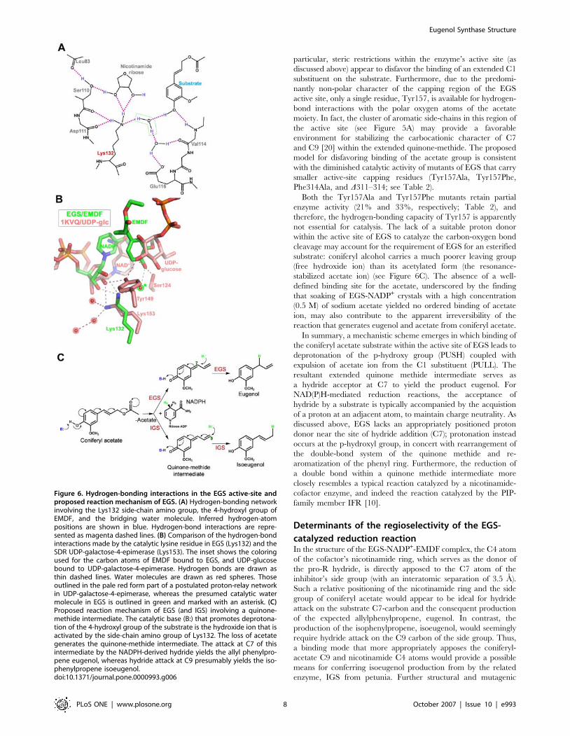
The formation of a quinone methide can be promoted by
abstraction of the proton from the p-hydroxyl group of substrate.
Detailed inspection of the hydrogen-bonding network involving
the e-amino group of Lys132 (Figure 6A) suggests that this group
exists formally in the unprotonated–NH2 state and is the donor in
hydrogen-bond interactions with the 29-hydroxyl group of the
nicotinamide-ribose and the backbone carbonyl oxygen of residue
110. With an available lone pair of electrons, the amino group can
serve as the acceptor in a hydrogen bond with the bridging water
molecule, and most importantly, thereby act as a general base.
The water molecule (as a hydroxide ion) can in turn facilitate
deprotonation of the substrate’s p-hydroxyl group. Intruigingly, in
the monoclinic structure of unliganded EGS-NADP+, a nitrate
anion from the crystallization medium occupies the site of the
bridging water molecule, and may mimic the hydroxide ion that
develops during the catalytic reaction.
The loss in activity of EGS Lys132-mutants can be interpreted
in terms of the proposed mechanistic model, in conjunction with
results from structural analyses. In the EGS (Lys132Gln)-NADP+-
EMDF complex, the Gln132 side chain retains an interaction with
the nicotinamide ribose (also through an intervening water
molecule), but is unable to form a direct or water-mediated
interaction with the p-hydroxyl group of EMDF. Likewise, Ala132
obviously lacks a catalytic group capable of promoting deprotona-
tion of the substrate’s p-hydroxyl group, and therefore, the
observed inactivity of the Lys132Ala and Lys132Gln mutants can
be readily explained. The Lys132Arg mutant is partially active,
and in this case, with the higher pKa and greater length of the Arg
side chain, the positively charged guanidinium moiety could
possibly participate directly (i.e. without the requirement for an
intervening water molecule) in lowering the pKa of the substrate’s
p-hydroxyl group. Interestingly, preliminary structural analysis of
the holo and EMDF-bound forms of the Lys132Arg mutant shows
that the Arg132 guanidinium moiety is displaced slightly by the
EMDF guaiacol ring (Figure 5D), thus diminishing the potential
influence of the Arg132 on catalysis.
One difficulty with the proposed catalytic role for a lysine
residue is the relatively high pKa (normally ,10.4 in solution) of
the side-chain amino group, which would disfavor acquisition of
the initial free-base state required for proton abstraction from the
substrate. However, the pKas of ionizable groups in proteins can
be greatly influenced by the local structural environment, in
particular, involvement in hydrogen-bonding networks and
hydrophobic interactions. Such factors have been suggested to
account for the catalytic-base activity of the lysine e-amino group
in a number of enzymes. A notable example is isochorismate
synthase [18], in which a catalytic lysine is proposed to
deprotonate and thereby activate a nucleophilic water molecule.
Furthermore, from theoretical calculations on the conserved Lys-
Tyr-Ser catalytic triad in an SDR-type alcohol dehydrogenase, the
catalytically important lysine residue is suggested to exist in
a partially unprotonated state, and in this state, participate in
a proton-relay network that involves hydroxyl groups on the
catalytic tyrosine and nicotinamide ribose [19]. This network
functions ultimately to abstract a proton from the alcohol
substrate. Intriguingly, although EGS lacks the catalytic tyrosine
of the SDR enzyme, Lys132 in EGS corresponds exactly to the
catalytic lysine of the SDRs, and the p-hydroxyl group of EGS-
bound EMDF occurs at roughly the same position as the Tyr f-
hydroxyl group of the catalytic tyrosine of SDRs (Figure 6B).
For the EGS-catalyzed reaction with the coniferyl acetate
substrate, formation of the quinone-methide intermediate would
be concomitant with displacement of an acetate ion (Figure 6C). In
concert with proton abstraction from the p-hydroxyl group, EGS
may therefore exploit an additional driving force for generation of
the reaction intermediate—promoting the loss of the acetate. In
Table 2. Relative activity of EGS variants. . . . . . . . . . . . . . . . . . . . . . . . . . . . . . . . . . . . . . . . . . . . . . . . . . . . . . . . . . . . . . . . . . . . . .
EGS variant Relative activity* (%)
Wild type 100
Lys132Ala 0
Lys132Gln 0
Lys132Arg 71 (66)
Tyr157Phe 32.5 (64.4)
Tyr157Ala 21 (63)
Ile261His 3.1 (60.2)
Phe314Tyr 112 (68)
Phe314Ala 54 (65)
Phe314Tyr-Ala-Gln-Pro-Ser-Thr 115 (611)
D311–314 34.9 (60.7)
*The standard error of each activity measurement (derived from duplicate ortriplicate determinations) is given in parentheses.
doi:10.1371/journal.pone.0000993.t002....
....
....
....
....
....
....
....
....
....
....
....
....
....
....
..
Eugenol Synthase Structure
PLoS ONE | www.plosone.org 7 October 2007 | Issue 10 | e993
particular, steric restrictions within the enzyme’s active site (as
discussed above) appear to disfavor the binding of an extended C1
substituent on the substrate. Furthermore, due to the predomi-
nantly non-polar character of the capping region of the EGS
active site, only a single residue, Tyr157, is available for hydrogen-
bond interactions with the polar oxygen atoms of the acetate
moiety. In fact, the cluster of aromatic side-chains in this region of
the active site (see Figure 5A) may provide a favorable
environment for stabilizing the carbocationic character of C7
and C9 [20] within the extended quinone-methide. The proposed
model for disfavoring binding of the acetate group is consistent
with the diminished catalytic activity of mutants of EGS that carry
smaller active-site capping residues (Tyr157Ala, Tyr157Phe,
Phe314Ala, and D311–314; see Table 2).
Both the Tyr157Ala and Tyr157Phe mutants retain partial
enzyme activity (21% and 33%, respectively; Table 2), and
therefore, the hydrogen-bonding capacity of Tyr157 is apparently
not essential for catalysis. The lack of a suitable proton donor
within the active site of EGS to catalyze the carbon-oxygen bond
cleavage may account for the requirement of EGS for an esterified
substrate: coniferyl alcohol carries a much poorer leaving group
(free hydroxide ion) than its acetylated form (the resonance-
stabilized acetate ion) (see Figure 6C). The absence of a well-
defined binding site for the acetate, underscored by the finding
that soaking of EGS-NADP+ crystals with a high concentration
(0.5 M) of sodium acetate yielded no ordered binding of acetate
ion, may also contribute to the apparent irreversibility of the
reaction that generates eugenol and acetate from coniferyl acetate.
In summary, a mechanistic scheme emerges in which binding of
the coniferyl acetate substrate within the active site of EGS leads to
deprotonation of the p-hydroxy group (PUSH) coupled with
expulsion of acetate ion from the C1 substituent (PULL). The
resultant extended quinone methide intermediate serves as
a hydride acceptor at C7 to yield the product eugenol. For
NAD(P)H-mediated reduction reactions, the acceptance of
hydride by a substrate is typically accompanied by the acquistion
of a proton at an adjacent atom, to maintain charge neutrality. As
discussed above, EGS lacks an appropriately positioned proton
donor near the site of hydride addition (C7); protonation instead
occurs at the p-hydroxyl group, in concert with rearrangement of
the double-bond system of the quinone methide and re-
aromatization of the phenyl ring. Furthermore, the reduction of
a double bond within a quinone methide intermediate more
closely resembles a typical reaction catalyzed by a nicotinamide-
cofactor enzyme, and indeed the reaction catalyzed by the PIP-
family member IFR [10].
Determinants of the regioselectivity of the EGS-
catalyzed reduction reactionIn the structure of the EGS-NADP+-EMDF complex, the C4 atom
of the cofactor’s nicotinamide ring, which serves as the donor of
the pro-R hydride, is directly apposed to the C7 atom of the
inhibitor’s side group (with an interatomic separation of 3.5 A).
Such a relative positioning of the nicotinamide ring and the side
group of coniferyl acetate would appear to be ideal for hydride
attack on the substrate C7-carbon and the consequent production
of the expected allylphenylpropene, eugenol. In contrast, the
production of the isophenylpropene, isoeugenol, would seemingly
require hydride attack on the C9 carbon of the side group. Thus,
a binding mode that more appropriately apposes the coniferyl-
acetate C9 and nicotinamide C4 atoms would provide a possible
means for conferring isoeugenol production from by the related
enzyme, IGS from petunia. Further structural and mutagenic
Figure 6. Hydrogen-bonding interactions in the EGS active-site andproposed reaction mechanism of EGS. (A) Hydrogen-bonding networkinvolving the Lys132 side-chain amino group, the 4-hydroxyl group ofEMDF, and the bridging water molecule. Inferred hydrogen-atompositions are shown in blue. Hydrogen-bond interactions are repre-sented as magenta dashed lines. (B) Comparison of the hydrogen-bondinteractions made by the catalytic lysine residue in EGS (Lys132) and theSDR UDP-galactose-4-epimerase (Lys153). The inset shows the coloringused for the carbon atoms of EMDF bound to EGS, and UDP-glucosebound to UDP-galactose-4-epimerase. Hydrogen bonds are drawn asthin dashed lines. Water molecules are drawn as red spheres. Thoseoutlined in the pale red form part of a postulated proton-relay networkin UDP-galactose-4-epimerase, whereas the presumed catalytic watermolecule in EGS is outlined in green and marked with an asterisk. (C)Proposed reaction mechanism of EGS (and IGS) involving a quinone-methide intermediate. The catalytic base (B:) that promotes deprotona-tion of the 4-hydroxyl group of the substrate is the hydroxide ion that isactivated by the side-chain amino group of Lys132. The loss of acetategenerates the quinone-methide intermediate. The attack at C7 of thisintermediate by the NADPH-derived hydride yields the allyl phenylpro-pene eugenol, whereas hydride attack at C9 presumably yields the iso-phenylpropene isoeugenol.doi:10.1371/journal.pone.0000993.g006
Eugenol Synthase Structure
PLoS ONE | www.plosone.org 8 October 2007 | Issue 10 | e993

studies and analyses of additional, newly discovered IGS-type
enzymes (Koeduka et al., submitted) are underway to probe the
enzymic determinants of the regioselectivity of the EGS/IGS
catalyzed reactions.
Modeling of NADP(H) and substrate binding in other
IFR-like proteinsThe EGS residues that are involved in interactions with the
NADP(H) cofactor (see above) are highly conserved in the other
IFR-like enzymes, and thus these enzymes can be expected to
maintain a cofactor-binding site very similar to that observed in
EGS. Most of these other enzymes have also been characterized as
A-type reductases. On the basis of the holo-EGS structures, the
NADP(H) cofactor can be readily modeled into the apo-structures
of the other IFR-like enzymes, although small, accommodating
adjustments to the surrounding protein are necessary.
The substrates of the PLR, PCBER and IFR enzymes all possess
a phenyl ring with a C4-hydroxyl group, and the expected sites of
hydride addition occur near the C7 atom of the substrate. For
these enzymes, substrate binding can be modeled on the basis of
the positioning of the guaiacol moiety of EMDF bound to EGS. As
expected, in all cases, the substrate C7-atom is positioned very
close to the C4 atom of the cofactor’s nicotinamide ring. The
substrate-binding pocket in general appears less enclosed in the
other IFR-like proteins than in EGS, consistent perhaps with the
larger size of the C1 substituents of the cognate substrates. The
more open binding pockets are due primarily to the absence of the
C-terminal tail that occurs in EGS, as well as the substitution of
smaller residues within the active-site capping region.
Moreover, the substrates for PLR and PCBER contain a cyclic-
ether linkage adjacent to the site of hydride addition, and will
therefore generate a transient alkoxide intermediate upon carbon-
oxygen bond cleavage and ring opening. These enzymes may
employ two means to promote protonation of the alkoxide
intermediate: a potential proton donor or hydrogen-bonding
group within the active site (His271 in PLR; Ser263 and Ser267 in
PCBER), and a more open binding site that may allow access to
the intermediate by bulk water.
MATERIALS AND METHODS
Protein expression and purificationA DNA fragment encoding the entire amino-acid sequence
(residues 1–314) of Ocimum basilicum EGS1 [4] was inserted
between the NcoI and BamH1 sites of the expression vector
pHIS8, which, under the control of a T7 promoter, yields the
target protein fused to an N-terminal octahistidine tag [21]. For
heterologous over-expression of the EGS protein, the plasmid
pHIS8(EGS) was transformed into the expression host E. coli
BL21(DE3) (Novagen). E. coli cultures in TB medium were grown
at 37uC to an optical density (600 nm) of 1.5, induced with 1 mM
isopropyl-b-D-thiogalactoside, and allowed to grow for an
additional 6 hrs at 20uC. Bacterial cells were harvested by
centrifugation, resuspended in lysis buffer (50 mM TrisHCl,
pH 8.0; 0.5 M NaCl; 20 mM imidazole; 1% v/v Tween20;
10% v/v glycerol; and 20 mM 2-mercaptoethanol), and lysed by
sonication. The EGS protein was isolated from the E. coli lysate by
affinity chromatography with nickel-nitrilotriacetic-acid coupled
agarose (Qiagen), and eluted with lysis buffer supplemented with
0.25 M imidazole. The partially purified EGS protein was treated
with thrombin for cleavage of the octahistidine tag, and then
further purified by gel-exclusion chromatography using a Superdex
200 HR26/60 column (Pharmacia Biosystems).
Site-directed mutagenesis of O. basilicum EGSSite-directed mutants of the EGS gene were created in the plasmid
pHIS8(EGS) with the PCR method [22]. The DNA sequences of
the mutant constructs were confirmed by sequencing of the entire
EGS insert in both the forward and reverse directions.
Chemoenzymatic synthesis of coniferyl acetate ((E)-
4-hydroxy-3-methoxycinnamylacetate)Coniferyl alcohol (180.2 mg, 1.0 mmol), Candida antarctica lipase B
(CAL-B, 25 mg, Aldrich, $10,000 U/g), vinyl acetate (401 mL,
5.0 mmol), and dry diethyl ether (Et2O, 50 mL, 0.2 M) were
stirred in a 125-mL Erlenmeyer flask at room temperature for 2 h
(Figure 7). The reaction mixture was filtered through glass wool
and concentrated in vacuo. The reaction yielded quantitatively the
desired product (222 mg colorless oil). 1H NMR (500 MHz,
CDCl3) d 6.91 (m, 3H), 6.59 (d, J = 15.92 Hz, 1H), 6.15 (dt,
J = 6.62, 15.92 Hz, 1H), 4.72 (d, J = 6.62 Hz, 2H), 3.92 (s, 3H),
2.11 (s, 3H); 13C NMR (125 MHz, CDCl3) d 171.2, 146.8, 146.1,
134.7, 129.0, 121.0, 120.9, 114.7, 108.6, 65.5, 56.1, 21.3. LCMS
[M-H]- calculated for C12H13O4: 221.08, found: 221.2.
Chemical synthesis of (7S,8S)-ethyl (7,8-methylene)-
dihydroferulate (EMDF)The compound (7S,8S)-ethyl (7,8-methylene)-dihydroferulate
(EMDF, (1S,2S)-ethyl 2-(4-hydroxy-3-methoxyphenyl) cyclo-pro-
panecarboxylate) was synthesized in three steps (Figure 8).
4-((2-methoxyethoxy)methoxy)-3-methoxy benzaldehyde (step 1). K2CO3
(1.38 g, 10.0 mmol, 1.0 eq.) was added to solution of vanillin
(1.52 g, 10.0 mmol, 1.0 eq.) in dry acetone (70 mL, 0.15 M), and
the mixture was stirred under argon at 0uC for 30 min. Then
MeOCH2CH2OCH2Cl (MEM-Cl, 1.49 g, 12.0 mmol, 1.2 eq.)
was added drop wise, and the mixture was stirred an additional
5 h. The mixture was then concentrated in vacuo to ,20 mL,
combined with 20 mL of water, and extracted with diethyl ether
(3625 mL). The organic layers were pooled, washed with brine,
dried with MgSO4, filtered and concentrated in vacuo. The
resulting yellow oil (2.38 g, 98% yield) was used without further
purification. 1H NMR (500 MHz, CDCl3) d 9.86 (s, 1H), 7.43 (s,
1H), 7.42 (d, J = 8.5 Hz, 2H), 7.32 (d, J = 8.5 Hz, 2H), 5.41 (s,
2H), 3.93 (s, 3H), 3.86 (t, J = 4.7 Hz, 2H), 3.54 (t, J = 4.7 Hz, 2H),
3.35 (s, 3H); 13C NMR (125 MHz, CDCl3) d 191.0, 152.0, 150.0,
131.1, 126.5, 114.8, 109.4, 94.0, 71.4, 68.2, 59.0, 56.0; LCMS
[M+Na]+ calculated for C12H16O5Na: 263.09, found: 263.3.
1-((2-methoxyethoxy)methoxy)-2-methoxy-4-vinylbenzene (step 2). To
a suspension of methyltriphenylphosphonium bromide
(MeP(Ph)3Br, 3.57 g, 10.0 mmol, 2.0 eq.) in anhydrous tetrahy-
drofuran (THF, 30 mL, 0.33 M), under argon at room temper-
ature, was added potassium bis-trimethylsilylamide (KN(TMS)2,
4.5 mL, 9.0 mmol, 1.8 eq., 0.5 M in toluene). After 30 min,
a solution containing 4-((2-methoxyethoxy)methoxy)-3-methoxy
benzaldehyde (1.20 g, 5.0 mmol, 1.0 eq.) was added to the yellow
colored ylide solution via cannula. The reaction was completed
within 30–60 min (as monitored by TLC, hexanes/ethyl acetate,
Figure 7. Chemoenzymatic synthesis of coniferyl acetate. Coniferylacetate was obtained as a colorless oil with a yield of nearly 100%.doi:10.1371/journal.pone.0000993.g007
Eugenol Synthase Structure
PLoS ONE | www.plosone.org 9 October 2007 | Issue 10 | e993

7:3). The reaction mixture was quenched with saturated NH4Cl
and extracted with diethyl ether (36100 mL). The organic layers
were combined, washed with water (26100 mL), brine
(26100 mL), dried over Na2SO4, filtered, concentrated in vacuo,
and subjected to Dry Column Vacuum Chromatography (90%
hexanes/ethyl acetate) to afford a colorless oil (2.01 g, 84% yield).1H NMR (500 MHz, CDCl3) d 7.15 (d, J = 8.3 Hz, 1H), 6.96 (s,
1H), 6.93 (d, J = 8.3 Hz, 1H), 6.65 (dd, J = 10.9, 17.5 Hz, 1H),
5.63 (d, J = 17.4 Hz, 1H), 5.32 (s, 2H), 5.17 (d, J = 10.9 Hz, 1H),
3.90 (s, 3H), 3.87 (t, J = 4.7 Hz, 2H), 3.56 (t, J = 4.7 Hz, 2H), 3.37
(s, 3H); 13C NMR (125 MHz, CDCl3) d 149.7, 146.4, 136.4,
132.3, 119.4, 116.2, 112.4, 109.1, 94.4, 71.5, 67.8, 59.0, 55.8;
LCMS [M+Na]+ calculated for C13H18O4Na: 261.1, found:
260.9.
(1S,2S)-ethyl 2-(4-hydroxy-3-methoxyphenyl) cyclo-propanecarboxylate
(step 3). To a solution of copper-ligand complex prepared from
CuIOTf (toluene)0.5 (129.3 mg, 0.25 mmol, 2.0 mol%) and ligand
(A) (75.3 mg, 0.25 mmol, 2.0 mol%) in 50 mL of dry dichlor-
omethane (DCM) was added 1-((2-methoxyethoxy)methoxy)-2-
methoxy-4-vinylbenzene (1.0 g, 4.2 mmol, 1.0 eq.). Ethyl diazoa-
cetate (CHN2CO2Et, 485.8 mL, 4.62 mmol, 1.1 eq.) was dissolved
in 0.5 mL dry dichloromethane and added to the stirring reaction
mixture by slow addition via syringe pump over 10 h. The crude
reaction mixture was filtered through a plug of silica gel and
concentrated in vacuo. 1H NMR and LCMS showed a complex
product mixture consisting of 80% conversion to cyclopropane
products with a trans to cis ratio of 2.5:1. Chiral HPLC analysis
(Chiralcel-OD) determined 95% ee for the trans product. In
addition to the MEM-protected trans- and cis-cyclopropane
products, the crude reaction mixture contained the desired final
product, the deprotected trans-cyclopropane, (1S,2S)-ethyl 2-(4-
hydroxy-3-methoxyphenyl) cyclo-propanecarboxylate (EMDF),
which was isolated by column chromatography (90% hexanes/
ethyl acetate R 100% ethyl acetate) (99.2 mg as a colorless oil,
10% yield). Subsequent X-ray structure analysis of EMDF
complexed with EGS confirmed the compound’s absolute
stereochemistry. 1H NMR (500 MHz, CDCl3) d 6.82 (d,
J = 8.2 Hz, 1H), 6.64 (s, 1H), 6.59 (d, J = 8.2 Hz, 1H), 5.56, ( br
s, Ar-OH, 1H), 4.17 (q, J = 7.0 Hz, 2H), 3.87 (s, 3H), 2.47 (ddd,
J = 4.6, 6.4, 9.6 Hz, 1H), 1.82 (ddd, J = 4.6, 5.6, 8.8 Hz, 1H), 1.54
(ddd, J = 4.8, 5.6, 9.2 Hz, 1H), 1.28 (t, J = 7.1 Hz, 3H)1.25 (ddd,
buried, 1H); 13C NMR (125 MHz, CDCl3) d 173.6, 146.5, 144.3,
131.9, 118.8, 114.3, 109.4, 60.7, 55.9, 26.1, 23.8, 16.7, 14.3;
LCMS [M-H]- calculated for C13H15O4Na: 235.1, found: 234.8.
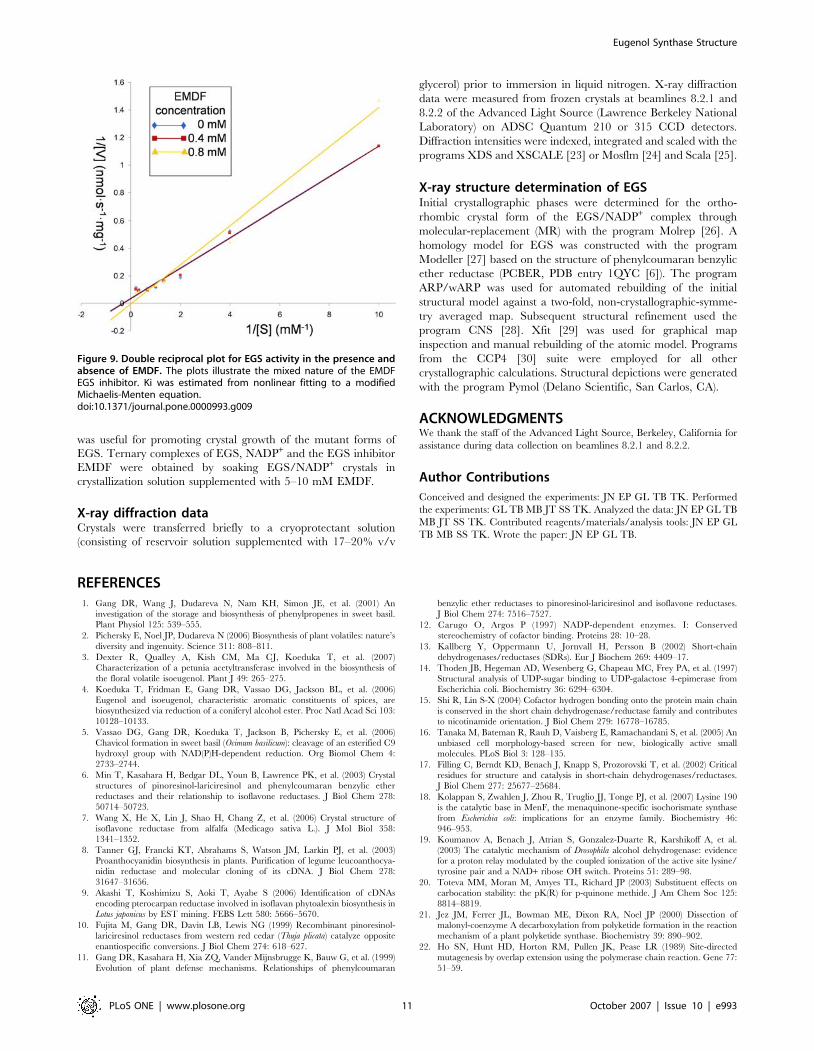
EGS enzyme assayEGS enzyme activity was measured by gas chromatography/mass
spectrometry as described previously [4]. The assay mixture (total
volume 0.15 mL) contained 0.05 M MES-KOH (pH 6.5), 1 mM
NADPH, 1 mM coniferyl acetate, and 2 mg of EGS. Reaction
mixtures were incubated at 25uC for 15 min followed by
extraction with 1 mL of hexane. For determination of the specific
activities of crude preparations of EGS, enzyme concentrations
were assessed from western blots with an EGS antibody. For
detailed kinetic analyses, substrate concentrations ranged from 0.1
to 5.0 mM, and for EMDF-inhibition determinations, the in-
hibitor concentrations used were 0, 0.4 and 0.8 mM (Figure 9).
Crystallization of basil EGSWild-type EGS from basil (Ocimum basilicum) in complex with
NADP+ was crystallized at 4uC from buffered solutions of protein
mixed with polyethylene glycol (PEG) and a salt. The typical
crystallization solutions employed were 0.1 M sodium succinate
(pH 5.5), 5 mM NADP+ (Sigma Aldrich), 0.3 M KCl, 2 mM
dithiothreitol and 21% (w/v) PEG 3350; or 0.1 M MOPSO
(pH 6.5-7.0), 5 mM NADP+, 0.3 M KNO3, 2 mM dithiothreitol
and 28% (w/v) PEG monomethylether 5000. These conditions
yielded a number of distinct (but related) crystal forms.
Morphologically, all of the crystal forms grew as thin plates. The
two most commonly observed forms were monoclinic (space group
P21, with unit-cell parameters a = 53.8, b = 85.9, c = 76.2 A,
b = 107.3u) and orthorhombic (space group P212121, with unit-cell
dimensions a = 79.3, b = 85.9, c = 99.2 A), and both diffracted X-
rays to high resolution (typically 1.6 to 2.0 A). Similar
crystallization conditions were also employed for wild-type EGS
complexed with NADPH (the reduced form of the cofactor) or
without added cofactor (apo-EGS), and for Lys132-mutant EGS
proteins complexed with NADP+. (It should be noted that the apo-
EGS protein likely contained a small amount of nicotinamide
cofactor incorporated during expression in E. coli.) Micro-seeding
Figure 8. Chemical synthesis of (7S,8S)-ethyl (7,8-methylene)-dihydroferulate (EMDF). EMDF was obtained with an overall yield of 10%.doi:10.1371/journal.pone.0000993.g008
Eugenol Synthase Structure
PLoS ONE | www.plosone.org 10 October 2007 | Issue 10 | e993
was useful for promoting crystal growth of the mutant forms of
EGS. Ternary complexes of EGS, NADP+ and the EGS inhibitor
EMDF were obtained by soaking EGS/NADP+ crystals in
crystallization solution supplemented with 5–10 mM EMDF.
X-ray diffraction dataCrystals were transferred briefly to a cryoprotectant solution
(consisting of reservoir solution supplemented with 17–20% v/v
glycerol) prior to immersion in liquid nitrogen. X-ray diffraction
data were measured from frozen crystals at beamlines 8.2.1 and
8.2.2 of the Advanced Light Source (Lawrence Berkeley National
Laboratory) on ADSC Quantum 210 or 315 CCD detectors.
Diffraction intensities were indexed, integrated and scaled with the
programs XDS and XSCALE [23] or Mosflm [24] and Scala [25].
X-ray structure determination of EGSInitial crystallographic phases were determined for the ortho-
rhombic crystal form of the EGS/NADP+ complex through
molecular-replacement (MR) with the program Molrep [26]. A
homology model for EGS was constructed with the program
Modeller [27] based on the structure of phenylcoumaran benzylic
ether reductase (PCBER, PDB entry 1QYC [6]). The program
ARP/wARP was used for automated rebuilding of the initial
structural model against a two-fold, non-crystallographic-symme-
try averaged map. Subsequent structural refinement used the
program CNS [28]. Xfit [29] was used for graphical map
inspection and manual rebuilding of the atomic model. Programs
from the CCP4 [30] suite were employed for all other
crystallographic calculations. Structural depictions were generated
with the program Pymol (Delano Scientific, San Carlos, CA).
ACKNOWLEDGMENTSWe thank the staff of the Advanced Light Source, Berkeley, California for
assistance during data collection on beamlines 8.2.1 and 8.2.2.
Author Contributions
Conceived and designed the experiments: JN EP GL TB TK. Performed
the experiments: GL TB MB JT SS TK. Analyzed the data: JN EP GL TB
MB JT SS TK. Contributed reagents/materials/analysis tools: JN EP GL
TB MB SS TK. Wrote the paper: JN EP GL TB.
REFERENCES1. Gang DR, Wang J, Dudareva N, Nam KH, Simon JE, et al. (2001) An
investigation of the storage and biosynthesis of phenylpropenes in sweet basil.
Plant Physiol 125: 539–555.
2. Pichersky E, Noel JP, Dudareva N (2006) Biosynthesis of plant volatiles: nature’s
diversity and ingenuity. Science 311: 808–811.
3. Dexter R, Qualley A, Kish CM, Ma CJ, Koeduka T, et al. (2007)Characterization of a petunia acetyltransferase involved in the biosynthesis of
the floral volatile isoeugenol. Plant J 49: 265–275.
4. Koeduka T, Fridman E, Gang DR, Vassao DG, Jackson BL, et al. (2006)
Eugenol and isoeugenol, characteristic aromatic constituents of spices, arebiosynthesized via reduction of a coniferyl alcohol ester. Proc Natl Acad Sci 103:
10128–10133.
5. Vassao DG, Gang DR, Koeduka T, Jackson B, Pichersky E, et al. (2006)
Chavicol formation in sweet basil (Ocimum basilicum): cleavage of an esterified C9hydroxyl group with NAD(P)H-dependent reduction. Org Biomol Chem 4:
2733–2744.
6. Min T, Kasahara H, Bedgar DL, Youn B, Lawrence PK, et al. (2003) Crystal
structures of pinoresinol-lariciresinol and phenylcoumaran benzylic etherreductases and their relationship to isoflavone reductases. J Biol Chem 278:
50714–50723.
7. Wang X, He X, Lin J, Shao H, Chang Z, et al. (2006) Crystal structure of
isoflavone reductase from alfalfa (Medicago sativa L.). J Mol Biol 358:1341–1352.
8. Tanner GJ, Francki KT, Abrahams S, Watson JM, Larkin PJ, et al. (2003)
Proanthocyanidin biosynthesis in plants. Purification of legume leucoanthocya-nidin reductase and molecular cloning of its cDNA. J Biol Chem 278:
31647–31656.
9. Akashi T, Koshimizu S, Aoki T, Ayabe S (2006) Identification of cDNAs
encoding pterocarpan reductase involved in isoflavan phytoalexin biosynthesis inLotus japonicus by EST mining. FEBS Lett 580: 5666–5670.
10. Fujita M, Gang DR, Davin LB, Lewis NG (1999) Recombinant pinoresinol-lariciresinol reductases from western red cedar (Thuja plicata) catalyze opposite
enantiospecific conversions. J Biol Chem 274: 618–627.
11. Gang DR, Kasahara H, Xia ZQ, Vander Mijnsbrugge K, Bauw G, et al. (1999)
Evolution of plant defense mechanisms. Relationships of phenylcoumaran
benzylic ether reductases to pinoresinol-lariciresinol and isoflavone reductases.
J Biol Chem 274: 7516–7527.
12. Carugo O, Argos P (1997) NADP-dependent enzymes. I: Conservedstereochemistry of cofactor binding. Proteins 28: 10–28.
13. Kallberg Y, Oppermann U, Jornvall H, Persson B (2002) Short-chain
dehydrogenases/reductases (SDRs). Eur J Biochem 269: 4409–17.
14. Thoden JB, Hegeman AD, Wesenberg G, Chapeau MC, Frey PA, et al. (1997)
Structural analysis of UDP-sugar binding to UDP-galactose 4-epimerase fromEscherichia coli. Biochemistry 36: 6294–6304.
15. Shi R, Lin S-X (2004) Cofactor hydrogen bonding onto the protein main chain
is conserved in the short chain dehydrogenase/reductase family and contributesto nicotinamide orientation. J Biol Chem 279: 16778–16785.
16. Tanaka M, Bateman R, Rauh D, Vaisberg E, Ramachandani S, et al. (2005) An
unbiased cell morphology-based screen for new, biologically active smallmolecules. PLoS Biol 3: 128–135.
17. Filling C, Berndt KD, Benach J, Knapp S, Prozorovski T, et al. (2002) Criticalresidues for structure and catalysis in short-chain dehydrogenases/reductases.
J Biol Chem 277: 25677–25684.
18. Kolappan S, Zwahlen J, Zhou R, Truglio JJ, Tonge PJ, et al. (2007) Lysine 190is the catalytic base in MenF, the menaquinone-specific isochorismate synthase
from Escherichia coli: implications for an enzyme family. Biochemistry 46:
946–953.
19. Koumanov A, Benach J, Atrian S, Gonzalez-Duarte R, Karshikoff A, et al.
(2003) The catalytic mechanism of Drosophila alcohol dehydrogenase: evidencefor a proton relay modulated by the coupled ionization of the active site lysine/
tyrosine pair and a NAD+ ribose OH switch. Proteins 51: 289–98.
20. Toteva MM, Moran M, Amyes TL, Richard JP (2003) Substituent effects oncarbocation stability: the pK(R) for p-quinone methide. J Am Chem Soc 125:
8814–8819.
21. Jez JM, Ferrer JL, Bowman ME, Dixon RA, Noel JP (2000) Dissection ofmalonyl-coenzyme A decarboxylation from polyketide formation in the reaction
mechanism of a plant polyketide synthase. Biochemistry 39: 890–902.
22. Ho SN, Hunt HD, Horton RM, Pullen JK, Pease LR (1989) Site-directed
mutagenesis by overlap extension using the polymerase chain reaction. Gene 77:
51–59.
Figure 9. Double reciprocal plot for EGS activity in the presence andabsence of EMDF. The plots illustrate the mixed nature of the EMDFEGS inhibitor. Ki was estimated from nonlinear fitting to a modifiedMichaelis-Menten equation.doi:10.1371/journal.pone.0000993.g009
Eugenol Synthase Structure
PLoS ONE | www.plosone.org 11 October 2007 | Issue 10 | e993

23. Kabsch W (1993) Automatic processing of rotation diffraction data from crystals
of initially unknown symmetry and cell constants. J Appl Crystallog 26: 795–800.
24. Leslie AG (2006) The integration of macromolecular diffraction data. Acta
Crystallogr D62: 48–57.
25. Evans P (2006) Scaling and assessment of data quality. Acta Crystallogr D62:
72–82.
26. Vagin A, Teplyakov A (1997) MOLREP: an automated program for molecular
replacement. J Appl Crystallog 30: 1022–1025.
27. Sali A, Blundell TL (1993) Comparative protein modeling by satisfaction of
spatial restraints. J Mol Biol 234: 779–815.28. Brunger AT, Warren GL (1998) Crystallography and NMR system: a new
software suite for macromolecular structure determination. Acta Crystallog D54:
905–921.29. McRee DE (1999) XtalView/Xfit: a versatile program for manipulating atomic
coordinates and electron density. J Struct Biol 125: 156–165.30. Collaborative Computational Project Number 4 (1994) The CCP4 suite:
programs for protein crystallography. Acta Crystallog D50: 760–763.
Eugenol Synthase Structure
PLoS ONE | www.plosone.org 12 October 2007 | Issue 10 | e993