structure and function of flavin-containing monoxygenases
TRANSCRIPT

Structure and Function of Flavin-containing
Monoxygenases 3 and 5
Dissertation
zur Erlangung des Doktorgrades
der Mathematisch Naturwissenschaftlichen Fakultät
der Christian Albrechts-Universität
zu Kiel
vorgelegt von
Meike Motika
Kiel 2010

List of Abbreviations
Referent:
Korreferent:
Tag der mündlichen Prüfung:
Zum Druck genehmigt:
Prof. Dr. B. Clement
Prof. Dr. A. Scheidig
12.03.2010
12.03.2010
Prof. Dr. Lutz Kipp
(Dekan)

List of Abbreviations
List of Abbreviations
5-DPT 10-(N,N-Dimethylamino pentyl)-2-trifluoromethyl) phenothiazine
8-DPT 10-(N,N-Dimethylamino octyl)-2-trifluoromethyl) phenothiazine
Abs Absorption
ACES N-(2-Acetamido)-2-aminoethanesulfonic acid
APS Ammonium persulfate
AUC Area under the curve
BCHP 4’-(4”-Bromophenyl)-ω-[4-chlorophenyl)-4-hydroxypiperidinyl]-
butyrophenone
BDAB 4’-(4-Bromophenyl)-ω-dimethylaminobutyrophenone
Bis-tris Bis-(2-hydroxyethyl)-imino-tris-(hydroxymethyl)-methan
bp Base pair
BPPB 4’-(4”-Bromophenyl)-ω-(4-phenylpiperazinyl)butyrophenone
BSA Bovine serum albumine
C12E8 Octa(ethylene glycol) dodecylmonoether
C8E4 Tetra(ethylene glycol) monooctylether
cDNA Complementary DNA
cGMP Cyclic guanosine monophosphate
CHAPS 3-[(3-Cholamidopropyl)dimethylammonio]-1-propane sulfonate
C-HEGA®-10 Cyclohexylbutanoyl-N-hydroxyethylglucamide
CIP Calf intestinal phosphatase
CMC Critical micelle concentration
CTAB Cetyltrimethylammonium bromide
Cymal®-1-7 Cyclohexyl-alkyl-β-D-maltoside
DDAO N,N-Dimethyl-1-dodecanamine-N-oxide
DDM n-Dodecyl-β-D-maltoside
DETAPAC Diethylenetriaminepentaacetic acid
DLS Dynamic light scattering
DM n-Decyl-β-D-maltoside
DNA Desoxyribonucleic acid
dNTP Desoxyribonucleotide triphosphate
DTT 1,4-Dithiothreitol

List of Abbreviations
E. coli Escherichia coli
EDTA Ethylenediaminetetraacetic acid
ESI Electrospray ionization
FA Facial amphiphile
FMO Flavin-containing monooxygenase
FOS-CHOLINE®10 n-Decylphosphocholine
FOS-CHOLINE®12 n-Dodecylphosphocholine
FPLC Fast performance liquid chromatography
fXa Factor Xa
GSH Glutathione
HECAMEG Methyl-6-O-(N-heptylcarbamoyl)-α-D-glucopyranoside
Hox Homeodomain protein
HPLC High performance liquid chromatography
HT High-throughput
IEF Isoelectric focusing
IPTG Isopropyl-β-D-thiogalactopyranoside
kb Kilo bases
kDa Kilo dalton
LB Lysogenic broth (Luria-Bertani broth)
MBP Maltose-binding protein
MeDDC S-Methyl-N,N-diethyldithiocarbamate
MEGA-8 Octanoyl-N-methylglucamide
MMI Mercaptoimidazole
MPTP 1-Methyl-4-phenyl-1,2,3,6-tetrahydropyridine
MPDP+ 1-Methyl-4-phenyl-2,3-dihydropyridinium cation
MPP+ 1-Methyl-4-phenylpyridinium cation
mRNA Messenger ribonucleic acid
MW Molecular weight
NADP Nicotinamide adenine dinucleotide phosphate, oxidized form
NADPH Nicotinamide adenine dinucleotide phosphate, reduced form
NFY Nuclear transcription factor Y
NO Nitric oxide
ODG n-Octyl-β-D-glucoside
P450 Cytochrome P450
List of Abbreviations
Pbx2 Pre-B-cell leukemia transcription factor 2
PDI Polydispersity index
PEG Polyethylene glycol
PEG MME Polyethylene glycol monomethylether
pI Isoelectric point
PMSF Phenylmethylsulfonyl fluoride
RT Room temperature
RT-PCR Reverse trancription polymerase chain reaction
SDS Sodium dodecyl sulfate
SDS-PAGE Sodium dodecyl sulfate polyacrylamide gelelectrophoreses
SEC Size exclusion chromatography
SNP Single-nucleotide polymorphism
SOC Super-optimal broth (SOB) with catabolite repression
S. pombe Schizosaccharomyces pombe
TEMED N,N,N',N'-Tetramethylethylenediamine
TMA Trimethylamine
TMAu Trimethylaminuria
TPN Total parenteral nutrition
Tris Tris-(hydroxylmethyl) aminomethan
Triton® X-100 α-[4-(1,1,3,3-Tetramethylbutyl)phenyl]-hydroxy-poly(oxy-1,2-
ethanediyl
USF1 Upstream transcription factor 1
YY1 YY1 transcription factor
ZWITTERGENT® 3-10 n-Decyl-N,N-dimethyl-3-ammonio-1-propanesulfonate

List of Abbreviations
Amino Acid Code
Amino acid 3-Letter-code 1-Letter-code
Alanine Ala A
Arginine Arg R
Asparagine Asn N
Aspartic acid Asp D
Cysteine Cys C
Glutamine Gln Q
Glutaminic acid Glu E
Glycine Gly G
Histidine His H
Isoleucine Ile I
Leucine Leu L
Lysine Lys K
Methionine Met M
Phenylalanine Phe F
Proline Pro P
Serine Ser S
Threonine Thr T
Tryptophan Trp W
Tyrosine Tyr T
Valine Val V

Abstract
Flavin-containing monooxygenases (FMOs) are a family of NADPH dependent enzymes
mainly catalyzing the oxygenation of heteroatom-containing nucleophilic xenobiotics. It
consists of five isoforms with FMO3 and 5 having highest mRNA levels in adult human liver.
Thus particular interest was paid to these two isoforms and their structural and functional
properties were characterized.
Several mutations of FMO3 have been reported to be associated with the disorder
trimethylaminuria (TMAu). In phenotyping and genotyping studies of self-reporting TMAu
patients at the Human BioMolecular Research Institute, a novel FMO3 variant (V187A) was
discovered that occurred in combination with E158K, E308G, and E305X. FMO3 V187A as
well as V187A/E158K were recombinantly expressed as maltose-binding fusion proteins
(MBP-FMO3) and characterized. In combination with the common mutations E158K and
E308G the novel variant impairs FMO3 oxygenation activity and leads to TMAu.
In order to investigate structure and function of human FMO5 (hFMO5) sufficient quantities of
highly purified, well characterized enzyme was needed. Thus, hFMO5 was successfully
expressed as MBP-fusion protein (MBP-hFMO5), purified and characterized in terms of
activity, purity, comparability to commercially available FMO5, stability at 4 °C, and
monodispersity. These studies showed that MBP-hFMO5 was suitable for further studies
including crystallization attempts. During these, MBP-hFMO5 proved to be stable without
detergent although detergent was inevitable for extraction from E. coli suggesting that FMO5
is not an integral membrane protein but rather only associated with the membrane. Further,
MBP-hFMO5 was crystallized. However, no satisfactory diffraction pattern could be obtained.
Crystallization of FMO5 seperated from the MBP-tag might improve crystals.
Also, species-dependent pKa differences of the FMO5 enzyme were investigated. pH
dependence studies of human and mouse MBP-FMO5 (MBP-mFMO5) were performed
showing that residues at positions 227 and 228 of MBP-hFMO5 were responsible for the
higher N-oxygenation activity of the human enzyme at low pH (i.e. pH 6).
Finally, in order to identify new substrates to further the knowledge of FMO5 active site
structure and to identify a possible physiological function of FMO5, a high-throughput
compatible enzyme activity assay was developed and several compounds were screened. Of
all compounds tested, one (i.e., 4’-(4-bromophenyl)-ω-dimethylaminobutyrophenone
HCl) was found to be N-oxygenated by MBP-hFMO5. This compound fits into the proposed
selectivity range of FMO5, having a tertiary amine group on a long carbon side-chain.
Overall, further compound screens will be needed to identify additional FMO5 substrates and
gain more knowledge of substrate specificity and of its structure-function relationship.
Zusammenfassung
Flavin-haltige Monooxygenasen (FMOs) gehören zu einer Familie von NADPH-abhängigen
Enzymen, die die Oxygenierung von hauptsächlich Heteroatom-haltigen nucleophilen Xenobiotika
katalysieren. Die Enzymfamilie besteht aus fünf Isoenzymen, von denen die mRNA Level von
FMO3 und 5 in der Leber von Erwachsenen am höchsten sind. Daher bestand ein besonderes
Interesse an diesen zwei Isoformen und an der Charakterisierung ihrer strukturellen und
funktionellen Eigenschaften.
Viele in der Literatur beschriebene Mutationen der humanen FMO3 werden mit der
Stoffwechselerkrankung Trimethylaminurie (TMAu) in Zusammenhang gebracht. In Phäno- und
Genotypisierungsstudien von TMAu-Patienten, durchgeführt am Human BioMolecular Research
Institute in San Diego, wurde eine neue FMO3 Variante (V187A) entdeckt, die in Kombination mit
E158K, E308G und E305X auftrat. Sowohl FMO3 V187A als auch V187A/E158K wurden
rekombinant als Maltose-bindende Fusionsproteine hergestellt (MBP-FMO3), gereinigt und näher
charakterisiert. Die durchgeführten Studien zeigten, dass diese neu entdeckte FMO3 Variante in
Kombination mit den häufig vorkommenden Mutationen E158K und E308G die
Oxygenierungsaktivität der FMO3 beeinträchtigt und so zu TMAu führt.
Um Struktur- und Funktionsstudien der humanen FMO5 (hFMO5) durchzuführen, wurden
ausreichende Mengen an hochgereinigtem, gut charakterisierten Enzym benötigt. Dazu wurde
hFMO5 als MBP-Fusionsprotein exprimiert, gereinigt und hinsichtlich Reinheit, Ausbeute,
Aktivität, Vergleichbarkeit mit kommerziell-erhältlicher FMO5, Stabilität und Monodispersität
charakterisiert. Diese Studien zeigten, dass MBP-hFMO5 für weitere Studien (u.a.
Kristallisationsstudien) geeignet ist. In diesen stellte sich heraus, dass MBP-hFMO5 auch in
Abwesenheit von Detergens stabil und aktiv ist, obwohl zu ihrer Extraktion aus E. coli Detergens
benötigt wird. Daher liegt die Vermutung nahe, dass FMO5 kein integrales Membranprotein ist
sondern vielmehr nur mit der Membran assoziiert ist. Weiterhin konnte MBP-hFMO5 kristallisiert
werden. Allerdings wurde kein angemessenes Diffraktionsmuster erhalten. Kristallisation mit dem
vom MBP-Tag getrennten FMO5 Protein könnte hierbei die Qualität der Kristalle verbessern.
In einer weiteren Studie wurden Spezies-abhängige pKa-Unterschiede der FMO5 untersucht,
indem pH-Abhängigkeitsstudien von humaner und muriner MBP-FMO5 (MBP-mFMO5)
durchgeführt wurden. Diese zeigten, dass die Aminosäuren an Position 227 und 228 der hFMO5
für die erhöhte N-Oxygenierungsaktivität bei niedrigem pH (d.h. bei pH 6) verantwortlich sind.
Zuletzt wurde zur Identifizierung von neuen Substraten ein ‘high-throughput’-fähiger
Enzymaktivitätstest entwickelt, mit dem zahlreiche Verbindungen getestet wurden. Ein neues
Substrat der FMO5 (4’-(4-Bromphenyl)-ω-dimethylaminobutyrophenon HCl) konnte auf diese
Weise identifiziert werden, welches gut in das bisher bekannte Selektivitätsmuster des Enzyms
paßt. Um weitere Erkenntnisse über die Struktur des aktiven Zentrums der FMO5 zu erhalten,
sind allerdings weitere Untersuchungen nötig.

Table of Contents
I
Table of Contents
1 Introduction 1
1.1 Introduction to Flavin-containing Monooxygenases 1
1.2 Nomenclature of FMO Enzymes 2
1.3 FMO Gene Organization 3
1.4 Regulation 4
1.4.1 Regulation of FMO Gene Expression 4
1.4.2 Species-, Tissue-, Age-, and Gender-Dependence of FMO Expression 4
1.4.3 Hormonal Regulation 7
1.4.4 Transcriptional Regulation 9
1.4.5 Posttranscriptional Regulation 10
1.5 Prominent FMO Polymorphisms 11
1.6 FMO Catalytic Mechanism 12
1.7 Differences between P450 and FMO Enzymes 14
1.8 Toxicity 15
1.9 Clinical Significance 19
1.10 Aim 21
2 Novel Variant of the Human FMO3 Gene Associated wit h Trimethylaminuria 24
2.1 Introduction 24
2.1.1 FMO3 Polymorphisms 24
2.1.2 Diseases and Disorders Associated with FMOs 24
2.1.3 Trimethylaminuria 25
2.1.4 Aim of the Study 31
2.2 Materials and Methods 32
2.2.1 Reagents 32
2.2.2 Genomic DNA Preparation and PCR Amplification 32 2.2.3 FMO3 Phenotyping by Urinary TMA and TMA-N-Oxide
Analysis 33
2.2.4 Cloning and cDNA Expression 34
2.2.5 Purification of MBP-FMO3 Fusion Proteins 34
2.2.6 Determination of MBP-FMO3 Concentration 35
2.2.7 Enzyme Assays 35
2.2.8 Data Analysis 39 2.3 Results 39
2.3.1 Phenotyping and Genotyping Results 39

Table of Contents
II
2.3.2 Cloning, Expression and Purification of Wild-type MBP-FMO3 and MBP-FMO3 Variants 40
2.3.3 Comparison of N- and S-Oxygenation Functional Activity of Wild-type MBP-FMO3 with MBP-FMO3 Variants 40
2.3.4 Kinetic Parameters for TMA N-Oxygenation by MBP-FMO3 and MBP-FMO3 Variants 41
2.3.5 Kinetic Parameters for MMI S-Oxygenation by MBP-FMO3 and MBP-FMO3 Variants 42
2.3.6 Stability of MBP-FMO3 and MBP-FMO3 Variants 43
2.4 Discussion 45
3 Expression, Purification, and Characterization of H uman FMO5 50
3.1 Introduction and Aim of the Study 50
3.2 Materials and Methods 51
3.2.1 Reagents 51
3.2.2 Expression of MBP-hFMO5 and Optimization of the Affinity Chromatography Purification Method 51
3.2.3 Development of an Ion Exchange Chromatography Method 56
3.2.4 Characterization of Purified MBP-hFMO5 58
3.2.5 Data Analysis 60
3.3 Results 61
3.3.1 Expression and Purification of MBP-hFMO5 via Affinity Chromatography 61
3.3.2 Purification of MBP-hFMO5 via Ion Exchange Chromatography 66
3.3.3 Characterization of Purified MBP-hFMO5 70
3.4 Discussion 77
4 Crystallography Studies of Human FMO5 81
4.1 Introduction 81
4.1.1 FMO Model Structure 81
4.1.2 FMO Protein Structure, Binding Sites, and Interaction with the Membrane 83
4.1.3 Aim of the Study 84
4.2 Materials and Methods 84
4.2.1 Reagents 84
4.2.2 Cloning and Expression 84
4.2.3 MBP-hFMO5 Purification 85 4.2.4 Crystallization of MBP-hFMO5 85
4.2.5 Crystallography Studies of hFMO5 without MBP-tag 89
4.3 Results 92
4.3.1 Crystallization of MBP-hFMO5 92
4.3.2 Crystallography Studies of hFMO5 without MBP-tag 99

Table of Contents
III
4.4 Discussion 105
5 pH Dependence of Human and Mouse FMO5 113
5.1 Introduction 113
5.1.1 pH Dependence of FMO Isoforms 113
5.1.2 Aim of the Study 114
5.2 Materials and Methods 115
5.2.1 Reagents 115
5.2.2 Chimera-Design of hm159, mh159, hm435, and mh435 115
5.2.3 Chimera-Design of hm229, mh229, hm370, and mh370 117
5.2.4 Site-directed Mutagenesis of Human and Mouse FMO5 Variants 121
5.2.5 Expression and Purification of Human - Mouse FMO5 Chimera and Human or Mouse FMO5 and their Variants. 124
5.2.6 Determination of Protein Concentrations of FMO5 Chimera 124
5.2.7 Enzyme Assays 124
5.2.8 Data Analysis 125
5.3 Results 126
5.3.1 pH Dependence of Human and Mouse FMO5 126
5.3.2 pH Dependence of hm159, mh159, hm435, and mh435 126
5.3.3 pH Dependence of hm229, mh229, hm370, and mh370 127
5.3.4 Summary: pH Dependence of Human and Mouse FMO5 Chimeras 128
5.3.5 pH Dependence of Human and Mouse FMO5 Variants 129
5.4 Discussion 133
6 Substrate Selectivity and Screens of Potential FMO5 Substrates 137
6.1 Introduction 137
6.1.1 FMO Substrates 137
6.1.2 Aim of the Study 139
6.2 Materials and Methods 140
6.2.1 Reagents 140
6.2.2 Cloning, Expression, and Purification of MBP-hFMO5 140
6.2.3 Determination of MBP-hFMO5 Concentration 141
6.2.4 Enzyme Assays 141
6.2.5 LC/MS Analysis 142
6.2.6 Development of a Photometric Activity Assay Method 143 6.2.7 Development of a Fluorimetric Activity Assay Method 144
6.2.8 Substrate Screens 145
6.3 Results 154
6.3.1 Comparison of Photometric and Fluorescent Activity Assay Methods 154

Table of Contents
IV
6.3.2 Substrate Screens 158
6.4 Discussion 160
7 Summary 162
8 References 168
9 Appendix 186
9.1 Constructs 186
9.2 Buffers and Reagents 187
9.3 Equipment List 188

1 Introduction
1
1 Introduction
1.1 Introduction to Flavin-containing Monooxygenase s
The flavin-containing monooxygenases (FMOs) (EC 1.14.13.8) are a family of
microsomal, NADPH dependent enzymes that catalyze the oxygenation of
nucleophilic nitrogen-, sulfur-, phosphorus-, and other heteroatom-containing
chemicals, drugs, and endogenous substrates [Cashman et al., 2006; Ziegler, 1980].
They belong to the group of oxygenases. Oxygenases incorporate oxygen into their
substrate and thus catalyze the most important oxidation reaction in the metabolism
of xenobiotics. They are subdivided in monooxygenases and dioxygenases,
depending whether one or two atoms of molecular oxygen are transferred into the
substrate, respectively [Testa, 1995]. The most prominent of monooxygenases in
regards of xenobiotic metabolism are the heme-coupled cytochrome P450
monooxygenases (P450s). In the early 1960s, it was believed that most, if not all
NADPH dependent, heteroatom-containing compound oxidations were catalyzed by
P450s. After the isolation, characterization, and purification of pig liver FMO by
Ziegler and colleagues [Ziegler, 1980], it was clear that FMO could oxygenate many
compounds previously thought to be exclusively oxidized by P450. It has been shown
that the family of enzymes that collectively constitute mammalian FMOs contribute
significanty to the oxygenation of nucleophilic xenobiotics, generally converting
lipophilic heteroatom-containing compounds to polar, readily excreted, oxygenated
metabolites [Cashman, 1995]. Of course, it should be recognized that FMO-mediated
oxygenation is only one part of the myriad of biotransformation steps that can befall a
xenobiotic, and the final disposition of a chemical will depend upon further metabolic
processes, both oxidative and reductive.
Various names have been given to FMOs over the years. Initially the FMO enzyme
was known as “Ziegler’s enzyme”, “dimethylaniline monooxygenase”, and “amine
oxidase”. These terms were soon recognized to be a too restrictive description
because at least some forms of the enzyme accept substrates as diverse as
hydrazines, phosphines, boron-containing compounds, sulfides, selenides, iodide, in
addition to primary, secondary, and tertiary amines. Later, the enzyme was more
generally appreciated as a multi-substrate FMO [Ziegler, 1993].

1 Introduction
2
In 1984 a new FMO was isolated from rabbit lung. This form possessed many
properties that the well-studied pig liver FMO did not possess and it became
apparent that FMO enzymes comprised a small family of enzymes [Tynes et al.,
1985; Williams et al., 1984]. In the early 1990s three further FMO enzymes were
described [Lawton et al., 1993b] and today there is evidence for the existence of five
forms of mammalian FMO enzymes with conserved amino acid sequences ranging
between 50 % and 58 %. In addition, 6 FMO pseudogenes, i.e., genes that lost their
ability to code for a functional protein [Vanin, 1985], have been described [Hernandez
et al., 2004; Hines et al., 2002].
1.2 Nomenclature of FMO Enzymes
In 1994 a nomenclature was developed to provide a systematic guideline for the
FMOs following the lead for the P450 enzymes [Lawton et al., 1994]. The
nomenclature is based on amino acid sequence comparison. To be considered a
member of the mammalian FMO family, a sequence identity of 40 % or higher is
required. Since other nonmamalian flavoenzymes have a lower sequence identity
(e.g., the cyclohexane monooxygenase shares only 25 % amino acid sequence
identity with other mammalian FMOs), they do not belong to this family. Within a
subfamily, amino acid sequence identity of ≥ 80 % is required. Thus, the five human
forms of FMO have 82 – 87 % sequence identity with their known orthologues in
other mammals but only 50 – 58 % similarity to each other [Cashman, 2005; Phillips
et al., 1995]. The flavin-containing monooxygenase gene family is designated as
“FMO”. Individual genes are distinguished by Arabic numbers (i.e., FMO1 through
FMO5). Genes and cDNA designations are italicized while mRNA and protein
designations are non-italicized [Lawton et al., 1994]. Because all literature published
before 1994 used several different methods naming FMO enzymes, Table 1.1 may
serve as a guide to the nomenclature.

1 Introduction
3
Table 1.1 Summary of mammalian flavin-containing monooxygenases.
Designation Old name Species Accession number
FMO1 1A1 Rabbit M32030
FMO1 Ziegler’s enzyme Pig M32031
FMO1 FMO-1 Human M64082
FMO2 1B1 Rabbit M32029
FMO2 Lung enzyme Rabbit
FMO3 1D1 Rabbit L10037
FMO3 HLFMO II Human M83772
FMO4 1E1 Rabbit L10392
FMO4 FMO2 Human Z11737
FMO5 1C1 Rabbit L08449
Adapted from Hines et al. [Hines et al., 1994] and Cashman et al. [Cashman et al.,
manuscript in preparation].
1.3 FMO Gene Organization
It is thought that gene duplication of a common ancestral gene that took place long
before the divergence of mammals led to all members of the FMO gene family
[Phillips et al., 1995]. Therefore, in all mammalian species, orthologues of each of the
FMO forms should be found. Thus, the individual genes have remained on the same
chromosomal arm, (i.e., the long arm of human chromosome 1) [Phillips et al., 1995].
All FMOs share a similar pattern of intron/exon organization. FMO2, 3, and 5 contain
eight coding exons (2 through 9), and the size and boundaries of these are highly
conserved. They also contain at least one non-coding exon (numbered 1). In
contrast, human FMO1 and 4 contain an additional non-coding exon (numbered 0)
[Dolphin et al., 1997b; Ziegler, 1991].
In addition, six human FMO pseudogenes have been described (FMOs 6P, 7P, 8P,
9P, 10P and 11P) [Hernandez et al., 2004; Hines et al., 2002]. FMOs 1, 2, 3, 4, and 6
are located on the long arm of human chromosome 1, in a 220 kb cluster of region
1q23 – 25 [Hernandez et al., 2004]. FMO5 is located outside this cluster in region

1 Introduction
4
1q21.1 [Gelb et al., 1997; Hernandez et al., 2004]. Approximately 4 Mb centromeric
of the original FMO gene cluster is another cluster with five of the FMO pseudogenes
[Hernandez et al., 2004]. The pseudogene cluster presumably arose through a series
of independent gene duplication events and not through complete duplication of the
gene cluster because the nucleotide sequences of members of the human
pseudogene cluster (FMOs 7P, 8P, 9P, 10P and 11P) are more similar to each other
than to members of the known gene cluster (FMOs 1, 2, 3, 4 and 6) [Hernandez et
al., 2004].
1.4 Regulation
1.4.1 Regulation of FMO Gene Expression
FMO enzymes can be regulated by a number of different factors such as enzyme
expression and physiological and dietary influences. FMO expression is dependent
on the tissue, species, and developmental stage [Hines et al., 1994; Ziegler, 1993].
These factors have been characterized in a number of animal species, such as
humans [Cashman et al., 2006; Zhang et al., 2006], mice [Janmohamed et al., 2004],
rats [Lattard et al., 2001; Lattard et al., 2002a; Lattard et al., 2003a; Lattard et al.,
2002b], pigs [Gasser et al., 1990], and rabbits [Lawton et al., 1990]. Results show
that FMO expression profiles are quite distinct among different species. Therefore,
studies concerning FMO in animal models (e.g., toxicology and metabolism studies
on drugs) are not always easily translatable to humans. Thus, knowledge of FMO
expression profiles and their regulation in small animals is essential to interpret data
useful for establishing animal models correctly as well as predicting the data for use
in studies of drug metabolism in humans.
1.4.2 Species-, Tissue-, Age-, and Gender-Dependenc e of FMO
Expression
FMO1
In humans, all FMOs, with the exception of FMO1, are expressed at greater levels in
adult liver and adult brain compared to fetal liver and fetal brain [Cashman et al.,
2006; Zhang et al., 2006]. Human FMO1 is 83 % sequence identical with mouse
FMO1 [Cherrington et al., 1998] and 82 % sequence identical with rat FMO1. It
shares the highest primary structure identity with rabbit (86 %) and pig FMO1 (88 %)

1 Introduction
5
[Lawton et al., 1994]. In humans, FMO1 is the most prevalent FMO in adult kidney.
FMO1 expression in fetal liver, small intestine, and lung is only 10.4, 6.9, and 2.8 %
of that in adult kidney, respectively [Cashman et al., 2006; Zhang et al., 2006]. While
in adult liver FMO1 is almost non-existent (i.e., less than 1 % of that in adult kidney),
other mammals, such as pigs, rabbits, rats, and mice, express FMO1 in a significant
amount not only in kidney, but also in adult liver. Further, FMO1 was found in the
lung of guinea pigs, hamsters, mice, and rats [Atta-Asafo-Adjei et al., 1993] as well
as in mouse brain [Janmohamed et al., 2004].
FMO2
FMO2 is the dominant FMO form in adult human lung as well as in human heart
[Nishimura et al., 2006]. Also, it is the prominent FMO form expressed at high levels
in lung of nonhuman primates and other mammals. However, due to a C�T
transition in codon 472, it is not expressed as a full-length active enzyme in most
humans [Dolphin et al., 1998; Whetstine et al., 2000; Yueh et al., 1997]. Only a small
portion of the population, mainly from African descent, has one normal allele and
therefore expresses an active form of this enzyme [Krueger et al., 2004]. FMO2
expression in kidney, small intestine, and adult liver is only 13.9, 2.3, and 1.8 % of
that in lung, respectively [Cashman et al., 2006; Zhang et al., 2006]. FMO2 is found
in the lung of rabbits, guinea pigs, and hamsters and in small amounts in the lung of
mice [Atta-Asafo-Adjei et al., 1993]. In contrast to other mammals, certain rat species
only encode a non-functional protein as described for humans [Lattard et al., 2002b].
FMO3
FMO3 is the major drug-metabolizing FMO form in adult human liver. It is expressed
at a similar magnitude as P450 2C9 that represents around 20 % of total liver P450
[Klick et al., 2007; Shimada et al., 1994]. In lung, kidney, and fetal liver FMO3 is
present at 4.5 %, 3.7 %, and 2.1 %, respectively, of the amount in adult liver. Small
intestine and brain FMO3 constitute less than 1 % of adult liver FMO3 [Cashman et
al., 2006; Zhang et al., 2006]. FMO3 expression is very low in fetal liver. Birth seems
necessary, but not sufficient for the onset of FMO3 expression [Klick et al., 2007].
During childhood, FMO3 expression increases to approximately 30 % of adult values.
In contrast to mice, FMO3 expression increases further, in a gender-independent
mechanism, approaching adult levels by 18 years of age. In some animals (e.g.,

1 Introduction
6
mice, rats, and dogs), FMO3 expression is gender-dependent and with the exception
of few species, all other mammals analyzed to date, including other primates, do not
express FMO3 as the dominant adult hepatic form [Cashman, 1995; Janmohamed et
al., 2004]. Therefore most small animals represent poor models for human FMO3-
mediated metabolism. Although there is no gender-difference in FMO3 levels in
rabbits, rabbit liver contains slightly more FMO3 than human liver. Thus, rabbits do
not represent good models because FMO3-contribution to human liver metabolism
might be over-predicted [Cashman, 2000; Ripp et al., 1999b]. One of the most
common animals used in pharmacological and toxicological models to predict
metabolism, toxicity, and effects in humans are rats that are about two months old
and in a period of acquisition of sexual maturity. Rats only display gender
dependence at a young age, but a significant gender difference is not observed in
FMO3 expression in adult rats [Lattard et al., 2002a] although it was reported that
adult male rat liver contains slightly more FMO3 than adult female rat liver [Dannan et
al., 1986; Ripp et al., 1999b]. Nevertheless, in rats, FMO3 levels are significantly
lower than those in humans and thus metabolism in rats might under-predict the
human situation. The liver of female mice and female dogs has much higher FMO3
activity than that of their male counterparts and relative FMO3 levels are comparable
to those in human liver. Thus, these animals could serve as suitable models for
human FMO3 activity. Nevertheless, it is important to keep in mind that both species,
unlike humans, display gender specific FMO3 expression. Also, mice continue to
express FMO1 in adult liver whereas the FMO1-content of adult human liver is almost
non-existent [Janmohamed et al., 2004].
FMO4
Compared to other FMO isoforms, FMO4 is detected in low amounts in several
human tissues. It is most prevalent in adult liver and kidney, whereas fetal liver, small
intestine, and lung contain about 10.9, 10.8, and 7 %, respectively, compared to
FMO4 of adult liver [Cashman et al., 2006; Zhang et al., 2006]. In mice, FMO4 mRNA
is also expressed in liver and kidney while very low amounts of FMO4 mRNA can be
detected in lung and brain [Janmohamed et al., 2004]. In rats, FMO4 was detected in
kidney and brain [Lattard et al., 2003a].
1 Introduction
7
FMO5
In human as well as mouse liver, FMO5 mRNA is the most abundantly expressed
FMO mRNA [Cashman et al., 2006; Janmohamed et al., 2004]. This is in contrast to
the long held assumption that FMO3 is the major form in adult human liver. A
considerable amount of FMO5 is also found in human fetal liver, small intestine,
kidney, and lung (18.1 %, 12.8 %, 9.8 %, and 4 %, respectively, of the amount
present in adult human liver) [Cashman et al., 2006; Zhang et al., 2006]. Human
FMO5 is 84 % sequence identical with mouse FMO5 [Cherrington et al., 1998]. In
mice, FMO5 mRNA levels are also relatively prominent in male kidney (and to a
lesser extent in female mouse kidney), lung, and brain [Janmohamed et al., 2004].
FMO5 is also found in the liver and kidney of rabbits, rats, guinea pigs, and hamsters
[Atta-Asafo-Adjei et al., 1993]. Although FMO5 represents ≥ 50 % of the total FMO
transcripts in adult human liver, the contribution of FMO5 enzyme functional activity
has not been clearly established primarily due to a paucity of selective substrates.
1.4.3 Hormonal Regulation
The mechanisms controlling the expression of FMO have not been fully elucidated.
However, the effects of hormones on FMO activity have been described in various
animal models (e.g., rats [Coecke et al., 1998; Dannan et al., 1986; Lemoine et al.,
1991] and mice [Falls et al., 1995; Falls et al., 1997]). Female mice have a greater
FMO activity than male mice. Testosterone decreasees FMO1 activity and abolishes
FMO3 activity in female mice and castrated male mice [Coecke et al., 1998; Falls et
al., 1995]. Progesterone and estradiol do not seem to have an effect on FMO activity
in mice [Coecke et al., 1998; Falls et al., 1995; Falls et al., 1997].
In rats, hepatic FMO is apparently positively regulated by testosterone [Dannan et al.,
1986; Lemoine et al., 1991] and repressed by 17β-estradiol [Coecke et al., 1998;
Dannan et al., 1986]. Another study [Coecke et al., 1998] suggested no involvement
of testosterone in rat FMO, contradicting earlier reports. However, age-dependence
was observed that supported the involvement of testosterone and 17β-estradiol
regulation of FMO in rats [Lattard et al., 2002a]. In male rat liver, FMO3 levels and
functional activity increases significantly during puberty whereas FMO1 remaines
unchanged. However, the FMO3 levels in the liver of female rats stay stable while
FMO1 undergoes an 85 % decrease as a function of age. The decrease in FMO1

1 Introduction
8
might be due to an increase of 17β-estradiol during puberty [Coecke et al., 1998;
Dannan et al., 1986; Lattard et al., 2002a]. Overall, the age-dependent change
results in almost no gender difference in rat hepatic FMO3 expression although a
slightly higher FMO3 level in male rats than in female rats has been reported
[Dannan et al., 1986; Ripp et al., 1999b].
Compared to liver, rat kidney displays a very different FMO expression pattern. In
female and male rat kidney, FMO1 does not change with age. Levels of FMO3
mRNA increase significantly in female rat kidney. In male rat kidney FMO3 mRNA
increases during puberty but decreases to the level of young rats thereafter [Lattard
et al., 2002a].
Other native and synthetic hormones that appear to influence FMO activity include
cortisol, progesterone, and dexamethasone. Through its diurnal secretion, cortisol
appears to control hepatic FMO activity in female mice [Dixit et al., 1984].
Progesterone and the glucocorticoid dexamethasone increase FMO2 protein levels in
rabbit lung [Lee et al., 1995]. In the lung of pregnant rabbits FMO2 mRNA and
protein expression correlate with the plasma peak of progesterone during mid- and
late-gestation [Hines et al., 1994; Lee et al., 1995]. In rabbit kidney, only
dexamethasone induces FMO2 protein levels and activity [Lee et al., 1993; Lee et al.,
1995]. Rabbit liver FMO1 may also be regulated during gestation by progesterone or
glucocorticoids because administration of these steroids resulted in a 4-fold
enhancement of FMO1 mRNA levels [Lee et al., 1995]. Up to a 20-fold variation of
FMO activity has been observed in the corpora lutea of the pig during estrous
[Heinze et al., 1970].
Diet may also have an influence on FMO activity. In rats receiving total parenteral
nutrition (TPN) with addition of choline, FMO activity was increased after 5 days
[Cashman et al., 2004]. The amount of FMO4 increased 1.6-fold when animals were
given TPN and choline compared to rats receiving TPN alone [Cashman et al., 2004].
In fish, osmoregulation was reported to play a role in FMO expression. In trout, FMO
expression and activity in osmoregulatory organs like gills, kidney, and gut increase
in a salinity-dependent manner [Larsen et al., 2001]. When euryhaline fish were
exposed to hypersaline environments, FMO was induced by trimethylamine N-oxide

1 Introduction
9
(TMA N-oxide) and urea that acts as an organic osmolyte to counterbalance
increases of osmotic pressure. Overall, much more work is needed to clarify the role
of different hormones in the expression of FMO enzymes.
1.4.4 Transcriptional Regulation
Several transcriptional factors have been described that might influence FMO
expression. In view of the developmental regulation of FMO3 and FMO1 in mice and
humans, this is of importance. For example, nuclear transcription factor Y (NFY),
upstream transcription factor 1 (USF1), an unidentified GC box binding element, and
yin yang 1 (YY1) were found to be important for regulating FMO3 transcription, but
do not appear to have an impact on temporal or tissue-specific regulation of FMO3
[Klick et al., 2007]. A possible transcription factor that may participate in FMO3
developmental- and tissue-specific regulation is pre-B-cell leukemia factor 2 (Pbx2), a
heterodimer with an not yet identified homeodomain protein (Hox) isoform. Pbx2
appears to be widely expressed in many tissues during and after embryonic
development [Selleri et al., 2004]. However, Pbx2 DNA specificity and activity is
dependent upon dimerization with one of the 39 human Hox isoforms that can be
expressed at different times during development, in different tissues, and act as
either an activator or repressor. Human FMO3 hepatic expression is restricted to
postnatal tissue [Koukouritaki et al., 2002] and because it was shown that only eight
of the 39 Hox isoforms, (i.e., A2, A4, A5, and B2 – B6), are expressed in human adult
liver [Takahashi et al., 2004], the Pbx2 partner involved in binding the FMO3
promoter is possibly one or more of these eight Hox isoforms [Klick et al., 2007].
Rabbit FMO1 is apparently regulated by the homeodomain-containing hepatic
nuclear factor HNF1α and the orphan nuclear receptor HNF4α [Luo et al., 2001].
HNF1α and HNF4α might be responsible for the FMO1 tissue-selective expression
pattern because there is a good correlation between the tissue-selective expression
patterns of HNF1α, HNF4α (i.e., expressed in liver, kidney, intestine, and stomach)
[Kuo et al., 1990; Sladek et al., 1990] and FMO1 (expressed in fetal liver, adult
intestine, and kidney) [Luo et al., 2001]. Also, it was suggested that HNF1α and
HNF4α are likewise important in regulating human FMO1 expression, because the
regulatory elements identified for rabbit FMO1 share high identity with human FMO1
and, with the exception of one of the two HNF4α sites, are also able to compete with

1 Introduction
10
the rabbit sequences for specific nuclear protein binding [Klick et al., 2007; Luo et al.,
2001]. It was reported that rabbit FMO1 promoter activity might be negatively
regulated by the YY1 transcription factor [Luo et al., 2001]. However, a later study
identified an upstream single-nucleotide polymorphism (SNP) (a C�A transversion)
for human FMO1 that lies within the conserved core binding sequence for the YY1
transcription factor. This SNP was shown to account for significant loss of FMO1
promoter activity through elimination of YY1 binding. Genotype analysis showed
individuals of Caucasian, African, and Hispanic descent possessed 11 %, 13 %, and
30 % frequency, respectively, leading to the proposal that this variant may account
for the observed interindividual variation of FMO1 expression. The study also
showed, as has been described earlier [Thomas et al., 1999], that YY1 may act as a
negative as well as positive regulator for rabbit and human FMO1, respectively
[Hines et al., 2003].
1.4.5 Posttranscriptional Regulation
Posttranscriptional regulation of FMO enzymes requires additional studies. It is not
known which factors affect FMO mRNA stability or transcript translation. FMO1 was
shown to be N-glycosylated at amino acid Asn 120 [Korsmeyer et al., 1998]. This
residue is well conserved suggesting that it may be important for enzyme structure
and function. However, FMO expression in bacterial cultures showed that
N-glycosylation is not required for functional enzyme activity. Expression of FMO
enzymes as N-terminal maltose-binding fusion proteins in E. coli resulted in a more
stable, active enzyme isolable in high purity [Brunelle et al., 1997]. Coupling poly-His
to the C-terminus of the protein also resulted in a very stable, readily purified enzyme
[Lattard et al., 2003b].
Nitric oxide (NO) appears to modify FMO posttranslationally. It was shown that NO
suppresses FMO1 activity directly and in a cGMP-independent matter by decreasing
the half-life of FMO1 mRNA rather than by decreasing its transcription. Under
treatment with lipopolysaccharides and cytokines that result in conditions of NO-
overproduction, mRNA levels of FMO1 in cultured rat hepatocytes were decreased.
Treatment with an NO-donor, spermine NONOate, also resulted in decreased FMO
protein levels and functional activity [Ryu et al., 2004].

1 Introduction
11
1.5 Prominent FMO Polymorphisms
Although FMO1 is the major FMO form in the liver of most adult mammals, in adult
humans it is more prominent in extra-hepatic drug metabolism. Some allelic variants
(e.g., R502X, I303T) have been described, but most of them are rare [Furnes et al.,
2003; Hines et al., 2003]. A relatively common variant is FMO1*6, a -9,536C�A
transversion that lies within the binding sequence for the YY1 transcription factor and
eliminates binding of YY1 resulting in a significant loss of FMO1 promoter activity
[Hines et al., 2003].
The most common FMO2 mutation is a 1414C�T mutation that leads to a premature
stop codon (Q472X). The expressed protein, designated hFMO2*2A, is truncated
and non-functional. All Caucasians and Asians genotyped to date express this
inactive protein. Only 26 % of individuals from African descent [Dolphin et al., 1998;
Whetstine et al., 2000] and 5 % from Hispanic descent [Krueger et al., 2004] possess
at least 1 allele coding for the catalytically active full length FMO2 protein designated
hFMO2*1. For the individuals carrying the hFMO2*1 allele this may have an impact
on drug-metabolism and toxicity, because FMO2 is known to metabolize and
preferentially bioactivate certain sulfur-containing chemicals such as substituted
thioureas to reactive metabolites [Krueger et al., 2002].
FMO3 represents the major drug-metabolizing FMO form in adult human liver and is
responsible for the conversion of the strong neuro-olfactant TMA derived from certain
foods, to its non-odorous N-oxide (TMA N-oxide). SNPs may lead to an FMO3
enzyme that is less active or inactive and therefore not capable of N-oxygenating
TMA. The metabolic disorder in which the odorous unmetabolized TMA is excreted in
body fluids is called trimethylaminuria (TMAu) and is likely the reason why FMO3 is
the best-studied FMO isoform in regards to SNPs. Over 300 SNPs have been
reported and deposited in the SNP database
(http://www.ncbi.nlm.nih.gov/projects/SNP/). Many mutations have been found to
decrease or even abolish FMO3 catalytic activity and lead to TMAu (e.g., E32K,
A52T, N61S, N61K, M66I, P153L, I199T, R238Q, E305X, E314X, R387L, G475D,
R500X, M82T, R223Q, and R492W). Most of these variants are rare and were found
only in certain ethnic groups. The more common genetic variants (i.e., E158K,
V257M, and E308G) are often linked to each other and can lead to decreased

1 Introduction
12
catalytic activity and mild symptoms of TMAu. The polymorphic variant L360P, to
date found only in individuals of African descent, is worth noting because it was
found to increase FMO catalytic activity [Borbas et al., 2006b]. Polymorphic variants
that alter FMO3 activity may also effect an individual’s drug metabolism as was
shown for a number of drugs in several studies including benzydamine [Mayatepek et
al., 2004], ranitidine [Kang et al., 2000], cimetidine [Cashman et al., 1993a],
tamoxifen [Krueger et al., 2006; Shibutani et al., 2003], and sulindac [Hisamuddin et
al., 2004; Hisamuddin et al., 2005].
1.6 FMO Catalytic Mechanism
The catalytic steps of pig FMO1 are known in some detail and have been reported by
the laboratories of Ballou [Beaty et al., 1981a; Beaty et al., 1981b; Jones et al., 1986]
and Ziegler [Poulsen et al., 1979; Ziegler, 1988]. Presumably, the other FMO forms
also follow a similar mechanism. The major steps in the FMO1 catalytic cycle are
shown in Figure 1.1.
Figure 1.1 Schematic representation of the catalytic steps of pig FMO1.
S and S-O are the substrate and oxygenated substrate, respectively. Adapted from
Cashman [Cashman, 1995].
In the first step of the enzyme reaction (step A), the fully oxidized flavoprotein (i.e.,
FMO-Flox) reacts with NADPH in a fast step to give the enzyme in the reduced form
+ H+

1 Introduction
13
(i.e., FMO-FlH2). The NADP+ produced remains at the active site of the enzyme. The
reaction of the reduced enzyme with molecular oxygen (step B) is also rapid as
shown in model studies, and generates the oxidant used in the enzyme reaction (i.e.,
the C4a-hydroperoxyflavin of FAD, FMO-FlOOH). The FMO structure stabilizes this
hydroperoxyflavin intermediate and considerable spectral evidence is available that
supports this stability, especially at low temperature [Beaty et al., 1981a]. Its
formation is remarkable because it is unusually resistant to decomposition, and it is
remarkably long lived. These observations suggest that non-nucleophilic FMO active
site amino acids are present to provide an appropriate lipophilic environment to
preserve this highly reactive species. Unlike P450 that only forms oxidizing agents
after substrate binding, the preloaded FMO active site oxidant, the
C4a-hydroperoxyflavin, waits in a ready position to oxygenate substrate (S). The
prediction is that FMO will oxygenate any nucleophilic heteroatom-containing
substrate that can be readily oxidized by hydrogen peroxide or peracids. The
exception are highly sterically hindered substrates that cannot reach the active site.
Oxygenation of substrate again proceeds rapidly (step C) with attack on the terminal
flavin peroxide oxygen to produce the oxygenated product (i.e., S-O) and the
C4a-hydroxyflavin form of FAD (i.e., FMO-FlHOH). The next step is supposed to be
rate-limiting and must involve either dehydration of the FMO-FlHOH or release of
NADP+ (step D). Because NADP+ is a competitive inhibitor of the pig FMO1 cofactor
NADPH, kinetic studies suggest that NADP+ leaves the flavoprotein last. Kinetic
analysis of a good substrate such as dimethylaniline showed that the slow step in the
overall catalytic cycle is not the release of oxygenated product which has important
consequences for the kinetics of good substrates, because the rate-determining step
occurs after product release. The mechanism of Figure 1.1 predicts that all good
substrates possess similar and large Vmax values. Solvent deuterium isotope effects
on the kinetics of dimethylaniline N-oxygenation with pig FMO1 suggest participation
of general acid catalysis [Fujimori et al., 1986] that would tend to support dehydration
as the rate-limiting step in the overall reaction. However, this point is controversial.
NADP+ appears to play a "gate-keeper" role in that the FMO-FlH2 that reacts with
molecular oxygen in the absence of NADP+ produces significant amounts of H2O2
that is otherwise not normally formed [Beaty et al., 1981a; Beaty et al., 1981b]. FMO
is generally tightly coupled and only minute amounts of H2O2 "leak" away from the
monooxygenase under normal conditions. If this were not the case, FMO would

1 Introduction
14
serve as an NADPH oxidase that would produce copious amounts of H2O2 in the
absence of substrate, and expose the cell to the untoward effects of oxidative stress.
The kinetics and proposed mechanism of FMO action is in accord with this
suggestion, and such a paradigm does not violate principles of enzyme saturation
(i.e., Michaelis-Menten) kinetics. However, a number of studies have shown that not
all substrates precisely obey the above model. This is especially true when
considering the stereoselectivity of FMO, where it appears that the nature of the
substrate in some cases can have a significant effect on the velocity of the reaction.
1.7 Differences between P450 and FMO Enzymes
When discussing enzymes involved in xenobiotic metabolism and especially when
discussing the group of monooxygenase enzymes, P450 enzymes come to mind
immediately, being the most prominent enzyme family in this field. P450s represent
typical characteristics of xenobiotic-metabolizing enzymes such as a mostly low
substrate and product specificity and a susceptibility to being induced or inhibited by
many xenobiotics, particularly by some of their own substrates. In general, this
behavior is useful when it comes to the elimination of physiologically useless
compounds, however it also leads to several problems including drug-drug
interactions. Although both FMOs and P450s are able to catalyze similar and in some
cases even the same biotransformation reactions, there are quite a few differences
between the two monooxygenase families.
For example, the first step of the catalytic mechanism of P450 is substrate binding
and only thereafter is P450 able to form oxidizing agents. In contrast, FMO pre-forms
an oxygenating agent and is generally then ready to accept a substrate. The 4a-
hydroperoxyflavin formed after addition of NADPH and molecular oxygen is very
stable and only insignificant amounts of H2O2 are formed. P450 however, forms an
unstable ferrous-O2 complex that can decompose and lead to the generation of O2- or
H2O2. Another significant difference in the catalytic cycle is the apparent one- vs. two-
electron nature of P450 and FMO, respectively, and a number of examples exists
that show that FMO metabolites in many cases do not inactivate FMO but can leave
the active site and migrate to other proteins nearby, and inhibit or covalently modify
those proteins. This observation points out that the active site of FMO that generated

1 Introduction
15
the highly reactive material is relatively immune to the electrophilic nature of the
metabolite.
In contrast to the P450 catalyzed biotransformation reactions, FMO mediated
metabolism may be utilized to the possibility of making future drugs safer through
fewer adverse drug-drug interactions by utilization of alternative drug metabolism
routes, i.e., FMO catalyzed drug metabolism.
Usually, the structure of the metabolite can be predicted with a great deal of certainty
based on the products from treating a substrate of FMO with peracids or hydrogen
peroxide. Although exceptions to this rule are known (i.e., N-oxide or S-oxide
metabolites may undergo rearrangements or elimination reactions to give products
that are not readily identifiable with FMO products), in general this chemical model
provides an important way to predict ahead of time whether a reaction is catalyzed by
FMO. Another advantage of FMO compared to P450 is that FMO enzymes are not
readily inhibited or induced. Only few inhibitors of FMO enzymes (e.g., aminostilbene
carboxylates) have been reported in the literature [Clement et al., 1996]. Variation is
mainly due to genetic differences and FMO inhibition is usually due to alternate
substrate competitive inhibition. In contrast CYP is induced or inhibited by a wider
variety of xenobiotics which in many cases leads to adverse drug-drug interactions.
Therefore it is advantageous to develop drugs that are metabolized by FMO.
In summary FMO mediated metabolism is advantageous compared to P450
mediated reactions because firstly, FMO enzymes are not readily induced or inhibited
and therefore metabolism of the compound is predictable. Secondly, the likelihood of
adverse drug-drug interactions is decreased because CYP is less dominant and most
drugs are metabolized by CYP and not FMO. Thus, even in combination with other
drugs, medication will not lead to drug-drug interactions [Cashman, 2005].
1.8 Toxicity
In general, oxygenation of lipophilic xenobiotics by FMO enzymes leads to more
polar, less toxic, and readily excreted metabolites. N-Oxygenation of tertiary amines,
for example, leads to pharmacological inactivation. N-Oxygenation of (S)-nicotine to
its trans N’-oxide by liver FMO3 constitutes a detoxication route in animals and

1 Introduction
16
humans, shunting alkaloid substrate from the metabolic pathway mediated by P450
that generate the electrophilic (S)-nicotine ∆1’,5’-iminium ion [Cashman et al., 1992b;
Damani et al., 1988; Park et al., 1993]. The neurotoxicant MPTP
(1-methyl-4-phenyl-1,2,3,6-tetrahydropyridine) is a good substrate for FMO1
[Cashman, 1988; Cashman et al., 1986] and tertiary amine N-oxygenation of MPTP
affords a polar metabolite that represents a major route for detoxication [Chiba et al.,
1990; Chiba et al., 1988]. In mice, MPTP N-oxide is the major metabolite observed in
the urine of animals treated with MPTP. Presumably, monoamine oxidase-catalyzed
oxidation of MPTP to the Parkinson-inducing neurotoxins MPDP+ (1-methyl-4-phenyl-
2,3-dihydropyridinium cation) and MPP+ (1-methyl-4-phenylpyridinium cation)
represent minor metabolic pathways. A study comparing metabolism of MPTP in rat
brain with that in the brain of Suncus murinus (S. murinus) showed significant
differences in MPTP metabolism. In contrast to rat brain, FMO activity was extremely
low in S. murinus brain and therefore MPTP was able to penetrate into the brain to a
higher extent. Presumably this leads to an accumulation of neurotoxic MPP+ in the
brain of S. murinus [Mushiroda et al., 2001]. Thus, MPTP metabolism is species
dependent and the relative contribution of oxidative and reductive pathways may help
determine the relative neurotoxicity of the compound [Chiba et al., 1990; Chiba et al.,
1988; Di Monte et al., 1991]. Other examples of metabolic detoxication mediated by
an FMO enzyme include N-oxygenation of 1,1-dialkylhydrazines (some of the most
toxic synthetic chemicals known to humans) or S-oxygenation of thiones [Prough et
al., 1981].
FMO enzymes also catalyze the N-oxygenation of a wide array of secondary and to
some extent primary amines. In some cases, this leads to bioactivation of these
compounds to more reactive metabolites [Cashman, 1989; Cashman et al., 1992a;
Cashman et al., 1988; Cashman et al., 1990b; Mani et al., 1991; Vyas et al., 1990].
For example, amphetamine and methamphetamine are oxidized by FMO3 to their
N-hydroxylamines, but are not very efficiently N-oxygenated further to their oxime or
nitrone, respectively. Because these hydroxylamines are more cytotoxic than the
parent compounds this metabolic route is considered a metabolic activation event
[Cashman et al., 1999b]. The N-desacetyl metabolite of the anti-fungal drug
ketoconazole, a secondary amine, has been described to be a more potent
cytotoxicant than the parent compound and is metabolized further by FMO to three

1 Introduction
17
metabolites. Two of them were identified to be a secondary N-hydroxylamine and a
nitrone and may be capable of reacting with proteins or glutathione (GSH) [Rodriguez
et al., 1997a; Rodriguez et al., 1997b; Rodriguez et al., 2003; Rodriguez et al., 2000;
Rodriguez et al., 1999]. Also, N-arylamines can be N-oxygenated by FMO to
N-hydroxyarylamines and subsequent metabolic activation of these metabolites to
reactive esters are implicated in the carcinogenic properties of arylamines in animals
[Ziegler et al., 1988]. An example for the bioactivation of arylamines is the
N-oxygenation of dapsone and sulfamethoxazole by FMO3 to their
arylhydroxylamines in human epidermal keratinocytes. These metabolites are further
metabolized to the corresponding arylnitroso metabolites that can bind to cellular
proteins [Vyas et al., 2006].
The sulfur atom of sulfur-containing xenobiotics and drugs is the preferred site for
FMO oxygenation, presumably because of the enhanced nucleophilicity of the
heteroatom [Ziegler, 1980; Ziegler, 1988; Ziegler, 1990; Ziegler, 1993]. Thus, this
class of compounds provides more examples of reactive metabolites produced by
FMO. For example, thiols, thioamides, 2-mercaptoimidazoles, thiocarbamates, and
thiocarbamides can be efficiently S-oxygenated by FMO to electrophilic reactive
intermediates. These reactive metabolites do not inactivate FMO, but may covalently
bind to other proteins. Thioamides are among the best substrates for FMO and
sequentially form mono- and di-S-oxides [Hanzlik et al., 1983]. Remarkably, even
thiobenzamide S,S-dioxides do not inactivate FMO, but efficiently covalently modify
other microsomal proteins, [Hanzlik, 1986] presumably by acylation of the amide
carbon atom [Cashman et al., 1983; Dyroff et al., 1981; Hanzlik et al., 1983].
Thioacetamide [Lee et al., 2003], and thiobenzamide [Hanzlik et al., 1983] are
S-oxygenated to their hepatotoxic sulfines and sulfenes. The structurally related
ethionamide, an agent used to treat tuberculosis, is a prodrug that is bioactivated by
S-oxygenation in Mycobacterium tuberculosis [Vannelli et al., 2002]. Thus, in this
case the cytotoxicity is utilized to destroy the bacterium.
2-Mercaptoimidazoles are efficiently S-oxygenated to sulfenic acids by FMO as well
as chemical oxidants that are subsequently S-oxygenated again to sulfinic acids
[Decker et al., 1992b; Miller et al., 1988; Ziegler, 1980]. The intermediate sulfenic
acid readily forms thiol adducts resulting in disulfides that serve as subsequent sites
for disulfide exchange and net thiol oxidation and substrate regeneration [Krieter et
1 Introduction
18
al., 1984]. Ziegler has shown that thiols that establish such a futile cycle catalyzing
the oxidation of cellular thiols (i.e., GSH) and NADPH may render the cell susceptible
to the toxic properties of other chemicals [Ziegler, 1993]. Thioureas are another class
of nucleophilic compounds that are extremely efficiently S-oxygenated by FMO
[Decker et al., 1992b; Guo et al., 1991; Kedderis et al., 1985; Krieter et al., 1984;
Miller et al., 1988]. Depending on the substituents on the nitrogen atom or whether
the thiourea moiety is part of an aromatic ring system, sequential S-oxygenation by
FMO may result in electrophilic sulfine metabolites. Sulfines are either rapidly
hydrolyzed (and detoxicated) or sufficiently stable to react with biological
macromolecules, and thus toxic [Decker et al., 1992a; Hines et al., 1994; Hui et al.,
1988]. In summary, the relative rate of sulfenic acid oxidation (i.e., to reactive
electrophilic sulfines) compared with the propensity for attack by a thiol (or hydrolysis
of the corresponding sulfines) probably determines the toxic potential of thioureido-
containing chemicals and drugs. FMO2 is mainly responsible for the S-oxygenation
of thioureas. Therefore, individuals carrying the catalytically active full length FMO2*1
enzyme are possibly at enhanced risk for toxicity stemming from thiourea-containing
compounds [Henderson et al., 2004b]. The thiourea-containing antitubercular
prodrug thiacetazone acts in the same manner as the thioamide ethionamide,
utilizing its toxicity against the bacterium. It was shown that human FMO1 and 3
catalyze the reaction to the reactive sulfenic acid species and subsequently to its
sulfinic acid and carbodiimide that may lead to the reported hepatotoxicity [Qian et
al., 2006]. Nevertheless, there are also many examples of detoxication by FMO
enzymes (e.g., the thioether-containing organophosphonate insecticides disulfoton
and phorate) through S-oxygenation by the full-length FMO2. Again, the full length
FMO2 enzyme is only expressed in certain mammals and a small portion of the
human population and therefore only certain individuals may be at reduced risk of
toxicity when exposed to these compounds [Henderson et al., 2004a].
There are a few examples where FMO may promote the formation of electrophilic
metabolites due to nonenzymatic rearrangement of enzymatically-generated tertiary
amine N-oxides [Cashman et al., 1988; Mani et al., 1991]. For example, verapamil N-
oxide is efficiently formed by FMO from the tertiary amine verapamil but the N-oxide
is not indefinitely stable and undergoes decomposition to a hydroxylamine and
3,4-dimethoxystyrene [Cashman, 1989]. It is possible that formation of these

1 Introduction
19
unanticipated metabolites of verapamil may contribute to the cardiotoxicity observed
with the parent drug.
1.9 Clinical Significance
In adult humans, FMO serves a role in the metabolism of many tertiary amine-
containing xenobiotics (e.g., trimethylamine [Ayesh et al., 1993], (S)-nicotine [Park et
al., 1993], tamoxifen [Krueger et al., 2006], ranitidine [Chung et al., 2000],
benzydamine [Mayatepek et al., 2004], itopride [Mushiroda et al., 2000], and
olopatadine [Kajita et al., 2002]) to polar, readily excreted tertiary amine N-oxides.
Tamoxifen, a breast cancer therapeutic, is hydroxylated by P450 3A4 and
subsequently sulfated producing a metabolite capable of binding to DNA whereas the
N-oxygenation by FMO1 and to a lesser extend FMO3 represents a detoxification
pathway [Krueger et al., 2006; Mani et al., 1991].
Heterocyclic amines metabolized by FMO enzymes include clozapine [Tugnait et al.,
1997], olanzapine [Ring et al., 1996], and xanomeline [Ring et al., 1999].
Xanomeline, a tetrahydropyridine and selective M1-muscarinic agonist, is
metabolized to xanomeline N-oxide in kidney and liver by FMO1 and 3 although
FMO3 has a much higher Km than FMO1. The antipsychotic drug clozapine is
N-oxygenated by FMO3 but also metabolized by P450 enzymes including P450 1A2
and 3A4 [Tugnait et al., 1997]. In the brain of rats administered clozapine, clozapine
N-oxide was found to be the major metabolite [Fang, 2000]. The structurally related
antipsychotic olanzapine is metabolized by FMO to its N-oxide, but also by P450
enzymes 2D6 and 1A2 to its 2-hydroxymethyl and 4’-N-desmethyl metabolite,
respectively [Ring et al., 1996].
Sulphur-containing drugs metabolized by FMO enzymes include sulfides or
thioethers that are S-oxygenated to their corresponding sulfoxides (e.g., albendazole
[Molina et al., 2007], cimetidine [Cashman et al., 1993a], methionine [Duescher et al.,
1994; Ripp et al., 1999a], sulindac sulfide [Hamman et al., 2000; Hisamuddin et al.,
2004; Hisamuddin et al., 2005], and tazarotenic acid [Attar et al., 2003]), sulfoxides
that are oxygenated to sulfones (e.g., ethionamide [Krueger et al., 2005], flosequinan
[Kashiyama et al., 1994], S-methyl esonarimod [Ohmi et al., 2003; Zhang et al.,
2007a], and other S-containing drugs such as methimazole and

1 Introduction
20
S-methyl N,N-diethyldithiocarbamate [Pike et al., 2001]. S-Methyl esonarimod, an
active metabolite of the antirheumatic drug esonarimod, is mainly deactivated
through FMO catalyzed S-oxygenation and P450 2C9-catalyzed 4-hydroxylation and
excreted as sulfoxide and 4-hydroxy sulfoxide, respectively [Ohmi et al., 2003].
FMO1 and FMO5 seem to be the major FMO forms involved in this oxygenation
reaction. Studies with recombinant mouse FMO1, 3 and 5 showed that mFMO1 and
mFMO5, but not mFMO3 catalyze S-oxygenation with a similar Km value for both
FMO1 and 5, but with a 3-fold higher Vmax value for mFMO1 [Zhang et al., 2007a].
Because recombinant FMO5 and human liver microsomes have the same Km value
and because FMO5 is the major FMO isoform in human liver, FMO5 is believed to be
the major enzyme catalyzing this reaction in human liver [Ohmi et al., 2003]. S-Methyl
N,N-diethyldithiocarbamate (MeDDC), a metabolite of the alcohol deterrent
disulfiram, is S-oxygenated in human kidney by FMO1 to MeDDC sulfine, a proposed
necessary intermediate metabolite for the in vivo inhibition of aldehyde
dehydrogenase by disulfiram, whereas in liver P450 is the major catalyst. Although
the contribution of human kidney microsomal FMO1-mediated S-oxygenation of the
S-methyl metabolite is 2- to 3-fold greater than P450, the clinical significance is not
clear because the human kidney has at least 14-fold less metabolic capacity than the
human liver [Pike et al., 2001].
Many of these FMO substrates are stereoselectively metabolized and the
stereoselectivity of the S-oxygenation by FMO is often distinct from that of P450
enzymes [Cashman et al., 1993a; Cashman et al., 1990a]. Also, for some substrates,
a particular FMO could be highly stereoselective, and for other FMO orthologues the
same substrate could be oxygenated with only modest stereoselectivity. Sulindac
sulfide, the active metabolite of sulindac, is stereoselectively oxygenated by FMO1,
2, and 3 mainly to R-sulindac sulfoxide. This is consistent with the finding, that this
enantiomer is enriched in human serum and urine [Hamman et al., 2000]. (S)-
Nicotine and cimetidine are probably the best studied in vivo stereoselective probes
of FMO function. In the presence of (S)-nicotine, FMO2 and FMO3 exclusively form
trans-(S)-nicotine N-1'-oxide [Cashman et al., 1992b; Park et al., 1993], whereas
FMO1 as well as P450 enzymes form a mixture of cis- and trans-(S)-nicotine
N-1′-oxide [Damani et al., 1988; Park et al., 1993]. Constraints on the binding
channels of especially FMO2 and 3 as well as additional interactions could be at

1 Introduction
21
work to produce the stereoselectivity observed. In vitro studies with adult human liver
microsomes showed that N-oxygenation was solely dependent on FMO3 [Cashman
et al., 1992b] and exclusively resulted in trans-(S)-nicotine N-1'-oxide formation
[Cashman et al., 1992b; Cashman et al., 1993b]. Thus, in adult male humans,
(S)-nicotine is N-1'-oxygenated with absolute stereoselectivity to produce only
trans-(S)-nicotine N-1'-oxide and formation of this metabolite is a selective functional
marker of adult human liver FMO3. The fact that no cis-(S)-nicotine N-1'-oxide was
observed suggests that neither extra-hepatic (i.e., kidney, intestine, or elsewhere)
(S)-nicotine N-1'-oxygenation metabolism in humans (e.g., catalyzed by FMO1) nor
autooxidation is occurring [Park et al., 1993]. Cimetidine S-oxygenation represents
another example in which FMO enzyme structural differences are manifested in
functional differences in enzyme stereoselectivity [Cashman et al., 1993a; Stevens et
al., 1993]. In vitro studies with adult human liver microsomes showed a clear
stereopreference of FMO3 toward (-)-cimetidine S-oxide (i.e., (-):(+) 84:16) formation
[Cashman et al., 1995; Cashman et al., 1993a] whereas FMO1 S-oxygenation
resulted in almost equal amounts of (+)- and (-)-cimetidine S-oxide (i.e., (+):(-) 57:43)
[Cashman et al., 1993a]. In human urine samples cimetidine S-oxygenation
stereopreference was (+):(-), 75:25. This is in relatively good agreement with the
enantiomeric composition of cimetidine S-oxide found with human liver microsomes.
The general conclusion is that stereoselective formation of cimetidine S-oxide (or
formation of trans-(S)-nicotine N-1'-oxide) may be a useful bioindicator of the
functional contribution of FMO3 in the human, or FMO1 and FMO3 in a particular
species. In summary, knowledge of the stereoselective oxygenation of cimetidine
and/or (S)-nicotine has been used as a diagnostic indicator of functional FMO activity
in humans and animals.
1.10 Aim
The overall aim of this study was the characterization of structural and functional
relations of FMO 3 and 5. The family of FMO enzymes consists of five isoforms
(FMO1 – FMO5); the best studied of these are FMO1 and 3. In case of FMO1, the
most prevalent FMO enzyme in adult kidney, this is due to its early purification from
pig liver [Cashman et al., 2006; Zhang et al., 2006; Ziegler, 1980]. FMO3 is also well
studied because of its association with the disorder TMAu and because of its high

1 Introduction
22
expression in adult human liver. Nevertheless, there are still large gaps of knowledge
in certain areas concerning FMO3. Although this isozyme has been studied to some
extend and although TMAu is a disorder with a long history, many aspects need
further investigation.
Self-reporting TMAu patients need to be examined because new polymorphic
variants of the FMO3 gene with unknown effect on metabolism may be found in
these cases that need further investigation. Findings from these studies may provide
important new information to our understanding of factors contributing to TMAu. In
addition, it may help identify functionally important residues of human FMO3, and
thus further our knowledge of the relationship between FMO enzyme structure and its
function. Herein, a novel mutation observed from phenotyping and genotyping
studies of self-reporting TMAu patients should be characterized.
For a long time FMO3 had been considered the major form in adult human liver.
However, it has now been verified by more recent studies that FMO5 mRNA is the
most abundantly expressed FMO mRNA in adult human liver [Cashman et al., 2006;
Janmohamed et al., 2004]. Thus, besides FMO3, the largely understudied FMO5 is
of special interest and investigations concerning its structure and function should be
done within this thesis. Three main areas were of special interest: The first goal was
to get an insight of the structure of FMO5. Since the underlying principle of function is
structure, this will advance the knowledge of FMO5s catalytic mechanism as well as
its substrate specificity and its general function. Solving the three-dimensional
structure of FMO5 will in addition help advance our knowledge of FMO enzymes in
general. Secondly, species differences of FMO5 and associated differences in pKa
values were of interest. This is also an important part when investigating the
connection between structure and function of FMO enzymes. Although mouse and
human FMO5 share a sequence identity of 84 % [Cherrington et al., 1998], there are
distinct differences in their pH dependent activity profile which should be compared in
this study in order to identify and analyze the amino acids responsible for the
difference. The third main area of interest was the development of a rapid and easy
method to screen for possible FMO5 substrates. Although FMO5 is highly expressed,
to date only very few substrates have been identified. Thus, the identification of new
substrates will help further the knowledge of FMO5 and possibly help determine a
physiological function of FMO5. In order to facilitate these studies, it was crucial to

1 Introduction
23
have sufficient quantities of purified and well characterized enzyme. Thus,
recombinant maltose-binding protein (MBP)-tagged FMO5 should be produced by
expression in bacteria and subsequent purification and characterization.
The work will advance the knowledge of how SNPs and resulting amino acid
changes may alter catalytic enzyme activity. It will provide additional knowledge
regarding function of FMO enzymes through structure analysis and aid defining the
substrate structure activity relationship for FMO enzymes further. Also, it will provide
an understanding as to how the human family of FMOs work together to detoxicate
drugs and chemicals.

2 Novel Variant of the Human FMO3 Gene Associated with Trimethylaminuria
24
2 Novel Variant of the Human FMO3 Gene Associated w ith
Trimethylaminuria
2.1 Introduction
2.1.1 FMO3 Polymorphisms
More than 300 SNPs of FMO3 are deposited in the SNP database and/or reported
elsewhere spanning the 26.92 kb human FMO3 gene region. Only a small portion of
those SNPs have been reported to be associated with interindividual differences in
the expression and/or function of FMO enzymes that potentially contribute to an
individual’s susceptibility to toxicants and drug response. Further, in vivo studies of
drugs like benzydamine [Mayatepek et al., 2004], ranitidine [Kang et al., 2000],
cimetidine [Cashman et al., 1993a], tamoxifen [Krueger et al., 2006], and sulindac
[Krueger et al., 2005] showed a close connection between certain common FMO3
polymorphic variants and drug metabolism. The most prominent example of a direct
causative relationship between mutations of the FMO3 gene and disease is the
disorder TMAu, which will be discussed in more detail below.
Some FMO gene variants appear to be restricted to certain ethnic populations. In
most cases this is due to founder effects, meaning that a small number of individuals
carrying only a fraction of the originals population’s genetic variation establishes a
new population, with only a few exceptions where ethnic specific association has
been demonstrated [Cashman et al., 2003]. Overall, these interethnic differences
contribute to the variability of FMO enzyme activity. Thus FMO-mediated drug
metabolism and possible differences among ethnic groups are probably due to more
common FMO3 genetic variants such as E158K, V257M, E308G and not rare
polymorphic variants.
2.1.2 Diseases and Disorders Associated with FMOs
TMAu is the metabolic disorder most studied that has been associated with FMO
enzymes. TMAu patients suffer from a strong body odor that is due to a decreased
ability of the FMO3 enzyme to metabolize the odorous TMA to its non-odorous TMA

2 Novel Variant of the Human FMO3 Gene Associated with Trimethylaminuria
25
N-oxide. TMA is subsequently excreted in body fluids. The disorder will be discussed
in more detail in section 2.1.3.
Changes in FMO functional activity have been associated with type I and II diabetes.
Streptozotocin-induced diabetic (i.e., insulin deficient) rats and mice and congenital
insulin resistant Ob/Ob mice were shown to express FMO with increased specific
activity [Krueger et al., 2005; Rouer et al., 1988]. FMO3 mRNA increased
dramatically in genetically modified male Db/Db mice with Type II diabetes compared
with normal (Db/+) male mice, whereas female Db/Db mice showed lower
mRNA-levels for FMO1, 3, 4, and 5 compared to female Db/+ mice. Thus, FMO3 is
likely to be responsible for the previously reported increase in FMO activity. In
diabetic rats, hepatic FMO1 activity increased and was restored after insulin-
treatment [Borbas et al., 2006a]. Also, hepatic FMO1 activity correlated with average
blood glucose concentration. Thus, insulin appears to be involved in hepatic FMO1
regulation and blood glucose may serve as a good marker for FMO induction. In rats,
FMO1 appears to be responsible for the observed increase in FMO activity.
Other diseases associated with FMOs include primary and secondary
hemochromatosis [Barber et al., 2000; Muckenthaler et al., 2003] and hypertension
[Cashman et al., 2003; Cashman et al., 2002; Dolan et al., 2005; Larsen et al., 2001],
but much more work is needed in these fields.
2.1.3 Trimethylaminuria
TMAu is a metabolic disorder characterized by the inability of the affected individual
to metabolize the odorous TMA to its non-odorous N-oxide (TMA N-oxide). Often
individuals with TMAu have a fish-like body odor, therefore the disorder is also known
as ‘fish odor syndrome’.
2.1.3.1 History
Although there is no clinical report of TMAu until the 20th century, it is a disorder that
can be found in numerous ancient anecdotal descriptions. The first time TMAu is
mentioned was in 1000 BC in an epic of the Bharata Dynasty from India. In this story,
a young woman named Satyavata is cast away from society to live a solitary life as a
ferry woman because she stank like ‘rotting fish’. The next entry of TMAu is from

2 Novel Variant of the Human FMO3 Gene Associated with Trimethylaminuria
26
around 1500 AD. Thai folklore of this time says that the fish odor syndrome was the
major cause of suicide among concubines in the Sukhothai period. In William
Shakespeare’s ‘The Tempest’ from the 16th century the jester Trinculo speaks of the
slave Caliban smelling strongly of old fish [Mitchell et al., 2001]. In the early 17th
century, the physician John Arbuthnot described in his ‘Nature of Aliments’ that
certain individuals smelled rancid when their diet was mostly fish. Together with two
other early papers published in the Lancet this was the first scientific description of
the disorder [Cashman et al., 2003]. In addition, several reports by physicians and
chemists have also described patients with strong fish-like body odor, that could not
be avoided just by paying more attention to cleanliness but instead seemed to
decrease when omitting certain foods like fish [Mitchell et al., 2001]. The first clinical
report of TMAu was by Humbert and colleagues in 1970 who reported a case of a girl
that had a fish-like odor. Biochemical studies showed that after a TMA challenge, the
excretion of TMA increased and a subsequent biopsy of a liver sample revealed that
the TMA N-oxidizing system was defective. Later reports showed that the disorder
could affect children as well as adults and was associated with the excretion of an
increased amount of TMA relative to TMA N-oxide [Mitchell et al., 2001].
2.1.3.2 Cause and Severity
TMAu is due to excretion of TMA in body fluids such as sweat and urine. TMA is a
strong neuro-olfactant that can be detected by the human nose in concentrations as
low as 1 ppm. It is formed endogenously from the breakdown of precursors such as
choline and carnitine or by reduction of TMA N-oxide through gut bacteria. Normally,
over 95 % of TMA is metabolized in the liver by FMO3 to its non-odorous N-oxide.
However, a number of genetic variants of the FMO3 gene leads to enzymes not
capable of oxygenating the odorous TMA. At least 40 genetic polymorphisms have
been reported to cause TMAu, many of them associated with the incidence and
severity of the disorder [Shimizu et al., 2007a]. Some gene mutations lead to FMO
enzymes that are still partly capable of oxygenating TMA. In these cases the
individual has a mild form of TMAu and the disease is often only discovered by a
choline challenge. Other genetic variants produce a totally inactive FMO3 leading to
a severe form of TMAu where no or almost no TMA is N-oxidized. In general, an
individual is recognized as not affected when more than 90 % of TMA is metabolized.
When 10 – 39 % of the TMA remains unmetabolized, the disease is considered to be

2 Novel Variant of the Human FMO3 Gene Associated with Trimethylaminuria
27
mild. Individuals having over 40 % unmetabolized TMA or more than 10 mg/ml TMA
in their urine are considered to be severely affected [Cashman et al., 2003].
2.1.3.3 Incidence
The National Institute of Health (NIH) classifies this disease as rare, but the actual
incidence is probably underreported because it is often mistaken for poor hygiene
[Krueger et al., 2005]. The incidence seems to be higher in females [Lambert et al.,
2001] and symptoms worsen for some but not all women during menstruation.
Apparently, owing to a founder effect, the incidence is higher in areas that have
populations derived from British, Scottish, and Irish descent like certain regions in the
United States and Australia [Akerman et al., 1999; Mitchell et al., 2001]. Also, it has
been stated that there is an elevated incidence of TMAu in tropical regions that
possibly arises from the advantage of TMA as an anti-insect secretion [Mitchell et al.,
1997].
2.1.3.4 Clinical Manifestation
Clinically, TMAu is closely associated with several social and psychological problems
no matter if adults or children are affected [Mitchell et al., 2001]. Thus, it cannot
simply be considered a ‘social’ or benign condition. The strong offensive body odor
can be highly disruptive for an individual’s personal life and work. Ayesh et al. [Ayesh
et al., 1993] described various psychological reactions noticed in a group of TMAu
patients including shame, embarrassment, low self-esteem, and frustration.
Associated social exclusion and isolation may further cause anxiety, paranoia, and
depression. Also, suicidal personalities were described and it was noticed that many
individuals had problems with addiction to cigarettes, alcohol, and drugs. Whether
the depression that is seen in some cases is due to the social isolation or due to a
dysfunctional metabolism of other endogenous substrates is not quite clear yet.
Besides the body odor it was reported that some male patients showed adverse
reactions to choline loading, with fever and vomiting and that these symptoms were
also observed on a number of occasions during periods of excessive malodor
[Chalmers et al., 2006]. McConnell et al. presented a boy with diagnosed TMAu
whose TMA levels were associated to the seizures and behavioral disturbances he
suffered from. This case suggests that not all of the psychiatric disturbances seen in

2 Novel Variant of the Human FMO3 Gene Associated with Trimethylaminuria
28
this disorder are necessarily related to a psychosocial reaction to the odor itself
[McConnell et al., 1997]. Another observation of this and other reports is that some
individuals with TMAu are aware of their strong body odor while others remain
unaware of the smell [Ayesh et al., 1993].
2.1.3.5 Diagnosis
Accurate diagnosis is essential for appropriate genetic counseling and long-term
management so that the affected individual can be treated as soon as possible
[Chalmers et al., 2006]. For diagnosis, the urinary ratios of TMA N-oxide to TMA
should be measured. In healthy individuals the TMA N-oxide:TMA+TMA N-oxide ratio
in the urine should be ≥95 %. It is not always entirely satisfactory to test for TMAu if
the person is on normal diet because the intake of TMA precursors may not be
known. Therefore it was suggested that the urine should be investigated after a
challenge dose of the TMA precursor choline [Murphy et al., 2000]. In addition,
genotyping the individuals and studying mutations in the FMO3 gene that cause
TMAu is also important.
2.1.3.6 Classification of TMAu
TMAu is generally divided into primary and secondary forms. The primary form is due
to a dysfunction of the FMO3 enzyme that may be either a result of a mutation of the
FMO3 gene or due to hormonal or inhibitory chemical influences. The secondary
form is the result of an overload of TMA precursor or TMA itself while the individual
has normal or only slightly decreased FMO3 enzyme activity. Secondary TMAu may
be observed in patients with chronic liver disease or in those individuals with bacterial
overgrowth that results in an increased production of TMA from its dietary precursors
[Cashman et al., 2003; Fraser-Andrews et al., 2003].
The primary form that accounts for the majority of TMAu reported cases is the
genetic form. It is due to an autosomal recessively inherited gene defect that leads to
FMO3 enzyme deficiency [Ayesh et al., 1993; Mitchell, 1999]. Some of the
polymorphic variants of FMO3 lead to enzymes that have abolished (severe TMAu)
or decreased catalytic activity (mild TMAu). Combinations of common genetic
polymorphisms may also lead to a mild form of TMAu [Akerman et al., 1999;
Cashman et al., 2003; Zschocke et al., 1999]. In case the disorder is present from

2 Novel Variant of the Human FMO3 Gene Associated with Trimethylaminuria
29
birth it becomes apparent as foods containing high amounts of choline or TMA
N-oxide are introduced into the diet [Chalmers et al., 2006; Mitchell et al., 2001].
TMAu is further divided into various sub-forms, some of which are discussed below.
The acquired form may emerge in adulthood without any genetic familial background
[Cashman et al., 2003; Mitchell, 1999; Mitchell et al., 2001]. In several cases an
incidence of hepatitis in adult life was reported that might have been responsible for
the manifestation of the disorder. Possibly, viral DNA has been inserted into the
genome affecting normal FMO expression [Cashman et al., 2003; Mitchell et al.,
2001].
The transient childhood form may appear in early childhood when the child is fed a
choline-containing diet. This form of TMAu is only temporary though. FMO3 is
induced after birth (0 – 8 month), but is absent in fetal liver. The FMO3 expression
levels increase afterwards, reaching significantly high levels at age 1 – 2 years and
mature levels at age 11 – 18 years. TMAu disappears when FMO3 is fully expressed
[Cashman et al., 2003; Cashman et al., 2002].
Another transient form of TMAu is associated with menstruation [Shimizu et al.,
2007a; Yamazaki et al., 2004; Zhang et al., 1996]. Further, it was proposed that even
in otherwise healthy women, a mild form of TMAu may occur around time of
menstruation that is due to a change in metabolic capacity. In those suffering from
TMAu, the symptoms seem to intensify during menstruation. First observations for
the decreased TMA metabolism were from self-reporting TMAu suffering patients.
These were supported in studies on same individuals [Shimizu et al., 2007a;
Yamazaki et al., 2004; Zhang et al., 1996].
Another cause of TMAu may be an overload of precursors such as TMA N-oxide,
choline, or carnitine [Zhang et al., 1996]. The disorder may become apparent
especially in individuals with certain FMO3 variants [Mitchell et al., 2001]. Enzymatic
oxidation capacity is usually not exceeded when on a normal diet [Zhang et al.,
1996], but it was reported that patients suffering from Huntington’s disease who were
treated with a daily oral dose of 8 – 20 g choline complained of a fishy odor in their
urine, sweat, and breath [Growdon et al., 1979]. Further, patients that suffer from
impaired hepatocellular function have problems with the clearance of TMA as well as

2 Novel Variant of the Human FMO3 Gene Associated with Trimethylaminuria
30
those with bacterial overgrowth in the small intestine due to an increased TMA
liberation from its precursors [Cashman et al., 2003; Mitchell, 1999].
Indole-containing materials that may be formed in the gastrointestinal system after
consuming Brussels sprouts are potent inhibitors of human FMO3. They decrease
FMO3 activity [Cashman et al., 1999a; Mitchell, 1999] and may exacerbate the TMAu
condition.
The fact that TMA metabolism decreases in some but not all women at the onset of
menstruation and the observation that the fishy odor is aggravated around puberty,
after taking oral contraceptives, and around menopause [Zhou et al., 2006] support
the hypothesis that FMO activity may be modulated by hormones [Mitchell, 1996;
Mitchell et al., 2001; Shimizu et al., 2007a] (see also chapter 1.4.3).
2.1.3.7 Treatment
Currently, there are only few ways to treat TMAu. On the one hand restriction of
foods with a high content of TMA and its precursors such as TMA N-oxide (i.e.,
saltwater fish [Zhang et al., 1999], cephalopods and crustaceans) and choline (eggs,
liver, kidney, peas, beans, peanuts, soy products, and other legumes) is an option
[Chalmers et al., 2006; Mitchell, 1996]. It was reported though that dietary restriction
primarily helped individuals that suffered from the mild form of TMAu. It was reported
that breastfeeding infants after consumption of TMA precursors also had an effect on
the baby [Mitchell, 1996]. Nevertheless, it is important not to over-restrict choline
intake in young children and pregnant or nursing women because choline is crucial
for the fetus’ and young infant’s nerve and brain development. For some individuals
suffering from severe TMAu, adverse tyramine reactions have been described in the
literature [Treacy et al., 1998]. Therefore, individuals with a severe form of TMAu
should also be careful with tyramine-containing foods such as aged cheese, red
wine, meats, and yeast products [Cashman, 2002].
Also, temporary treatment with antibiotics like neomycin, metronidazole, or amoxicillin
to inhibit the enterobacterial reduction of the TMA N-oxide in the intestine may help
some patients. Antibiotics are able to decrease the production of TMA from choline to
a limited extent and also slow the rate of TMA production, but antibiotics do not
completely prevent formation of TMA from choline. Antibiotics should only be used in

2 Novel Variant of the Human FMO3 Gene Associated with Trimethylaminuria
31
exceptional cases and with caution. They should only be applied temporarily and as
an adjunct to dietary treatment due to the risk of developing multi-antibiotic drug
resistances. Temporary antibiotic treatment may be helpful if TMAu symptoms are
exacerbated when the patient undergoes periods of great stress, emotional upset,
exercise, or infection, or in women at the onset of menstruation, or whenever dietary
restriction cannot be realized. To avoid resistance to antibiotics it is imperative to
alternate the therapy of different antibiotics [Chalmers et al., 2006; Mitchell et al.,
2001].
Treatment with charcoal or copper chlorophyllin may be effective to decrease free
urinary TMA levels and increase TMA N-oxide levels to normal values. The effects of
copper chlorophyllin appear to last longer [Yamazaki et al., 2004]. Supplementation
with folate is necessary because dietary choline restriction increases
folate-requirements. To enhance residual hepatic FMO3 activity, addition of riboflavin
(that might help stabilize FMO3 and increase its half-life) may be supplemented
[Cashman et al., 2003]. The use of soap with a pH value of 5.5 – 6.5 can remove
traces of free TMA from the skin. At this pH odorous TMA (pKa 9.8) is protonated and
will be retained in a less volatile salt form and the salt can be washed off with water
[Chalmers et al., 2006; Mitchell, 1996].
2.1.4 Aim of the Study
TMAu often manifests itself in a body odor for individuals affected which is due to
decreased metabolism of dietary-derived TMA. Several SNPs of the FMO3 gene
have been described and result in an enzyme with decreased or abolished functional
activity for TMA N-oxygenation thus leading to TMAu. Herein, a novel mutation
associated with TMAu that was observed from phenotyping and genotyping
self-reporting individuals will be reported and characterized.
In a 33 year old woman, in addition to the common polymorphisms E158K and
E308G, a SNP at position 187 (i.e., V187A) that had not been described to date and
a truncation mutation E305X reported previously were observed. Examination of both
biological parents showed that the biological mother carried the
E158K/V187A/E308G allele, and the biological father carried the E305X allele. While
it is known that E305X will abolish FMO3 function, the V187A mutation has not been

2 Novel Variant of the Human FMO3 Gene Associated with Trimethylaminuria
32
reported nor previously characterized. Aim of this study was to characterize this new
and unusual variant. Thus, the V187A and the V187A/E158K variants of FMO3
should be cloned, expressed, and purified as maltose-binding fusion proteins. The
triple mutant E158K/V187A/E308G reflecting the genotype of one allele of the
affected individual examined was not studied because of difficulties in expression
and characterization of the enzyme. The variants will be tested in vitro for the
oxygenation of selective functional substrates for the FMO3 enzyme
(i.e., 10-(N,N-dimethylamino pentyl)-2-trifluoromethyl)phenothiazine (5-DPT),
10-(N,N-dimethylamino octyl)-2-trifluoromethyl)phenothiazine (8-DPT),
mercaptoimidazole (MMI), TMA, and sulindac sulfide). The thermal stability of the
variant FMO3s will also be examined and compared to wild-type enzyme.
2.2 Materials and Methods
2.2.1 Reagents
Chemicals and reagents used in this study were purchased from Sigma-Aldrich
Chemical Co. (St Louis, MO, USA) in appropriate purity. Buffers and other reagents
were purchased from VWR Scientific, Inc. (San Diego, CA, USA). The synthesis of
the phenothiazines 5-DPT and 8-DPT has been previously described [Lomri et al.,
1993b; Nagata et al., 1990; Zhang et al., 2007a] and was done by Dr. Karl
Okolotowicz (HBRI, San Diego, USA).
2.2.2 Genomic DNA Preparation and PCR Amplification
Genomic DNA preparation and polymerase chain reaction (PCR) amplification had
been done previously at the HBRI (San Diego, CA, USA) as described before [Zhang
et al., 2003]. Briefly, blood samples from individuals with self-reported TMAu
symptoms were collected by their primary care physicians and sent to the HBRI
laboratory for genotype analysis. All human samples were approved by the
Independent Review Consultant Inc. (San Anselmo, CA, USA) institutional review
board. The ethnicity of the individuals was defined by a self-report questionnaire that
indicated the race of both biological parents. The individuals tested in this study were
U.S. citizens from Northern European descent. Genomic DNA was prepared from
whole blood using the Qiagen QIAmp Blood Kit (Valencia, CA, USA) following the
manufacturer’s protocol. Eight coding exons of FMO3 and neighboring flanking

2 Novel Variant of the Human FMO3 Gene Associated with Trimethylaminuria
33
intronic regions were amplified from genomic DNA and sequenced using the primers
listed in Table 2.1 and PCR conditions reported previously [Zhang et al., 2003]. DNA
sequences were analyzed with Sequencher Software (Gene Code Corporation, Ann
Arbor, MI, USA) by procedures that could resolve heterozygotes under reliable
quality-control conditions. Genebank sequence NT_004487 was used as the wild-
type FMO3 reference sequence.
Table 2.1 Polymerase chain reaction and sequencing primers for coding exons of
FMO3.
Exon Length Primer Sequence (5’-3’)
2 498 bp forward TCAAACTCCTGGGCTCAAGT
reverse TTTCCAACCTGCTCTTGACA
3 581 bp forward CAGATTCAACCCACCATTGA
reverse TTCTTCAGCATTATGACAAGAGC
4/5 651 bp forward ATCTGCCAAAACCATTTGCT
reverse ACGAGAGTCACCCGAGTACC
6 408 bp forward GGGGTGCTCACCAGAATATC
reverse AAAAGCCAGCAGGCATATCA
7 429 bp forward TCCAATAATTGTCTCTGTTTTCCA
reverse TTCATCTTCGCAATCCATGA
8 473 bp forward GGAAAATTACAGGCTGGTCCT
reverse CATTCCAATGATGTCATTCAGG
9 475 bp forward GCGAGCCATTTTCTCTGTTC
reverse CCCCTGTCTGGGTATTGTCA
2.2.3 FMO3 Phenotyping by Urinary TMA and TMA- N-Oxide Analysis
FMO3 phenotyping by urinary TMA and TMA-N-oxide analysis had been done
previously at the HBRI (San Diego, CA, USA). A selective functional method was
used to characterize the FMO3 phenotype in humans by determining the amount of

2 Novel Variant of the Human FMO3 Gene Associated with Trimethylaminuria
34
TMA and TMA N-oxide from urine. After collection of first void morning urine sample
the TMA and TMA N-oxide concentrations following a normal diet were determined.
The urine was cooled to 4 °C with ice and acidified to pH 1 with 6 N HCl and
immediately stored at -80 °C until thawing for assa y. TMA and TMA N-oxide
concentration was determined by electrospray ionization mass spectrometry with a
deuterated TMA internal standard as previously described [Cashman et al., 2001].
2.2.4 Cloning and cDNA Expression
The expression vector for FMO V187A and E158K/V187A constructs were cloned
into pMAL-2c (New England BioLabs, Ipswich, MA, USA) by site directed
mutagenesis methods as described previously [Brunelle et al., 1997; Zhang et al.,
2003]. Cloning was done by Kiersten Riedler (HBRI, San Diego, CA, USA). Briefly,
wild-type FMO3, FMO3 V187A, and FMO3 V187A/E158K were expressed as
N-terminal maltose-binding fusion proteins (i.e., MBP-FMO3, MBP-FMO3 V187A,
and MBP-FMO3 V187A/E158K). After transformation of E. coli DH1α cells with
pMAL-MBP-FMO3 plasmid, cells were grown at 37 °C in SOC medium to an
absorbance of 0.4 - 0.5 at 600 nm and then 0.2 mM IPTG, 0.05 mM riboflavin, and
100 µg/ml ampicillin were added and the cells were further shaken at room
temperature overnight and harvested by centrifugation for 10 minutes at 6,000 g.
2.2.5 Purification of MBP-FMO3 Fusion Proteins
All of the following procedures including the purification process were carried out at
4 °C. The cell pellet was resuspended in lysis buff er as described previously [Lattard
et al., 2003b]. After stirring the cell suspension on ice for 30 minutes, the cells were
disrupted by sonication (i.e., five 8-seconds bursts separated by periods of cooling)
on a Sonics Vibracell ultrasonic processor (Sonics and Materials Inc., Newtown, CT,
USA). The solution was centrifuged and the resulting supernatant was loaded onto
an amylose column (New England BioLabs, Ipswich, MA). In order to obtain a higher
yield of protein, the pellets were extracted a second time as described above and the
resulting supernatant of the second extraction was also loaded onto the amylose
column. Protein purification was carried out on a low pressure chromatography
system, Biologic LP (Bio-Rad, Hercules, CA, USA). After loading the supernatants at
0.75 ml/min onto an amylose column that was equilibrated with 10 column volumes

2 Novel Variant of the Human FMO3 Gene Associated with Trimethylaminuria
35
of buffer A (i.e., 50 mM Na2HPO4 pH 8.4 and 0.5 % Triton® X-100 containing
15 µg/ml FAD) the column was washed with at least 10 column volumes of buffer A.
Bound MBP-FMO3 protein was then eluted with a linear maltose gradient: 0 - 100 %
10 mM maltose in buffer A. Eluted fractions (5 ml each) were analyzed and the
fractions with the highest enzyme activity were pooled and concentrated with a
Centriprep centrifugal filter unit with Ultracel-30 membrane (Millipore, Billerica, MA,
USA).
2.2.6 Determination of MBP-FMO3 Concentration
MBP-FMO3 and variants were quantified by SDS-PAGE and Coomassie Blue
staining and compared with a bovine serum albumin (BSA) standard. Briefly,
MBP-FMO3 proteins and different quantities of standard BSA (2, 1.5, 1.0, 0.5, and
0.1 µg per lane) were fractionated by electrophoresis on a 10 % polyacrylamide gel
under denaturing conditions and stained with Coomassie Blue. After destaining,
MBP-FMO quantification was done by densitometry analysis employing Kodak
molecular imaging software (Eastman Kodak Company, Rochester, NY, USA).
2.2.7 Enzyme Assays
Oxygenation of mercaptoimidazole (MMI) was determined spectrophotometrically by
measuring the rate of MMI S-oxygenation via the reaction of the oxidized product
with nitro-5-thiobenzoate (TNB) to generate 5,5’-dithiobis(2-nitrobenzoate) (DTNB).
The reaction is shown in Figure 2.1. The assay contained 50 mM sodium phosphate
buffer, pH 8.5, 0.5 mM NADP+, 0.5 mM glucose-6-phosphate, 1.5 IU/ml glucose-6-
phosphate dehydrogenase, 0.06 mM DTNB, 0.04 mM dithiothreitol (DTT), and
depending on the variant 45 - 360 µg/ml MBP-FMO3. Reactions were initiated by the
addition of 2 mM final concentration of substrate and the disappearance of the yellow
color was followed spectrophotometrically at 412 nm. Kinetic parameters (i.e., Vmax
and Km) for MBP-FMO3-mediated MMI S-oxygenation were determined by initiating
the enzyme reaction with different amounts of substrate. The final substrate
concentrations were 800, 400, 200, 100, 40, 10, and 1 µM for wild-type MBP-FMO3,
800, 200, 100, 40, 20, 10, 5, and 1 µM for the MBP-FMO3 V187A variant, and 200,
100, 40, 10, and 5 µM for the MBP-FMO3 V187A/E158K variant.

2 Novel Variant of the Human FMO3 Gene Associated with Trimethylaminuria
36
Figure 2.1 S-Oxygenation of MMI by FMO and reaction of the oxidized product with
TNB to DTNB.
MMI, mercaptoimidazole; TNB, nitro-5-thiobenzoate; DTNB, 5,5’-dithiobis(2-
nitrobenzoate). Adapted from Dixit et al. [Dixit et al., 1984].
Kinetic parameters (i.e., Vmax and Km) for MBP-FMO3-mediated TMA N-oxygenation
were determined by spectrophotometrically monitoring the oxidation of NADPH
associated with TMA N-oxygenation. The assay medium contained 50 mM sodium
phosphate buffer, pH 8.5, 0.5 mM diethylenetriaminepentaacetic acid (DETAPAC),
0.2 mM NADPH, and 160 - 640 µg/ml MBP-FMO3. Incubations were initiated by the
addition of different amounts of substrate and monitored at 340 nm for NADPH
depletion. The final substrate concentration was 100, 40, 20, 10, and 5 µM for wild-
type MBP-FMO3 and the MBP-FMO3 V187A variant and 800, 400, 200, 100, and
40 µM for MBP-FMO3 V187A/E158K.
N-Oxygenation of 5- and 8-DPT was determined by HPLC analysis as previously
described [Lattard et al., 2003b]. The reaction is shown in Figure 2.2. Both
compounds are mainly N-oxygenated by FMO enzymes, but not S-oxygenated as
described previously for phenothiazine drugs [Clement et al., 1993] because of the
electron withdrawing effect of the trifluoromethyl group inseted at the 2 position of the
N
NSH
N
NSOH
N
NS S
COO-
NO2
N
N
N
NS S SO2N
HOOC
S
COOH
NO2
SO2N
HOOC
S
COOH
NO2
NADPH+H+
H2O, NADP+
TNB
MMI
TNB
MMI
2TNB
2MMI
MMI
DTNB
DTNB
+ + O2
FMO

2 Novel Variant of the Human FMO3 Gene Associated with Trimethylaminuria
37
phenothiazine ring system. Briefly, a standard incubation mixture of 250 µl final
volume contained 50 mM potassium phosphate buffer, pH 8.4, 0.4 mM NADP+,
0.4 mM glucose-6-phosphate, 4 U glucose-6-phosphate dehydrogenase, 0.25 mM
DETAPAC, and 40 µg/ml wild-type MBP-FMO3 or its variant at 4 °C. Inc ubations
were initiated by the addition of substrate to a final concentration of 200 µM at 37 °C.
After incubation for 20 minutes shaking under aerobic conditions, enzyme reactions
were stopped by the addition of 4 volumes of cold dichloromethane. About 20 mg of
Na2CO3 was added and the incubations were mixed and centrifuged to partition
metabolites and remaining substrate into the organic fraction. The organic phase was
collected and evaporated under a stream of argon. Metabolites and remaining
substrate were dissolved in methanol, mixed thoroughly, centrifuged and analyzed
with a Hitachi HPLC system (Hitachi L-7200 autosampler and L-7100 pump
interfaced to a Hitachi L-7400 UV detector). Chromatographic separation of analytes
was done on an Axxi-Chrom normal phase analytical column (250 x 4.6 mm 5 µm,
silica) with a mobile phase of 80 % MeOH/20 % isopropanol/0.025 % HClO4 (v/v/v).
The flow rate was 1.6 ml/min and the total run time was 10 minutes for 5-DPT and
8.5 minutes for 8-DPT. The wavelength for UV detection was set to 243 nm. The
retention times for 5-DPT and 5-DPT N-oxide, and 8-DPT and 8-DPT N-oxide were
6.4, 4.6, 5.8, and 4.1 minutes, respectively.
N
S
CF3
(CH2)
N
N
S
CF3
(CH2)
N+
O
+ NADPH+H+ + O2
FMO+ NADP+ + H2O
Figure 2.2 N-Oxygenation of 5- or 8-DPT by FMO.
n equals 5 or 8 for 5- and 8-DPT, respectively.
The S-oxygenation of sulindac sulfide was determined by HPLC analysis as
previously described with slight modifications [Hamman et al., 2000; Shimizu et al.,
2007b]. The reaction is shown in Figure 2.3. A standard incubation mixture of 250 µl
final volume contained 50 mM potassium phosphate buffer, pH 8.4, 0.4 mM NADP+,
0.4 mM glucose-6-phosphate, 4 U glucose-6-phosphate dehydrogenase, 0.25 mM
n n

2 Novel Variant of the Human FMO3 Gene Associated with Trimethylaminuria
38
DETAPAC, and 40 µg/ml wild-type MBP-FMO3 or MBP-FMO3 variant. Incubations
were initiated by the addition of sulindac sulfide to a final concentration of 200 µM.
After incubation for 20 minutes with continuous shaking under aerobic conditions at
37 °C, the incubation was stopped by addition of 20 µl cold 25 % phosphoric acid and
4 volumes of ethyl acetate. The incubations were mixed and centrifuged to partition
metabolites and remaining substrate into the organic fraction. The organic fraction
was collected and evaporated under a stream of argon. Metabolites and remaining
substrate were dissolved in methanol, mixed thoroughly, centrifuged, and analyzed
with a Hitachi HPLC system (Hitachi L-7200 autosampler and L-7100 pump
interfaced to a Hitachi L-7400 UV detector). Chromatographic separation of analytes
was performed on an Axxi-Chrom reverse phase analytical column (250 x 4.6 mm 5
µm, Supelco) with a mobile phase consisting of 70 % acetonitrile and 30 %
phosphate buffer, pH 3. The flow rate was 1.0 ml/min and the total run time was 12
minutes. The wavelength for UV detection was set to 360 nm. The retention times for
sulindac sulfide and sulindac S-oxide were 9.6 and 5.0 minutes, respectively.
F
S
COOHF
S
COOH
O
sulindac sulfide
+ NADPH+H+ + O2
FMO
sulindac sulfoxide
+ +NADP+ H2O
Figure 2.3 S-Oxygenation of sulindac sulfide by FMO.
To determine the thermal stability of wild-type MBP-FMO3, MBP-FMO3 V187A, and
MBP-FMO3 V187A/E158K, all three FMO enzymes were incubated at 40 °C for 0, 1,
and 5 minutes in the presence or absence of an NADPH-regenerating system prior to
the addition of the completed reaction mixture. N-Oxygenation of 8-DPT was
determined using the HPLC method described above. Mean velocity calculated for
0 minutes incubation at 40 °C was designated as 100 % for each recombinant
enzyme, and the velocities for heat-treated enzymes were normalized accordingly.

2 Novel Variant of the Human FMO3 Gene Associated with Trimethylaminuria
39
2.2.8 Data Analysis
The kinetic parameters for TMA and MMI were determined by examining the data
from incubation of at least five different substrate concentrations with MBP-FMO3
variants. Incubations were done in triplicate and for data analysis a nonlinear
regression curve fit tool using a Michaelis-Menten model (Y= (Vmax · X)/(Km + X) with
Vmax = Ymax and Km = Xmid) in Graphpad software (Graphpad Prism, Version 3.00,
San Diego, CA, USA) was utilized. Data obtained was presented as the best fit value
± standard error. Statistical analysis was also done using Graphpad Prism software
and statistical significance was judged at P<0.05.
2.3 Results
2.3.1 Phenotyping and Genotyping Results
Diagnosis of TMAu included measurement of the urinary ratios of TMA N-oxide to
TMA and genotyping of the affected individuals. A urine and blood sample from a
young woman of Northern European descent with a history of unpleasant body odor
was examined and the phenotype as well as genotype was determined. When
determining TMA and TMA N-oxide levels in the urine of this sample, TMA could be
detected whereas no TMA N-oxide was detectable indicating severely abnormal TMA
metabolism.
Genotyping of the 33 year old female showed several mutations at a number of
different loci. One heterozygous missense mutation identified was Val (GTT) to Ala
(GCT) at position 187 in exon 4 of the FMO3 gene. This is the first time this mutation
was detected after genotyping over 100 individuals with self-reported body odor at
HBRI. The V187A mutation has not been reported in the literature by any other group
genotyping and phenotyping individuals for FMO3. The individual was also identified
as heterozygous for E305X, a known mutation leading to TMAu due to abolished
FMO3 function. In addition, two common polymorphisms (i.e., E158K and E308G)
were also identified and these SNPs have been shown to generally decrease FMO3
activity when observed together [Cashman et al., 2003; Dolan et al., 2005; Zschocke
et al., 1999]. Sometimes, TMAu observed from individuals harboring the 158/308
mutations was modest and only observable under challenge situations such as

2 Novel Variant of the Human FMO3 Gene Associated with Trimethylaminuria
40
conditions of large dietary intake of TMA or TMA precursors. Other mutations were
observed in the intronic regions with unknown biological significance and are not
described here. Both biological parents of the young woman were also genotyped.
The father was found to be heterozygous for E305X whereas the mother was
heterozygous at three positions E158K/V187A/E308G. Thus, it was clear that
E158K/V187A/E308G is on the same chromosome and inherited from the mother,
and E305X is on the other chromosome inherited from the father. Unfortunately, the
urine samples from the parents were not available for analysis.
2.3.2 Cloning, Expression and Purification of Wild- type MBP-FMO3 and
MBP-FMO3 Variants
To study the effect of the novel V187A mutation on FMO3 enzyme function, and its
effect on FMO3 enzyme activity in combination with the common E158K
polymorphism, wild-type, V187A mutant, and V187A/E158K double-mutant FMO3s
were expressed in E. coli as MBP-fusion proteins and assayed for their ability to
catalyze the N- and S-oxygenation of various typical FMO substrates. When
expressing the protein, we observed that the expression of both mutants (i.e., MBP-
FMO3 V187A and MBP-FMO3 V187A/E158K) was less efficient than that of wild-type
MBP-FMO3, yielding only about 20 % of the wild-type protein expressed. Due to the
significant decrease in expression and enzyme activity for both, V187A/E158K and
E158K/E308G, the triple mutant E158K/V187A/E308G reflecting the genotype of one
allele of the affected individual examined was not studied because of the difficulty in
expression and characterization of the enzyme.
2.3.3 Comparison of N- and S-Oxygenation Functional Activity of Wild-
type MBP-FMO3 with MBP-FMO3 Variants
To compare the N- and S-oxygenation of wild-type MBP-FMO3 with MBP-FMO3
V187A and MBP-FMO3 V187A/E158K, selective functional substrates (i.e., 5- and
8-DPT) were used to examine N-oxygenation and sulindac sulfide was used as
substrate to investigate differences in S-oxygenation. The results (Figure 2.1)
showed that wild-type MBP-FMO3 had the highest N-oxygenation activity followed by
MBP-FMO3 V187A with a specific activity of 69 % of wild-type N-oxygenation activity
for 5- and 8-DPT. MBP-FMO3 V187A/E158K had the lowest specific activity with only

2 Novel Variant of the Human FMO3 Gene Associated with Trimethylaminuria
41
15 and 17 % of wild-type activity for 5- and 8-DPT, respectively. MBP-FMO3 V187A
had an S-oxygenation activity of 102 % for sulindac sulfide compared to wild-type
MBP-FMO3. For MBP-FMO3 V187A/E158K the S-oxygenation was decreased to
only 31 % of wild-type values for sulindac sulfide S-oxygenation.
0
25
50
75
5-DPT
8-DPT
Sulin
dac su
lfide
Su
bst
rate
Oxy
ge
na
tio
n
(nm
ol/
min
/mg
of
FMO
3)
Figure 2.1 Specific activity for N-oxygenation of 5-DPT and 8-DPT and
S-oxygenation of sulindac sulfide by wild-type MBP-FMO3 (white bars), MBP-FMO3
V187A (grey bars), and MBP-FMO3 V187A/E158K (black bars) in nmol/min/mg of
FMO3 enzyme.
Statistically significant differences between wild-type MBP-FMO3 and MBP-FMO3
V187A or wild-type MBP-FMO3 and MBP-FMO3 V187A/E158K are identified with
* for P<0.05, ** for P<0.01, and *** for P<0.001.
2.3.4 Kinetic Parameters for TMA N-Oxygenation by MBP-FMO3 and
MBP-FMO3 Variants
To determine the kinetic parameters for TMA N-oxygenation by MBP-FMO3 V187A
and MBP-FMO3 V187A/E158K, the enzymes were incubated with different
concentrations of TMA. The single mutant V187A had a similar Vmax and Km value as
the wild-type enzyme. However, the double mutant MBP-FMO3 V187A/E158K had a
much lower Vmax value and a Km that was 25-fold greater than that of MBP-FMO3
**
** *** ***

2 Novel Variant of the Human FMO3 Gene Associated with Trimethylaminuria
42
V187A or wild-type MBP-FMO3, causing a 65-fold decrease in catalytic efficiency for
TMA (Table 2.2).
Table 2.2 N-Oxygenation of trimethylamine (TMA) by wild-type MBP-FMO3,
MBP-FMO3 V187A, and MBP-FMO3 V187A/E158K.
WT FMO3 FMO3 V187A FMO3 V187A/E158K
Km (µM) 15.3 ± 5.1 17.5 ± 5.3 430 ± 150
Vmax (nmol min -1 mg -1) 11.9 ± 1.3 19.3 ± 2.0 6.3 ± 1.1
Vmax/Km (10-3 min -1 mg -1) 0.8 ± 0.2 1.1 ± 0.2 0.02 ± 0.01
Data are best fit values ± standard error. WT MBP-FMO3 data was calculated from
one or two assays from two separate enzyme preparations. Data of MBP-FMO3
V187A was calculated from four assays from pooled enzyme of three enzyme
preparations. MBP-FMO3 V187A/E158K data was calculated from two assays from
pooled enzyme of four enzyme preparations.
2.3.5 Kinetic Parameters for MMI S-Oxygenation by MBP-FMO3 and
MBP-FMO3 Variants
To determine the kinetic parameters for MMI S-oxygenation by MBP-FMO3 V187A
and MBP-FMO3 V187A/E158K, the enzyme was incubated with different
concentrations of MMI. All three enzymes had similar Km values. The single mutant
V187A also had a similar Vmax value as the wild-type enzyme, but the Vmax value of
the double mutant MBP-FMO3 V187A/E158K was only 24 % of that of the wild-type
enzyme (Table 2.3).

2 Novel Variant of the Human FMO3 Gene Associated with Trimethylaminuria
43
Table 2.3 S-Oxygenation of mercaptoimidazole (MMI) by wild-type MBP-FMO3,
MBP-FMO3 V187A, and FMO3 V187A/E158K.
WT FMO3 FMO3 V187A FMO3 V187A/E158K
Km (µM) 12.7 ± 2.0 9.7 ± 1.9 11.7 ± 1.7
Vmax (nmol min -1 mg -1) 27.8 ± 0.9 30.3 ± 1.4 6.8 ± 0.2
Vmax/Km (10-3 min -1 mg -1) 2.2 ± 0.3 3.1 ± 0.5 0.6 ± 0.1
Data are best fit values ± standard error. WT MBP-FMO3 data was calculated from
two enzyme preparations. Data of MBP-FMO3 V187A was calculated from two
assays from pooled enzyme of three enzyme preparations. MBP-FMO3
V187A/E158K data was calculated from pooled enzyme of four enzyme preparations.
2.3.6 Stability of MBP-FMO3 and MBP-FMO3 Variants
The N-oxygenation of 5-DPT was determined by HPLC analysis as described under
enzyme assays after incubation of the three FMO enzymes at 40 °C for 0, 1, 2, and 5
minutes in the presence or absence of an NADPH-regenerating system. In Figure 2.2
only wild-type MBP-FMO3 and MBP-FMO3 V187A are shown, because the
functional activity of MBP-FMO3 V187A/E158K was non-detectable after incubation
at 40 °C regardless of the presence or absence of a n NADPH regeneration system.

2 Novel Variant of the Human FMO3 Gene Associated with Trimethylaminuria
44
0
50
100
150
200
0 m
in1
min
5 m
in
1 m
in +
NADPH
5 m
in +
NADPH
Time of Pre-Incubation
% E
nzym
e A
ctiv
ity
Figure 2.2 N-Oxygenation of 5-DPT by wild-type MBP-FMO3 (white bars) and
MBP-FMO3 V187A (hatched bars) after incubation at 40 °C for 1 and 5 minutes with
or without cofactor added expressed as % remaining activity.
The 0 minutes time point is set 100 % and equals 72 and 19 nmol/min/mg of FMO3
for wild-type MBP-FMO3 and MBP-FMO3 V187A, respectively. Statistically
significant differences between wild-type MBP-FMO3 and MBP-FMO3 V187A are
identified with * for P<0.05, ** for P<0.01, and *** for P<0.001.
While no significant difference between the relative loss of activity for the wild-type
enzyme compared to MBP-FMO3 V187A was observed after pre-incubation at 40 °C,
a distinct difference was notable when both enzymes were pre-incubated at elevated
temperature in the presence of NADPH for 1 and 5 minutes (Figure 2.2, P<0.001).
About 70 % of the wild-type MBP-FMO3 activity was retained whereas the enzyme
activity of MBP-FMO3 V187A increased to 140 and 170 % of its original activity after
1 and 5 minutes preincubation with NADPH, respectively.
***
***

2 Novel Variant of the Human FMO3 Gene Associated with Trimethylaminuria
45
2.4 Discussion
Comprehensive biochemical characterization of recombinant variant FMO3 enzymes
based on data from genotype and phenotype analysis of individuals with
self-reported symptoms of TMAu can potentially reveal important new information
about structure and function of human FMO3 [Akerman et al., 1999; Cashman, 2002;
Cashman et al., 2002; Dolphin et al., 1997a; Shimizu et al., 2007b; Treacy et al.,
1998; Yeung et al., 2007]. Findings from the studies herein provide important new
information to our understanding of factors contributing to the primary genetic form of
TMAu, and identify a functionally important residue of human FMO3. For the samples
examined from the 33 year old woman, the novel mutation V187A in combination
with the two common polymorphisms, E158K and E308G, were causative for
decreased TMA metabolism and the resultant severe TMAu was confirmed by
phenotyping studies.
Despite the subtlety of the V187A mutation, in combination with the E158K and
E308G polymorphisms, a major impact on FMO3 enzyme functional activity could be
observed that led to an enzyme with significantly decreased activity. To confirm this,
not only the FMO3 V187A mutant enzyme, but also the double mutant, FMO3
V187A/E158K were expressed and purified and the enzyme function of both
enzymes was characterized and compared with wild-type FMO3 using selective
functional substrates. As shown in Figure 2.1, FMO3 enzyme activity observed from
wild-type MBP-FMO3 confirmed that all substrates examined were efficiently
oxygenated by human FMO3. The V187A mutation decreased the catalytic efficiency
of the enzyme for 5-DPT and 8-DPT, and the double mutant V187A/E158K further
decreased enzyme activity significantly for all substrates tested. Kinetic parameters
for TMA N-oxygenation by MBP-FMO3 V187A and MBP-FMO3 V187A/E158K (Table
2.2) showed that although Vmax/Km for MBP-FMO3 V187A did not differ from wild-type
MBP-FMO3, the Vmax/Km for MBP-FMO3 V187A/E158K was significantly decreased
(65-fold). The kinetic parameters of MMI S-oxygenation by FMO3 (Table 2.3) showed
similar Vmax/Km values for wild-type MBP-FMO3 and MBP-FMO3 V187A whereas the
Vmax/Km for V187A/E158K for MMI S-oxygenation was about 4-fold lower than that of
wild-type MBP-FMO3.

2 Novel Variant of the Human FMO3 Gene Associated with Trimethylaminuria
46
It has been reported previously that mutations that did not have a significant impact
on specific activity of FMO3 by themselves decrease its oxygenation activity
significantly in combination with other SNPs [Akerman et al., 1999; Cashman et al.,
2003; Zschocke et al., 1999]. Similarly, the effect of the V187A mutation on FMO3
functional activity is not very distinct, but in combination with the common mutation
E158K the enzymes oxygenation activity is drastically decreased for all substrates
tested.
As shown in 2.3 A, position V187 is a highly conserved residue within the FMO gene
family (i.e., FMO1 to 5) and across species (i.e., chimpanzee, rhesus monkey, dog,
cattle, rabbit, chicken, rat, and mouse). From the primary sequence, the mutation is
immediately upstream of the essential FMO3 NADPH binding domain (GXGXXG)
(Figure 2.3). According to the human FMO3 homology structure model we developed
based on four related proteins [Borbas et al., 2006b], the residue V187 resides at the
beginning part of a β-sheet leading to the NADPH binding domain. The residue is
also conserved in FMO from Schizosaccharomyces pombe and Methylophaga sp.
strain SK1, two FMO related enzymes with crystal structures recently solved [Alfieri
et al., 2008; Eswaramoorthy et al., 2006], and phenylacetone monooxygenase from
Thermobifida fusca, the first Baeyer-Villiger monooxygenase crystallized [Malito et
al., 2004] (Figure 2.3 B). Based on the crystal structure of these related enzymes, the
hypothesis is that the Val resides in the second Rossmann fold involved in
NADP+/NADPH binding. From the structure of Methylophaga FMO with NADP+, the
Val side chain does not directly interact with NADP+. We hypothesize that the mutant
amino acid of FMO3 V187A interferes with NADPH and NADP+ binding indirectly
through affecting the Rossmann fold conformation. Alternatively, it is possible that the
enzyme adopts large conformational changes during catalytic processes to
accommodate substrate binding and hydroperoxyflavin formation [Alfieri et al., 2008].
However, no crystal structure is available yet to illustrate those conformations. During
such putative conformational changes, the Val could directly interact with NADPH
and/or NADP+ binding, and therefore the V187A can possibly have direct interference
with NADPH and/or NADP+ binding as well. As expected, the data from heat
treatment (Figure 2.2) showed that wild-type MBP-FMO3 is not stable under elevated
temperature when incubated in the absence of NADPH. Interestingly, a significantly
higher activity was observed after pre-incubation of the V187A mutant

2 Novel Variant of the Human FMO3 Gene Associated with Trimethylaminuria
47
Figure 2.3 A Alignment of amino acids 179 to 200 for various FMOs.
V187 is highly conserved in FMO family members (FMO1, 3, 4, and 5) and across species
lines. Amino acids adjacent to V187 including the nearby NADPH-binding domain (GxGxxG).
Human FMO family members (FMO1 – 5) and FMO3 from different species including
chimpanzee, rhesus monkey, dog, cattle, rabbit, chicken, rat, and mouse, are aligned based
on homology. The GenBank accession number for each gene is indicated. The NADPH
binding domain (GxGxxG) located downstream of V187 is marked. The V187 residue is
conserved in all FMO genes listed except for human FMO2.
B Alignment of FMO3 amino acids 179 to 200 with the bacterial FMO from Methylophaga
sp., a yeast FMO from Schizosaccharomyces pombe as well as a Baeyer-Villiger
monooxygenase, PAMO (Phenylacetone monooxygenase).
V187 is highly conserved in all aligned flavoprotein monooxygenases. The GenBank
accession number for each gene is indicated. The NADPH binding domain (GxGxxG) located
downstream of V187 is marked.

2 Novel Variant of the Human FMO3 Gene Associated with Trimethylaminuria
48
enzyme with NADPH at 40 °C. This also points to a d ecreased or slower interaction
with the enzyme’s cofactor. Based on the literature, the departure of NADP+ is
proposed to be the rate-limiting step in the catalytic cycle of FMO enzymes [Beaty et
al., 1981a; Beaty et al., 1981b; Jones et al., 1986; Poulsen et al., 1979; Ziegler,
1988]. It is possible that the V187A mutation interferes with NADPH binding and the
enzyme binds the NADPH less efficiently. To verify the possibility that wild-type
FMO3 and FMO3 V187A kinetics differ from each other the effect of NADP+ on the
selective functional activity of both enzymes was examined. NADP+ has been
reported in the literature to be a non-competitive inhibitor of FMO1 against the
xenobiotic substrate and a competitive inhibitor of NADPH binding to FMO1 [Beaty et
al., 1981b; Poulsen et al., 1979]. The effect of NADP+ on the selective functional
activity of wild-type MBP-FMO3 and MBP-FMO3 V187A was examined. For MBP-
FMO3 V187A, increasing concentrations of NADP+ not only led to a decrease in Vmax,
but also resulted in a significant decrease in Km (data not shown), suggesting that this
variant follows an un-competitive model rather than a non-competitive model as
proposed for pig-liver FMO1. Unfortunately, the photometric assay readouts in the
presence of NADP+ reduced the range of assay detection limit and a conclusion of
whether the inhibition mechanism is significantly different for the V187A variant
compared to wild-type FMO3 cannot be reliably made. Alternative stopped-flow
kinetic analysis will be necessary to clarify this issue. Thus, NADP+ could not be
confirmed as a non-competitive inhibitor of MMI S-oxygenation in the presence of
wild-type FMO3. Characterization of the V187A mutant in this report may prompt
additional studies to test the hypothesis of cofactor interaction once additional FMO3
structural information becomes available and stopped-flow kinetic studies are
performed.
It is also notable that enzyme expression for the recombinant protein is significantly
lower for the V187A containing variants, suggesting that protein folding may not be
as efficient as the wild-type enzyme. Whether this is occurring in the in vivo situation
and results in lower overall FMO3 protein concentration in the adult human liver of
affected individuals remains to be determined.
It has been reported previously that patients carrying heterozygotic FMO3 mutations
show the TMAu phenotype, [Fujieda et al., 2003; Zhang et al., 2003; Zschocke et al.,
1999]. The possibility that other mutations in intronic regions of the allele that encode

2 Novel Variant of the Human FMO3 Gene Associated with Trimethylaminuria
49
for the functional enzyme can affect the enzyme expression level cannot be
excluded. However, in the presence of different detergents formation of stable and
catalytically active oligomeric forms of purified recombinant human FMO3 was
observed (chapter 3) and similar observations were reported earlier [Ziegler et al.,
1972]. Whether FMO3 mutants can interfere with self oligomer-formation and/or
hetero oligomer formation with wild-type enzymes and hence lead to lower overall
enzyme activity is not known. The characterization of these effects in the in vivo
setting that may affect oligomerization of FMO3 expressed from different alleles
remains to be investigated.
In summary, a novel FMO3 gene mutation associated with TMAu was recombinantly
expressed and proved to influence substrate oxygenation characteristics. This novel
mutation of human FMO3, V187A, in combination with the common polymorphism
E158K leads to an enzyme that has less than 3 % TMA N-oxygenating activity
compared with wild-type FMO3. Generally, the common polymorphisms E158K and
E308G alter FMO3 enzyme activity only slightly [Cashman et al., 2000], but often, if
the two polymorphisms occur together, they can lead to an FMO3 enzyme with
decreased activity [Cashman et al., 2000; Cashman et al., 2003; Dolan et al., 2005;
Zschocke et al., 1999] and this has been reflected in in vivo functional activity leading
to elevated unmetabolized TMA [Lambert et al., 2001; Zschocke et al., 1999].
Similarly, the novel V187A mutation in conjunction with the common polymorphisms
E158K and E308G significantly impairs FMO3 resulting in an enzyme with drastically
decreased function that manifests itself in severe TMAu.

3 Expression, Purification, and Characterization of MBP-hFMO5
50
3 Expression, Purification, and Characterization of Human
FMO5
3.1 Introduction and Aim of the Study
FMOs are, after cytochromes P450, the most important monooxygenase system in
humans and are involved in metabolism of many xenobiotics. Nevertheless, there is
still a need to further the knowledge of this enzyme family, especially of to date
neglected but possibly important isoforms such as FMO5.
In order to study structure and function of FMO5, the protein needs to be expressed
and purified successfully. It is further important to characterize the purified FMO5.
Detailed characterization becomes indispensable when attempting crystallography
studies (chapter 4). The protein has to fulfill several requirements including sufficient
purity and monodispersity. Also, it has to be functionally active and stable over a
reasonable time period.
For future characterization studies of FMO5 the enzyme should be expressed as
N-terminal maltose-binding fusion protein in E. coli (i.e., MBP-FMO5), solubilized
from the bacterial membrane, and purified. The MBP-fusion construct has proven to
be successful in expression of FMO enzymes yielding stable and highly active
enzyme at greater purity than was readily possible before [Brunelle et al., 1997].
Solubilization studies should be done to determine a suitable detergent for extraction
of FMO5 from cells and subsequent purification utilizing different methods (i.e.,
affinity chromatography, ion exchange chromatography) to obtain highly pure FMO5.
Further, the purified enzyme should be analyzed in various ways. Firstly, the
expressed and purified recombinant produced MBP-tagged hFMO5 should be
compared to commercially available hFMO5 (BD Gentest Supersomes, BD
Biosciences, San Jose, CA, USA) in order to show that MBP-FMO5 may be used
instead of commercially available FMO5 for kinetic and structural studies. Secondly,
stability, oligomerization state as well as monodispersity of the purified FMO5 should
be examined.

3 Expression, Purification, and Characterization of MBP-hFMO5
51
3.2 Materials and Methods
3.2.1 Reagents
Chemicals and reagents used in this study were purchased from Sigma-Aldrich
Chemical Co. (St Louis, MO, USA) in appropriate purity. Buffers and other reagents
were purchased from VWR Scientific, Inc. (San Diego, CA, USA). The synthesis of
the phenothiazine 8-DPT has been previously described [Lomri et al., 1993b; Nagata
et al., 1990; Zhang et al., 2007a] and was done by Dr. Karl Okolotowicz (HBRI, San
Diego, USA). Detergents were purchased from Anatrace (Maumee, OH, USA). MBP
column material was purchased from New England BioLabs (Ipswich, MA, USA).
Fast performance liquid chromatography (FPLC) columns were purchased from GE
Healthcare (Uppsala, Sweden). Baculovirus-insect cell expressed FMO5 was
purchased from BD Biosciences (BD Gentest Supersomes, BD Biosciences, San
Jose, CA, USA).
3.2.2 Expression of MBP-hFMO5 and Optimization of t he Affinity
Chromatography Purification Method
3.2.2.1 Cloning and Expression
Human FMO5 was expressed as N-terminal maltose-binding fusion protein (MBP-
FMO5). As described previously for mouse FMOs [Zhang et al., 2007a], the hFMO5
gene was amplified at HBRI (San Diego, CA, USA) via reverse transcription (RT-)
PCR from RNA using the following primers: 5’-hFMO5
GATCTCTAGAATGACTAAGAAAAGAATTGCTGTGA (XbaI site underlined) and
3’-FMO5 GATCCTGCAGCCAATGAAAAACAGGGCAGT (PstI site underlined). The
hFMO5 gene was then subcloned into the expression vector pMAL-c2 (New England
BioLabs, Ipswich, MA, USA) through the corresponding cloning sites XbaI and PstI.
The construct is shown in Figure 3.1.

3 Expression, Purification, and Characterization of MBP-hFMO5
52
Figure 3.1 pMAL Vector with human FMO5.
The ampicillin-resistant site not shown in the Figure.
E. coli DH1α cells transfected with pMAL-MBP-FMO5 plasmid were grown at 37 °C
and 300 rpm in SOC medium containing 100 µg/ml ampicillin to an absorbance of
0.4 – 0.5 at 600 nm. Induction solution (containing 0.2 mM isopropyl
β-thiogalactopyranoside and 0.05 mM riboflavin) was added, flasks were covered
with aluminum foil to keep riboflavin from light induced degradation, and the cells
were further incubated shaking for 20 hours at room temperature. Cells were
harvested by centrifugation at 6,000 g for 10 minutes. All of the following procedures
were carried out at 4 °C. The cell pellet was resus pended in lysis buffer (consisting of
50 mM Na2HPO4, pH 8.4, containing 0.5 % Triton® X-100, 0.2 %
pMal-hFMO5
8273 bp
hFMO5
m alE
Ptac
lacI
Pst I (4353)
BsiWI (1819)
Nde I (7397)
Xba I (2708)
Ava I (2672)
Ava I (6121)
Bam HI (2702)
Bam HI (3372)
Hin dIII (3190)
Hin dIII (4357)
Nco I (2490)
Nco I (4017)
Eco RI (2696)
Eco RI (3295)
Eco RI (3629)
Apa LI (412)
Apa LI (1242)
Apa LI (5163)
Apa LI (6904)
Apa LI (7402)

3 Expression, Purification, and Characterization of MBP-hFMO5
53
L-α-phosphatidylcholine, 0.5 mM phenylmethylsulfonylfluoride, and 100 mM FAD) as
described previously [Lattard et al., 2003b]. The resuspended cells were disrupted by
sonication (i.e., five 8-seconds bursts separated by periods of cooling on a Sonics
vibracell ultrasonic processor (Sonics and Materials Inc., Newtown, CT, USA)). The
solution was centrifuged for 25 minutes at 18,000 g and 4 °C.
3.2.2.2 Extraction and Solubilization Studies
Effect of Different Detergents on N-Oxygenation Activity of MBP-hFMO5
The effects of a variety of detergents on N-oxygenation activity of FMO5 protein were
tested by measuring enzyme activity of FMO5 after precipitation and re-solubilization
with 8-DPT as substrate.
For precipitation polyethylene glycol (PEG) was used as previously described [Lomri
et al., 1993a]. An equal amount of 40 % PEG 8000 was added to the enzyme
solution, mixed thoroughly, and centrifuged at 15,000 g in a bench top centrifuge for
20 minutes at 4 °C. Supernatant was discarded and t he remaining pellet was
resuspended in 50 mM potassium phosphate buffer, pH 8.4, containing 1 or 2 x
critical micelle concentration (CMC) of one of the following detergents: the zwitter-
ionic detergents CHAPS (3-[(3-Cholamidopropyl)dimethylammonio]-1-
propanesulfonate) or FOS-CHOLINE®-12 (n-Dodecylphosphocholine, FC12), or the
non-ionic detergents, n-decyl-β-D-maltoside (DM), n-dodecyl-β-D-maltoside (DDM),
n-Octyl-β-D-glucoside (ODG), Cymal®-5, 6, and 7 (5-Cyclohexyl-1-pentyl-β-D-
maltoside, C5), and Triton® X-100 (α-[4-(1,1,3,3-Tetramethylbutyl)phenyl]-hydroxy-
poly(oxy-1,2-ethanediyl, TX100) (Table 3.1). The different batches were analyzed
afterwards for 8-DPT N-oxygenation activity.

3 Expression, Purification, and Characterization of MBP-hFMO5
54
Table 3.1 Detergents tested in solubilization studies.
Detergent CMC (H2O)1 Structure
CHAPS
8 mM
(0.49 %)
FC12 1.5 mM
(0.047 %)
DM 1.8 mM
(0.087 %)
DDM 0.17 mM
(0.0087 %)
ODG 18-20 mM
(0.53 %)
C5-7
C5 = 2.4-5 mM
(0.12 %)
C6 = 0.56 mM
(0.028 %)
C7 = 0.19 mM
(0.0099 %)
TX 100 0.23 mM
0.010 - 0.016 %
1CMC values taken from supplier (Anatrace, Maumee, OH, USA); % given as (w/v).
FOS-CHOLINE®-12, FC12; n-decyl-β-D-maltoside, DM; n-dodecyl-β-D-maltoside,
DDM; n-Octyl-β-D-glucoside, ODG; Cymal®-5, 6 and 7, C5-7, and Triton® X-100,
TX100.
O
NH
NOH
OH OH
SO3+-
OP
O
O
ON
+
-
OHO
OHOH
OHO
OHOH
OHO O
OHO
OHOH
OHO
OHOH
OHO O
OHO
OHOH
OH O
OHO
OHOH
OHO
OHOH
OHO O CH2 x
x = 5 - 7
O CH2 CH2 x OH
x = 9 - 10

3 Expression, Purification, and Characterization of MBP-hFMO5
55
Comparison of Extraction Efficiency of DDM and DM
E. coli with human pMAL-MBP-FMO5 was grown, induced, and pelleted. The pellets
were than resuspended in lysis buffer containing either 0.1 – 0.2 % DM or 0.01 –
0.02 % DDM and disrupted by sonication. After addition of NADPH-regenerating
system (see chapter 3.2.4.2) the solution was mixed thoroughly and centrifuged. To
determine extraction efficiency, the supernatant was afterwards incubated with
8-DPT for 20 minutes for determination of enzyme activity (see chapter 3.2.4.2).
Extraction Efficiency of Triton ® X-100 vs. DDM
E. coli with human pMAL MBP-FMO5 was grown, induced, and pelleted. The pellets
were than resuspended in lysis buffer containing either 0.01 % DDM or 0.5 %
Triton® X-100 and disrupted by sonication. As control, the pellet resulting from lysis
with DDM was extracted a second time after centrifugation, this time with
Triton® X-100. For batch-wise purification, 300 µl amylose column material
equilibrated with 50 mM sodium phosphate buffer, pH 8.5, was added to all three
supernatants (each 5 ml). After 3 hours incubation at 4 °C, the column material was
washed with equilibration buffer and finally MBP-hFMO5 was eluted with 50 mM
sodium phosphate buffer, pH 8.5, containing 10 mM maltose by incubation at 4 °C for
20 minutes and subsequent centrifugation. Afterwards, the supernatant was analyzed
for enzyme activity as described in chapter 3.2.4.2.
3.2.2.3 Affinity Chromatography
The first purification step after cell lysis exploited the MBP-affinity tag connected to
the FMO5 enzyme. Thus, after centrifugation of the lyzed cells, the supernatant was
loaded onto an amylose column (New England BioLabs, Ipswich, MA, USA). In order
to improve the yield of protein, the pellets were extracted one more time as described
above (chapter 3.2.2.1) and the resulting supernatant was also loaded onto the same
amylose column. Protein purification was carried out on a low pressure
chromatography system, Biologic LP (Bio-Rad, Hercules, CA, USA). For a 6 l
bacterial culture, a 10 ml amylose column was used. After loading the supernatants
at 1 ml/min onto a column equilibrated with ten column volumes of buffer A (i.e.,
50 mM Na2HPO4, pH 8.4, and 0.5 % Triton® X-100 containing 15 µg/ml FAD) the
column was washed with at least ten column volumes of buffer A. Bound
MBP-hFMO5 protein was then eluted with 3 mM maltose in buffer A at 1 ml/min.

3 Expression, Purification, and Characterization of MBP-hFMO5
56
Eluted fractions were analyzed via absorption at 280 nm (Figure 3.5), SDS-PAGE
(chapter 3.2.4.1), or HPLC-based enzyme activity assay (chapter 3.2.4.2). Fractions
containing the fusion protein were concentrated with an Amicon Ultra-15 centrifugal
filter unit with an Ultracel-50 filter (Millipore, Billerica, MA, USA).
3.2.3 Development of an Ion Exchange Chromatography Method
3.2.3.1 Buffer and Column Selection
A protocol for further protein purification utilizing ion exchange chromatography was
developed. 700 mg of MBP-hFMO5 from 4 separate 6 l bacterial cultures was pooled
after affinity chromatography and used to develop such a protocol. 2.5 mg of this
protein was loaded at 1 ml/min onto a 1 ml DEAE, Q, or ANX column (HiTrap FF
columns, GE Healthcare, Uppsala, Sweden) at four different pHs (i.e., pH 6, 7, 8, and
9). Elution profiles showing absorption at 280 nm and SDS gels of eluted fractions
were evaluated regarding capture of MBP-hFMO5 protein.
3.2.3.2 Determination of a Salt Gradient
After choosing a column, an adequate salt gradient was determined. Affinity column
purified MBP-hFMO5 was loaded onto a 1 ml HiTrap Q FF column at 1 ml/min and
washed with 50 mM Bis-Tris , pH 6 (buffer B) containing 0.01 % DDM. A linear salt
gradient of first 0 – 50 % and later 1 – 30 % 1 M NaCl in buffer B containing 0.01 %
DDM was applied over 20 column volumes. Salt concentrations for a suitable step
gradient for separation of contaminating protein and MBP-hFMO5 was roughly
estimated from the first two linear gradients. The approximated step gradient was
refined by repetitive ion exchange purification and SDS gel analysis of resulting
fractions varying only the salt concentration of the different steps in the gradient.
3.2.3.3 Scale-Up Experiments
For larger amounts of purified protein the ion exchange chromatography method
developed was transfered to a 5 ml Q HP column (GE Healthcare, Uppsala,
Sweden). 30 mg of affinity purified MBP-hFMO5 was loaded onto a 5 ml Q HP
column at 5 ml/min and eluted with a step gradient of 6.5 % and 25 % 1 M NaCl.
Likewise, ion exchange purification could be adapted to 1 ml HiTrap Q HP columns

3 Expression, Purification, and Characterization of MBP-hFMO5
57
(GE Healthcare, Uppsala, Sweden) using a flow rate of 1 ml/min and a step gradient
of 65 mM and 250 mM NaCl.
An overview of the purification process including columns, buffers, and flow rates is
given in Table 3.2.
Table 3.2 Overview over the parameters in the purification process.
Chromatography method
Affinity Anion exchange Size exclusion
Column 10 ml amylose
column
1 ml HiTrap Q
FF or HP
5 ml HiTrap Q
HP
Superose 6
10/300 GL
Equilibration
buffer
50 mM Na2HPO4
pH 8.4, 15 µg/ml
FAD, ± 0.5 %
Triton® X-100
50 mM Bis-Tris
pH 6 ± 0.01 %
DDM
50 mM Bis-Tris
pH 6 ± 0.01 %
DDM
50 mM Bis-Tris
pH 6, 0.01 %
DDM
Elution buffer 50 mM Na2HPO4
pH 8.4, 15 µg/ml
FAD, 3 mM
maltose, ± 0.5 %
Triton® X-100
50 mM Bis-Tris
pH 6 ± 0.01 %
DDM,
stepgradient:
0, 65, 250 and
1000 mM NaCl
50 mM Bis-Tris
pH 6 ± 0.01 %
DDM,
stepgradient:
0, 65, 250 and
1000 mM NaCl
Flow rate 1 ml/min 1 ml/min 5 ml/min 0.5 ml/min
Fraction size 5 ml 1 ml 5 ml 1 ml
Detection
wavelength
280 nm 280 and
450 nm
280 and
450 nm
280 and
450 nm

3 Expression, Purification, and Characterization of MBP-hFMO5
58
3.2.4 Characterization of Purified MBP-hFMO5
3.2.4.1 Determination of MBP-hFMO5 Concentration
Concentration of purified MBP-hFMO5 was determined by SDS-PAGE and
Coomassie Blue staining and compared with bovine serum albumin (BSA) standard.
MBP-hFMO5 proteins and different quantities of standard BSA (i.e., 2, 1.5, 1.0, 0.5,
and 0.1 µg per lane) were fractionated by electrophoresis on a 10 % polyacrylamide
gel under denaturing conditions and stained with Coomassie Blue. After destaining,
FMO5 quantification was done by densitometry analysis employing Kodak molecular
imaging software (Eastman Kodak Company, Rochester, NY, USA) or Adobe
Photoshop Elements (Version 7).
For protein concentration measurements of Q column purified fractions containing
DDM instead of Triton® X-100 or no detergent at all, the absorbance at 280 nm was
measured on a NanoDropTM ND-1000 UV/VIS Spectrophotometer (Thermo Fisher
Scientific, Wilmington, DE, USA) using a calculated extinction coefficient of
121 M-1cm-1 estimated after the method of Gill and von Hippel [Gill et al., 1989] using
the online Protein Calculator v3.3.
3.2.4.2 8-DPT N-Oxygenation Activity and Stability of MBP-hFMO5
N-Oxygenation of 8-DPT HCl was determined by HPLC analysis as previously
described [Lattard et al., 2003b]. A standard incubation mixture of 250 µl final volume
contained 50 mM potassium phosphate buffer at pH 8.5, 0.4 mM NADP+, 0.4 mM
glucose-6-phosphate, 4 U glucose-6-phosphate dehydrogenase, 0.25 mM
DETAPAC, and 40 µg MBP-hFMO5. Reactions were initiated by addition of substrate
to a final concentration of 400 µM. After incubation for 20 minutes shaking under
aerobic conditions at 37 °C, enzyme reactions were stopped by addition of 4 volumes
of cold dichloromethane. About 20 mg of sodium carbonate was added to incubations
and they were mixed and centrifuged in order to partition metabolites and remaining
substrate into the organic fraction. The organic phase was collected and evaporated
under a stream of argon. Metabolites and remaining substrate were dissolved in
methanol, mixed thoroughly, centrifuged, and analyzed with a Hitachi HPLC system
(Hitachi L-7200 autosampler and L-7100 pump interfaced to a Hitachi L-7400 UV
detector). Chromatographic separation of analytes was done on an Axxi-Chrom’s

3 Expression, Purification, and Characterization of MBP-hFMO5
59
normal phase analytical column (250 x 4.6 mm 5 µm, silica) with a mobile phase of
80 % methanol/ 20 % isopropanol/ 0.025 % perchloric acid (v/v/v). The flow rate was
1.6 ml/min and the total run time was 11 minutes. The wavelength for UV detection
was set to 243 nm. Retention times for 8-DPT and 8-DPT N-oxide were 5.8, and
4.3 minutes, respectively, and the enzyme activity was determined by calculation of
the ratio of 8-DPT to 8-DPT N-oxide peak.
Comparison of recombinant expressed MBP-hFMO5 and commercially available
FMO5 (BD Gentest) was done by evaluating kinetic parameters and enzyme stability
over 30 minutes at 37 °C for both enzymes. The kine tic parameters (i.e., Vmax and
Km) for N-oxygenation of 8-DPT HCl by FMO5 was determined by HPLC analysis as
described above. Incubations were initiated by the addition of different amounts of
substrate. The final substrate concentrations were in the range of 10 µM – 2 mM.
Stability was determined by incubating the enzyme in presence of cofactor and
400 µM 8-DPT HCl for 5, 10, 20, and 30 minutes. 8-DPT HCl and its N-oxide were
then analyzed on a HPLC system as described above.
Stability of MBP-hFMO5 was determined by incubating 5 ml HP Q column-purified
MBP-hFMO5 at 4 °C for 5 days. The 8-DPT N-oxygenation activity of MBP-hFMO5
was determined for three time-points (i.e., day 0, day 3, and day 5). 500 mM NADP+
and/or 20 % glycerol was added to determine whether these supplements improve
the enzyme’s stability.
3.2.4.3 Native Gel Electrophoreses
MBP-hFMO5 and five standard proteins (i.e., thyroglobulin (669 kDa), ferritin
(440 kDa), aldolase (158 kDa), conalbumin (75 kDa), and ovalbumin (43 kDa)) were
fractionated by electrophoresis on a NativePAGE Novex 3 – 12 % Bis-Tris Gel with
Native-PAGE Running Buffer Kit (Invitrogen Corporation, Carlsbad, CA, USA)
following the vendors instructions. After destaining, molecular weight determination of
sample protein was done by comparison of MBP-hFMO5 bands with standard protein
bands employing Kodak molecular imaging software (Eastman Kodak Company,
Rochester, NY, USA).

3 Expression, Purification, and Characterization of MBP-hFMO5
60
3.2.4.4 Size Exclusion Chromatography
MBP-hFMO5 purified via affinity chromatography or ion exchange chromatography
following affinity chromatography was also analyzed utilizing size exclusion
chromatography. Peak fractions of the protein from either column were concentrated
and loaded onto a Superose 6 10/300 GL size exclusion column equilibrated with
50 mM Bis-Tris, pH 6, containing 0.01 % DDM.
3.2.4.5 Dynamic Light Scattering
Protein was analyzed via dynamic light scattering (DLS) in order to determine the
size distribution profile of the protein sample and ensure monodispersity before
setting up crystallography experiments (chapter 4). For this purpose, 60 µl protein
solutions in different concentrations and with various additives were analyzed with a
Zetasizer Nano-S (Malvern Instruments Ltd, Malvern, United Kingdom). DLS
measurements were done at the Zentrum für Biochemie und Molekularbiologie
(ZBM), University of Kiel. FMO5 fractions analyzed included affinity column purified
MBP-hFMO5, Q-purified MBP-hFMO5 with and without 20 % glycerol. Some
MBP-hFMO5 fractions were treated with Calbiosorb Adsorbent beads (Merck,
Darmstadt, Germany) in order to evaluate the effect of removal of excess detergent
on monodispersity. Also, Calbiosorb Adsorbent beads treated Q-purified FMO5 was
examined in different concentrations (i.e., 25, 10, 5, 2.5, and 0.25 mg/ml).
3.2.5 Data Analysis
Kinetic parameters for 8-DPT N-oxygenation activity were determined by examining
the data from incubations of seven different substrate concentrations with
MBP-hFMO5 (i.e., 0, 10, 40, 100, 200, 400, 800 µM, and 2 mM). Incubations were
done in duplicates and for data analysis a nonlinear regression curve fit tool using a
Michaelis-Menten model (Y= (Vmax · X)/(Km + X) with Vmax = Ymax and Km = Xmid) in
Graphpad software (Graphpad Prism, Version 3.00, San Diego, CA, USA) was
utilized. Time dependence of MBP-hFMO5 was also done in duplicates and data was
analyzed with linear regression using Graphpad software. Data obtained was
presented as the best fit value ± standard error. Statistical analysis was also done
using Graphpad Prism software.

3 Expression, Purification, and Characterization of MBP-hFMO5
61
3.3 Results
3.3.1 Expression and Purification of MBP-hFMO5 via Affinity
Chromatography
3.3.1.1 Extraction and Solubilization
Effect of Different Detergents on N-Oxygenation Activity of MBP-hFMO5
In order to determine a suitable detergent for MBP-hFMO5 solubilization and
purification, the effect of nine different detergents (i.e., Triton® X-100, CHAPS, DM,
DDM, ODG, FC12, and Cymal®-5, 6 and 7) on N-oxygenation activity of MBP-hFMO5
were tested.
In control experiments with MBP-hFMO5 solubilized in Triton® X-100 and
MBP-hFMO5 after PEG precipitation and re-solubilization in Triton® X-100 no
significant difference in enzyme activity between the two was observed (Figure 3.2).
Overall, results showed that DM and DDM performed best even in concentrations of
2 x CMC. Thus, these detergents were chosen for further analyzation.

3 Expression, Purification, and Characterization of MBP-hFMO5
62
0
5
10
15
20
25
30
TX100
CHAPSDM
DDMODG
FC12 C5 C6 C7
8-D
PT
N-O
xyg
enat
ion
(n
mo
l/min
/mg)
Figure 3.2 Effect of a variety of detergents on MBP-hFMO5 activity.
Amylose column purified MBP-hFMO5 was precipitated with 40 % PEG and
resuspended in 50 mM phosphate buffer (pH 8.4) containing a variety of different
detergents in two different concentrations: 1 x (grey bars) and 2 x CMC (black bars).
8-DPT N-oxygenation activity of the enzyme was determined. For comparison,
regular amylose column-purified MBP-hFMO5 in buffer containing 0.5 %
Triton® X-100 that was not precipitated with 40 % PEG is shown as a white bar.
Triton® X-100, TX100; n-decyl-β-D-maltoside, DM; n-dodecyl-β-D-maltoside, DDM;
n-Octyl-β-D-glucoside, ODG; FOS-CHOLINE®-12, FC12; Cymal®-5, 6 and 7, C5-7.
Extraction Efficiency of DDM and DM in Comparison
Comparison between DM and DDM showed a very slight advantage of DDM over DM
(data not shown). Thus, extraction efficiency of DDM was afterwards compared to
that of Triton® X-100 anticipating replacement of the latter.
Extraction Efficiency of Triton ® X-100 and DDM
Comparison of MBP-hFMO5 extraction efficiencies from bacterial cell pellets with
Triton® X-100 and DDM showed that although DDM seemed to have no negative
influence on enzyme activity, it was not suitable for extraction (Figure 3.3). When

3 Expression, Purification, and Characterization of MBP-hFMO5
63
extracting the bacterial cells with lysis buffer containing Triton® X-100 a larger
amount of MBP-hFMO5 was obtained after purification with amylose resin in
comparison to bacterial cells extracted with lysis buffer containing DDM.
Only after the second extraction (i.e., with Triton® X-100 instead of DDM) of the
bacterial pellet already extracted with DDM, an appropriate MBP-hFMO5 band could
be observed on the SDS gel. Thus, Triton® X-100 was chosen for extraction of
MBP-hFMO5 from bacterial cells and was used in the first purification step.
Triton® X-100 is rather heterogeneous and thus not suitable for crystallography
studies. In order to remove it, a buffer exchange was done on the ion exchange
column switching to DDM as detergent (see also Figure 3.15) because although
DDM did not extract the protein very efficiently, it was superior with regards to
enzyme activity (Figures 3.2 and 3.3).
MW (kDa) 250 148
98 64 50
36
1 2 3 marker
Figure 3.3 Extraction efficiency of Triton® X-100 and DDM.
Coomassie stained SDS gel of: 1, pellet extracted with Triton® X-100; 2, pellet
extracted with DDM; 3, second extraction of already with DDM extracted pellet with
Triton® X-100; molecular weight (MW) marker, SeeBlue® Plus2 (Invitrogen
Corporation, Carlsbad, CA, USA). The arrow points to the MBP-hFMO5 band.
Later, after ensuring that no interference of DDM with the amylose column would
occur, Triton® X-100 was replaced with DDM in the chromatography buffer in this first
purification step (Figure 3.15). Subsequent studies showed that treatment of MBP-
hFMO5 with an excess amount of polystyrene beads to remove residual detergents

3 Expression, Purification, and Characterization of MBP-hFMO5
64
from the protein sample did not result in precipitation of MBP-hFMO5 (chapters
4.2.4.3 and 4.3.1.3). Additional experiments also showed that MBP-hFMO5 could be
purified in the absence of detergent. Thus, in the final purification protocol,
Triton® X-100 was used to extract MBP-hFMO5 from E. coli cells, but all purification
steps were done in the absence of detergent. An overview of the development and
design of purification process including usage of detergents is given in Figure 3.15 at
the end of this chapter.
3.3.1.2 Affinity Chromatography
In the first purification step utilizing an amylose column, a total of about 700 mg
MBP-hFMO5 was obtained from four separate 6 l bacterial cultures (Figure 3.4). A
representative elution chromatogram from this affinity purification step is shown in
Figure 3.5. Protein concentration and the 8-DPT N-oxygenation activity were
determined by HPLC analysis (Table 3.3). The obtained protein preparations were
pooled and stored at -80 °C until used for ion exch ange chromatography method
development and initial crystallography screens.
MW (kDa) 250 148
98
64
50
36
marker 2µg 0.8 µg
Figure 3.4 Representative Coomassie stained SDS gel with 2 and 0.8 µg of affinity
chromatography purified MBP-hFMO5 protein.
SeeBlue® Plus2 (Invitrogen Corporation, Carlsbad, CA, USA) was used as MW
protein marker. The arrow points to the MBP-hFMO5 band.

3 Expression, Purification, and Characterization of MBP-hFMO5
65
Figure 3.5 Representative elution profile of affinity chromatography of MBP-hFMO5.
0 – 93 minutes, loading of protein; 93 – 188 minutes, wash step; 188 – 236 minutes,
elution step with 3 mM maltose.
Table 3.3 MBP-hFMO5 protein preparations of four separate 6 l bacterial cultures
after affinity purification.
Batch 8-DPT assay Yield
Specific activity
(nmol/(min·mg))
Protein
(mg/ml)
Total volume of
fraction (ml)
Total amount of
protein (mg)
1 12.5 54 2.7 146
2 5.0 39 3.2 126
3 7.2 69 3.6 249
4 7.1 57 3.5 199
Time (min)
Elu
tion
Abs
280
nm
Was
h

3 Expression, Purification, and Characterization of MBP-hFMO5
66
3.3.2 Purification of MBP-hFMO5 via Ion Exchange Ch romatography
3.3.2.1 Columns, Buffers, and pH
After affinity chromatography, a protocol for further protein purification utilizing ion
exchange chromatography was developed using affinity column purified
MBP-hFMO5 (section 3.2.2.3). The most promising conditions for further purification
with an FPLC-system (Äkta-Purifier, GE Healthcare, Uppsala, Sweden) were
determined by testing three different columns (i.e., DEAE, Q, and ANX) at different
pHs (6 – 9). Most protein was captured at pH 6 on the ANX column followed by the Q
column (Figure 3.6). For further purification, 1 ml or 5 ml HiTrap Q HP columns (GE
Healthcare, Uppsala, Sweden) with Bis-Tris buffer at pH 6 were chosen because
high performance columns are available for the Q column. Also, choosing a strong
ion exchanger such as Q over a weak one (i.e., DEAE or ANX) is advantageous
because it does not show variation in ion exchange capacity with change of pH.
0
5
10
15
20
25
30
35
40
45
pH 6 pH 7 pH 8 pH 9
AU
C F
MO
5 p
ea
k o
f to
tal
AU
C (
%)
Figure 3.6 Determination of a suitable anion exchange column and pH.
% Area under the curve (AUC) of MBP-hFMO5 peaks in relation to AUC of all peaks
eluted from the column (= 100 %) observed is shown for Q (black bars), ANX (grey
bars), and DEAE (white bars) columns at pH 6, 7, 8, and 9.

3 Expression, Purification, and Characterization of MBP-hFMO5
67
3.3.2.2 Salt Gradient
To separate protein impurities from MBP-hFMO5, a step gradient was determined on
1 ml HiTrap Q FF column with affinity chromatography purified protein. Most
impurities were eluted with 65 mM NaCl in buffer B. MBP-hFMO5 was then eluted
with 250 mM NaCl in buffer B (Figure 3.7).
3.3.2.3 Scale-Up Experiments
For purification of larger amounts of MBP-hFMO5, a 5 ml HiTrap Q HP column was
utilized. This column had been successfully used with 30 mg of affinity column
purified MBP-hFMO5 and the previously determined step-gradient (elution of most
impurities with 65 mM NaCl in buffer B followed by elution of MBP-hFMO5 with 250
mM NaCl in buffer B). The elution profile as well as the SDS gel of the fractions
obtained (Figure 3.8) look similar to those obtained with the 1 ml HiTrap Q FF
column.

3 Expression, Purification, and Characterization of MBP-hFMO5
68
A
0 5 10 15 20 25 30 35 40-250
0
250
500
750
1000
-25
25
75
125
65 mM
Time (min)
Abs
280
nm
(m
AU
) Abs
450 nm
(mA
U)
250 mM 1 M
B
MW (kDa)
250 148
98
64
50
36
Peak marker 1 2 3 4
NaCl gradient (mM) 0 65 250 1000
Figure 3.7 A Elution profile of MBP-hFMO5 purified on a 1 ml HiTrap Q FF column.
Absorbance at 280 nm is shown as solid line, absorbance at 450 nm as dashed line.
B Coomassie stained SDS gel of fractions from 1 ml HiTrap Q FF column eluted with
a NaCl step-gradient as shown in A.
The column was loaded with 7 mg concentrated amylose column-purified
MBP-hFMO5. The washing buffer consisted of 50 mM Bis-Tris buffer, pH 6,
containing 0.01 % DDM. SeeBlue® Plus2 (Invitrogen Corporation, Carlsbad, CA) was
used as MW protein marker. The arrow points to the MBP-hFMO5 band.
1 2 3 4

3 Expression, Purification, and Characterization of MBP-hFMO5
69
A
0 5 10 15 20 25 30
0
1000
2000
3000
0
50
100
150
200
Time (min)
Abs
280
nm
(m
AU
) Abs
450 nm
(mA
U)
65 mM
250 mM
1M
B
MW (kDa) 250 148
98
64
50
36
Peak marker 1 2 3 4
NaCl gradient (mM) 0 65 250 1000
Figure 3.8 A Elution profile of MBP-hFMO5 purified on a 5 ml HiTrap Q HP column.
Absorbance at 280 nm is shown as solid line, absorbance at 450 nm as dashed line.
B Coomassie stained SDS gel of fractions from 5 ml HiTrap Q HP column eluted with
a NaCl step-gradient as shown in A.
The column was loaded with 30 mg concentrated amylose column-purified
MBP-hFMO5. The washing buffer consisted of 50 mM Bis-Tris buffer, pH 6,
containing 0.01 % DDM. SeeBlue® Plus2 (Invitrogen Corporation, Carlsbad, CA) was
used as MW protein marker. The arrow points to the MBP-hFMO5 band.
1 2 3 4

3 Expression, Purification, and Characterization of MBP-hFMO5
70
3.3.3 Characterization of Purified MBP-hFMO5
3.3.3.1 Comparison between Recombinant Expressed MB P-hFMO5 and
Commercially Available FMO5
8-DPT HCl Substrate Dependence
Kinetic parameters (i.e., Vmax and Km) for N-oxygenation of 8-DPT HCl by FMO5 were
determined by HPLC analysis and the results are shown in Figure 3.9 and Table 3.4.
Both enzymes had very similar Km values (i.e., 120 ± 21 µM for Gentest FMO5 and
117 ± 21 µM for MBP-hFMO5). The Vmax values were also similar, with a slightly
higher value for MBP-hFMO5.
0 500 1000 1500 2000 25000
1
2
3
4
8-DPT HCl (µµµµM)
8-D
PT
HC
lN
-Oxy
gena
tion
(nm
ol/m
in/m
g FM
O5)
Figure 3.9 Determination of kinetic parameters for 8-DPT HCl N-oxygenation with
MBP-hFMO5 and commercially available FMO5.
MBP-hFMO5 is shown as solid line (■), commercially available FMO5 as dashed line
(▲). r2 Values are 0.96 and 0.97 for MBP-hFMO5 and commercially available FMO5,
respectively.
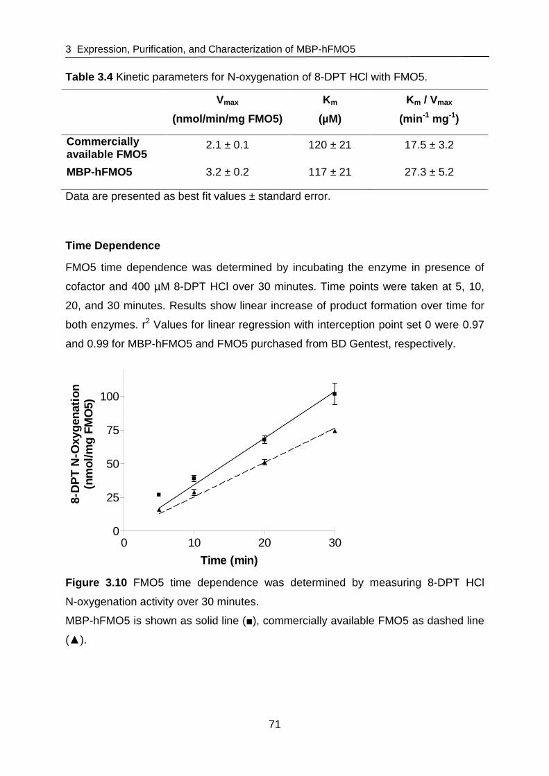
3 Expression, Purification, and Characterization of MBP-hFMO5
71
Table 3.4 Kinetic parameters for N-oxygenation of 8-DPT HCl with FMO5.
Vmax
(nmol/min/mg FMO5)
Km
(µM)
Km / Vmax
(min -1 mg -1)
Commercially available FMO5
2.1 ± 0.1 120 ± 21 17.5 ± 3.2
MBP-hFMO5 3.2 ± 0.2 117 ± 21 27.3 ± 5.2
Data are presented as best fit values ± standard error.
Time Dependence
FMO5 time dependence was determined by incubating the enzyme in presence of
cofactor and 400 µM 8-DPT HCl over 30 minutes. Time points were taken at 5, 10,
20, and 30 minutes. Results show linear increase of product formation over time for
both enzymes. r2 Values for linear regression with interception point set 0 were 0.97
and 0.99 for MBP-hFMO5 and FMO5 purchased from BD Gentest, respectively.
0 10 20 300
25
50
75
100
Time (min)
8-D
PT
N-O
xyge
natio
n(n
mol
/mg
FMO
5)
Figure 3.10 FMO5 time dependence was determined by measuring 8-DPT HCl
N-oxygenation activity over 30 minutes.
MBP-hFMO5 is shown as solid line (■), commercially available FMO5 as dashed line
(▲).

3 Expression, Purification, and Characterization of MBP-hFMO5
72
3.3.3.2 Stability of MBP-hFMO5 at 4 °C
The stability of MBP-hFMO5 at 4 °C was checked over 5 days and NADP+ and/or
20 % glycerol was added to determine whether these supplements improve the
enzyme’s stability. The addition of NADP+ and/or glycerol decreased loss of enzyme
activity at 4 °C over 5 days. The specific activity of MBP-hFMO5 was still at 65 %,
73 %, and 80 % of that at day zero after addition of NADP+, glycerol, or both,
respectively, whereas a 50 % loss in enzyme specific activity was observed when
neither substance was added. Results are shown in Figure 3.11 and Table 3.5.
Therefore, the NADP+ as well as 20 % glycerol was added to the enzyme to improve
its stability in future crystallization studies (chapter 4). The observed increase of
enzyme activity upon addition of glycerol at day 0 is probably due to the
cryoprotective properties of glycerol.
0
2
4
6
8
10
12
14
16
18
Ø + NADP + Glycerol + NADP + Glycerol
8-D
PT
N-o
xyge
natio
n (n
mol
/min
/mg
FM
O5)
Figure 3.11 Stability of MBP-hFMO5 at 4 °C.
8-DPT N-oxygenation of MBP-hFMO5 at day 0 (black bars) and after incubation at
4 °C for 3 (grey bars) and 5 days (white bars) with and without addition of 500 mM
NADP+ and/or 20 % glycerol.
+ +

3 Expression, Purification, and Characterization of MBP-hFMO5
73
Table 3.5 Stability of MBP-hFMO5 at 4 °C.
Ø + NADP+ + Glycerol + NADP+
+ Glycerol
Day 0 100.0 ± 7.7 100.0 ± 12.0 100.0 ± 0.4 100.0 ± 4.5
Day 3 79.6 ± 16.0 82.1 ± 5.5 75.1 ± 3.1 91.4 ± 11.2
Day 5 47.4 ± 2.8 65.3 ± 2.6 73.3 ± 2.8 79.2 ± 3.4
8-DPT N-oxygenation of hFMO5 at day 0 and after incubation at 4 °C for 3 and
5 days with and without addition of 500 mM NADP+ and/or 20 % glycerol are given in
% with day 0 set 100 %.
3.3.3.3 Oligomerization State of MBP-hFMO5
The oligomerization state of MBP-hFMO5 was determined by native gel
electrophoreses and gel filtration. After amylose column-purification, the enzyme
existed mainly as mono-, tri-, and hexamer (Figure 3.12, lane 1) whereas it was
primary found to be in a hexameric state after Q and SEC purification (Figure 3.12,
lanes 2 and 3). After addition of 0.5 % Triton® X-100 (~30 x CMC) or 10 x CMC of
DDM (0.1 %) to the Q and SEC-purified protein, the multimerization state changed
towards coexistence of mono-, tri-, and hexamers (Figure 3.12, lanes 4-7).
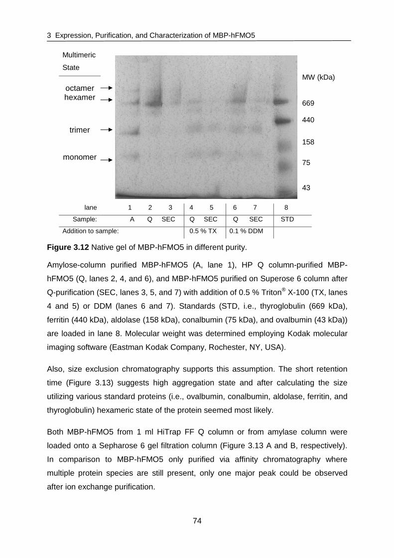
3 Expression, Purification, and Characterization of MBP-hFMO5
74
Multimeric
State
octamer hexamer
trimer
monomer
lane 1 2 3 4 5 6 7 8
Sample: A Q SEC Q SEC Q SEC STD
Addition to sample: 0.5 % TX 0.1 % DDM
Figure 3.12 Native gel of MBP-hFMO5 in different purity.
Amylose-column purified MBP-hFMO5 (A, lane 1), HP Q column-purified MBP-
hFMO5 (Q, lanes 2, 4, and 6), and MBP-hFMO5 purified on Superose 6 column after
Q-purification (SEC, lanes 3, 5, and 7) with addition of 0.5 % Triton® X-100 (TX, lanes
4 and 5) or DDM (lanes 6 and 7). Standards (STD, i.e., thyroglobulin (669 kDa),
ferritin (440 kDa), aldolase (158 kDa), conalbumin (75 kDa), and ovalbumin (43 kDa))
are loaded in lane 8. Molecular weight was determined employing Kodak molecular
imaging software (Eastman Kodak Company, Rochester, NY, USA).
Also, size exclusion chromatography supports this assumption. The short retention
time (Figure 3.13) suggests high aggregation state and after calculating the size
utilizing various standard proteins (i.e., ovalbumin, conalbumin, aldolase, ferritin, and
thyroglobulin) hexameric state of the protein seemed most likely.
Both MBP-hFMO5 from 1 ml HiTrap FF Q column or from amylase column were
loaded onto a Sepharose 6 gel filtration column (Figure 3.13 A and B, respectively).
In comparison to MBP-hFMO5 only purified via affinity chromatography where
multiple protein species are still present, only one major peak could be observed
after ion exchange purification.
MW (kDa) 669 440
158
75
43

3 Expression, Purification, and Characterization of MBP-hFMO5
75
A
0 25 50 75
0
10
20
30
40
50
0
10
20
30
40
50
Time (min)
Abs
280
nm
(m
AU
) Abs
450 nm
(mA
U)
B
0 10 20 30 40 50 60
0
25
50
75
100
0
5
10
15
20
Time (min)
Abs
280
nm
(m
AU
) Abs
450 nm
(mA
U)
Figure 3.13 Elution profile of fractions from SEC column (Sepharose 6) loaded with
MBP-hFMO5.
Absorption at 280 nm is shown as solid line, absorption at 450 nm as dashed line.
A SEC elution profile of MBP-hFMO5 previously purified on an amylose column.
B SEC elution profile of MBP-hFMO5 previously purified on Q column.
3.3.3.4 Monodispersity of MBP-hFMO5
DLS analysis data of affinity purified MBP-hFMO5 is shown and either size (Figure
3.14 A) or volume (Figure 3.14 B) is plotted against intensity. When size is plotted
against intensity two large peaks are observed, showing contamination or at least
two species of proteins. However, when size is plotted against volume, it becomes
apparent that the contamination is not as dominant as it seemed in Figure 3.14 A.

3 Expression, Purification, and Characterization of MBP-hFMO5
76
The polydispersity index (PdI) values for amylose pure MBP-hFMO5 were
0.394 ± 0.006.
A B
C D
Figure 3.14 Dynamic light scattering of affinity (A, B) and ion exchange (C, D)
purified MBP-hFMO5.
Dynamic light scattering of affinity purified and Q purified MBP-hFMO5 showed an
increased monodispersity of the protein with only one peak visible in both graphs
(Figure 3.14 C and D) and improved PdI values of 0.134 ± 0.002.

3 Expression, Purification, and Characterization of MBP-hFMO5
77
DLS analysis done with Q-purified MBP-hFMO5 where glycerol was added and
Q-purified MBP-hFMO5 that was treated with Calbiosorb Adsorbent beads (Merck,
Darmstadt, Germany) did not show significant differences. Further, Calbiosorb
Adsorbent treated Q-purified MBP-hFMO5 was examined in various concentrations
(i.e., 25, 10, 5, 2.5, and 0.25 mg/ml), but again no significant differences between the
different concentrations could be observed (data not shown). For all Q-purified MBP-
hFMO5 samples measured average PdI values were 0.141 ± 0.037.
3.4 Discussion
For structural and functional characterization of MBP-hFMO5, highly-purified and well
characterized protein was needed. Especially crystallography experiments (chapter
4) require not only pure, but also monodispers and stable protein. Herein, a two-step
purification method was developed and obtained protein was thoroughly
characterized (Figure 3.15).
Figure 3.15 Overview of MBP-hFMO5 purification protocol development.
Dashed lines and white boxes stand for earlier steps in purification protocol
development, solid lines and grey boxes represent final purification protocol as used
for crystallography studies in chapter 4.
Human FMO5 was successfully expressed as N-terminal MBP-fusion protein
(MBP-hFMO5) in E. coli DH1α cells. After expression, a number of different
detergents was tested for extraction of MBP-hFMO5, but only Triton® X-100
solubilized FMO5 sufficiently. The degree in which detergents are able to cross
Expression in E. coli
Solubilization with Triton X-100
Amylose column (buffer containing Triton X-100)
Amylose column (buffer without detergent)
Amylose column (buffer containing DDM)
Q column (buffer without detergent)
Q column (buffer without detergent)
Q column (buffer containing DDM)

3 Expression, Purification, and Characterization of MBP-hFMO5
78
membranes is said to be affecting the extend to which efficient solubilization occurs
[le Maire et al., 2000]. Triton® X-100 showed best solubilization ability. This might be
due to the hydrophobic-hydrophilic properties of its polyethylene chains [Israelachvili,
1997] allowing Triton® X-100 to ‘flip-flop’ rapidly across membranes. In contrast, DDM
has a strong hydrophilic head averting fast ‘flip flop’ across the membrane [Kragh-
Hansen et al., 1998; le Maire et al., 2000] and thus proved unsuitable for extraction of
MBP-hFMO5 as seen in this study. For subsequent studies, Triton® X-100 was
chosen for extraction of MBP-hFMO5 from E. coli.
Affinity chromatography was used to purify the supernatant after cell lysis. This was
done on an amylose column that binds to the MBP tagged to FMO5. Impurities could
thus be eluted from the column during an extended washing step. Afterwards, the
MBP and therefore the protein of interest was eluted with 3 mM maltose.
After this first purification step, a second step was added. MBP-hFMO5 was further
purified on an ion exchange column. At pH 6, MBP-hFMO5 was bound to the resin of
a 1 ml HiTrap Q column. Following a washing step with 65 mM NaCl in which
impurities were removed, the protein of interest was eluted at a concentration of
250 mM NaCl.
Purified MBP-hFMO5 was characterized in order to confirm its suitability for kinetic
and crystallization studies. Firstly, MBP-hFMO5 was supposed to be compared to
commercially available hFMO5 (BD Gentest) in order to show that MBP-hFMO5 may
be used for kinetic and structural studies instead of expensive purchasable FMO5.
The kinetic parameters of recombinant expressed MBP-hFMO5 and commercially
available FMO5 were determined and time dependence studies were carried out.
These studies showed very similar kinetic behavior for both enzymes, with a Km of
roughly 120 µM for both enzymes and a Vmax of 2.1 and 3.2 for Gentest FMO5 and
MBP-hFMO5, respectively. Time dependence studies also showed a linear behavior
for both enzymes over 30 minutes validating the standard incubation time of
20 minutes. Thus, MBP-hFMO5 is very similar to commercially available FMO5 and
may be used to study kinetic as well as structural parameters. In addition to lower
costs, MBP-hFMO5 has the advantage that it can be purified easily utilizing its MBP-
tag. Further, it can be produced in large quantities and higher purity may be achieved
which is especially advantageous when attempting to crystallize the protein.

3 Expression, Purification, and Characterization of MBP-hFMO5
79
Enzyme stability was determined under various conditions at 4 °C. Results from this
study showed that addition of glycerol and NADP+ to the sample improved
MBP-hFMO5 stability 1.6-fold. Therefore, in subsequent crystallization studies these
additives were added to the enzyme preparations.
For characterization of the protein, native gel electrophoresis and size exclusion
chromatography were used to determine the oligomerization state of MBP-hFMO5.
Compared to affinity purified MBP-hFMO5, the monodispersity improved significantly
upon further purification via ion exchange chromatography. As shown on native gels,
high detergent content seemed to separate protein molecules and MBP-hFMO5 that
was primary found to exist in a hexameric state after ion exchange chromatography
and size exclusion chromatography changed its oligomerization state towards
coexistence of mono-, tri-, and hexamers. Results from size exclusion
chromatography of ion exchange purified MBP-hFMO5 also suggested hexameric
state of the protein. The observation that FMO proteins possibly exist in higher
ordered complexes has been made previously [Brunelle et al., 1997] and a
hexameric state of a native FMO enzyme was first presumed in 1972 by Ziegler and
Mitchell [Ziegler et al., 1972]. Although an MBP fusion could be affecting the
oligomerization state of FMO, this was not the case for other MBP-tagged proteins
like MBP-phenylalanin hydroxylase [Martinez et al., 1995]. This was an important
observation because multimerization could have a significant effect on crystallization.
Another important factor in crystallization studies is monodispersity of the protein.
Thus, MBP-hFMO5 was characterized via DLS analysis. DLS is a method to
determine particle size in solution and thus gives an estimate of the monodispersity
of a given protein sample. It is a technique based on Brownian motion of particles. A
laser beam is scattered by particles in solution and the time dependent change of the
signal intensity compared to itself is used to determine the size of the particles. In
case of small particles this change is fast whereas larger particles give ‘speckle
patterns’ that fluctuate at a slower rate. Thus, the time gives an indication of the
mean particle size. In addition, large particles will scatter more light than small ones
and therefore, in case more than one peak is observed, the importance of the second
peak will be represented more realistically if intensity is correlated to volume. This
can also be observed with affinity column purified MBP-hFMO5 where two large
peaks are visible when size is plotted against intensity whereas the second peak

3 Expression, Purification, and Characterization of MBP-hFMO5
80
(particles of larger size) decreases when size is plotted against volume. In
comparison to only affinity-purified MBP-hFMO5 where impurities are obvious, DLS
with Q-purified samples showed only a single peak. No significant difference could be
observed between ion exchange purified MBP-hFMO5 and samples of the same
purity that were treated with Calbiosorb Adsorbent beads added to remove residual
detergent in the sample. Also, addition of 20 % glycerol and different concentrations
of MBP-hFMO5 did not alter DLS results.
Studies showed that E6 oncoprotein from human Papillomavirus strain 16 fused to
MBP formed soluble high molecular weight aggregates [Zanier et al., 2007]. The size
and amount of aggregated particles varied depending on expression conditions and
E. coli strain, suggesting that aggregation took place in vivo. However, the
aggregates were supposedly relatively monodispers and the E6 protein possessed a
native-like fold. These features are advantageous in view of FMO5 crystallization
attempts and seemed to be transferable to MBP-hFMO5. As shown for E6,
MBP-hFMO5 seemed to be correctly folded, retaining its enzymatic activity. Also,
DLS data suggested relatively monodispers protein, which is also in accordance with
MBP-tagged E6 protein.
Overall, a cost-efficient way to obtain large quantities of highly purified MBP-hFMO5
for future structural and functional enzyme characterization studies was successfully
developed and optimized.

4 Crystallography Studies of Human FMO5
81
4 Crystallography Studies of Human FMO5
4.1 Introduction
X-ray crystallography studies of proteins and enzymes are important tools to
determine the 3D structure of a protein of interest. The three-dimensional structure is
key to understanding the function of any protein. A crystal structure can give
information about the location of the active site, substrate- and cofactor-binding
domains, and about conformational changes a protein may undergo. This leads to a
better understanding of the enzyme in respect of substrate binding, mechanism of
action, as well as possible influence of different mutations. In case of FMO enzymes
little is known about these properties because to date, the tertiary structure of the
mammalian FMO enzymes is not known. Solving the three-dimensional structure of a
mammalian FMO isozyme would help tremendously in the understanding of
mammalian FMOs in general because they contain a large percentage of identical
residues (50 – 58 % amino acid sequence identity in human FMO isozymes).
Especially amino acids involved in FAD and NADPH binding are conserved
throughout human FMOs and across species lines [Cashman, 1995].
4.1.1 FMO Model Structure
Although the tertiary structure of mammalian FMO enzymes has not been solved to
date, model predictions by Cashman [Cashman, 2002] as well as Ziegler and
Poulsen [Ziegler et al., 1998] have been made for human FMO3. These were based
on comparison with solved structures of other flavoproteins (Figure 4.1).

4 Crystallography Studies of Human FMO5
82
Figure 4.1 Proposed human FMO3 model.
The model is based on homology modeling with the protein structures 1get (green),
1vqw (yellow), 1w4x (red), and 1npx (blue). FAD (magenta) and NADPH are also
shown. There are two domains in the structure, one being relevant for FAD, the other
for NADPH binding. These two domains are relatively similar in all models whereas
the rest of the structure is very divergent (cyan). Taken from [Cashman et al.,
manuscript in preparation].
In addition, the crystal structure of FMO from Schizosaccharomyces pombe
(S. pombe) has been solved [Eswaramoorthy et al., 2006]. However, this information
cannot be transferred to mammalian FMO enzymes without caution because
S. pombe FMO consists of only 447 amino acids whereas human FMO1 – 5 are 532
to 558 amino acids in length and it shares only 21 to 23 % sequence identity with
human FMOs.
4 Crystallography Studies of Human FMO5
83
4.1.2 FMO Protein Structure, Binding Sites, and Int eraction with the
Membrane
An overview of the different well conserved motives and their postulated functions
was given by Krueger and Williams [Krueger et al., 2005]. These functions were
proposed based on the PRINTS database that catalogues protein fingerprints,
conserved motives characterizing the protein family [Attwood et al., 2003], and rabbit
FMO2 as reference sequence for fingerprint analysis [Lawton et al., 1990]. The main
regions of interest for FMO3 are listed below and compared to the crystal structure of
S. pombe.
4.1.2.1 FAD Binding
The FAD binding motif, GXGXXG, is supposed to be within the region of amino acids
3 – 26 and amino acids 27 – 51 are supposed to stabilize FAD binding through a
EXXXXXGG motif [Krueger et al., 2005]. Both regions seem essential and mutations
in either one have been reported to result in loss of activity for FMO3 [Lawton et al.,
1993b]. Likewise, the nucleotide binding motif GXGXXG makes hydrogen-bonding
contacts with the adenine nucleotide of FAD and the N3 of adenine binds to Arg 39
that is part of the EXXXXXGG motif in S. pombe.
4.1.2.2 NADPH Binding
The NADPH binding site is located in the region of residues 186 and 213. This
proposal also seems consistent with the S. pombe structure, where NADPH is bound
to the second nucleotide binding motif (GXGXX(G/A)). Here the nicotinamide is
stacked with the isoalloxazine ring of the FAD so interaction between the bases of
nicotinamide and flavin is possible [Eswaramoorthy et al., 2006].
4.1.2.3 Substrate Binding
The substrate in the S. pombe structure (i.e., methimazole) is supposed to stack with
the isoalloxazine ring of bound FAD and replace the NADPH in the structure. In case
of mammalian FMO enzymes, this seems unlikely, because kinetic studies with
FMO1 suggested that NADP+ does not leave the flavoprotein until after substrate
oxygenation. Maybe a different binding mechanism applies here.

4 Crystallography Studies of Human FMO5
84
4.1.2.4 The FATGY-Motif and Membrane Interaction Si tes
One other very prominent motif is the FATGY motif. It is a motif that is conserved in
nearly all mammalian FMO enzymes and is located between amino acids 315 and
342 which represent the linkage between NADPH and the active site. Only FMO4
has a FTTGY and S. pombe FMO contains an YCTGY-motif.
The sites of membrane interaction are still unknown although considerable
information about protein structure is already available [Krueger et al., 2005; Lawton
et al., 1993a]. Maybe a crystal structure of a mammalian FMO enzyme will explain
where FMO is bound to membranes.
4.1.3 Aim of the Study
The objective was to purify FMO5 in order to crystallize it and finally obtain a
three-dimensional structure of a mammalian FMO enzyme. This would give answers
to the location of binding sites for FAD, NADPH, and substrate and would lead to a
better understanding of the mechanistic pathway of FMO mediated metabolism.
4.2 Materials and Methods
4.2.1 Reagents
Chemicals and reagents used in this study were purchased from Sigma-Aldrich
Chemical Co. (St Louis, MO, USA) in appropriate purity. Buffers and other reagents
were purchased from VWR Scientific, Inc. (San Diego, CA, USA). Polyethylene
glycols (PEGs), crystallography screens, and crystallography plates (24 or 96 well
sitting drop plates) were purchased from Hampton Research Corp. (Aliso Viejo, CA,
USA).
4.2.2 Cloning and Expression
Human FMO5 was expressed as N-terminal maltose-binding fusion protein
(MBP-hFMO5). Cloning and expression was done as described in section 3.2.2.1
with few changes. MBP-hFMO5 was expressed in E. coli DH1α or E. coli BL21 cells.
After induction, cells were incubated shaking either at room temperature for 20 hours
or at 20 °C for 24 hours.

4 Crystallography Studies of Human FMO5
85
4.2.3 MBP-hFMO5 Purification
The purification procedure has already been described in chapter 3, but was
optimized for crystallization trials.
After expression, E. coli cells were harvested by centrifugation at 6,000 g for
10 minutes and the cell pellet was frozen in order to increase yield. After
resuspending the cell pellet in lysis buffer as described in section 3.2.2.1, cells were
either disrupted via sonication as described above or by passage through a French
press (Thermo Electron Corp., Needham Heights, MA, USA) operating at 20,000 psi
depending on what method was available. For crystallization studies the cell pellet
was only extracted once and after centrifugation the supernatant was loaded at
1 ml/min onto an amylose column (New England BioLabs, Ipswich, MA, USA)
equilibrated with ten column volumes of buffer A’ (i.e., 50 mM Na2HPO4, pH 8.4,
containing 15 µg/ml FAD). The column was washed with at least ten column volumes
of buffer A’ and bound MBP-hFMO5 protein was eluted with 3 mM maltose in buffer
A’ at 1 ml/min. Eluted protein was concentrated with an Amicon Ultra-15 centrifugal
filter unit with an Ultracel-50 filter (Millipore, Billerica, MA, USA).
Further protein purification was carried out on an FPLC system (Äkta-Purifier, GE
Healthcare, Uppsala, Sweden) using a 1 ml Q HP column (GE Healthcare, Uppsala,
Sweden) with a flow rate of 1 ml/min and a step gradient of 65 mM and 250 mM NaCl
as described in chapter 3.3.2.
Refinement of the Lysis Protocol
The lysis buffer was changed to 50 mM phosphate buffer, pH 8.4, containing 0.5 %
Triton® X-100, 0.1 % L-α-phosphatidylcholine, 0.5 mM phenylmethylsulfonylfluoride,
100 mM FAD, and 1 tablet /15 ml lysis buffer of Roche Mini Complete (EDTA-free)
protease inhibitor mix (Roche, Indianapolis, IN, USA). This did not seem to improve
crystallization and the original method was used for further MBP-hFMO5 purification.
4.2.4 Crystallization of MBP-hFMO5
4.2.4.1 Screening Kits
For Crystallization trials, laboratory space and equipment was generously provided
by Prof. Dr. C. D. Stout at the Scripps Research Institute, La Jolla, CA, USA and

4 Crystallography Studies of Human FMO5
86
Prof. Dr. A. Scheidig at the ZBM, University of Kiel. Initial crystallization conditions for
MBP-hFMO5 were screened using different commercially available 96-well screening
kits with conditions ranging from pH 4.6 – 8.5, various precipitants including PEGs
ranging from 200 to 20,000, and a wide variety of salts. Four 96-well crystal
screening kits were set up in sitting drops using the vapor diffusion method: EasyXtal
PEGs Suite (Qiagen Inc., Valencia, CA, USA), Crystal Strategy Screen I and II, and
PACT premier (Molecular Dimensions, Apopka, FL, USA). The protein used was
Q-purified MBP-hFMO5, in a concentration of 15 mg/ml containing 200 µM NADP+
and 20 % glycerol. Protein solution was mixed with equal volumes of the reservoir
solution.
4.2.4.2 Refinement Screens
The most promising results from initial 96-well screens were refined in terms of
protein concentration as well as type and concentration of PEG and salt. 24-well
plates were set up in sitting drops using the vapor diffusion technique.
Protein
In order to determine optimal protein concentration, plates were set up examining
dependence of protein concentration vs. PEG concentration. Plates contained 0.3 M
Na-acetate, 0.1 M Na-cacodylate, pH 6.5, PEG 2K monomethyl ether (PEG 2K
MME). PEG concentrations screened were in the range of 20 – 30 %. Q-purified
MBP-hFMO5 was mixed with an equal volume of reservoir solution in concentrations
between 5 and 40 mg/ml.
PEG
Initial 96-well screens showed PEG to be the most successful precipitant. Thus, type
and concentration was refined in further studies. Different concentrations of PEGs in
the range of 400 – 20K were tested. Concentrations tested were 10 – 32 % for PEG
2K MME, 2 – 28 % for PEG 6K and 10K, and 2 – 22 % for PEG 20K. The plates were
set up with 0.1 or 0.2 M sodium acetate as salt and 0.1 M Bis-Tris propane at pH 6.5.
Protein solution in concentrations of 20 – 25 mg/ml was mixed with equal volumes of
reservoir solution.

4 Crystallography Studies of Human FMO5
87
Salts
The best performing salts from the initial screens (i.e., Na-acetate, Na-formate,
Na-tartrate, Na-phosphate, K-citrate, KSCN, MgCl2, NH4Cl, LiCl, and NaBr) were
analyzed further. Conditions for salt screens were 0.1 M Na-cacodylate or 0.1 M
Bis-Tris propane, pH 6.5, 22 – 26 % PEG 2K MME, and 0.1 or 0.2 M salt. Also, plates
were set up containing 0.1 M Bis-Tris propane, pH 6.5, 24 – 28 % PEG 2K MME, and
Na-acetate in the range of 0 - 1.5 M in order to determine the most advantageous
salt concentration. Q-purified MBP-hFMO5 solution at 25 – 30 mg/ml was mixed with
equal volumes of reservoir solution.
4.2.4.3 Influence of Detergents
Bio-Beads ®-Treatment
In some cases, Q column-purified MBP-hFMO5 was treated with Bio-Beads®
(Bio-Beads® SM Media, Bio-Rad, Hercules, CA, USA) in order to remove excess
detergent (Triton® X-100 and/or DDM). Bio-Beads® were activated with methanol and
washed thoroughly with 50 mM Bis-Tris buffer, pH 6.5. Afterwards, Bio-Beads® were
slowly added to Q-purified protein.
For comparison between Bio-Beads®-treated and untreated MBP-hFMO5, plates
were set up with 22 – 26 % PEG 2 K MME, 0.1 M Na-cacodylate or 0.1 M Bis-Tris
propane at pH 6.5, and Na-formate, Na-acetate, MgCl2, or NH4Cl. Protein solution at
a concentration of 30 mg/ml was left untreated or was treated with activated Bio-
Beads® and crystallization drops were set up with either protein solution mixed with
equal volumes of reservoir solution.
Detergent Refinement in the Purification Protocol
In order to remove Triton® X-100 quantitatively, the purification protocol was refined:
The protein was still extracted in Triton® X-100, but after binding to amylose resin the
protein was washed and further purified with buffer devoid of detergent.
Influence of Detergents
The influence of addition of a wide variety of detergents to crystallization drops was
studied. Detergents tested are listed in Table 4.1. The reservoir solution consisted of
26 % PEG 2K MME, 0.2 M Na-acetate, and 0.1 M Bis-Tris propane, pH 6.5.
Crystallization drops consisted of equal volumes of reservoir solution and Bio-

4 Crystallography Studies of Human FMO5
88
Table 4.1 Detergents used in crystal screens to determine influence of detergents on
crystallization of MBP-hFMO5.
Detergent Concentration Detergent Concentration
217 FA 1 and 2 x CMC Cymal®-1 1 x CMC
231 FA 1 and 2 x CMC Cymal®-2 1 x CMC
234 FA 1 and 2 x CMC Cymal®-5 1 x CMC
235 FA 1 and 2 x CMC Cymal®-6 1 x CMC
238 FA 1 and 2 x CMC Cymal®-7 1 x CMC
C-HEGA®-10 1 and 2 x CMC n-Nonyl-β-D-glucopyranoside 1 and 2 x CMC
Sucrose-monolaurate 1 x CMC C8E4 1 x CMC
CTAB 1 x CMC C12E8 1 x CMC
DDAO 1 x CMC FOS-CHOLINE® -10 1 x CMC
n-Octanyl-sucrose 1 x CMC FOS-CHOLINE® -12 2 x CMC
HECAMEG 1 and 2 x CMC β-Octylglucoside 1 x CMC
n-Decanoyl-sucrose 1 x CMC n-Octyl-β-D-glucopyranoside 1 and 2 x CMC
MEGA-8 1 x CMC CHAPS 1 x CMC
ZWITTERGENT® 3-10 1 x CMC n-Decyl-maltoside 1 x CMC
1-s-Nonyl-β-D-thio-glucoside 1 x CMC n-Octyl-maltoside 1 x CMC
n-Nonyl-β-D-thio-maltoside 1 x CMC
FA, facial amphiphile; C-HEGA®-10, Cyclohexylbutanoyl-N-hydroxyethylglucamide; CTAB,
Cetyltrimethylammoniumbromid; DDAO, N,N-Dimethyl-1-dodecanamine-N-oxide;
HECAMEG, Methyl-6-O-(N-heptylcarbamoyl)-α-D-glucopyranoside; MEGA-8, Octanoyl-N-
methylglucamide; ZWITTERGENT® 3-10, n-Decyl-N,N-dimethyl-3-ammonio-1-
propanesulfonate; Cymal®-1-7, Cyclohexyl-alkyl-β-D-maltoside; C8E4, tetra(ethylene glycol)
monooctylether; C12E8, octa(ethylene glycol) dodecyl monoether; FOS-CHOLINE®-10, n-
Decylphosphocholine; FOS-CHOLINE®-12, n-Dodecylphosphocholine; CHAPS, 3-[(3-
Cholamidopropyl)-dimethylammonio]-1-propane sulfonate

4 Crystallography Studies of Human FMO5
89
Beads®-treated Q-pure MBP-hFMO5 (30 mg/ml) spiked with detergent to yield a final
concentration of 1 or 2 times their CMC.
4.2.4.4 X-Ray Diffraction Measurement
X-ray diffraction measurement was done by Dr. Andrew Annalora (Scripps Research
Institute, La Jolla, CA, USA). Several crystals were screend at the Stanford
Synchotron Radiation Laboratory (SSRL, Palo Alto, CA, USA) on Beamline 7-1 and
11-1. Prior to flash freezing and storage, several preparation conditions were tested:
A quick (10 – 30 seconds) or long (30 seconds – 2 minutes) soak of the crystals in
buffer identical to the reservoir condition but with addition of 20 % glycerol, and/or
17 % ethylene glycol was done for cryoprotection. Also, crystals were frozen directly
from the drop with and without a quick soak in 100% parrafin oil.
4.2.5 Crystallography Studies of hFMO5 without MBP- tag
4.2.5.1 Cleavage of MBP-hFMO5 with Factor Xa
250 µg MBP-hFMO5 were incubated in a total volume of 100 µl 100 mM phosphate
buffer, pH 7.6, containing 0.2 mM NADPH and 5 µg factor Xa (New England BioLabs,
Ipswich, MA, USA) for 1.5 or 3 hours at 37 °C. In o rder to prevent potential
precipitation of cleaved FMO5, incubations were also done in the presence of
detergent (i.e., CHAPS). To compare rate of degradation or loss of activity during the
1.5 or 3 hour incubation period, 3-hour control incubations were done without factor
Xa at 37 °C and at 4 °C. Again, control incubations were also done in the presence of
CHAPS. The incubation scheme is shown in Table 4.2.

4 Crystallography Studies of Human FMO5
90
Table 4.2 Incubation scheme of factor Xa cleavage reaction.
Sample Incubation conditions
A incubation for 1.5 hrs at 37 °C + factor Xa
B incubation for 3 hrs at 37 °C + factor Xa
C incubation for 1.5 hrs at 37 °C + factor Xa + CHA PS
D incubation for 3 hrs at 37 °C + factor Xa + CHAPS
E incubation for 3 hrs at 37 °C
F incubation for 3 hrs at 4 °C
G incubation for 3 hrs at 37 °C + CHAPS
H incubation for 3 hrs at 4 °C + CHAPS
After 1.5 or 3 hour incubation, samples were put on ice and immediately assayed for
8-DPT N-oxygenation activity as described in section 3.2.4.2 with minor changes.
Briefly, a standard incubation mixture contained 100 mM potassium phosphate
buffer, pH 8.4, 0.2 mM NADPH, 0.25 mM DETAPAC, and 75 µg/ml MBP-hFMO5.
After extraction of metabolites and remaining substrate in dichloromethane, HPLC
analysis was done on a Waters HPLC system (Waters 600E controller and Waters
Autosampler 700 Satellite WISP interfaced to a Waters 486 Absorbance Detector).
Chromatographic separation of analytes was done on a LiChrospher-Si 60 (250 x
4.6 mm, 5 µm; Merck, Darmstadt, Germany) with a mobile phase of 80 %
methanol/20 % isopropanol/0.025 % HClO4 (v/v/v). The flow rate was 1.5 ml/min and
the total run time was 15 minutes. The wavelength for UV detection was set to
243 nm. The retention times for 8-DPT and 8-DPT N-oxide were 5.5 and 4.0 minutes,
respectively.
In addition, immediately after incubation with factor Xa, samples were analyzed by
SDS-PAGE and Coomassie Blue staining. 2 µl of protein samples were fractionated
by electrophoresis on a 10 % polyacrylamide gel under denaturing conditions and
stained with Coomassie Blue. Protein bands were analyzed by densitometry using
Adobe Photoshop Elements (Version 7).

4 Crystallography Studies of Human FMO5
91
4.2.5.2 Purification of hFMO5 from MBP
After cleavage of MBP-hFMO5, hFMO5 had to be separated from remaining factor
Xa, MBP-tag and non-cleaved MBP-hFMO5. This was attempted by various methods
including ion exchange chromatography, affinity chromatography, and size exclusion
chromatography.
Affinity Chromatography
MBP-hFMO5 was cleaved with factor Xa after affinity purification and subsequent ion
exchange purification. The ion exchange purification step also served as buffer
exchange to eliminate remaining maltose from the first affinity purification step.
A second affinity chromatography step was performed after cleaving the protein.
500 µl amylose resin (New England BioLabs, Ipswich, MA, USA) equilibrated with
50 mM phosphate buffer, pH 8.5, was incubated with 5 mg cleaved MBP-hFMO5
over night rotating at 4 °C. The next day, amylose resin was removed by filtration and
bound protein was eluted with 3 mM maltose. Flow through (supposedly containing
cleaved FMO5) and eluted protein (which should contain the MBP-tag) was then
analyzed via SDS-PAGE and 8-DPT HCl N-oxygenation activity assay as described
in section 3.2.4.2.
Ion Exchange Chromatography
Purification was carried out on an FPLC system (Äkta-Purifier, GE Healthcare,
Uppsala, Sweden) at the ZBM, University of Kiel. Cleaved MBP-hFMO5 protein was
loaded onto a HiTrap Q HP column equilibrated with 50 mM Bis-Tris buffer, pH 6, at
0.5 ml/min. The column was washed with at least ten column volumes of equilibration
buffer. A linear salt gradient of 0 – 65 % 50 mM Bis-Tris buffer, pH 6 containing 1 M
NaCl was applied to elute bound protein.
Size Exclusion Chromatography
Another attempt to purify hFMO5 from uncleaved MBP-hFMO5, MBP, and factor Xa
was purification via size exclusion chromatography. Purification was carried out on a
FPLC system (Äkta-Purifier, GE Healthcare, Uppsala, Sweden) at the ZBM,
University of Kiel, Germany. Cleaved protein was loaded at 0.5 ml/min onto a
Superdex 200 10/300GL size exclusion column (GE Healthcare, Uppsala, Sweden)

4 Crystallography Studies of Human FMO5
92
equilibrated with 50 mM Bis-Tris buffer, pH 6. Eluted fractions were concentrated with
Microcon centrifugal filter units (Ultracel YM-30, Millipore, Billerica, MA, USA) and
analyzed via SDS-PAGE.
4.3 Results
4.3.1 Crystallization of MBP-hFMO5
4.3.1.1 Screening Kits
Crystallization involves phase transition where an initially solubilized protein comes
out of solution to form crystals as solution is slowly brought to supersaturation. Once
nuclei have formed, protein concentration in the solution drops and no new crystals
will form whereas already existing crystals one may grow [Chayen, 1998]. Vapor
diffusion is the most widely used technique and involves a drop composed of a
mixture of protein samle and crystallization reagent. The concentration within the
drop is usually lower than that needed for crystallization. The drop is equilibrated
against a liquid reservoir with higher concentrations. To achive equilibrium, water
vapor leaves the drop and the concentration within the drop will slowly increase
[Chayen, 1998]. At best, crystals will form, but also amorphous precipitate or
coadunate crystals may be observed.
The two most common set-ups for crystallization studies are hanging and sitting drop
techniques. In the hanging drop technique, the drop of protein and reagent hangs
from a coverslip above the reservoir solution. In case of the sitting drop technique,
the protein-reservoir drop sits on a surface closer to the reservoir [Chayen, 1998].
The results of all screens showed that overall a lower pH, i.e., 6 - 6.5, seemed
favorable. Also, PEGs were the preferred precipitant. A variety of salts was tested
and those that performed best were: Na-acetate, Na-formate, Na-tartrate,
Na-phosphate, K-citrate, KSCN, MgCl2, NH4Cl, LiCl, and NaBr. Especially chloride-
and acetate-salts gave promising results.
4.3.1.2 Refinement Screens
The crystallization conditions were refined starting with the most promising conditions
found in the 96-well screens. Optimal protein concentration as well as PEG type and
concentration were determined and influence of salt concentration was evaluated.

4 Crystallography Studies of Human FMO5
93
Protein
Results from screens where protein concentration was screened against PEG 2K
MME concentration showed that higher protein concentrations of 20 – 30 mg/ml gave
better results than lower concentrations (Figure 4.2).
Figure 4.2 Protein concentration vs. PEG 2K MME concentration.
Crystallization conditions: 20 – 30 % PEG 2K MME, 0.3 M Na-acetate, and 0.1 M
Na-cacodylate, pH 6.5. Drops consisted of equal volumes of reservoir solution and
5 – 40 mg/ml MBP-hFMO5.
PEG
The best results when evaluating various PEGs at different concentrations were
obtained with PEG 2K MME, PEG 6K and PEG 10K. Lower molecular weight-PEGs
as well as PEG 20K mostly led to heavy precipitation or no precipitation at all. For
PEG 2K MME the preferred concentration range was 24 – 28 %, for PEG 6K it was
20 – 28 % and PEG 10K performed best at concentrations between 23 and 27 %.
With these PEGs dense yellow oil-like phases as shown in Figure 4.3 were obtained.
Pro
tein
Con
cent
ratio
n
light oil or light precipitate
light oil or light precipitate
dense yellow oil
dense yellow oil
heavy precipitate
PEG Concentration
light oil or light precipitate

4 Crystallography Studies of Human FMO5
94
Figure 4.3 Picture of the yellow oil-like phase.
Crystallization condition: 0.1 M Bis-Tris propane, pH 6.5, 0.2 M Na-acetate, and 26 %
PEG 2K MME. Drops consisted of equal volumes of reservoir solution and 30 mg/ml
MBP-hFMO5.
Salts
The best performing salts from initial screens (Na-acetate, Na-formate, Na-tartrate,
Na-phosphate, K-citrate, KSCN, MgCl2, NH4Cl, LiCl, and NaBr) were analyzed further
and the most promising were Na-formate, MgCl2, NH4Cl, and Na-acetate. The latter
was then used to set up further crystallography experiments. Evaluation of
Na-acetate concentrations did not show an obvious trend of whether low or high salt
concentrations are preferable.
Maltose
As previously reported, MBP may adopt distinct conformational states depending on
presence or absence of maltose [Boos et al., 1998; Spurlino et al., 1991]. Thus, it is
important to avoid partial occupancy of maltose that might result in mixed
conformational states and interfere with crystallization by inhibiting formation of well-
ordered crystals [Smyth et al., 2003]. In general, addition of maltose to the protein
solution gave more “dense yellow oil” (Figure 4.2). Thus, in addition to 200 µM
NADP+ and 20 % glycerol, maltose was routinely added to protein preparations in a
concentration of 100 µM.
4.3.1.3 Influence of Detergents
Bio-Beads ®-Treatment
Crystallization trials showed that preparation of MBP-hFMO5 without detergent on
Q column did not lead to crystals but rather resulted again in dense yellow oil phases

4 Crystallography Studies of Human FMO5
95
as shown in Figure 4.3. These were probably due to residual Triton® X-100 from the
first purification step. Also, addition of different amounts (0.05 – 1 x CMC) of DDM to
crystallization drops did not result in crystal growth.
It was found that Triton® X-100 is strongly adsorbed onto Bio-Beads® SM-2, a neutral
porous polystyrene-divinylbenzene adsorbent [Holloway, 1973]. Thus, in order to
remove excess Triton® X-100 from the sample, Bio-Beads® were added. It is
remarkable, that even after addition of excess amounts of Bio-Beads®, MBP-hFMO5
stayed in solution. From this experiment and the solubilization study it can be
concluded that detergent (i.e., Triton® X-100) is necessary for protein extraction from
E. coli, but that for subsequent purification procedures, no further addition of
detergent is required.
A comparison was made between crystallization plates containing protein treated
with Bio-Beads® and untreated protein. Overall, Bio-Beads®-treatment of MBP-
hFMO5 resulted in more dense yellow oil phases and brown spheres (Figure 4.4).
Figure 4.4 Example of dense yellow oil and brown spheres as seen after Bio-
Beads®-treatment of purified MBP-hFMO5.
Crystallization condition: 0.1 M Bis-Tris propane, pH 6.5, 0.1 M Na-acetate, and 30 %
PEG 2K MME. Drops consisted of equal volumes of reservoir solution and 25 mg/ml
MBP-hFMO5 treated with Bio-Beads®.
The brown spheres seem to be a pre-stage of crystals because they cracked when
shattered producing sharp edges (Figure 4.5). Thus, MBP-hFMO5 is not only stable
without detergent, it also seems that detergent removal is important for crystal
growth.
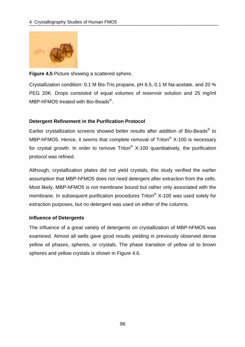
4 Crystallography Studies of Human FMO5
96
Figure 4.5 Picture showing a scattered sphere.
Crystallization condition: 0.1 M Bis-Tris propane, pH 6.5, 0.1 M Na-acetate, and 20 %
PEG 20K. Drops consisted of equal volumes of reservoir solution and 25 mg/ml
MBP-hFMO5 treated with Bio-Beads®.
Detergent Refinement in the Purification Protocol
Earlier crystallization screens showed better results after addition of Bio-Beads® to
MBP-hFMO5. Hence, it seems that complete removal of Triton® X-100 is necessary
for crystal growth. In order to remove Triton® X-100 quantitatively, the purification
protocol was refined.
Although, crystallization plates did not yield crystals, this study verified the earlier
assumption that MBP-hFMO5 does not need detergent after extraction from the cells.
Most likely, MBP-hFMO5 is not membrane bound but rather only associated with the
membrane. In subsequent purification procedures Triton® X-100 was used solely for
extraction purposes, but no detergent was used on either of the columns.
Influence of Detergents
The influence of a great variety of detergents on crystallization of MBP-hFMO5 was
examined. Almost all wells gave good results yielding in previously observed dense
yellow oil phases, spheres, or crystals. The phase transition of yellow oil to brown
spheres and yellow crystals is shown in Figure 4.6.
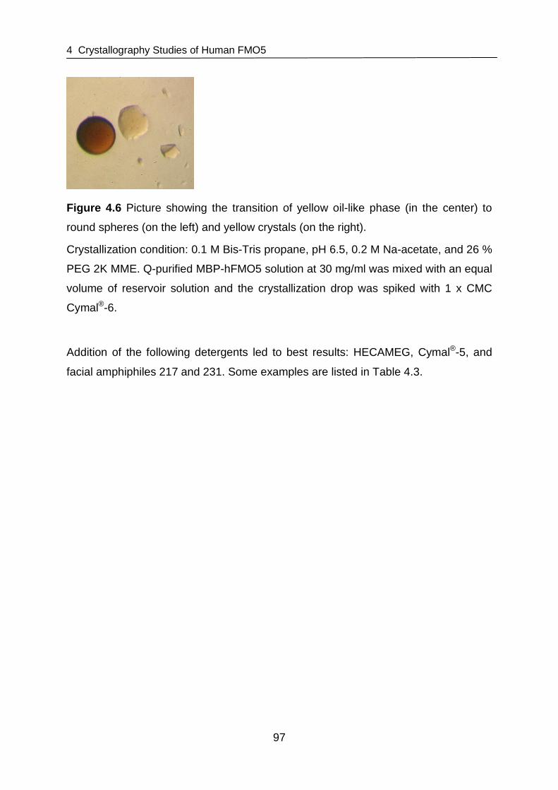
4 Crystallography Studies of Human FMO5
97
Figure 4.6 Picture showing the transition of yellow oil-like phase (in the center) to
round spheres (on the left) and yellow crystals (on the right).
Crystallization condition: 0.1 M Bis-Tris propane, pH 6.5, 0.2 M Na-acetate, and 26 %
PEG 2K MME. Q-purified MBP-hFMO5 solution at 30 mg/ml was mixed with an equal
volume of reservoir solution and the crystallization drop was spiked with 1 x CMC
Cymal®-6.
Addition of the following detergents led to best results: HECAMEG, Cymal®-5, and
facial amphiphiles 217 and 231. Some examples are listed in Table 4.3.

4 Crystallography Studies of Human FMO5
98
Table 4.3 Examples of wells from screens with drops containing MBP-hFMO5 that
were spiked with different detergents.
Detergent added Picture of well
HECAMEG
O NH
OOOH
OHOH
OH
Cymal®-5
OO
OH
OHOH
O
OOHOH
OH
OH
217 FA
231 FA
Crystallization conditions were 26 % PEG 2K MME, 0.2 M Na-acetate, and 0.1 M Bis-
Tris propane buffer at pH 6.5. Drops consisted of equal volumes of reservoir solution
and 30 mg/ml Bio-Beads®-treated Q-pure MBP-hFMO5 spiked with detergent at 1 x
(Cymal®-5, 217 FA and 231 FA) or 2 x (HECAMEG) their CMC. 231 FA (facial
amphiphile), 3α-hydroxy-7α,12α-bis[(β-D-maltopyranosyl)ethyloxy]cholane; 217 FA,
7α,12α-bis[(β-D-maltopyranosyl)]cholane.
OH
O
O
O
OO
OHOH
OHOH
OH
OH
OH
O
O
O
OHOH
OHOH
OH
OH
OH
O
O
O
OHOH
OHOH
OH
OH
OHO
OO
OHOH
OHOH
OH
OH
OH
O
O

4 Crystallography Studies of Human FMO5
99
As shown in the SDS gel (Figure 4.7) the crystals proved to be MBP-tagged FMO5.
Crystals dissolved in buffer were run on SDS gel and a band could be obsered
around 100 kDa. These results show that MBP-tagged human FMO5 is crystallizable.
MW (kDa)
204
123 80 48
MBP-hFMO5 marker
Figure 4.7 SDS gel of redissolved MBP-FMO5 crystal.
High Molecular Weight Prestained marker (Bio-Rad, Hercules, CA, USA) was used
as MW protein marker. Arrow points to MBP-hFMO5.
However, these coadunate crystals did not yield satisfactory diffraction patterns and
none of the preparation conditions prior to flash-freezing that were tested improved
diffraction. The crystals obtained lacked in optical clarity, smooth faces and had only
few sharp edges. Also, arrays were probably not ordered well enough to obtain
evaluable diffraction patterns.
4.3.2 Crystallography Studies of hFMO5 without MBP- tag
Crystallization studies with purified MBP-hFMO5 led to a number of crystals in
various conditions. Unfortunately, no satisfactory diffraction pattern could be obtained
from these crystals. Thus, crystallization or purification conditions needed to be
altered in order to improve crystal growth. Further refinement of purification
conditions such as including additional protease inhibitors (Roche Mini Complete
(EDTA-free) protease inhibitor mix) in the lysis buffer, usage of an extended linear
NaCl gradient in the ion exchange purification step, addition of NADP+ after
purification but before concentrating the protein, or air-fuging the protein before
crystallization plates are set up, were tested but did not lead to improved
crystalization.

4 Crystallography Studies of Human FMO5
100
Although expression and purification as MBP-fusion protein has numerous
advantages, for crystallography experiments an MBP-tag might be problematic. It has
been suggested that MBP hinders crystal growth by introducing conformational
heterogeneity [Smyth et al., 2003]. Since FMO5 had been cloned into a pMAL-c2
vector that contains a protease sensitive site right after the MBP sequence (i.e.,
IEGR), the MBP-tag may be removed from FMO5 by proteolytic splicing of the fused
protein from MBP with factor Xa. Consequently, FMO5 could be separated from its
tag and crystallization studies with purified FMO5 could be attempted that might lead
to good diffracting crystals.
4.3.2.1 Cleavage of MBP-hFMO5 with Factor Xa
After 1.5 or 3 hour incubation of MBP-hFMO5 with factor Xa, the samples were
analyzed in regards to 8-DPT N-oxygenation activity and extent of cleavage. SDS-
PAGE showed 67 % cleavage after 1.5 and 73 % after 3 hours incubation (Figure
4.8).
MBP-hFMO5 hFMO5 MBP
MW (kDa)
116.0
66.2 45.0 35.0
25.0 18.4 14.4
1.
5 h,
37
°C, f
Xa
3 h,
37
°C, f
Xa
1.5
h, 3
7 °C
, fX
a, C
HA
PS
3 h,
37
°C, f
Xa,
CH
AP
S
3 h,
37
°C
3 h,
4 °C
3 h,
37
°C, C
HA
PS
3 h,
4 °C
, CH
AP
S
mar
ker
Figure 4.8 Coomassie stained SDS gel of fractions A-H (see also Table 4.2).
peqGOLD Protein-Marker I (Peqlab, Erlangen, Germany) was used as MW protein
marker. Arrows point to MBP-hFMO5 and the cleaved products, hFMO5 and MBP.

4 Crystallography Studies of Human FMO5
101
In the presence of NADPH, hFMO5 retains almost 100 % of its 8-DPT N-oxygenation
activity after 1.5 or 3 hour incubation at 37 °C co mpared to enzyme stored at 4 °C
(Figure 4.9). Also, cleavage did not precipitate or inactivate the enzyme. Thus,
including CHAPS in the incubation mixture was not necessary. It even seems
disadvantageous to add CHAPS because 8-DPT N-oxygenation activity decreased
about 65 % compared to MBP-hFMO5 stored at 4 °C (Fi gure 4.9).
0
2
4
6
8
10
12
14
16
18
20
1.5
h, 3
7 °C
+fX
a
3 h
, 37
°C +
fXa
1.5
h, 3
7 °C
+fX
a +
CH
AP
S
3 h,
37
°C +
fXa
+ C
HA
PS
3 h
, 37
°C
3 h,
4 °C
3 h
, 37
°C +
CH
AP
S
3 h,
4 °C
+C
HA
PS
8-D
PT
N-O
xyge
natio
n (n
mol
/min
/mg
FMO
5)
Figure 4.9 8-DPT N-oxygenation of fractions A – H (see also Table 4.2).
4.3.2.2 Purification of hFMO5 from MBP
Affinity Chromatography
Separation of hFMO5 from MBP was attempted via affinity chromatography on
amylose resin. SDS-PAGE as well as HPLC based enzyme activity analysis showed
no separation of hFMO5 and MBP (Figure 4.10). HPLC analysis showed the main
activity in the eluate rather than the flow through which is in agreement with the SDS
gel. It is possible that cleaved hFMO5 stays somewhat associated with the cleaved
MBP-tag.

4 Crystallography Studies of Human FMO5
102
0
2
4
6
8
10
12
Q pure FMO5 flow through eluate
8-D
PT
N-O
xyge
natio
n (n
mol
/min
/mg
FM
O5)
MW (kDa) 116
66.2
45
35
25
M
BP
-FM
O5
‘flow
thro
ugh’
‘elu
ate’
mar
ker
Figure 4.10 Results of 8-DPT N-oxygenation activity assays and Coomassie stained
SDS gel of amylose purified, cleaved MBP-hFMO5.
peqGOLD Protein-Marker I (Peqlab, Erlangen, Germany) was used as MW protein
marker. Arrows point to Q-pure MBP-hFMO5 and the cleaved products, hFMO5 and
MBP.
Ion Exchange Chromatography
Another attempt to separate hFMO5 from MBP, MBP-hFMO5, and factor Xa was
purification on a HiTrap Q HP anion exchange column. The resulting chromatogram
is shown in Figure 4.11. The fractions were analyzed with SDS-PAGE (Figure 4.12).
Both, chromatogram and SDS gel show good separation of MBP (peak 1) and FMO5
(peak 2), however, hFMO5 co-elutes with uncleaved MBP-hFMO5.
MBP-hFMO5
hFMO5
MBP

4 Crystallography Studies of Human FMO5
103
MBP-hFMO5
hFMO5
MBP
MW (kDa)
116 66.2 45 35
25
18.4
P
eak
1
Pea
k 2
Cle
aved
MB
P-F
MO
5
mar
ker
Figure 4.12 Coomassie stained SDS gel of fractions of Q column-purified cleaved
MBP-hFMO5.
peqGOLD Protein-Marker I (Peqlab, Erlangen, Germany) was used as MW protein
marker. Arrows point to Q-pure MBP-hFMO5 and the cleaved products, hFMO5 and
MBP.
Figure 4.11 Chromatogram of Q-column purified cleaved MBP-hFMO5.
Absorbance at 280 nm is shown as solid line, absorbance at 450 nm as dotted line,
and conductivity is shown as dashed line.
-200
0
200
400
600
800
1000
1200
1400
0 10 20 30 40 50
Volume (ml)
Abs
(28
0 nm
/450
nm
)
0
5
10
15
20
25
30
35
40
45
Con
duct
ivity
(m
S/c
m)

4 Crystallography Studies of Human FMO5
104
Size Exclusion Chromatography
As third method for FMO5 purification, size exclusion chromatography was tested.
The chromatogram obtained from SEC is shown in Figure 4.13 and the SDS gel of
the corresponding fractions is shown in Figure 4.14. In a first peak, hFMO5 elutes
clearly visible with a band at ~ 60 kDa. In a second peak, MBP elutes giving a band
at ~ 40 kDa in the SDS gel. However, as with anion exchange chromatography,
hFMO5 co-elutes with MBP-hFMO5.
0 10 20 30
0
250
500
750
1000
0
25
50
75
Volume (ml)
Abs
280
nm
(m
AU
) Abs
450 nm
(mA
U)
Figure 4.13 Elution profile of factor Xa cleaved MBP-hFMO5 on Sephadex 200
10/300GL size exclusion column.
Peak 1 Peak 2 Peak 3

4 Crystallography Studies of Human FMO5
105
MW (kDa)
250 148
98
64
50
36
22 16
mar
ker
Pea
k 2
P
eak
1
Cle
aved
MB
P-
FM
O5
Figure 4.14 Coomassie stained SDS gel of fractions of cleaved MBP-hFMO5 purified
on SEC column.
SeeBlue® Plus2 (Invitrogen Corporation, Carlsbad, CA, USA) was used as MW
protein marker. Arrows point to Q-pure MBP-hFMO5 and the cleaved products,
hFMO5 and MBP.
4.4 Discussion
It had been reported that fusion of a protein of interest to a large soluble affinity-tag
such as MBP may improve several features of the protein like solubility [Donnelly et
al., 2006; Kapust et al., 1999], yield [Butt et al., 1989], and folding [Kapust et al.,
1999]. In addition, it could give protection from proteolysis and resulting fusion
proteins may be easily purified via affinity chromatography [Smyth et al., 2003].
Although less efficiently, even without the context of a fusion protein, MBP has been
shown to interact preferentially with unfolded proteins and to promote their folding in
vitro [Richarme et al., 1997]. Further, it was proposed that MBP has chaperone-like
qualities when fused to the N-terminus of a protein of interest assisting in correct
protein folding and enhancing solubility [Kapust et al., 1999; Sachdev et al., 1998].
This might be partly due to its maltose binding site, a deep hydrophobic cleft that,
MBP-hFMO5 hFMO5
MBP

4 Crystallography Studies of Human FMO5
106
together with other hydrophobic sites located on the surface of the protein, could
serve as binding sites to incompletely folded protein [Kapust et al., 1999].
In accordance with these reported theories, a fusion of hFMO5 to MBP led to soluble,
active, and stable protein as described in chapter 3. This protein was also obtained at
a high yield and could easily be purified via affinity chromatography. Crystallization
experiments were done screening various commercially available crystal screen kits
for suitable crystallization conditions. These were refined further and the conditions
that finally led to MBP-hFMO5 crystals were 26 % PEG 2K MME and 0.2 M sodium
acetate in 0.1 M Bis-Tris propane buffer, pH 6.5. Interestingly, these crystallization
conditions are analog to those reported for other MBP-tagged proteins that were
successfully crystallized. Comparison of different crystals obtained as MBP-fusion
proteins showed that, as for MBP-hFMO5, PEG and related molecules were most
successful as precipitants and that the pH was generally low [Center et al., 1998;
Kukimoto et al., 2000; Liu et al., 2001; Smyth et al., 2003].
When working with membrane associated proteins such as FMO enzymes use of
detergents is unavoidable. As expected, surfactants play a dominant role in
purification and crystallization of FMO5. Detergents are indispensable for
solubilization of MBP-hFMO5 from E. coli (see chapter 3). Also in crystallography
screens, surfactants play a major role, but the use of detergents introduces many
problems. Detergent micelles have rough heterogeneous surfaces, their hydrophobic
tails have considerable contact with the surrounding solution [Bogusz et al., 2000],
and they are fluid systems exchanging micellar components with the solvent
[Garavito et al., 2001; Wennerstrom et al., 1979]. They have a major impact on
protein crystallization, because detergent-detergent and detergent-protein
interactions influence the behavior of membrane proteins. In addition, interactions
with any remaining lipids have an effect on how a protein behaves [Garavito et al.,
2001]. In mixed systems (considering only detergents and lipids) phase behavior as
well as changes in micelle shape, size, and CMC will be hard to predict because
these systems never behave like solutions of the pure components [Wennerstrom et
al., 1979]. The behavior will be altered again when effects of solvent components like
salts, PEG, or pH are considered [Arnold et al., 2008; Garavito et al., 2001].

4 Crystallography Studies of Human FMO5
107
Since usage of detergents introduces a completely new layer of unpredictability, a
detergent used in crystallization studies preferably has a defined head group size
and type. Especially mixtures like Triton® do not seem to work well [Arnold et al.,
2008]. In case of FMO5 where usage of Triton® X-100 was inevitable for extraction of
the protein from E. coli (see chapter 3), complete removal of detergent and possibly
substitution with other detergents before crystallization seemed necessary. Removal
of detergent from protein samples previous to crystallography screens has been
done by hydroxyl apatite column chromatography [Chen et al., 1999] or by
precipitation [Askolin et al., 2004], dialysis, or dilution of the sample [Rigaud et al.,
1997]. Another method that proved to be successful in production of two-dimensional
[Rigaud et al., 1997] or three-dimensional crystals [Bron et al., 1999] was surfactant
removal utilizing Bio-Beads®. In 1973, Holloway et al. already described the
successful removal of Triton® X-100 by adsorbance onto the polystyrene based
Bio-Beads® [Holloway, 1973]. Interestingly, upon addition of excess amounts of
Bio-Beads® to the protein solution, MBP-hFMO5 did not precipitate suggesting that
FMO5 is not an integral membrane protein, but rather only associated with the
membrane. As shown in the study, removal of Triton® X-100 with Bio-Beads®
improved results from crystallization trials of MBP-hFMO5 leading to thick yellow oil
phases and premature crystals with spherical shape (Figure 4.4).
Experiments in which a variety of different detergents was added after removal of
Triton® X-100 led to further improvement of this first pre-stage of crystals (Table 4.3).
The transition of the routinely obtained thick yellow oil to spherical shapes obtained
after Triton® X-100 removal and to yellow crystals is shown in Figure 4.6.
In several wells that yielded crystals, detergents were added that belong to a new
group of amphiphilic agents that is supposed to stabilize membrane proteins and
lower the risk of denaturing or aggregation. The structure of these new types of
amphiphilic molecules is based on cholic acid, but they exhibit facial amphiphilicity,
meaning that their polar and nonpolar groups are located on opposite faces instead
of at opposite ends as usual in other detergents [Zhang et al., 2007b; Zhong et al.,
2005]. The terminal carboxylate of cholate was removed and uncharged polar groups
were introduced at the parallel hydroxyl groups in the center of the cholic acid
skeleton [Zhang et al., 2007b]. The advantage of this structure is that these
surfactants are supposed to self-assemble face-to-face. Thus the hydrophobic parts

4 Crystallography Studies of Human FMO5
108
of two molecules have similar dimensions as that of the lipid bilayer. In initial stability
tests, the facial amphiphiles performed much better than previously used detergents,
keeping the protein in solution and active for a longer time [Zhang et al., 2007b]. Also
in case of MBP-hFMO5, these detergents seemed to improve crystallization although
others like HECAMEG and Cymals® also yielded crystals. Latter detergents have
previously aided in crystallization studies, e.g., in case of hydrophobin HFBI from
Trichoderma reesei that yielded crystals in the presence of Cymal®-5 [Askolin et al.,
2004]. In general, controlled addition of detergent and thus employing the concept of
mixed micelles seems beneficial for MBP-hFMO5 crystal growth.
Unfortunately, no satisfactory diffraction pattern could be obtained from MBP-hFMO5
crystals. Several features of the MBP-hFMO5 construct could be argued to have
been a cause for the poor condition of the crystals obtained. The MBP affinity tag
itself might be problematic in crystallization experiments. Although expression and
purification as MBP-fusion protein has numerous advantages, it has been suggested
to hinder crystal growth by conformational heterogeneity introduced by the fusion tag
[Smyth et al., 2003]. Circumventing cleavage of MBP-hFMO5 and leaving the
MBP-tag in place for crystallization studies has the advantage of avoiding potential
problems often associated with proteolytic splicing including low yield, precipitation of
target protein, cumbersome optimization of cleavage and subsequent purification
conditions, high cost of proteases such as factor Xa, or potential loss of activity of the
target protein during cleavage. However, leaving the tag attached to the protein of
interest results in a multi-domain protein that might be less likely to form well-ordered,
diffracting crystals, which is probably due to conformational heterogeneity allowed by
the flexible linker region between MBP and the target protein [Smyth et al., 2003]. In
fact, most crystal structures of MBP-tagged proteins previously reported contained a
shorter, more rigid linker between the affinity tag and the target protein [Center et al.,
1998; Ke et al., 2003; Kukimoto et al., 2000; Liu et al., 2001]. These short, two to five
amino acid long, mostly alanine containing linkers used instead of the typical longer
linker in the MBP-hFMO5 fusion protein might be one reason for improved
crystallization [Kapust et al., 1999; Smyth et al., 2003].
Also, in contrast to hFMO5, most proteins that led to crystals upon fusion with MBP
were small in comparison with the rather large MBP-tag they were fused to. This
could also play a role in successful crystallization as the ratio of MBP/protein might

4 Crystallography Studies of Human FMO5
109
be significant. Smaller proteins could be able to allow the larger MBP-tag to direct the
construction of a crystal lattice whereas larger ones might be less susceptible to let
MBP conduct crystal formation [Smyth et al., 2003].
These potential problems implicated by leaving the large affinity tag fused to hFMO5
during crystallization trials have also been encountered in this study and although
crystals could be grown from MBP-hFMO5, no satisfactory diffraction pattern could
be obtained. Therefore, the next step was to cleave off the MBP-tag and perform
crystallization experiments with only hFMO5. Successive experiments attempted
proteolytic splicing of MBP from hFMO5 and subsequent purification of the latter. The
separation of MBP and hFMO5 was done utilizing a previously inserted protease
sensitive site right after the MBP sequence that may be cleaved by factor Xa. After
incubation of three hours at 37 °C, cleavage was la rgely completed and addition of
NADPH to the incubation mixture prevented the enzyme from being heat-inactivated.
It is notable that similar experiments had been done with MBP-hFMO3 [Brunelle et
al., 1997]. However, after cleaving MBP from hFMO3, the latter showed intractable
solubility and enhanced instabilities [Brunelle et al., 1997] as previously described for
a number of purified FMO enzymes [Cashman, 1995; Guan et al., 1991]. In contrast
to expectations, after proteolytical separation from MBP, human FMO5 stays in
solution and is active.
After successful cleavage, the enzyme had to be separated from remaining factor Xa,
MBP, and residual uncleaved MBP-hFMO5. A variety of methods to purify MBP from
the target protein have been described including affinity purification [Riggs, 2000], ion
exchange chromatography [de Pieri et al., 2004; Guan et al., 2002; Hao et al., 2007;
Riggs, 2000; Yan et al., 2006], and size exclusion chromatography [Branco et al.,
2008]. Herein, these methods were carried out and evaluated.
Firstly, purification via amylose resin was attempted. In general, a disadvantage of
this procedure is that although MBP can be removed from the cleavage mixture, the
protease and other contaminants cannot be removed. Nevertheless, it has been
described to be effective for MBP removal [Riggs, 2000]. After an ion exchange
chromatography step to remove residual maltose from the elution of the first affinity
column, the fusion protein (i.e., MBP-hFMO5) was cleaved and applied to the
amylose resin. The target protein should be located in the flow through fractions and

4 Crystallography Studies of Human FMO5
110
freed of MBP. In this study however, MBP could not be separated from hFMO5. Both
proteins were found mainly in the eluate after washing with 3 mM maltose but the
flow through also contained both proteins to some degree. Potential difficulties might
have arisen if MBP was denatured or otherwise damaged in the purification process,
because than it presumably looses its affinity to the amylose resin [Riggs, 2000].
Maltose might also be more difficult to separate from the MBP than expected
because it is buried in a deep groove almost inaccessible to solvent. Also, extensive
hydrogen-bonding and van-der-Waals interactions keep the maltose in place
[Spurlino et al., 1991]. Thus, incomplete removal of maltose might have led to
ineffective purification on amylose resin.
Secondly, separation of hFMO5 and the affinity tag was attempted by size exclusion
chromatography. Successful purification of target protein from MBP after proteolytic
splicing has been described for recombinant Lassa virus (LASV) proteins via SEC
[Branco et al., 2008]. hFMO5 could be successfully separated from MBP, however, it
co-elutes with MBP-hFMO5.
Therefore, a third purification method to separate hFMO5 and MBP has been
attempted, i.e., ion exchange chromatography. Especially anion exchange
chromatography utilizing DEAE chromatography [de Pieri et al., 2004; Guan et al.,
2002; Hao et al., 2007; Riggs, 2000; Yan et al., 2006] or Q columns [de Pieri et al.,
2004; Riggs, 2000] has been used several times. Purification of MBP from hFMO5
was successful using a 1ml HiTrap Q HP column, but as in case of SEC purification,
hFMO5 co-eluted with uncleaved MBP-hFMO5.
Further studies are needed to succeed in purification of hFMO5 and subsequent
crystallization experiments. Potential methods include cation exchange
chromatography or antibody affinity chromatography. Theoretical calculation of the
isoelectric point (pI) of MBP-hFMO5, MBP, and hFMO5 suggested that cation
exchange purification on a HiTrap SP column (GE Healthcare, Uppsala, Sweden) at
pH 7 could be successful (pIMBP 5, pIFMO5 8, and pIMBP-FMO5 6). To verify these
calculated pI values, isoelectric focusing (IEF) could be performed. Afterwards, cation
exchange should be used to purify hFMO5. Another approach to separate the MBP-
tag from hFMO5 is the usage of MBP specific monoclonal antibodies that could be
conjugated to Sepharose beads [Park et al., 1998]. Also, a poly His site could be

4 Crystallography Studies of Human FMO5
111
introduced into the protein vector and protein could be purified via Ni2+-column affinity
chromatography after proteolytic splicing of MBP from the target protein. This has
been described previously for several other proteins [Feher et al., 2004;
Ramachandran et al., 2007]. Using antibodies as well as using an additional His-tag
have the disadvantage that they only remove MBP, but not the protease and other
remaining contaminants. However, usage of biotin-labeled factor Xa has been
reported to be advantageous because the protease can be removed after cleavage
from the separated proteins by binding to streptavidin gel [Salek-Ardakani et al.,
2002].
Another approach to remove MBP from hFMO5 could exploit in-situ proteolysis. This
is usually done by incubating trace amounts of protease with the protein of interest in
crystallization trials [Dong et al., 2007; Wernimont et al., 2009]. The method was
suggested to improve crystallization conditions because stable domains crystallize
more readily and result in better diffracting crystals [Wernimont et al., 2009] and
might not be effective in case of MBP-hFMO5. However, for hFMO5, this in-situ
proteolysis method might be applied to the cleavage of the large MBP-tag from
hFMO5 to improve crystals by incubation with various amounts of factor Xa and
screening at different conditions.
There are several of other suggestions to improve crystallization of either proteins
that fail to crystallize or in case poorly ordered crystals are obtained. These
improvements mainly aim at lowering molecular flexibility that may arise from motion
of large domains, surface loops, or chain termini. Mostly, in these cases redesign of
the protein construct is required. For instance, removal of a disoriented terminal
extension has been successful. In case of FMOs though this may lead to a loss of
function as has been shown for hFMO3 where even a truncation of only 22 amino
acids led to a significant decrease in functional enzyme activity and further truncation
(65 amino acids or more) resulted in abolished activity [Yamazaki et al., 2007]. Thus,
truncation of the hydrophobic C-terminus is not an option for crystallization studies of
FMO enzymes.
Another approach is changing the surface properties of a protein. For example,
methylation of lysine or site-directed mutagenesis of large charged residues that lie
on the surface of the protein to smaller hydrophobic residues may be useful because

4 Crystallography Studies of Human FMO5
112
even small motions of these flexible, solvent-exposed amino acid side chains can be
disruptive to a well-ordered crystal lattice [Walter et al., 2006]. Controlled dehydration
of poorly ordered crystals has been described to be successful [Heras et al., 2003].
This method might afford an opportunity to improve the poor quality crystals already
obtained.
In summary, this study showed that MBP-hFMO5 is crystallizable. However, further
studies are needed to seperate hFMO5 from MBP after factor Xa cleavage in order to
start crystallization experiments with the tag-free protein and obtain better diffracting
crystals.

5 pH Dependence of Human and Mouse FMO5
113
5 pH Dependence of Human and Mouse FMO5
5.1 Introduction
5.1.1 pH Dependence of FMO Isoforms
FMO5 shows several unique features compared to other FMO enzymes. When
comparing the pH dependence of three functional mouse FMOs (i.e., mFMO1,
mFMO3, and mFMO5) and human FMO5 (hFMO5), it was found that the pH profile
of FMO5 differed significantly from that of all other FMO isoforms examined in the
study [Zhang et al., 2007a] (Figure 5.1). The pH optima of FMO1 and 3 lie around
mFMO1
7.0 8.0 9.0 10.0 11.00
50
100
150
pH
% p
H 8
act
ivity
mFMO3
7.0 8.0 9.0 10.0 11.00
50
100
150
pH
% p
H 8
act
ivity
mFMO5
7.0 8.0 9.0 10.0 11.00
50
100
150
pH
% p
H 8
act
ivity
Figure 5.1 pH Dependence of mFMO1, mFMO3, mFMO5, and hFMO5.
Plots of mouse FMOs are shown as white bars, human plots as dotted bars. n.d., not
detectable. Bar-graphs adapted from Zhang et al. [Zhang et al., 2007a].
hFMO5
5.0 6.0 7.0 8.0 9.0 10.0 11.00.0
0.5
1.0
1.5
pH
% p
H 8
act
ivity
% p
H 8
act
ivity
150
100
50
0 n.d.

5 pH Dependence of Human and Mouse FMO5
114
pH 8 through 10, whereas for FMO5 8-DPT N-oxygenation activity continues to
increase from pH 7 to pH 11. Further, comparison of pH profiles from human and
mouse FMO5 show significant differences in the lower pH range (Figure 5.1).
5.1.2 Aim of the Study
A comparison of FMOs showed that the pH dependent activity profile of FMO5
differed significantly from that of other FMO isoforms. The objective of this study was
to examine the pH dependence of FMO5 to gain insight into the mechanism of action
of FMO5 enzymes. Determination of pH dependent activity profiles and subsequent
calculation of pKa values for certain ionizable groups within the enzyme have been
performed previously in order to reveal the roles of these amino acid residues in
substrate binding and catalysis [Adachi et al., 2010; Cook et al., 1981; Grimshaw et
al., 1981; Viggiani et al., 2004]. For the catalytic mechanism of FMO enzymes the
theory is that the dehydration of the C4a-hydroxyflavin or the release of NADP+, but
not the release of oxygenated substrate represents the rate-limiting step (see also
section 1.6). Thus, pKa values calculated from pH dependent enzyme activity profiles
of FMOs would have to attributed to titrable groups on amino acid residues involved
in the rate-limiting step (e.g., NADP+ release) or in conformational changes allowing
NADP+ release. Therefore these residues could be identified via pH studies.
Functional recombinant human and mouse FMO5 were expressed as maltose-
binding fusion proteins (i.e., MBP-hFMO5 and MBP-mFMO5) from E. coli, purified
with affinity chromatography, and examined with 8-DPT as substrate for their N-
oxygenation functional activity at different pH values. Results showed differences in
enzyme activity at low pH between the two enzymes. In subsequent studies
chimeras, i.e., enzymes composed of one part human and one part mouse
sequence, should be expressed as MBP-fusion proteins and purified. To identify the
region in that amino acid residues involved in an altered pH profile of hFMO5 and
mFMO5 functional enzyme activity lie, pH dependent activity studies should be
performed with these human-mouse chimeras. Once a manageable region of interest
is located, human and mouse sequences will be aligned to locate amino acid
differences between the two species within this region of interest. Amino acid/s
involved in the pH shift between human and mouse FMO5 should be identified by
swapping these amino acids utilizing site-directed mutagenesis. First, the amino
acids will only be mutated in human FMO5 to those found in the mouse enzyme. To

5 pH Dependence of Human and Mouse FMO5
115
verify initial results from this study, amino acid positions that seem to impact the pH
profile in human FMO5 will also be mutated in the mouse enzyme. The results of
these studies may help explain the mechanism of FMO function.
5.2 Materials and Methods
5.2.1 Reagents
All chemicals and reagents were purchased from Sigma-Aldrich Chemical Co. (St
Louis, Missouri, USA) in appropriate purity. Buffers and other reagents were
purchased from VWR Scientific, Inc. (San Diego, California, USA). The phenothiazine
8-DPT was synthesized by Dr. Karl Okolotowicz (HBRI, San Diego, USA) as
previously described [Lomri et al., 1993b; Nagata et al., 1990; Zhang et al., 2007a].
Plasmids pMAL-hFMO5 and pMAL-mFMO5 [Zhang et al., 2007a] were prepared as
previously described at HBRI.
5.2.2 Chimera-Design of hm159, mh159, hm435, and mh 435
Chimeric FMO5 hm159, mh159, hm435, and mh435 expression plasmids were
created by swapping homologous restriction fragments of pMAL-hFMO5 and pMAL-
mFMO5 plasmid DNAs. This had been previously done by Kiersten Riedler (HBRI,
San Diego, CA, USA). Restriction enzymes used to cut both hFMO5 and mFMO5,
with the cut sites at the same positions within the hFMO5 and mFMO5 open reading
frames, were HindIII and NcoI. HindIII cuts at position 3190 of both, the pMAL-
hFMO5 and pMAL-mFMO5 plasmids, between codon 159 and codon 160 of FMO5,
as well as a cut site downstream from the FMO5 sequence. NcoI cuts at position
4017 of both plasmids, between codons 435 and 436 of FMO5, and at position 2490,
upstream from the FMO5 sequence (Figure 5.2).

5 pH Dependence of Human and Mouse FMO5
116
Chimera
mFMO5
hFMO5
hm159
mh159
hm435
mh435
Figure 5.2 Scheme of human and mouse FMO5 and the four human/mouse
chimeras.
The human part is shown as solid line; the mouse part is represented by a dashed
line. Nomenclature of chimeras is due to the order of human and mouse sequence
followed by the amino acid position where the switch was made.
pMAL-hFMO5 and pMAL-mFMO5 plasmids were digested at 37 °C for two hours
with HindIII and NcoI in four separate digestion reactions. Restriction digestion
products were purified by agarose gel electrophoresis and extracted from gel using
the QIAquick Gel Extraction Kit (Qiagen Inc., Valencia, CA, USA). All vectors (i.e.,
pMAL-hFMO5 HindIII, pMAL-mFMO5 HindIII, pMAL-hFMO5 NcoI, and pMAL-
mFMO5 NcoI) were then treated for one hour at 37 °C with Calf Intestine
Phosphatase (CIP) in order to minimize recircularization of the vector DNA.
Afterwards, DNA was purified and concentrated by phenol:chloroform extraction to
denature and remove protein followed by ethanol precipitation of the DNA. After
redissolving DNA pellets in sterile water, pMAL-hFMO5 and pMAL-mFMO5 DNA
inserts were ligated into either the HindIII or NcoI sites of the vectors, in order to
produce plasmids containing chimeric FMO5 DNA. Finally, ligation products were
transformed into MAX Efficiency® DH5α competent E. coli cells (Invitrogen
Corporation, Carlsbad, CA, USA). In order to unambiguously identify the sequence of
each DNA insert, plasmids purified from single colonies of each construct
transformed into E. coli were sequenced by Eton Bioscience Inc. (San Diego, CA,

5 pH Dependence of Human and Mouse FMO5
117
USA) and chromatograms obtained from each sequencing reaction were analyzed
using Sequencher software (Gene Codes Corporation, Ann Arbor, MI, USA).
5.2.3 Chimera-Design of hm229, mh229, hm370, and mh 370
Due to the lack of convenient homologous restriction sites, the second set of
chimeras was constructed by swapping homologous regions of pMAL-hFMO5 and
pMAL-mFMO5 plasmid DNA through PCR amplification. The first swapping point was
located at position 3399/3400 of the pMAL-hFMO5 and pMAL-mFMO5 plasmids,
between codon 229 and codon 230 of FMO5. The other was located at position
3822/3823 of both plasmids, between codons 370 and 371 of FMO5. Chimeras were
constructed in two steps of PCR reactions utilizing the FastStart High Fidelity PCR
System, dNTPack (Roche Diagnostics GmbH, Mannheim, Germany): In a first PCR
step, four human and four mouse FMO5 parts were amplified separately, each with
25 extended bases of human and mouse FMO, respectively, at one end (Figure 5.3,
PCR reaction 1). Primers used are listed in Table 5.1. PCR reactions were run using
a Gene Amp PCR 9700 system (Perkin Elmer, Waltham, MA, USA). Each PCR
reaction was prepared in a volume of 50 µl and contained the following components:
5 µl 10 x FastStart High Fidelity reaction buffer (Roche Diagnostics GmbH,
Mannheim, Germany), 1 µl template DNA (i.e., mFMO5 or hFMO5), 2 µl gene
specific primer pair, 1 µl dNTP mix (Roche Diagnostics GmbH, Mannheim,
Germany), 0.5 µl FastStart High Fidelity enzyme mix (Roche Diagnostics GmbH,
Mannheim, Germany), and sterile water. The PCR was run at 95 °C for 2 minutes
followed by 30 cycles at 95 °C for 30 seconds, 55 ° C for 30 seconds, and 72 °C for
2 minutes. Afterwards a 10 minute-cycle was added at 72 °C.

5 pH Dependence of Human and Mouse FMO5
118
PCR reaction
1
2
Figure 5.3 Scheme of PCR reactions for constructing human/mouse chimeras:
hm229, mh229, hm370, and mh370.
Human parts are shown as solid lines, mouse parts as dashed lines. DNA is
represented as lines, primers as solid black and dashed arrows.

5 pH Dependence of Human and Mouse FMO5
119
Table 5.1 Polymerase chain reaction primers for human/mouse FMO5 chimeras.
FMO5 chimera
FMO5 part
Primer
hm229 human, bp 1-687
hF5fXbaI forward
5'- GAT CTC TAG AAT GAC TAA GAA AAG AAT TGC TGT GA -3'
reverse 5'- AGA GAG TAG CAG GTC AAT AGG ATA TCC GTA GTC CCC TAC ACG ATT CAG -3'
mouse, bp 688-1602
forward 5'- CTG AAT CGT GTA GGG GAC TAC GGA TAT CCT ATT GAC CTG CTA CTC TCT -3'
mF5rPstIn reverse
5'- GAT CCT GCA GCT AAA AAT AAG CCA GGA TGA CAG C -3'
mh229 mouse, bp 1-687
mF5fXbaIn forward
5'- GAT CTC TAG AAT GGC CAA AAA AAG GAT TGC T -3'
reverse 5'- AGA GAA CAA CAC ATC AGC AGG ATA TCC ATG CTT GCC TAC ACG GTT CAA -3'
human, bp 688-1602
forward 5'- TTG AAC CGT GTA GGC AAG CAT GGA TAT CCT GCT GAT GTG TTG TTC TCT -3'
hF5rPstI reverse
5'- GAT CCT GCA GCC AAT GAA AAA CAG GGC AGT -3'
hm370 human, bp 1-1110
hF5fXbaI forward
5'- GAT CTC TAG AAT GAC TAA GAA AAG AAT TGC TGT GA -3'
reverse 5'- AAT GGC TCC CAA GGG CTG AAT TAA GCC TAT GAT TGC AAG AGT TGG CCT -3'
mouse, bp 1111-1602
forward 5'- AGG CCA ACT CTT GCA ATC ATA GGC TTA ATT CAG CCC TTG GGA GCC ATT -3'
mF5rPstIn reverse
5'- GAT CCT GCA GCT AAA AAT AAG CCA GGA TGA CAG C -3'
mh370 mouse, bp 1-1110
mF5fXbaIn forward
5'- GAT CTC TAG AAT GGC CAA AAA AAG GAT TGC T -3'
reverse 5'- AAT GGC TCC TAA GGG CTG AAT CAA GCC GAT GAT TGC AAG TGT TGG TTT -3'
human, bp 1111-1602
forward 5'- AAA CCA ACA CTT GCA ATC ATC GGC TTG ATT CAG CCC TTA GGA GCC ATT -3'
hF5rPstI reverse
5'- GAT CCT GCA GCC AAT GAA AAA CAG GGC AGT -3'
Mouse sequence parts are underlined.

5 pH Dependence of Human and Mouse FMO5
120
After gel extraction and determination of DNA concentration of PCR products
employing Kodak molecular imaging software (Eastman Kodak Company, Rochester,
NY, USA), human and mouse parts were annealed with their counterparts and
amplified in a second PCR reaction (Figure 5.3, PCR reaction 2). PCR reactions
were run using a DNA Engine Peltier Thermal Cycler (Bio-Rad, Hercules, CA, USA)
with FastStart High Fidelity PCR System, dNTPack (Roche Diagnostics GmbH,
Mannheim, Germany). Each PCR reaction was prepared in a volume of 24 µl and
contained the following components: 2.5 µl 10 x FastStart High Fidelity reaction
buffer (Roche Diagnostics GmbH, Mannheim, Germany), 50 ng of each template
DNA, 0.5 µl dNTP mix (Roche Diagnostics GmbH, Mannheim, Germany), 0.5 µl
FastStart High Fidelity enzyme mix (Roche Diagnostics GmbH, Mannheim,
Germany), and sterile water. The PCR was run at 95 °C for 2 minutes followed by
8 cycles at 95 °C for 30 seconds, 55 °C for 30 seco nds, and 72 °C for 2 minutes to
anneal human and mouse parts with their counterparts. Afterwards a 2 minute-cycle
was inserted at 55 °C. During this cycle 1 µl of co rresponding end primers was added
to each reaction (Table 5.2). The PCR was then run for an additional 30 cycles at
95 °C for 30 seconds, 55 °C for 30 seconds, and 72 °C for 2 minutes followed by a
7 minute-cycle at 72 °C to amplify chimera.
Table 5.2 Setup of 2nd PCR reaction.
FMO5 chimera
Template DNA from 1 st PCR reaction used in PCR reaction
Primer used in PCR reaction
hm229 human, bp 1-687 mouse, bp 688-1602
hF5 f XbaI mF5 r Pst In
mh229 mouse, bp 1-687 human, bp 688-1602
mF5 f Xba In hF5 r Pst I
hm370 human, bp 1-1110 mouse, bp 1111-1602
hF5 f Xba I mF5 r Pst In
mh370 mouse, bp 1-1110 human, bp 1111-1602
mF5 f Xba In hF5 r Pst I
Mouse parts are shown as dashed lines, human parts as solid lines.

5 pH Dependence of Human and Mouse FMO5
121
The PCR products from this second PCR reaction were gel-purified and sequences
of human-mouse chimera constructs were verified by sequencing analysis (Eton
Bioscience Inc., San Diego, CA, USA). The chromatograms obtained from each
sequencing reaction were analyzed with Sequencher software.
In order to ligate the chimera constructs into the pMAL vector, the vector was purified
from overnight culture utilizing a QIAfilter Plasmid Midi Kit (Qiagen Inc., Valencia, CA,
USA). 43 µl Backbone vector or 40 µl of each insert were double digested over night
at 37 °C with each 1 µl PstI and 1 µl XbaI enzymes, and 5 µl 10 x buffer 3 (50 mM
Tris-HCl pH 7.9, 100 mM NaCl, 10 mM MgCl2, 1 mM DTT). 1 µl CIP enzyme was
added to the vector DNA and incubated at 37 °C for one hour to minimize
recircularization. After double digestion of both constructs and pMAL-backbone, both,
vector DNA and inserts, were run on a 1 % agarose gel and purified. Ligation of
inserts into the backbone vector was done by incubation of 2 µl ligase buffer with 2 µl
backbone, 3 µl insert (or water as control), 1 µl ligase T4, and 2 µl of sterile water at
16 °C for 48 hours. Ligated DNA was than purified a nd concentrated by ethanol
precipitation. To 10 µl ligation mix 25 µl ethanol was added. The mixture was
vortexed and incubated at -80 °C for 4 hours follow ed by centrifugation and drying.
Finally, ligation products were transformed into DH5α competent E. coli cells (easy
shock 10B electro-competent cells, Bio-Rad, Hercules, CA, USA). These were
cultured over night on ampicillin-resistant LB-agar plates. In order to unambiguously
identify the sequence of each DNA insert, plasmids purified from single colonies of
each construct transformed into E. coli were sequenced by Retrogen, Inc. (San
Diego, CA, USA) and chromatograms obtained from each sequencing reaction were
analyzed using the Sequencher program.
5.2.4 Site-directed Mutagenesis of Human and Mouse FMO5 Variants
Site-directed mutagenesis was done using the QuikChange Site-Directed
Mutagenesis Kit (Stratagene, La Jolla, CA, USA) following the manufacturer’s
instructions. Primer designed for site-directed mutagenesis in mFMO5 and hFMO5
are listed in Table 5.3. Each PCR reaction was prepared in a volume of 50 µl and
contained the following components: 5 µl 10 x reaction buffer for Pfu (Stratagene, La
Jolla, CA, USA), 1 µl of each template DNA, 2 µl gene specific primer pair, 1 µl dNTP
mix (Stratagene, La Jolla, CA, USA), 1 µl Pfu Turbo DNA polymerase (Stratagene,

5 pH Dependence of Human and Mouse FMO5
122
La Jolla, CA, USA), and sterile water. The PCR was run at 95 °C for 30 seconds
followed by 17 cycles at 95 °C for 30 seconds, 55 ° C for 1 minute, and 68 °C for
9 minutes. Afterwards 2 µl DpnI was mixed into each reaction and incubated for at
least 3 hours at 37 °C. PCR products were purified and concentrated by ethanol
precipitation (in case of hFMO5 Q170K, E181V, G182V, D227K, and Y228R) or with
QIAquick PCR Purification kit (Qiagen Inc., Valencia, CA, USA) following the
manufacturers instructions (in case of mFMO5 K227D, H228Y and hFMO5 Y228F,
Y228R, Y228K, and Y228A).
DNA was transformed into electro-competent cells (easy shock 10B electro-
competent cells, Bio-Rad, Hercules, CA, USA, in case of mFMO5 H206Q and
hFMO5 Q206H or DH5α electro-competent cells, New England BioLabs, Ipswich,
MA, USA, in case of all other FMO5 variants), and cultured over night on ampicillin-
resistant LB-agar plates. DNA, purified from selected colonies, was sent for
sequencing (Eton Bioscience Inc., San Diego, CA, USA). The chromatograms
obtained from each sequencing reaction were analyzed using the Sequencher
program in order to verify the sequence of each DNA insert. If the sequence was
correct, plasmids were transformed into DH1α cells. Afterwards the protein was
expressed and purified as described in section 5.2.5.

5 pH Dependence of Human and Mouse FMO5
123
Table 5.3 Polymerase chain reaction primers for human and mouse FMO5 variants.
FMO5 variant Primer Sequence
hFMO5 Q170K forward 5'- GAG AAG TTC AAA GGG AAG TAC TTC CAC AGT CGA G -3'
reverse 5'- CTC GAC TGT GGA AGT ACT TCC CTT TGA ACT TCT C -3'
hFMO5 E181V forward 5'- GAC TAT AAG AAC CCA GTG GGA TTC ACT GGA AAG AG -3'
reverse 5'- CTC TTT CCA GTG AAT CCC ACT GGG TTC TTA TAG TC -3'
hFMO5 G182E forward 5'- CTA TAA GAA CCC AGA GGA ATT CAC TGG AAA GAG AGT C -3'
reverse 5'- GAC TCT CTT TCC AGT GAA TTC CTC TGG GTT CTT ATA G -3'
hFMO5 Q206H forward 5'- GGC TGT AGA GAT TAG CCA CAC AGC CAA GCA GGT TTT C -3'
reverse 5'- GAA AAC CTG CTT GGC TGT GTG GCT AAT CTC TAC AGC C -3'
hFMO5 D227K forward 5'- CCT GAA TCG TGT AGG GAA GTA CGG ATA TCC TGC TG -3'
reverse 5'- CAG CAG GAT ATC CGT ACT TCC CTA CAC GAT TCA GG -3'
hFMO5 Y228H forward 5'- GAA TCG TGT AGG GGA CCA TGG ATA TCC TGC TGA TGT G -3'
reverse 5'- CAC ATC AGC AGG ATA TCC ATG GTC CCC TAC ACG ATT C -3'
hFMO5 Y228F forward 5’- CGT GTA GGG GAC TTC GGA TAT CCT GCT -3’
reverse 5’- AGC AGG ATA TCC GAA GTC CCC TAC ACG -3’
hFMO5 Y228R forward 5’- CGT GTA GGG GAC CGC GGA TAT CCT GCT -3’
reverse 5’- AGC AGG ATA TCC GCG GTC CCC TAC ACG -3’
hFMO5 Y228K forward 5’- CGT GTA GGG GAC AAG GGA TAT CCT GCT -3’
reverse 5’- AGC AGG ATA TCC CTT GTC CCC TAC ACG -3’
hFMO5 Y228A forward 5’- CGT GTA GGG GAC GCC GGA TAT CCT GCT -3’
reverse 5’- AGC AGG ATA TCC GGC GTC CCC TAC ACG -3’
mFMO5 K227D forward 5’- GAA CCG TGT AGG CGA CCA TGG ATA TCC T -3’
reverse 5’- AGG ATA TCC ATG GTC GCC TAC ACG GTT C -3’
mFMO5 H228Y forward 5’- CCG TGT AGG CAA GTA CGG ATA TCC TAT TG -3’
reverse 5’- CAA TAG GAT ATC CGT ACT TGC CTA CAC GG -3’
Mutant positions are underlined.

5 pH Dependence of Human and Mouse FMO5
124
5.2.5 Expression and Purification of Human - Mouse FMO5 Chimera and
Human or Mouse FMO5 and their Variants.
Expression vector for the eight FMO5 chimeras and hFMO5 and mFMO5 variants
were cloned into pMAL-2c (New England BioLabs, Ipswich, MA, USA) with site-
directed mutagenesis methods as described previously [Brunelle et al., 1997; Zhang
et al., 2003]. Expression and purification was done as described in section 3.2.2 with
minor changes. Briefly, the FMO5 enzymes were expressed as N-terminal maltose-
binding fusion proteins (i.e., MBP-FMO5). After transformation of E. coli DH1α cells
with pMAL-MBP-FMO5 plasmid, cells were grown, induced and disrupted as
described in section 3.2.2.1. The protein was then applied to a 10 – 15 ml amylose
column at 0.75 ml/min or 1 ml/min. Bound MBP-FMO5 protein was eluted with 3 mM
maltose or a linear maltose gradient: 0 – 100 % 10 mM maltose in buffer A over
100 minutes at 1 ml/min. Eluted fractions containing the fusion protein were pooled
and concentrated with a Centriprep centrifugal filter unit with Ultracel-30 membrane
or an Amicon Ultra-15 centrifugal filter unit with Ultracel-50 membrane (Millipore,
Billerica, MA, USA).
5.2.6 Determination of Protein Concentrations of FM O5 Chimera
Concentration of purified MBP-FMO5 chimeras was determined by SDS-PAGE and
Coomassie Blue staining and compared with BSA standard (see section 3.2.4.1).
5.2.7 Enzyme Assays
5.2.7.1 N-Oxygenation of 8-DPT by FMO5
N-Oxygenation of 8-DPT was determined by HPLC analysis as described in section
3.2.4.2 with incubation mixtures containing 100 mM potassium phosphate buffer at
different pHs (i.e., pH 6.0, 6.3, 6.7, 7.0, 7.3, 7.7, and 8.0), 0.4 mM NADP+, 0.4 mM
glucose-6-phosphate, 4 U glucose-6-phosphate dehydrogenase, 0.25 mM
DETAPAC, and 80 µg MBP-FMO5.
5.2.7.2 Optimization of Enzyme Assays
Assay conditions for 8-DPT N-oxygenation activity by FMOs have previously been
described and successfully used for various studies [Brunelle et al., 1997] and thus

5 pH Dependence of Human and Mouse FMO5
125
were adapted with only minor changes for the pH study described herein. However,
closer evaluation in terms of sensitivity to pH change and buffer strength showed that
the previously used conditions can be optimized. Also, results from the determination
of kinetic parameters (i.e., Vmax and Km) for FMO5-mediated 8-DPT HCl
N-oxygenation suggested that a higher substrate concentration is favorable for
routine analysis (Km = 117 µM and Vmax = 3.2 nmol/min/mg FMO5). However,
although buffer strength and pH seem to play an important role, the Km was not
expected to be increased at different pHs and thus, a substrate concentration of
400 µM was considered saturation condition for all pHs examined. Therefore, for
pH profile experiments of human and mouse FMO5 variants, a final 8-DPT HCl
concentration of 400 µM was used. The revised assay protocol is stated hereafter.
Nevertheless, pH profiles of human and mouse FMO5 were performed with both
methods and comparison showed no significant changes. Thus, profiles generated
with the original method were not repeated.
5.2.7.3 Optimized 8-DPT Assay
N-Oxygenation of 8-DPT was determined by HPLC analysis as described in section
3.2.4.2 with incubation mixtures containing a buffer mix consisting of 0.1 M ACES,
52 mM Tris, and 52 mM ethanolamine at different pHs (i.e., pH 6.0, 6.3, 6.7, 7.0, 7.3,
7.7, 8.0, and 9.0), 0.4 mM NADP+, 0.4 mM glucose-6-phosphate, 4 U glucose-6-
phosphate dehydrogenase, 0.25 mM DETAPAC, and 40 µg MBP-FMO5. The
advantage of this buffer system over phosphate buffer which was used earlier is that
a) its ionic strength is virtually constant over a wide range of pHs and b) its buffer
capacity is much better than that of phosphate buffer over the pH range tested [Ellis
et al., 1982]. Reactions were initiated by addition of substrate to a final concentration
of 400 µM. After 20-minute incubation the samples were processed and analyzed via
HPLC as described in section 3.2.4.2.
5.2.8 Data Analysis
Results were analyzed with Graphpad software (Graphpad Prism, Version 2.00 or
3.00, San Diego, CA, USA). For pKa determination, log-enzyme activities in
nmol/min/mg FMO5 were plotted against pH and the following equasion was applied:
Y=log(Vmax/(1+10pKa-X)) with Ka representing the dissociation constant of an ionizable

5 pH Dependence of Human and Mouse FMO5
126
group on the enzyme [Cook et al., 1981]. Data obtained was presented as best fit
value ± standard error. Statistical analysis was also done using Graphpad Prism
software and statistical significance was judged at P<0.05.
5.3 Results
5.3.1 pH Dependence of Human and Mouse FMO5
The pH dependence of human and mouse FMO5 was studied and pKa values were
determined in the range of pH 6 – 8. As shown previously, the pKa determined from
pH dependent enzyme activity profiles with mouse FMO5 was significantly higher
than that determined with human FMO5 (6.6 ± 0.1 and 7.2 ± 0.1, respectively).
5.3.2 pH Dependence of hm159, mh159, hm435, and mh4 35
The pH dependent 8-DPT N-oxygenation activity of MBP-FMO5 chimeras hm159,
mh159, hm435, and mh435 was analyzed and pH profiles were evaluated in regard
to activity drop at low pH values. Comparing hm159 and mh159 FMO5, it can be
concluded that the amino acid residues 1 – 159 of hFMO5 are not involved in the
increased activity observed at lower pH for hFMO5 because the pH dependent
activity profile of mh159 is close to that of human FMO5 whereas the pH drop of
hm159 is steeper at low pH resembling the pH profile of mouse FMO5. Comparing
the other two chimeras (i.e., hm435 and mh435), the difference in activity drop
around pH 6 is not as pronounced. Nevertheless, the pH profile of hm435 seems
closer to that of hm159 and thus to that of wild-type mouse FMO5, whereas the pH
profile of mh435 seems to follow more the outline of mh159 (Figure 5.5). Therefore,
the amino acid/s most likely to be responsible for the increased activity of hFMO5 at
lower pH in comparison to mFMO5 lies between amino acid residues 160 and 434.

5 pH Dependence of Human and Mouse FMO5
127
6.0 6.3 6.7 7.0 7.3 7.7 8.00
50
100
pH
8-D
PT
N-O
xyge
natio
n(%
)
Figure 5.5 Human/mouse chimeras and their pH dependent activity profiles derived
from 8-DPT N-oxygenation activity assays at pH 6 through 8.
hm159, white bars; mh159, black bars; hm435, hatched bars; mh435, grey bars.
5.3.3 pH Dependence of hm229, mh229, hm370, and mh3 70
With data from the first set of chimeras, the region of interest where the amino acid
residue/s involved in the observed higher activity of hFMO5 below pH 7 are located
could be narrowed down (i.e., aa 160 – 434). A set of four new chimeric FMO5
enzymes representing one part human FMO5 and one part mouse FMO5 were
designed, expressed, and purified in order to determine their pH profiles and localize
the amino acid/s of interest. The pH profile of hm229 FMO5 could not be determined
due to very poor expression rates. The pH dependent FMO5 activity profiles for the
other three chimeras analyzed do not differ significantly from each other and are
shown in Figure 5.6.
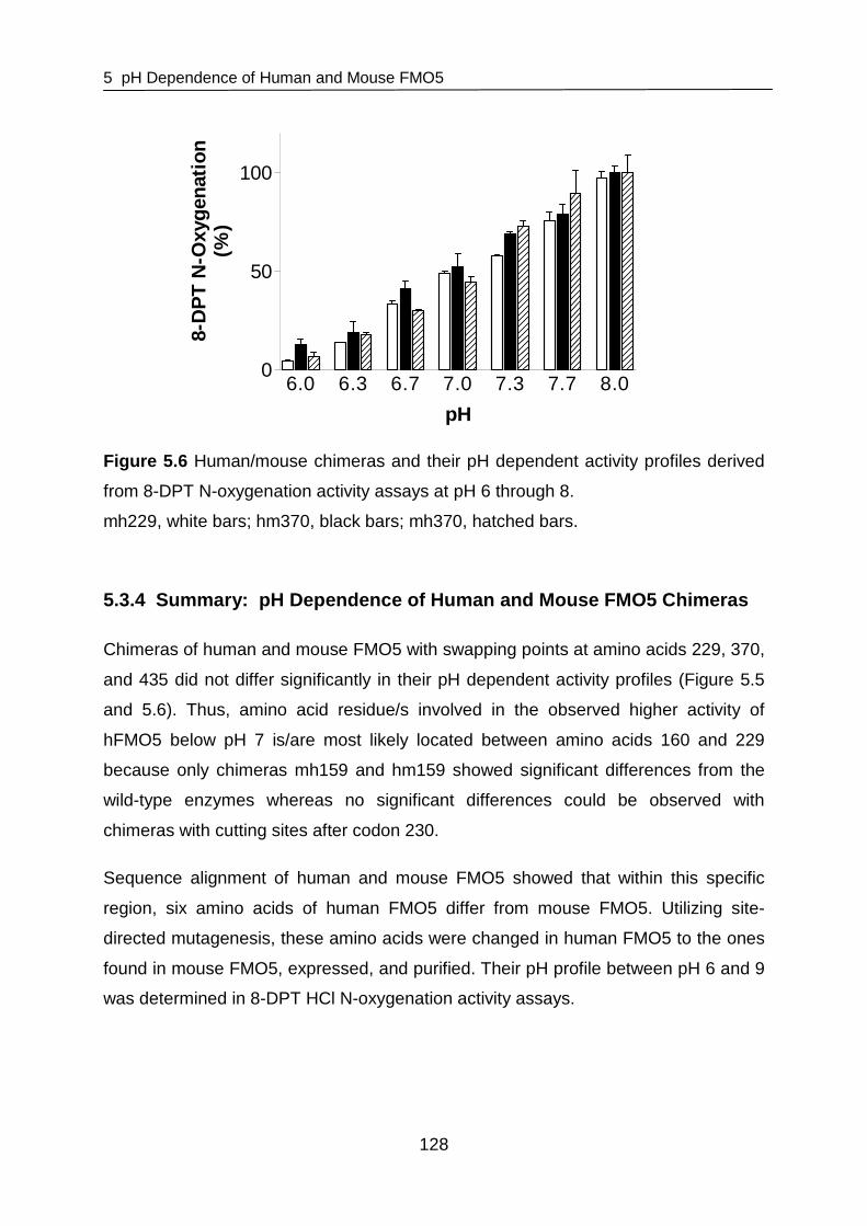
5 pH Dependence of Human and Mouse FMO5
128
6.0 6.3 6.7 7.0 7.3 7.7 8.00
50
100
pH
8-D
PT
N-O
xyge
natio
n(%
)
Figure 5.6 Human/mouse chimeras and their pH dependent activity profiles derived
from 8-DPT N-oxygenation activity assays at pH 6 through 8.
mh229, white bars; hm370, black bars; mh370, hatched bars.
5.3.4 Summary: pH Dependence of Human and Mouse FM O5 Chimeras
Chimeras of human and mouse FMO5 with swapping points at amino acids 229, 370,
and 435 did not differ significantly in their pH dependent activity profiles (Figure 5.5
and 5.6). Thus, amino acid residue/s involved in the observed higher activity of
hFMO5 below pH 7 is/are most likely located between amino acids 160 and 229
because only chimeras mh159 and hm159 showed significant differences from the
wild-type enzymes whereas no significant differences could be observed with
chimeras with cutting sites after codon 230.
Sequence alignment of human and mouse FMO5 showed that within this specific
region, six amino acids of human FMO5 differ from mouse FMO5. Utilizing site-
directed mutagenesis, these amino acids were changed in human FMO5 to the ones
found in mouse FMO5, expressed, and purified. Their pH profile between pH 6 and 9
was determined in 8-DPT HCl N-oxygenation activity assays.

5 pH Dependence of Human and Mouse FMO5
129
5.3.5 pH Dependence of Human and Mouse FMO5 Variant s
The pH profile of wild-type hFMO5 and wild-type mFMO5 as well as hFMO5 variants
purified was determined after optimization of assay conditions (section 5.2.7.3).
The results of wild-type human and mouse FMO5 are shown in Figure 5.7. The
results are similar to those obtained with the original assay conditions with pKa values
of 7.7 ± 0.1 and 6.9 ± 0.1 calculated from the pH dependent activity profiles of mouse
and human FMO5, respectively.
6 7 8 9-1
0
1
2
mFMO5hFMO5
pH
log(
N-O
xyge
natio
n(n
mol
/min
/mg
FMO
5))
Figure 5.7 pH Dependence of wild-type human and mouse FMO5.
hFMO5 is shown as solid line (■), mFMO5 as dashed line (∆).
Five hFMO5 variants were successfully expressed, purified, and analyzed. Results
for these variant hFMO5s are shown in Figure 5.8. The pKa values determined with
wild-type human and mouse FMO5 and variant hFMO5 are listed in Table 5.5.

5 pH Dependence of Human and Mouse FMO5
130
6 7 8 9
-1
0
1
pH
log(
N-O
xyge
natio
n(n
mol
/min
/mg
FMO
5))
Figure 5.8 pH Dependence of human FMO5 variants.
■ hFMO5 Q170K; ▲ hFMO5 G182E; ▼ hFMO5 Q206H; ● hFMO5 D227K; ∆ hFMO5
Y228H. hFMO5 Q170K, hFMO5 G182E, and hFMO5 D227K are shown as solid
lines, hFMO5 Q206H and hFMO5 Y228H are shown as dashed lines.
Table 5.5 pKa Values calculated from pH dependence profiles of human FMO5
variants compared to those of wild-type human and mouse FMO5.
Variant pKa
wild-type hFMO5 6.9 ± 0.1
wild-type mFMO5 7.7 ± 0.1
hFMO5 Q170K 6.6 ± 0.1
hFMO5 G182E 6.6 ± 0.1
hFMO5 Q206H 6.5 ± 0.05
hFMO5 D227K 7.3 ± 0.1
hFMO5 Y228H 7.9 ± 0.2
Data is presented as best fit value ± standard error.
The data clearly show that replacement of the tyrosine at position 228 with histidine
in hFMO5 increases the pKa and thus results in a pH profile similar to that observed

5 pH Dependence of Human and Mouse FMO5
131
with the wild-type mouse enzyme, whereas hFMO5 Q170K, G182E, and Q206H
mutations do not have a significant influence on pKa. D227K showed a slightly
increased pKa in comparison to that calculated from pH dependence profiles of wild-
type hFMO5, even though this increase was lower than hFMO5 Y228H. To confirm
this result, the mouse counterparts with the amino acid of the corresponding human
enzyme were made (i.e., mFMO5 K227D and H228Y) and as suspected, pH
dependence studies with mouse FMO5 K227D as well as mouse FMO5 H228Y led to
decreased pKa values compared to that determined with wild-type mouse enzyme
(Figure 5.9 and Table 5.6). Also, several variants of hFMO5 Y228 were made in
order to evaluate the effect of different amino acids at position 228. The results are
shown in Figure 5.10 and summarized in Table 5.6.
6 7 8 9-2
-1
0
1
2
pH
log(
N-O
xyge
natio
n(n
mol
/min
/mg
FMO
5))
Figure 5.9 pH Dependence of human and mouse FMO5 variants.
▼ hFMO5 D227K; ■ mFMO5 K227D; ● hFMO5 Y228H; ▲ mFMO5 H228Y. hFMO5
D227K and mFMO5 K227D are shown as solid lines, hFMO5 Y228H and mFMO5
H228Y are shown as dashed lines.

5 pH Dependence of Human and Mouse FMO5
132
6 7 8 9-2
-1
0
1
2
pH
log(
N-O
xyge
natio
n(n
mol
/min
/mg
FMO
5))
Figure 5.10 pH Dependence of human FMO5 Y228 variants.
■ hFMO5 Y228H; ▲ hFMO5 Y228F; ▼ hFMO5 Y228R; ∆ hFMO5 Y228K; ● hFMO5
Y228A. hFMO5 Y228F, hFMO5 Y228R, and hFMO5 Y228K are shown as solid lines,
hFMO5 Y228H and hFMO5 Y228A are shown as dashed lines.
Table 5.6 pKa Values calculated from pH dependence profiles of human and mouse
FMO5 variants compared to those of the wild-type FMO5 enzymes.
Variant pK a
wild-type hFMO5 6.9 ± 0.1
wild-type mFMO5 7.7 ± 0.1
hFMO5 D227K 7.3 ± 0.1
mFMO5 K227D 6.6 ± 0.05
hFMO5 Y228H 7.9 ± 0.2
mFMO5 H228Y 6.5 ± 0.05
hFMO5 Y228K 8.0 ± 0.1
hFMO5 Y228A 7.8 ± 0.1
hFMO5 Y228F 6.7 ± 0.05
hFMO5 Y228R 7.7 ± 0.1 Data is presented as best fit value ± standard error.
As expected, in mFMO5 replacement of a histidine at position 228 by tyrosine
(mFMO5 H228Y) significantly decreased the calculated pKa value in comparison to

5 pH Dependence of Human and Mouse FMO5
133
wild-type mFMO5. The same observation was made for the K227D variant of mouse
FMO5.
Changing the amino acid tyrosine in wild-type hFMO5 to three of the tested amino
acids (i.e., lysine, alanine, or arginine) also increased the pKa significantly. However,
changing tyrosine to phenylalanine did not make a significant difference in pKa value
(pKa of 6.9 and 6.7 determined with wild-type hFMO5 and hFMO5 Y228F,
respectively). Overall, the low pKa seemed to be due to the amino acids at position
227 and 228 in the human enzyme and thus they are at least in part responsible for
the increased enzyme activity at low pH.
5.4 Discussion
Within this study, the observed pH dependent activity change of human FMO5 was
compared to mouse FMO5. Interestingly, with the human enzyme a significantly
lower pKa was observed than with the mouse enzyme. Via chimera studies the region
in which the amino acids that are responsible for this pKa shift lie was determined.
Alignment of human and mouse FMO5 sequences showed that this region is only
distinguished by six amino acids. In additional site-directed mutagenesis studies
these amino acids were changed and the pH dependent activity profiles of the
resulting variants were determined. Resulting data clearly showed that the residues
in question lie at positions 227 and 228 of the enzyme.
It has been reported previously that pH dependent activity of enzymes is set mainly
by pKa values of one or a few key ionizable groups within the enzyme, primarily in the
active-site cleft [Joshi et al., 2000]. According to the human FMO3 homology
structure model developed based on four related proteins [Borbas et al., 2006b] (see
also chapter 4), the amino acid that influences the pH profile (i.e., Y228) lies on the
surface of the protein close to the entrance of the back passage connected to the
space between the two domains where NADPH binds. Therefore, in case of FMO5, it
is more likely that NADPH-binding or NADP+ release is altered rather than substrate
binding.
It might have been expected that an amino acid change of tyrosine to histidine, which
has a lower pKa value on its own, would also lead to a decrease in the pKa value
measured. However, this could not be observed in this study. Instead, it is possible

5 pH Dependence of Human and Mouse FMO5
134
that in wild-type human FMO5 the tyrosine at position 228 can make hydrogen
bonding with NADP+. If tyrosine is replaced by a histidine, this interaction may be
interrupted due to protonation at lower pH. The unpolar amino acids phenylalanine
and alanine might not be able to interact because of the missing polar hydroxylic
group. Indeed, with hFMO5 Y228A a significantly increased pKa value was observed
(7.8 ± 0.1). However, replacement of tyrosine by phenylalanine (hFMO5 Y228F) did
not alter the pKa significantly from that observed with the wild-type enzyme (6.7 ±
0.05). Also, a significantly increased pKa for pH profiles of both hFMO5 Y228R and
hFMO5 Y228K (i.e., 7.7 ± 0.1 and 8.0 ± 0.1, respectively) in comparison to wild-type
human FMO5 (6.9 ± 0.1) was observed showing that both lysine and arginine might
affect NADP+ binding.
Conformational changes may also have an influence on titration behavior. Thus, the
similar pKa values observed for pH dependence profiles of hFMO5 Y228 and F228
could be explained by sterical similarity between the two amino acid side chains. It is
possible that a benzene ring is needed in this position and that changing this might
have an effect on neighboring residues such as D227. As the tyrosine at position
228, aspartic acid at position 227 could possibly form hydrogen bonds with NADP+
and changing Asp227 to lysine resulted in an increased pKa (i.e., 7.3 ± 0.1). Shifting
this residue by changing the size of the amino acid at position 228 might also prevent
this interaction and lead to an altered pKa.
Amino acid residues may be sensitive to both electrostatic and structural changes of
others that are located in immediate vicinity. Thus, an amino acid change could have
an effect on pKa values of sterical neighboring residues as was described for the
substitution of asparagine with aspartic acid at position 35 in Bacillus circulans
xylanase [Joshi et al., 2000]. In this enzyme, substitution of the basically neutral
amino acid Asn with the acidic Asp (pKa 3.7) also influenced the pKa values of Glu78
and Glu172. Latter ones were elevated, probably due to charge repulsion. Overall,
the pH optimum of the xylanase was shifted from 5.7 to 4.6 [Joshi et al., 2000]. In
addition, protonation or deprotonation of amino acids may also help stabilize other
structurally close residues and substitution might lead to destabilization and an
altered pH. For example, for a polyketide synthase, stabilization and promotion of a
thiolate anion at cysteine 164 by histidine 303, as an imidazolium cation, was
reported. Thus, upon substitution of this His303, in addition to reactivity, the pKa
5 pH Dependence of Human and Mouse FMO5
135
value of the Cys164 changed [Jez et al., 2000]. It is possible that amino acid changes
in FMO5 at position 227 and 228 may also alter pKa values of surrounding residues
or stabilize/destabilize these depending on the charge of the amino acid in these
positions. Thus, although based on current three-dimensional models D227 and
Y228 lie close to the surface of the protein, they could have an impact on amino
acids that are closer to the substrate binding site or that influence NADPH binding or
NADP+ release. A crystal structure of a mammalian FMO may give more detailed
answers as to how these amino acids influence residues in or close to the active site.
The pI of the enzyme could also affect its pH dependent activity profile. The
theoretical pI of human and mouse MBP-FMO5 are 6.3 and 7.2, respectively
(http://www.expasy.ch/cgi-bin/protparam). It is possible that the enzyme has to be
negatively charged to function properly. Since human FMO5 has a lower pI than the
mouse enzyme, it will keep its negative charge longer at lower pH. Thus, human
FMO5 has a negative charge above pH 6 whereas mouse FMO5 is negatively
charged above pH 7. Therefore, its activity decreases faster in comparison to human
FMO5 below this pH as seen in the pH profiles in this study. Calculation of the
variants’ pIs shows an increase for human FMO5 D227K to 6.5 and a decrease for
mouse FMO5 K227D to 6.8 supporting this theory. FMO5 is negatively charged at its
pH optimum. Assuming the enzyme functions best when it is negatively charged, a
substitution of Asn with Asp and the resulting change of pI could lead to altered
charge of the enzyme at low pH. Also, the pI could simply be a requirement for
stability in the surrounding environment rather than affecting substrate binding or
turnover by determining the charge of the enzyme [Joshi et al., 2000].
In conclusion, the pH dependent activity change of human and mouse FMO5 were
compared and the amino acids responsible for an observed pKa shift between the
two were identified. Changing aspartic acid at position 227 to lysine or tyrosine at
position 228 to histidine of human FMO5 elevated the pKa observed in pH profiles
from 6.9 to 7.3 and 7.9, respectively. Accordingly, an amino acid switch of Lys227 to
Asp227 or His228 to Tyr228 in mouse FMO5 lowered the pKa from 7.7 to 6.6 and 6.5,
respectively. In order to address the question of how exactly the pH dependent
activity profile is changed by these amino acids and whether the residues at position
227 and 228 interact with the cofactor directly or whether the affect neighboring
residues will need further investigation. For example, crystallographic studies with

5 pH Dependence of Human and Mouse FMO5
136
both wild-type and mutant enzymes at acidic pH value or determination of pKa values
for specific residues via NMR may lead to more comprehensive explanations of the
mechanism of FMO5 catalysis and the influence of amino acids on pH dependent
enzyme activity.

6 Substrate Selectivity and Screens of Potential FMO5 Substrates
137
6 Substrate Selectivity and Screens of Potential FM O5 Substrates
6.1 Introduction
6.1.1 FMO Substrates
Generally, any compound containing a soft nucleophile can be a substrate to FMOs if
it is able to gain access to the 4a-hydroperoxyflavin and the catalytic site of the FMO
enzyme [Cashman, 1995; Krueger et al., 2005]. This leads to differences in substrate
specificity between the FMO isoforms because the substrate binding channel of
different FMOs are clearly distinct [Lomri et al., 1993b; Lomri et al., 1993c; Nagata et
al., 1990]. FMO1 possesses the broadest and shallowest substrate binding channel
of all mammalian FMOs. In contrast, FMO2 and FMO3 preferably N-oxygenate long-
chain aliphatic amines (i.e., those possessing a nucleophilic tertiary amine at least six
or seven carbon atoms away from a bulky moiety). The channel to the
4a-hydroperoxyflavin in pig FMO1 is about 3 Å deep and 12 Å wide [Cashman, 1995;
Nagata et al., 1990], but the binding site of human hepatic FMO1 is more restricted
than that of animals and lies about 5 Å below the surface and is only 4.5 Å in
diameter [Cashman et al., 2006; Kim et al., 2000; Krueger et al., 2005; Ziegler, 2002].
Human FMO1 is efficient in N-oxygenating tertiary amines such as chlorpromazine,
imipramine, and 10-[(N,N-dimethylamino)alkyl] phenothiazine derivatives (DPT), but
does not readily catalyze N-oxygenation of primary amines, although aliphatic
primary amines can act as positive effectors [Cashman, 1995; Cashman, 2000;
Krueger et al., 2005]. In contrast, the binding channel of rabbit and human FMO2 and
human FMO3 rests at least 6 to 8 Å below the mouth of the substrate binding
channel with a diameter of about 8 Å [Krueger et al., 2005; Lomri et al., 1993b;
Nagata et al., 1990]. FMO2 is the most size-restricted FMO isoform whereas FMO3
appears to be intermediate. FMO2 as well as FMO3 and 5 also N-oxygenate primary
amines such as n-octylamine and the Km decreases with an increasing length of the
chain between C8 and C12. The resulting hydroxylamine is a better substrate for
FMO2 and is usually N-oxygenated to the cis-isomer of the oxime [Poulsen et al.,
1986]. When examining rabbit, monkey, and human FMO2 for functional activity, it
appears that generally sulfur-containing chemicals are better substrates for FMO2
than nitrogen-containing substrates [Dolphin et al., 1998; Krueger et al., 2004;

6 Substrate Selectivity and Screens of Potential FMO5 Substrates
138
Krueger et al., 2002; Whetstine et al., 2000]. FMO3 enzymes generally prefer
substrates that are slightly smaller than those accepted by FMO1, but are also able
to N-oxygenate primary, secondary, and tertiary amines. Nitrogen atoms on longer
side chains are more efficiently oxygenated than those on shorter side chains,
apparently because the substrate binding site is buried deep within the enzyme.
Typical substrates for FMO3 include benzydamine, methimazole, trimethylamine, and
the probe substrate 5-DPT [Cashman, 2000; Cashman et al., 2006].
Although not clearly understood to date, FMO4 and 5 supposedly have very
restricted substrate specificities. FMO4 is very unstable and its cDNA expression is
problematic affording poorly active enzyme [Lattard et al., 2003a]. Thus, it has been
difficult to establish extensive FMO4 substrate specificity relations [Cashman et al.,
2006]. FMO5 does not oxygenate the typical FMO substrates such as MMI and TMA
[Overby et al., 1995; Zhang et al., 2007a]. In studies with a series of DPT-analogs an
8-fold increase in specific activity of mouse FMO5 was observed between 5-DPT and
8-DPT. Thus, it was suggested that the active site of mouse FMO5 lies about 6 Å
below the surface [Zhang et al., 2007a]. Known substrates of FMO5 are short-chain
aliphatic primary amines such as n-octylamine, thioethers with proximal carboxylic
acids (i.e., S-methyl esonarimod), and 5- and 8-DPT [Cashman et al., 2006; Overby
et al., 1995; Zhang et al., 2007a].
Endogenous substrates of FMOs include biogenic amines (i.e., tyramine and
phenethylamine) [Cashman, 2000]. Tyramine is N-oxygenated by FMO1 and 3 to its
trans-oxime, and FMO3 is predicted to be predominantly responsible for trans-oxime
formation. Phenethylamine is also metabolized by human hepatic FMO3 and to a
lesser extent by porcine liver FMO1. It is N-oxygenated to the phenethyl
hydroxylamine and subsequently to its trans-oxime and this terminates biological
activity [Lin et al., 1997a; Lin et al., 1997b]. FMO apparently precludes biologically
important nucleophiles including many endogenous thiols and other heteroatom-
containing compounds from the active site [Ziegler, 1990]. This is advantageous
because if cellular nucleophiles were to be continuously oxidized, this would
represent a tremendous drain on the ability of a cell to produce reducing equivalents
(i.e., NADPH) for normal cell function. In addition, oxygenation of endogenous thiols
by FMO would also produce a large amount of electrophilic metabolites that might
overwhelm the cells ability to detoxicate them. FMO stabilizes the peroxyflavin and

6 Substrate Selectivity and Screens of Potential FMO5 Substrates
139
does so with a molecular architecture that does not allow reactive metabolites
generated to covalently modify the active site or substrate binding region. Thus, the
substrate binding region is presumably composed of highly lipophilic and non-
nucleophilic amino acid residues. Overall, substrate specificities of FMO1 – 3 have
been examined thoroughly in the past. However, for FMO4 and FMO5 enzymes
there is still a large gap in knowledge of substrate specificity and the substrate
binding site dimensions.
6.1.2 Aim of the Study
As stated in 6.1.1, substrate specificity of FMO isozymes is mainly due to size
restriction of the substrate binding channel of the enzyme. A three-dimensional
structure of the FMO enzymes, as attempted in chapter 4 would certainly be useful to
define substrate specificity since computational modeling studies could be
performed. However, much can be learned about the size of the catalytic site from
comparison of accepted substrates, as was already done for FMO1 – 3. In case of
FMO5, however, the paucity of substrates prevents this approach.
A photometric HPLC based functional assay has been established for mouse and
human FMO5 [Zhang et al., 2007a]. In this HPLC analysis assay that has been
previously described [Lattard et al., 2003b] the ratio of a known FMO5 substrate
(i.e., 8-DPT) to product (8-DPT N-oxide) after a 20 minute incubation period at 37 °C
is determined. However, in order to screen and identify alternative substrates for
FMO5, an easy and quick high-throughput (HT) capable kinetic activity assay should
be developed to aid the search for new FMO5 substrates. This would help to gain a
clearer understanding of the substrate specificity of human FMO5 and the structure
and size of its catalytic site. Identification of structural groups oxygenated by FMO5
could suggest likely physiological substrates and contribute an explanation of the
physiological role of FMO5 in mammalian tissue.

6 Substrate Selectivity and Screens of Potential FMO5 Substrates
140
6.2 Materials and Methods
6.2.1 Reagents
Buffer reagents and substrates tested were purchased from Sigma-Aldrich Chemical
Co. (St Louis, MO, USA) in appropriate purity. 8-DPT HCl was synthesized by Dr.
Karl Okolotowicz (HBRI, San Diego, USA). 4’-(4-bromphenyl)-ω-
dimethylaminobutyrophenon HCl (BDAB HCl) was originally developed in our
research group as NO-inhibitor and was synthesized by Dr. Britta Gerig
(Pharmaceutical Institute, CAU Kiel, Germany). 4’-(4”-bromophenyl)-ω-[4-
chlorophenyl)-4-hydroxypiperidinyl]-butyrophenone (BCHP) as well as 4’-(4”-
bromophenyl)-ω-(4-phenylpiperazinyl)butyrophenone (BPPB) were developed as
antipsychotics and were synthesized by Nikola Klein (Pharmaceutical Institute, CAU
Kiel, Germany). All compounds synthesized were of analytical grade and
charaterized in the usual way.
6.2.2 Cloning, Expression, and Purification of MBP- hFMO5
Recombinant human FMO5 was expressed as an N-terminal maltose-binding fusion
protein (MBP-hFMO5) [Lattard et al., 2003b] and purified as described in section
3.2.2 with minor changes. MBP-FMO5 was expressed in E. coli BL21 cells. After
induction, cells were incubated shaking at 20 °C fo r 24 hours. After expression, E.
coli cells were harvested by centrifugation at 6,000 g for 10 minutes and the cell
pellet was frozen in order to increase yield. After resuspending the cell pellet in lysis
buffer as described in section 3.2.2.1, cells were disrupted by passage through a
French press (Thermo Electron Corp., Needham Heights, MA, USA) operating at
20,000 psi. The cell pellet was only extracted once and after centrifugation the
supernatant was loaded at 1 ml/min onto an amylose column (New England BioLabs,
Ipswich, MA, USA) equilibrated with ten column volumes of buffer A’ (i.e., 50 mM
Na2HPO4, pH 8.4, containing 15 µg/ml FAD). The column was washed with at least
ten column volumes of buffer A’ and bound MBP-hFMO5 protein was eluted with
3 mM maltose in buffer A’ at 1 ml/min. Eluted protein was concentrated with an
Amicon Ultra-15 centrifugal filter unit with an Ultracel-50 filter (Millipore, Billerica, MA,
USA).

6 Substrate Selectivity and Screens of Potential FMO5 Substrates
141
6.2.3 Determination of MBP-hFMO5 Concentration
Concentration of purified MBP-hFMO5 was determined by measuring the
absorbance at 280 nm with a NanoDropTM ND-1000 UV/VIS Spectrophotometer
(Thermo Fisher Scientific, Wilmington, DE, USA) using a calculated extinction
coefficient of 121 M-1cm-1 estimated after the method of Gill [Gill et al., 1989].
6.2.4 Enzyme Assays
N-oxygenation of 8-DPT HCl by MBP-hFMO5 was determined by HPLC analysis as
previously described (section 3.2.4.2).
As primary confirmatory assay for BDAB HCl as potential FMO5 substrate,
N-oxygenation of 8-DPT HCl by MBP-hFMO5 was determined in the presence of
BDAB HCl. A standard incubation mixture of 250 µl final volume contained 50 mM
potassium phosphate buffer, pH 8.5, 0.2 mM NADPH, 0.25 mM DETAPAC, and
60 µg MBP-hFMO5. Reactions were initiated by addition of 8-DPT HCl or a mixture
of 8-DPT HCl and BDAB HCl. For 8-DPT HCl, a final concentration of 400 µM was
chosen, BDAB was added to a final concentration of 200 µM. After incubation for
20 minutes shaking under aerobic conditions at 37 °C, enzyme reactions were
stopped and processed as described in section 3.2.4.2. HPLC analysis was done on
a Waters AllianceTM HPLC-System (Waters e2695 XC Separations Modul, Waters
2998 Photodiode Array Detector and EmpowerTM 2 Software). Chromatographic
separation of analytes was done on a LiChrospher-Si 60 (250 x 4.6 mm, 5 µm;
Merck, Darmstadt, Germany) with a mobile phase of 80 % methanol/20 %
isopropanol/0.025 % HClO4 (v/v/v). The flow rate was 1.5 ml/min and the total run
time was 18 minutes. The wavelength for UV detection was set to 243 nm. The
retention times for 8-DPT and 8-DPT N-oxide were 4.9 and 3.7 minutes, respectively.
Statistical analysis was also done using Graphpad Prism software and statistical
significance was judged at P<0.05.
As secondary confirmatory assay, N-oxygenation of BDAB HCl was determined by
HPLC analysis. A standard incubation mixture of 250 µl final volume contained
50 mM potassium phosphate buffer, pH 8.5, 0.2 mM NADPH, 0.25 mM DETAPAC,
and 60 µg MBP-hFMO5. Reactions were initiated by addition of substrate to a final
concentration of 200 µM. After incubation for 20 minutes shaking under aerobic

6 Substrate Selectivity and Screens of Potential FMO5 Substrates
142
conditions at 37 °C, enzyme reactions were stopped by addition of 4 volumes of cold
dichloromethane. About 20 mg of Na2CO3 was added and the incubations were
mixed and centrifuged to partition metabolites and remaining substrate into the
organic fraction. The organic phase was collected and evaporated. Metabolites and
remaining substrate were dissolved in methanol, mixed thoroughly, centrifuged and
analyzed with a Waters AllianceTM HPLC-System (Waters e2695 XC Separations
Modul, Waters 2998 Photodiode Array Detector and EmpowerTM 2 Software).
Chromatographic separation of analytes was done on a LiChrospher-Si 60 (250 x
4.6 mm, 5 µm; Merck, Darmstadt, Germany) with a mobile phase of 80 %
methanol/20 % isopropanol/0.025 % HClO4 (v/v/v). The flow rate was 1.5 ml/min and
the total run time was 18 minutes. The wavelength for UV detection was set to
293 nm. The retention times for BDAB and BDAB N-oxide were 10.7 and
5.7 minutes, respectively.
6.2.5 LC/MS Analysis
In order to identify the metabolite produced by incubation of BDAB HCl with FMO5 in
the presence of NADPH (section 6.2.4), an LC/MS-method was developed. After
incubation, metabolites and remaining substrate were dissolved in methanol, mixed
thoroughly, centrifuged and analyzed with an Esquire LC MS system (Bruker
Daltonics, Bremen, Germany) after separation with an HP HPLC-System (HP Series
1100 Binary Pump G1312A, HP 1100 VWD UV/VIS Detector and Agilent
ChemStation HPLC Software (Version A.09.01), and Esquire Control (Version 6.14)
and Data Analysis (Version 3.0) MS Software). Chromatographic separation of
analytes was done on a Symmetry C18 (250 x 4.6 mm, 5 µm; Waters) with a Guard
Pak C18 precolumn (3 x 4 mm; Waters) and a mobile phase of 70 % of 0.1 % formic
acid in acetonitrile, 15 % of 0.1 % acetic acid, and 15 % methanol (v/v/v). The flow
rate was 1.5 ml/min and the total run time was 12 minutes. The wavelength for UV
detection was set to 293 nm. The retention times for BDAB and BDAB N-oxide were
6.3 and 3.5 minutes, respectively. MS analysis was done using electrospray
ionization (ESI) with a dry temperature of 340 °C, a dry gas flow rate of 7 l/min, and a
spray pressure of 30 psi. The scan was done between 50 and 1000 m/z and peaks
were detected at m/z 346 and 362 for BDAB and BDAB N-oxide, respectively.
6 Substrate Selectivity and Screens of Potential FMO5 Substrates
143
6.2.6 Development of a Photometric Activity Assay M ethod
6.2.6.1 Assay Protocol Outline
The FMO5 activity assay was based on the photometric determination of NADPH
consumption. During substrate oxygenation by FMO5, NADPH is oxidized to NADP+
and the disappearance of NADPH can be monitored photometrically over time at
340 nm. A typical reaction mixture consisted of 50 mM phosphate buffer, pH 8.4,
200 µM NADPH, 250 µM DETAPAC to inhibit autooxidation, and 200 µg/ml
MBP-hFMO5 in a final volume of 100 µl. This mixture was prepared in advance and
dispensed into 96-well flat-bottom UV-Star microplates (Greigner Bio-One,
Frickenhausen, Germany). Afterwards, the reaction was initiated by addition of
substrate. Kinetic decrease of NADPH was measured over 20 minutes and each well
was read every 60 seconds using a Cary 50 Scan UV-Visible Spectrophotometer
(Varian, Palo Alto, CA, USA). For initial assay optimization studies, the known
substrate 8-DPT HCl was used to validate the high-throughput assay and the final
8-DPT HCl concentration in these initial studies was 400 µM.
6.2.6.2 Optimization of Assay Conditions
Protein Concentration
Purified MBP-hFMO5 was evaluated over a range of enzyme concentrations in
reactions containing 8-DPT HCl. The reaction mixture consisted of MBP-hFMO5 and
200 µM NADPH in 50 mM potassium phosphate buffer, pH 8.4, containing 0.25 mM
DETAPAC. The reactions were initiated by addition of 8-DPT HCl to a final
concentration of 400 µM. Slopes representing FMO activity generated by reactions
containing 8-DPT HCl were normalized to slopes of control reactions without the
substrate. Reactions were done in quadruplicates and enzyme was used in
concentrations between 0.05 and 0.8 mg/ml.
Substrate Dependence
Kinetic parameters (Vmax, Km) of 8-DPT HCl were evaluated. The reaction mixture
consisted of 200 µg/ml MBP-hFMO5 and 200 µM NADPH in 50 mM potassium
phosphate buffer, pH 8.4, containing 0.25 mM DETAPAC. Reactions were initiated
by addition of at least five varying concentrations of 8-DPT HCl between 5 and
400 µM. Reactions were done in quadruplicates and slopes reflecting rates of

6 Substrate Selectivity and Screens of Potential FMO5 Substrates
144
cofactor depletion were calculated by normalizing to slopes of reactions lacking
protein.
6.2.7 Development of a Fluorimetric Activity Assay Method
6.2.7.1 Assay Protocol Outline
The FMO5 activity assay was based on the fluorimetric determination of NADPH
consumption and was adapted from Simeonov et al. [Simeonov et al., 2008] who
established a fluorescence based NADPH depletion assay for inhibitor screening of
the Schistosoma mansoni redox cascade. During substrate oxygenation by FMO5,
NADPH is oxidized to NADP+ and the decrease of NADPH fluorescence can be
monitored kinetically over 40 minutes, reading each well every 60 seconds at
excitation/emission wavelengths of 365 nm/450 nm using a Perkin Elmer LS55
Fluorescence Spectrophotometer (Perkin Elmer, Waltham, MA, USA). A typical
reaction mixture consisted of 50 mM phosphate buffer, pH 8.4, 200 µM NADPH, 250
µM DETAPAC, and 200 µg/ml MBP-hFMO5. This mixture was prepared in advance
and dispensed into white 96-well flat-bottom microplates (OptiPlate-96, Perkin Elmer,
Waltham, MA, USA). Afterwards, the reaction was initiated by addition of substrate.
For initial assay optimization studies, the known substrate 8-DPT HCl was used to
validate the HT-assay and the final 8-DPT HCl concentration in these initial studies
was 400 µM.
6.2.7.2 Optimization of Assay Conditions
Protein Concentration
Purified MBP-hFMO5 was evaluated over a range of enzyme concentrations in
reactions containing a final concentration of 400 µM 8-DPT HCl. This was done
measuring the decrease of NADPH fluorescence kinetically at excitation/emission
wavelengths of 365 nm/450 nm over 50 minutes. Each well was read every
60 seconds. The reaction mixture consisted of MBP-hFMO5 in concentrations
between 0.05 and 0.8 mg/ml and 200 µM NADPH in 50 mM potassium phosphate
buffer, pH 8.4, containing 0.25 mM DETAPAC. The reactions were initiated by
addition of 8-DPT HCl to a final concentration of 400 µM. Reactions were done in
quadruplicates and slopes representing FMO5 activity generated by reactions

6 Substrate Selectivity and Screens of Potential FMO5 Substrates
145
containing 8-DPT HCl were normalized to slopes of control reactions without
substrate.
Substrate Dependence
Kinetic parameters (Vmax, Km) of 8-DPT HCl were evaluated. Again, the decrease of
NADPH fluorescence was measured kinetically at excitation/emission wavelengths of
365 nm/450 nm over 60 minutes and each well was read every 60 seconds. The
reaction mixture consisted of 300 µg/ml MBP-hFMO5 and 200 µM of its cofactor
NADPH in 50 mM potassium phosphate buffer, pH 8.4, containing 0.25 mM
DETAPAC. Reactions were initiated by addition of at least five varying concentrations
of 8-DPT HCl between 20 and 1600 µM. Reactions were done in quadruplicates and
slopes reflecting rates of cofactor NADPH depletion were calculated by normalizing
to slopes of reactions lacking protein.
6.2.8 Substrate Screens
80 Compounds were tested with the developed photometric assay method for
MBP-hFMO5 catalyzed oxygenation. These are listed in Table 6.1. Compounds were
either dissolved in methanol or water and reactions were started by addition of these
to yield final concentrations of 400 µM. Methanol concentrations in final incubations
did not exceed 1 %. At this concentration, no significant change in enzyme activity
was observed. Reactions were done in at least duplicates and slopes reflecting rates
of NADPH depletion were calculated by normalizing to slopes of reactions lacking
enzyme. In all plates at least one well contained 8-DPT HCl as positive control.

6 Substrate Selectivity and Screens of Potential FMO5 Substrates
146
Table 6.1 Compounds tested with the developed photometric assay method.
Acetazolamide Acetaminophen
S
N N
NH
O
SNH2O
O
NH
OH
O
Adenine Amitriptyline
N
N
NH
N
NH2
N
L-Arginine Astemizole
COOH
NH
NH NH2
NH2
N
N
F
NH
N
O
Benzocaine Benzydamine
NH2
O
O
N
N
O
N

6 Substrate Selectivity and Screens of Potential FMO5 Substrates
147
BCHP BDAB HCl
Br
N
O
Cl
OH
Br
N
O
HCl
BPPB Cadaverine 2·HCl
BrO
N N
NH2 NH2 2 HCl
Chloramphenicol Chloroquine
OH
NH
OH
O
Cl
Cl
O2N
NCl
NH
CH3
N
CH3
CH3
Chlorpromazine Chlorprothixene
N
S
Cl
N
S
N
Cl
Cimetidine Cocaine
NH
NSNH
N
NH
NC
N
O O
O
O

6 Substrate Selectivity and Screens of Potential FMO5 Substrates
148
Codeine L-Cysteine
O
OH
O
N
SH
COOHNH2
Desipramine 1,4-Diaminobutane 2·HCl
N
NH
NH2
NH2
2 HCl
8-DPT HCl Dimethyl sulfoxide
N
S
(CH2)8
CF3
N
S
O
Diphenhydramine Dithiothreitol
ON
SHSH
OH
OH
Dopamine HCl Drofenin
NH2
OH
OH
HCl
O
ON

6 Substrate Selectivity and Screens of Potential FMO5 Substrates
149
Ephedrine Glibenclamid
OH
NH
ClO
NH
SO O
NH
O
NH
OMe
Guanethidine Histamine 2·HCl
NNNH2
NH2
NH
N
NH2
2 HCl
L-Histidine HCl Imipramine
NH2
N
NH
COOH
HCl N
N
Isoaminile Isoprenalin
NCN
OHOH
OHNH
Isothipendyl Levodopa
N N
S
N
OHOH
NH2
COOH

6 Substrate Selectivity and Screens of Potential FMO5 Substrates
150
Lidocaine L-Lysine HCl
NH
ON
NH2 COOH
NH2 HCl
Mafenide Meprobamate
NH2
S
NH2
OO
ONH2
O
O NH2
O
L-Methionine Metoclopramid
NH2
SCOOH
Cl
NH2
O
NH
N
OMe
Metoprolol N,N-Dimethylbutylamine O
O NH
OH
N
N,N-Dimethylethylamine N,N-Dimethylhexylamine
N
N

6 Substrate Selectivity and Screens of Potential FMO5 Substrates
151
N,N-Dimethyloctylamine Nitrofurantoin
N
N NNHO
O
OO2N
n-Octylamine Orciprenaline
NH2
OH OH
OH
NH
Oxedrin Oxeladin
OH
OH
NH
O O
O
N
Perphenazin Phenacetin
N
S
Cl
NN
OH
NH
O
O
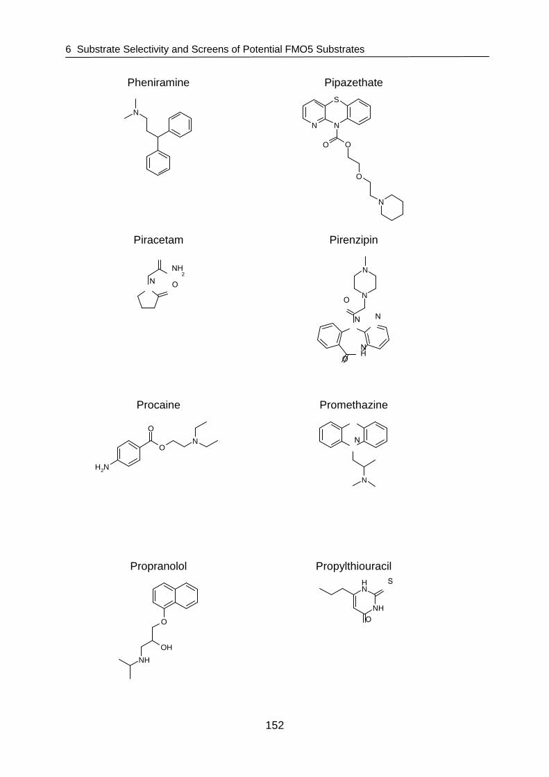
6 Substrate Selectivity and Screens of Potential FMO5 Substrates
152
Pheniramine Pipazethate
N
N N
S
O O
O
N
Piracetam Pirenzipin
N O
O
NH2
N
NH
N
O
ON
N
Procaine Promethazine
NH2
O
O
N
N
S
N
Propranolol Propylthiouracil
O
OH
NH
NH
NH
S
O

6 Substrate Selectivity and Screens of Potential FMO5 Substrates
153
Ranitidine Saccharin
O
N S NH
NH
O2N
NHS
O
OO
Spermidine Spermine 4·HCl
NH2
NH
NH2
NH2 NH
NH
NH2
4 HCl
Sulfaguanidine Sulfamethoxazol
NH2 S
O
O
N
NH2
NH2
NH2 SO
ONH
NO
Sulfanilamide Sulfathiazole
NH2 S NH2
O
O
NH2 S NH
O
ON
S
Sulfisomidin Tetracain
NH2 S NH
O
ON
N
NH
O
O
N
Thiamine cloride·HCl Thiopental
N
N
NH2
N+
S
OH
HCl
NH
NH
O
O S
Cl-

6 Substrate Selectivity and Screens of Potential FMO5 Substrates
154
Thioridazine Tolbutamide
N
S
S
N
S NH
O
O
NH
O
L-Tryptophan Verapamil
NH2N
COOH
N
O
O
O O
CN
6.3 Results
6.3.1 Comparison of Photometric and Fluorescent Act ivity Assay Methods
For the development of a suitable HT-adaptable FMO5 oxygenation activity assay,
two approaches were carried out and evaluated. Both methods were based on
NADPH consumption associated with FMO5 dependent oxygenation activity.
However, one was a photometric approach whereas the other exploits the inherent
fluorescence of NADPH.
6.3.1.1 Photometric Activity Assay Method
Time Dependence
Protein dependence as well as substrate dependence was measured over
20 minutes and graphs were plotted and evaluated in regards to signal stability over
time. A stable slope for NADPH depletion at 340 nm could be observed for the whole
time period measured.

6 Substrate Selectivity and Screens of Potential FMO5 Substrates
155
Protein Concentration
Results of protein dependence studies are shown in Figure 6.1. The protein
concentration dependent increase of activity is clearly visible. It is necessary to use
enough enzyme to consume sufficient amounts of NADPH to generate detectable
and reproducible slopes. However, at a concentration of 0.8 mg/ml enzyme the
activity decreases. This might be due to nonspecific binding of MBP-hFMO5 to the
substrate, leading to a decrease in effective concentration. For further studies, MBP-
hFMO5 concentrations between 0.2 and 0.4 mg/ml were used.
-0.001
0.001
0.003
0.005
0.007
0.009
0 0.05 0.1 0.2 0.4 0.8
MBP-hFMO5 (mg/ml)
dA/m
in
Figure 6.1 MBP-hFMO5 concentration dependence of the photometric plate-reader
based NADPH depletion assay with 8-DPT HCl as substrate.
Substrate Dependence
Data obtained from incubation of MBP-hFMO5 with varying amounts of 8-DPT HCl
was plotted as 8-DPT concentration vs. enzyme activity in nmol/min/mg FMO5 and a
nonlinear regression curve fit tool using a Michaelis-Menten model
(Y= (Vmax · X)/(Km + X) with Vmax = Ymax and Km = Xmid) in Graphpad software
(Graphpad Prism, Version 2.00, San Diego, CA, USA) was utilized to calculate the
kinetic parameters. The obtained Km was 12.3 ± 1.0 µM and Vmax was

6 Substrate Selectivity and Screens of Potential FMO5 Substrates
156
6.4 ± 0.1 nmol/min/mg FMO5 (Figure 6.2). As positive control in screening assays, a
concentration of 200 µM 8-DPT HCl was selected.
0 100 200 300 400 5000.0
2.5
5.0
7.5
8-DPT HCl (µµµµM)
8-D
PT
HC
lN
-Oxy
gena
tion
(nm
ol/m
in/m
g FM
O5)
Figure 6.2 8-DPT HCl concentration dependence for photometric plate-reader based
NADPH depletion assay with MBP-hFMO5.
6.3.1.2 Fluorimetric Activity Assay Method
Time Dependence
Protein dependence as well as substrate dependence was measured over
60 minutes and graphs were plotted and evaluated in regards to signal stability over
time. Signal stability was dependent on protein concentration. A clear decrease of
NADPH fluorescence could only be observed for 10 minutes at a MBP-hFMO5
concentration of 0.8 mg/ml. At 0.4 mg/ml, the signal was stable for 40 minutes and at
protein concentrations below 0.4 mg/ml, the decrease was linear within the range
tested (50 minutes) (data not shown).
Protein Concentration
Results of protein dependence studies are shown in Figure 6.3. The slopes were only
evaluated in the range of 0 to 30 minutes because linearity was not given afterwards
at higher enzyme concentrations (see also time dependence). The protein
concentration dependent increase of activity is clearly visible. However, although it is
necessary to use enough enzyme to consume sufficient amounts of NADPH to

6 Substrate Selectivity and Screens of Potential FMO5 Substrates
157
generate detectable and reproducible slopes, large amounts of enzyme will consume
substrate too quickly and the slope will decrease. Also, protein may nonspecifically
bind to a substrate, leading to a decrease in effective concentration. The optimal
concentration for further studies lies in the range of 0.2 and 0.4 mg/ml MBP-hFMO5.
0
0.5
1
1.5
2
2.5
0 0.05 0.1 0.2 0.4 0.8
MBP-hFMO5 (mg/ml)
Net
Dec
reas
e of
NA
DP
H F
luor
esce
nt S
igna
l/min
Figure 6.3 MBP-hFMO5 concentration dependence of the fluorimetric plate-reader
based NADPH depletion assay with 8-DPT HCl as substrate.
Substrate Dependence
Data from substrate dependence studies fit to Michaelis-Menten kinetic in the range
of 20 – 400 µM. However, higher concentrations of substrate led to decreased netto
fluorescence signal. This could be due to an inhibitory effect of substrate on the
enzyme. Thus, data obtained from incubation of MBP-hFMO5 was plotted in the
range of 20 - 400 µM as 8-DPT HCl concentration vs. netto decrease of NADPH
fluorescent signal and a nonlinear regression curve fit tool using a Michaelis-Menten
model (Y= (Vmax · X)/(Km + X) with Vmax = Ymax and Km = Xmid) in Graphpad software
(Graphpad Prism, Version 2.00, San Diego, CA, USA) was utilized to calculate kinetic
parameters. The obtained Km was 30.3 ± 4.5 µM and Vmax was 6.4 ± 0.2 nmol/min/mg
(Figure 6.4). As positive control in subsequent assays, a concentration of 200 µM
8-DPT HCl should be used.

6 Substrate Selectivity and Screens of Potential FMO5 Substrates
158
0 100 200 300 400 5000.0
2.5
5.0
7.5
8-DPT HCl (µµµµM)
8-D
PT
HC
lN-O
xyge
natio
n(n
mol
/min
/mg
FMO
5)
Figure 6.4 8-DPT HCl concentration dependence of fluorescence plate-reader based
NADPH depletion assay with MBP-hFMO5.
6.3.2 Substrate Screens
No significant differences in terms of protein and substrate concentration needed
could be observed between photometric and fluorescence enzyme activity assay. For
substrate screens, the photometric assay was chosen. Besides the positive control
substrate 8-DPT HCl, only 4’-(4-bromophenyl)-ω-dimethylaminobutyrophenone HCl
(BDAB HCl) showed considerable decrease in absorbance at 340 nm. For all other
compounds tested, no decrease in absorbance could be observed at the substrate
concentrations used in the assay. A few other substrates including chlorpromazine,
chlorprothixene, drofenin HCl, promethazine HCl, thioridazine, and phenacetin
appeared to be potential hits after the primary screen and were re-screened to
eliminate false-positives. None of these compounds proved to be a substrate for
FMO5 and thus only BDAB HCl was examined further. BDAB N-oxygenation activity
of FMO5 was supposed to be confirmed with an independent assay. Within this
assay, 8-DPT HCl was incubated with MBP-hFMO5 in the presence and absence of
BDAB HCl. As shown in Figure 6.5, conversion of 8-DPT was significantly inhibited
by addition of BDAB to the incubation mixture. This inhibition was possibly due to
BDAB HCl being a competitive substrate for MBP-hFMO5. Thus, in a secondary
confirmatory assay, an HPLC-based method was developed to separate BDAB HCl
from its metabolite/s after incubation with FMO5.

6 Substrate Selectivity and Screens of Potential FMO5 Substrates
159
0.00
5.00
10.00
15.00
20.00
25.00
8-DPT 8-DPT +BDAB
8-D
PT
HC
l N
-Oxy
gena
tion
(nm
ol/m
in/m
g FM
O5)
Figure 6.5 8-DPT HCl N-oxygenation by MBP-hFMO5 with and without BDAB.
Statistically significant differences between incubation with 8-DPT HCl and incubation
with a combination of 8-DPT HCl and BDAB HCl are identified with *** for P<0.001.
Only one additional peak was observed in HPLC analysis after incubation of BDAB
with MBP-hFMO5. This peak was analyzed via LC-MS and proved to be BDAB
N-oxide with m/z 362 (Figure 6.6).
Figure 6.6 HPLC Chromatogram of BDAB HCl and BDAB N-oxide.
***
BDAB N-oxide
BDAB
Mass spectrum of peak 1
6 Substrate Selectivity and Screens of Potential FMO5 Substrates
160
6.4 Discussion
Two different HT-compatible screening methods that are based on NADPH
consumption during enzyme catalysis have been developed and were compared. No
significant difference in terms of necessary protein and substrate concentrations
could be observed. The photometric assay was chosen to screen several potential
FMO5 substrates. Besides the control substrate 8-DPT HCl and BDAB HCl, none of
the compounds tested showed considerable decrease in 340 nm absorbance.
Further evaluation of BDAB HCl proved this compound to be a substrate for FMO5.
Several substrates have already been tested for oxygenation activity catalyzed by
FMO5 from various species (i.e., mouse, human, and rabbit FMO5) including
methimazole [Atta-Asafo-Adjei et al., 1993; Overby et al., 1995; Zhang et al., 2007a],
ranitidine and cimetidine [Overby et al., 1997], phorate [Cherrington et al., 1998],
trimethylamine [Atta-Asafo-Adjei et al., 1993; Zhang et al., 2007a], chlorpromazine,
perchlorperazine, imipramine, N,N-dimethylaniline, cysteamine, trimethylamine and
n-decylamine [Atta-Asafo-Adjei et al., 1993], L-arginine and Nδ-Methyl-D,L-arginine
[Kotthaus, 2008], n-octylamine [Atta-Asafo-Adjei et al., 1993; Cherrington et al.,
1998; Overby et al., 1995], n-nonylamine [Atta-Asafo-Adjei et al., 1993],
S-methylesonarimod [Ohmi et al., 2003; Zhang et al., 2007a] and 8-DPT [Zhang et
al., 2007a]. However, only the last four compounds were actually oxygenated by the
enzyme. Of these four known FMO5 substrates, besides 8-DPT, n-octylamine was
examind within this study. It is notable that upon incubation with n-octylamine no
decrease of NADPH dependent absorption at 340 nm could be observed in the
photometric assay although this compound had previously been reported by one
group [Overby et al., 1995] to be a substrate for human FMO5. This could not be
confirmed in this study.
Overall, the assay methods developed may be used to screen for further potential
substrates. Screening a library of several thousands of compounds in a HT-format
may help find additional FMO5 substrates and may lead to identification of structural
groups oxygenated by FMO5 that will suggest likely physiological substrates and
contribute an explanation of the role of FMO5.

6 Substrate Selectivity and Screens of Potential FMO5 Substrates
161
A potential problem of both screening methods is the requirement of high substrate
concentrations that might make it difficult to find a suitable library. An alternative
approach could be an inhibitor screen for competition against 8-DPT as FMO5
substrate. Lower compound concentrations would be needed for this approach and
the method may still identify groups capable of binding to the enzyme’s active site,
providing an indirect perspective on true substrate structure features. In general, the
assay protocol would be similar to that of the direct FMO5 substrate screen and the
inhibitor approach could also be combined with HPLC analysis of 8-DPT
N-oxygenation as a basis for secondary confirmatory assays.
A completely different approach to test the significance of FMO5 would be the
development of a knock-out or knock-in mouse model of FMO5 or the usage of short
interfering RNA (si-RNA) in order to suppress FMO5 expression. Such studies could
also give more information about the physiological role and importance of FMO5.

7 Summary
162
7 Summary
In this work, the structural and functional relations of flavin-containing
monooxygenases (FMOs) 3 and 5 were investigated. The family of FMO enzymes
represents, after the cytochromes P450, the most important monooxygenases in
humans. It consists of five isozymes (FMO1 – 5) which catalyze mainly the
oxygenation of nucleophilic N- and S-containing xenobiotics [Cashman et al., 2006;
Ziegler, 1980]. In the main drug-metabolizing organ, the adult human liver, FMO3
and FMO5 mRNA are the most abundantly expressed FMO mRNAs [Cashman et al.,
2006; Janmohamed et al., 2004]. Thus special interest was paid to these two
isoforms.
In the past, FMO3 gained considerable importance because of its association with
trimethylamineuria (TMAu). TMAu is a disorder that often manifests itself in a body
odor for individuals affected. It is due to decreased metabolism of dietary-derived
trimethylamine (TMA). In a healthy individual, 95 % or more of TMA is converted by
the FMO3 to non-odorous TMA N-oxide. Several SNPs of the FMO3 gene have been
described and result in an enzyme with decreased or abolished functional activity for
TMA N-oxygenation thus leading to TMAu.
The disorder may be diagnosed by genotyping or by measuring urinary ratios of TMA
N-oxide to TMA. Accurate diagnosis is essential for the affected individuals so that
they can be treated appropriately. Treatment comprises restriction of certain foods,
supplementation of folate, riboflavin, and, in very severe cases administration of
antibiotics to decrease TMA formation due to gut bacteria [Akerman et al., 1999;
Ayesh et al., 1993; Cashman et al., 2002; Mitchell, 1996; Mitchell et al., 2001].
Biochemical characterization of recombinant expressed variant FMO3 observed from
genotyping and phenotyping aids to reveal important information about structure and
function of human FMO3 [Akerman et al., 1999; Cashman, 2002; Cashman et al.,
2002; Dolphin et al., 1997b; Shimizu et al., 2007b; Treacy et al., 1998; Yeung et al.,
2007].
Herein, a novel mutation observed from phenotyping and genotyping of self-reporting
individuals was examined. This novel mutation was heterozygous at position 187

7 Summary
163
(V187A) and occurred in combination with mutations at position 158 (E158K), 308
(E308G), and 305 (E305X). Familial genetic analysis showed that the
E158K/V187A/E308G derived from the same allele from the mother, and the E305X
was derived from the father. Wild-type human FMO3 and its variants V187A and
V187A/E158K were expressed as maltose-binding fusion proteins (MBP-FMO3),
purified, and characterized for oxygenation of several common FMO3 substrates (i.e.,
5- and 8-DPT, mercaptoimidazole (MMI), TMA, and sulindac sulfide) and for their
thermal stability. The novel mutation in combination with the common polymorphism
E158K led to severely decreased enzyme activity. Similarly, although by itself, the
common polymorphisms E158K and E308G only slightly alter FMO3 activity, in
combination they may lead to decreased activity [Cashman, 2000; Cashman et al.,
2003; Dolan et al., 2005; Zschocke et al., 1999]. This has also been reflected in vivo
leading to TMAu [Lambert et al., 2001; Zschocke et al., 1999]. Likewise, the novel
V187A mutation in conjunction with the common polymorphisms E158K and E308G
significantly impairs FMO3 resulting in an enzyme with drastically decreased function
that manifests itself in severe TMAu.
Although FMO5 mRNA is most abundantly expressed in human adult liver, it is a
largely understudied enzyme. Therefore, besides FMO3, FMO5 was of particular
interest and was characterized structurally and functionally within this thesis.
To facilitate these studies, highly-purified and well characterized FMO5 enzyme was
produced. Human FMO5 was successfully expressed as maltose-binding fusion
protein (MBP-hFMO5). Solubilization studies showed Triton® X-100 to be superior in
extracting MBP-hFMO5 from E. coli. After cell lysis, protein was purified exploiting the
MBP-tag for amylose-column chromatography. Further purification was done on an
anion-exchange column and remaining impurities could be removed utilizing a NaCl
salt gradient. Comparison to commercially available FMO5 showed no significant
differences in kinetic behavior and since MBP-hFMO5 could be readily purified at low
cost and in high quantities, it was chosen for further studies. Also MBP-hFMO5 could
be obtained in high purity and was thus also suitable for crystallography studies.
Further, MBP-hFMO5 was characterized in terms of enzyme activity and purity,
stability at 4 °C, and oligomerization state and mo nodispersity. Especially for
crystallography studies these characterization studies are substantial. They showed
that addition of glycerol and NADP+ to the sample improved MBP-hFMO5 stability

7 Summary
164
1.6-fold and in subsequent crystallization studies these additives were added to
enzyme preparations. Results from size exclusion chromatography of ion exchange
purified MBP-hFMO5 suggested hexameric state for the protein which is in
accordance with previous publications [Brunelle et al., 1997; Ziegler et al., 1972].
Dynamic light scattering (DLS) data of ion exchange purified FMO5 samples showed
a single peak, suggesting pure, monodispers protein suitable for crystallography
studies.
When working with membrane associated proteins such as FMO enzymes use of
detergents is unavoidable. As expected, surfactants play a dominant role in
purification and crystallization of FMO5. Unfortunately, usage of detergents
introduces a high level of unpredictability, especially if detergents are used that are
heterogenic by themselves, such as Triton® X-100. However, the usage of this
detergent was inevitable for the extraction of MBP-hFMO5 from E. coli and complete
removal of Triton® X-100 and possibly substitution with other detergents seemed
necessary prior to crystallization trials. Removal was done using polystyrene based
beads (e.g., Bio-Beads®) and interestingly, upon addition of excess amounts of Bio-
Beads® to the protein solution, MBP-hFMO5 did not precipitate. This result suggests
that FMO5 is not an integral membrane protein, but rather only associated with the
membrane. Also, removal of Triton® X-100 with Bio-Beads® improved results from
crystallization trials of MBP-hFMO5 leading to thick yellow oil phases and premature
crystals with spherical shapes.
Crystallography experiments were done by initially screening various commercially
available 96-well plate crystal screen kits for suitable crystallization conditions. The
most promising conditions were refined further in 24-well plates evaluating protein
concentration, salts and buffers as well as precipitant type and concentration.
Conditions that finally led to MBP-hFMO5 crystals were 26 % PEG 2K MME and
0.2 M sodium acetate in 0.1 M Bis-Tris propane buffer, pH 6.5.
Experiments in which a variety of different detergents was added after removal of
Triton® X-100 led to further improvement of these first spherical shaped pre-stages of
crystals. Detergents that yielded best results mainly belonged to a new group of
amphiphilic agents that is supposed to stabilize membrane proteins and lower the
risk of denaturing or aggregation. These surfactants are based on cholic acid and

7 Summary
165
exhibit facial amphiphilicity [Zhang et al., 2007b; Zhong et al., 2005]. In addition,
detergents such as HECAMEG and the Cymal® series also yielded crystals. In
general, employing the concept of mixed micelles by controlled addition of detergent
seemed beneficial for FMO5 crystal growth.
Unfortunately, no satisfactory diffraction pattern could be obtained from MBP-hFMO5
crystals. This might be due to the flexible linker region between MBP-tag and FMO5
that possibly introduces conformational heterogeneity [Smyth et al., 2003]. Therefore,
MBP-hFMO5 was cleaved to perform crystallization experiments with only hFMO5.
Splicing of MBP from hFMO5 was done utilizing a previously inserted protease
sensitive site right after the MBP sequence that may be cleaved by factor Xa. After
incubation for three hours at 37 °C, cleavage was l argely completed and addition of
NADPH to the incubation mixture prevented the enzyme from being heat-inactivated.
It is notable that similar experiments had been done with MBP-hFMO3 [Brunelle et
al., 1997]. However, after cleaving MBP from hFMO3, the latter showed intractable
solubility and enhanced instabilities [Brunelle et al., 1997] as previously described for
a number of purified native FMO enzymes [Cashman, 1995; Guan et al., 1991]. In
contrast to expectations, after proteolytical separation from MBP, human FMO5
stayed in solution and retained the same level of enzyme activity as the tagged
protein. However, subsequent purification of hFMO5 from remaining factor Xa, MBP,
and residual uncleaved MBP-hFMO5 using a variety of previously described methods
including affinity purification [Riggs, 2000], ion exchange chromatography [de Pieri et
al., 2004; Guan et al., 2002; Hao et al., 2007; Riggs, 2000; Yan et al., 2006], and size
exclusion chromatography [Branco et al., 2008] failed.
In summary, this study showed that MBP-hFMO5 is crystallizable. However, further
studies are needed to purify hFMO5 from MBP in order to start crystallization
experiments with the tag-free protein and obtain well diffracting crystals.
FMO5 displays a significantly different pH dependent activity profile than the other
FMO isoforms [Zhang et al., 2007a]. While pH optima of mouse FMO1 and 3
(mFMO1 and 3) display normal bell shape curves peaking around pH 8 to 10, with
human FMO5 (hFMO5) and mFMO5, 8-DPT N-oxygenation activity continues to
increase from pH 7 to pH 11 [Zhang et al., 2007a]. A possible reason for the different
pH profile found in FMO5 could be a slower overall turnover rate indicating a possibly
7 Summary
166
different rate-limiting step in the catalytic cycle of FMO5. To date, the catalytic steps
of pig FMO1 are known in some detail [Beaty et al., 1981a; Beaty et al., 1981b;
Jones et al., 1986; Poulsen et al., 1979; Ziegler et al., 1988] and presumably, the
other FMO isoforms follow a similar mechanism. However, this might not be the case
for all FMO isoenzymes and the assumption is that FMO5 catalytical mechanism is
distinct from that of other FMO enzymes.
The objective of this study was to determine the pH dependence of FMO5 to gain an
insight into the mechanism of action of FMO5 enzymes. Therefore, functional
recombinant hFMO5 and mFMO5 were expressed as MBP-fusion proteins from
E. coli, purified with affinity chromatography, and examined for their 8-DPT
N-oxygenation activity at different pH values. MBP-hFMO5 showed a broader range
and greater functional activity from pH 6 to 11 compared to mFMO5. mFMO5 lost
nearly all functional activity at pH 6, while hFMO5 maintained almost normal enzyme
activity. In order to identify the amino acid residues involved in the effects of pH on
hFMO5 and mFMO5 functional enzyme activity, pH-studies in the range of pH 6
through 9 were done with MBP-tagged chimeras of recombinant human and mouse
FMO5 and variants of both. Results of this study showed that the residues
responsible for the pH profile distinction between MBP-hFMO5 and MBP-mFMO5 are
located at positions 227 and 228 of the enzyme. Further mutants were made to
investigate the role of these amino acids. Combined with additional structural,
functional and kinetic information, this study may add to provide insight into the
mechanism of FMO5 revealing important interactions between substrate or cofactors
of FMO and specific amino acid residues of the enzyme. In addition, results may be
relevant for other FMO enzymes as well if the catalytic mechanism of all FMOs
proves to be identical.
The last objective within this thesis was the development of a high-throughput (HT)
compatible enzyme activity assay in order to screen for new FMO5 substrates. Two
different HT-compatible screening methods that are based on a decrease of NADPH
during enzyme catalysis have been developed and were compared. One was based
on fluorescence whereas the other was a photometric assay. Since no significant
difference in terms of protein and substrate concentrations needed or sensitivity
could be observed, the photometric assay was chosen to screen 80 compounds as
possible FMO5 substrates. Potential hits from this first screen were re-screened in

7 Summary
167
order to eliminate false-positives. Besides the control substrate 8-DPT HCl only
4’-(4-bromophenyl)-ω-dimethylaminobutyrophenone (BDAB) HCl showed
considerable decrease in 340 nm absorbance. Thus, only this compound was
examined further. An HPLC method was developed to separate substrate from its
metabolite/s after incubation with MBP-hFMO5. The resulting BDAB metabolite
observed after incubation was analyzed via LC-MS and proved to be BDAB N-oxide.
Thus, results from the primary photometric assay could be confirmed. This compound
fits well into the proposed selectivity range of FMO5, having a tertiary amine group
on a long carbon side-chain away from a bulky moiety. It is still surprising that
compounds with very similar structures such as benzydamine or tetracaine did not
prove to be substrates of FMO5. Overall, further compound screens will be needed to
identify additional FMO5 substrates and gain more knowledge of substrate specificity
and thus the relationship between structure and function.

8 References
168
8 References
Adachi, M.S., Juarez, P.R., Fitzpatrick, P.F. 2010. Mechanistic studies of human spermine oxidase: kinetic mechanism and pH effects. Biochemistry 49:386-92
Akerman, B.R., Lemass, H., Chow, L.M., Lambert, D.M., Greenberg, C., Bibeau, C., Mamer, O.A., Treacy, E.P. 1999. Trimethylaminuria is caused by mutations of the FMO3 gene in a North American cohort. Mol Genet Metab 68:24-31
Alfieri, A., Malito, E., Orru, R., Fraaije, M.W., Mattevi, A. 2008. Revealing the moonlighting role of NADP in the structure of a flavin-containing monooxygenase. Proc Natl Acad Sci U S A 105:6572-7
Arnold, T., Linke, D. 2008. The use of detergents to purify membrane proteins. Curr Protoc Protein Sci Chapter 4: Unit 4 8 1-4 8 30
Askolin, S., Turkenburg, J.P., Tenkanen, M., Uotila, S., Wilson, K.S., Penttila, M., Visuri, K. 2004. Purification, crystallization and preliminary X-ray diffraction analysis of the Trichoderma reesei hydrophobin HFBI. Acta Crystallographica Section D-Biological Crystallography 60:1903-1905
Atta-Asafo-Adjei, E., Lawton, M.P., Philpot, R.M. 1993. Cloning, sequencing, distribution, and expression in Escherichia coli of flavin-containing monooxygenase 1C1. Evidence for a third gene subfamily in rabbits. J Biol Chem 268:9681-9
Attar, M., Dong, D., Ling, K.H., Tang-Liu, D.D. 2003. Cytochrome P450 2C8 and flavin-containing monooxygenases are involved in the metabolism of tazarotenic acid in humans. Drug Metab Dispos 31:476-81
Attwood, T.K., Bradley, P., Flower, D.R., Gaulton, A., Maudling, N., Mitchell, A.L., Moulton, G., Nordle, A., Paine, K., Taylor, P., Uddin, A., Zygouri, C. 2003. PRINTS and its automatic supplement, prePRINTS. Nucleic Acids Res 31:400-2
Ayesh, R., Mitchell, S.C., Zhang, A., Smith, R.L. 1993. The fish odour syndrome: biochemical, familial, and clinical aspects. Bmj 307:655-7
Barber, M., Conrad, M.E., Umbreit, J.N., Barton, J.C., Moore, E.G. 2000. Abnormalities of flavin monooxygenase as an etiology for sideroblastic anemia. Am J Hematol 65:149-53
Beaty, N.B., Ballou, D.P. 1981a. The oxidative half-reaction of liver microsomal FAD-containing monooxygenase. J Biol Chem 256:4619-25
Beaty, N.B., Ballou, D.P. 1981b. The reductive half-reaction of liver microsomal FAD-containing monooxygenase. J Biol Chem 256:4611-8
Bogusz, S., Venable, R.M., Pastor, R.W. 2000. Molecular dynamics simulations of octyl glucoside micelles: Structural properties. Journal of Physical Chemistry B 104:5462-5470
Boos, W., Shuman, H. 1998. Maltose/maltodextrin system of Escherichia coli: transport, metabolism, and regulation. Microbiol Mol Biol Rev 62:204-29

8 References
169
Borbas, T., Benko, B., Dalmadi, B., Szabo, I., Tihanyi, K. 2006a. Insulin in flavin-containing monooxygenase regulation. Flavin-containing monooxygenase and cytochrome P450 activities in experimental diabetes. Eur J Pharm Sci 28:51-8
Borbas, T., Zhang, J., Cerny, M.A., Liko, I., Cashman, J.R. 2006b. Investigation of structure and function of a catalytically efficient variant of the human flavin-containing monooxygenase form 3. Drug Metab Dispos 34:1995-2002
Branco, L.M., Matschiner, A., Fair, J.N., Goba, A., Sampey, D.B., Ferro, P.J., Cashman, K.A., Schoepp, R.J., Tesh, R.B., Bausch, D.G., Garry, R.F., Guttieri, M.C. 2008. Bacterial-based systems for expression and purification of recombinant Lassa virus proteins of immunological relevance. Virol J 5:74
Bron, P., Lacapere, J.J., Breyton, C., Mosser, G. 1999. The 9 A projection structure of cytochrome b6f complex determined by electron crystallography. J Mol Biol 287:117-26
Brunelle, A., Bi, Y.A., Lin, J., Russell, B., Luy, L., Berkman, C., Cashman, J. 1997. Characterization of two human flavin-containing monooxygenase (form 3) enzymes expressed in Escherichia coli as maltose binding protein fusions. Drug Metab Dispos 25:1001-7
Butt, T.R., Jonnalagadda, S., Monia, B.P., Sternberg, E.J., Marsh, J.A., Stadel, J.M., Ecker, D.J., Crooke, S.T. 1989. Ubiquitin fusion augments the yield of cloned gene products in Escherichia coli. Proc Natl Acad Sci U S A 86:2540-4
Cashman, J.R. 1988. Facile N-oxygenation of 1-methyl-4-phenyl-1,2,3,6-tetrahydropyridine by the flavin-containing monooxygenase. A convenient synthesis of tritiated [methyl-3H]-4-phenyl-2,3-dihydropyridinium species. J Med Chem 31:1258-61
Cashman, J.R. 1989. Enantioselective N-oxygenation of verapamil by the hepatic flavin-containing monooxygenase. Mol Pharmacol 36:497-503
Cashman, J.R. 1995. Structural and catalytic properties of the mammalian flavin-containing monooxygenase. Chem Res Toxicol 8:166-81
Cashman, J.R. 2000. Human flavin-containing monooxygenase: substrate specificity and role in drug metabolism. Curr Drug Metab 1:181-91
Cashman, J.R. 2002. Human flavin-containing monooxygenase (form 3): polymorphisms and variations in chemical metabolism. Pharmacogenomics 3:325-39
Cashman, J.R. 2005. Some distinctions between flavin-containing and cytochrome P450 monooxygenases. Biochem Biophys Res Commun 338:599-604
Cashman, J.R., Akerman, B.R., Forrest, S.M., Treacy, E.P. 2000. Population-specific polymorphisms of the human FMO3 gene: significance for detoxication. Drug Metab Dispos 28:169-73
Cashman, J.R., Camp, K., Fakharzadeh, S.S., Fennessey, P.V., Hines, R.N., Mamer, O.A., Mitchell, S.C., Nguyen, G.P., Schlenk, D., Smith, R.L., Tjoa, S.S., Williams, D.E., Yannicelli, S. 2003. Biochemical and clinical aspects of the human flavin-containing monooxygenase form 3 (FMO3) related to trimethylaminuria. Curr Drug Metab 4:151-70

8 References
170
Cashman, J.R., Celestial, J.R., Leach, A.R. 1992a. Enantioselective N-oxygenation of chlorpheniramine by the flavin-containing monooxygenase from hog liver. Xenobiotica 22:459-69
Cashman, J.R., Lattard, V., Lin, J. 2004. Effect of total parenteral nutrition and choline on hepatic flavin-containing and cytochrome P-450 monooxygenase activity in rats. Drug Metab Dispos 32:222-9
Cashman, J.R., Motika, M.S. manuscript in preparation. Monoamine Oxidases and Flavin-Containing Monooxygenases. In: Comprehensive Toxicology C. McQueen, editor. ELSEVIER, Kidlington, United Kingdom
Cashman, J.R., Parikh, K.K., Traiger, G.J., Hanzlik, R.P. 1983. Relative hepatotoxicity of ortho and meta mono substituted thiobenzamides in the rat. Chem Biol Interact 45:341-7
Cashman, J.R., Park, S.B., Berkman, C.E., Cashman, L.E. 1995. Role of hepatic flavin-containing monooxygenase 3 in drug and chemical metabolism in adult humans. Chem Biol Interact 96:33-46
Cashman, J.R., Park, S.B., Yang, Z.C., Washington, C.B., Gomez, D.Y., Giacomini, K.M., Brett, C.M. 1993a. Chemical, enzymatic, and human enantioselective S-oxygenation of cimetidine. Drug Metab Dispos 21:587-97
Cashman, J.R., Park, S.B., Yang, Z.C., Wrighton, S.A., Jacob, P., 3rd, Benowitz, N.L. 1992b. Metabolism of nicotine by human liver microsomes: stereoselective formation of trans-nicotine N'-oxide. Chem Res Toxicol 5:639-46
Cashman, J.R., Proudfoot, J., Pate, D.W., Hogberg, T. 1988. Stereoselective N-oxygenation of zimeldine and homozimeldine by the flavin-containing monooxygenase. Drug Metab Dispos 16:616-22
Cashman, J.R., Williams, D.E. 1990a. Enantioselective S-oxygenation of 2-aryl-1,3-dithiolanes by rabbit lung enzyme preparations. Mol Pharmacol 37:333-9
Cashman, J.R., Xiong, Y., Lin, J., Verhagen, H., van Poppel, G., van Bladeren, P.J., Larsen-Su, S., Williams, D.E. 1999a. In vitro and in vivo inhibition of human flavin-containing monooxygenase form 3 (FMO3) in the presence of dietary indoles. Biochem Pharmacol 58:1047-55
Cashman, J.R., Xiong, Y.N., Xu, L., Janowsky, A. 1999b. N-oxygenation of amphetamine and methamphetamine by the human flavin-containing monooxygenase (form 3): role in bioactivation and detoxication. J Pharmacol Exp Ther 288:1251-60
Cashman, J.R., Yang, Z., Yang, L., Wrighton, S.A. 1993b. Stereo- and regioselective N- and S-oxidation of tertiary amines and sulfides in the presence of adult human liver microsomes. Drug Metab Dispos 21:492-501
Cashman, J.R., Yang, Z.C., Hogberg, T. 1990b. Oxidation of N-hydroxynorzimeldine to a stable nitrone by hepatic monooxygenases. Chem Res Toxicol 3:428-32
Cashman, J.R., Zhang, J. 2002. Interindividual differences of human flavin-containing monooxygenase 3: genetic polymorphisms and functional variation. Drug Metab Dispos 30:1043-52
Cashman, J.R., Zhang, J. 2006. Human flavin-containing monooxygenases. Annu Rev Pharmacol Toxicol 46:65-100

8 References
171
Cashman, J.R., Zhang, J., Leushner, J., Braun, A. 2001. Population distribution of human flavin-containing monooxygenase form 3: gene polymorphisms. Drug Metab Dispos 29:1629-37
Cashman, J.R., Ziegler, D.M. 1986. Contribution of N-oxygenation to the metabolism of MPTP (1-methyl-4-phenyl-1,2,3,6-tetrahydropyridine) by various liver preparations. Mol Pharmacol 29:163-7
Center, R.J., Kobe, B., Wilson, K.A., Teh, T., Howlett, G.J., Kemp, B.E., Poumbourios, P. 1998. Crystallization of a trimeric human T cell leukemia virus type 1 gp21 ectodomain fragment as a chimera with maltose-binding protein. Protein Sci 7:1612-9
Chalmers, R.A., Bain, M.D., Michelakakis, H., Zschocke, J., Iles, R.A. 2006. Diagnosis and management of trimethylaminuria (FMO3 deficiency) in children. J Inherit Metab Dis 29:162-72
Chayen, N.E. 1998. Comparative studies of protein crystallization by vapour-diffusion and microbatch techniques. Acta Crystallogr D Biol Crystallogr 54:8-15
Chen, Z.W., Baruch, P., Mathews, F.S., Matsushita, K., Yamashita, T., Toyama, H., Adachi, O. 1999. Crystallization and preliminary diffraction studies of two quinoprotein alcohol dehydrogenases (ADHs): a soluble monomeric ADH from Pseudomonas putida HK5 (ADH-IIB) and a heterotrimeric membrane-bound ADH from Gluconobacter suboxydans (ADH-GS). Acta Crystallographica Section D-Biological Crystallography 55:1933-1936
Cherrington, N.J., Falls, J.G., Rose, R.L., Clements, K.M., Philpot, R.M., Levi, P.E., Hodgson, E. 1998. Molecular cloning, sequence, and expression of mouse flavin-containing monooxygenases 1 and 5 (FMO1 and FMO5). J Biochem Mol Toxicol 12:205-12
Chiba, K., Horii, H., Kubota, E., Ishizaki, T., Kato, Y. 1990. Effects of N-methylmercaptoimidazole on the disposition of MPTP and its metabolites in mice. Eur J Pharmacol 180:59-67
Chiba, K., Kubota, E., Miyakawa, T., Kato, Y., Ishizaki, T. 1988. Characterization of hepatic microsomal metabolism as an in vivo detoxication pathway of 1-methyl-4-phenyl-1,2,3,6-tetrahydropyridine in mice. J Pharmacol Exp Ther 246:1108-15
Chung, W.G., Park, C.S., Roh, H.K., Lee, W.K., Cha, Y.N. 2000. Oxidation of ranitidine by isozymes of flavin-containing monooxygenase and cytochrome P450. Jpn J Pharmacol 84:213-20
Clement, B., Lustig, K.L., Ziegler, D.M. 1993. Oxidation of desmethylpromethazine catalyzed by pig liver flavin-containing monooxygenase. Number and nature of metabolites. Drug Metab Dispos 21:24-9
Clement, B., Weide, M., Ziegler, D.M. 1996. Inhibition of purified and membrane-bound flavin-containing monooxygenase 1 by (N,N-dimethylamino)stilbene carboxylates. Chem Res Toxicol 9:599-604
Coecke, S., Debast, G., Phillips, I.R., Vercruysse, A., Shephard, E.A., Rogiers, V. 1998. Hormonal regulation of microsomal flavin-containing monooxygenase activity by sex steroids and growth hormone in co-cultured adult male rat hepatocytes. Biochem Pharmacol 56:1047-51

8 References
172
Cook, P.F., Kenyon, G.L., Cleland, W.W. 1981. Use of pH studies to elucidate the catalytic mechanism of rabbit muscle creatine kinase. Biochemistry 20:1204-10
Damani, L.A., Pool, W.F., Crooks, P.A., Kaderlik, R.K., Ziegler, D.M. 1988. Stereoselectivity in the N'-oxidation of nicotine isomers by flavin-containing monooxygenase. Mol Pharmacol 33:702-5
Dannan, G.A., Guengerich, F.P., Waxman, D.J. 1986. Hormonal regulation of rat liver microsomal enzymes. Role of gonadal steroids in programming, maintenance, and suppression of delta 4-steroid 5 alpha-reductase, flavin-containing monooxygenase, and sex-specific cytochromes P-450. J Biol Chem 261:10728-35
de Pieri, C., Beltramini, L.M., Selistre-de-Araujo, H.S., Vettore, A.L., da Silva, F.R., Arruda, P., Oliva, G., de Souza, D.H. 2004. Overexpression, purification, and biochemical characterization of GumC, an enzyme involved in the biosynthesis of exopolysaccharide by Xylella fastidiosa. Protein Expr Purif 34:223-8
Decker, C.J., Doerge, D.R. 1992a. Covalent binding of 14C- and 35S-labeled thiocarbamides in rat hepatic microsomes. Biochem Pharmacol 43:881-8
Decker, C.J., Doerge, D.R., Cashman, J.R. 1992b. Metabolism of benzimidazoline-2-thiones by rat hepatic microsomes and hog liver flavin-containing monooxygenase. Chem Res Toxicol 5:726-33
Di Monte, D.A., Wu, E.Y., Irwin, I., Delanney, L.E., Langston, J.W. 1991. Biotransformation of 1-methyl-4-phenyl-1,2,3,6-tetrahydropyridine in primary cultures of mouse astrocytes. J Pharmacol Exp Ther 258:594-600
Dixit, A., Roche, T.E. 1984. Spectrophotometric assay of the flavin-containing monooxygenase and changes in its activity in female mouse liver with nutritional and diurnal conditions. Arch Biochem Biophys 233:50-63
Dolan, C., Shields, D.C., Stanton, A., O'Brien, E., Lambert, D.M., O'Brien, J.K., Treacy, E.P. 2005. Polymorphisms of the Flavin containing monooxygenase 3 (FMO3) gene do not predispose to essential hypertension in Caucasians. BMC Med Genet 6:41
Dolphin, C.T., Beckett, D.J., Janmohamed, A., Cullingford, T.E., Smith, R.L., Shephard, E.A., Phillips, I.R. 1998. The flavin-containing monooxygenase 2 gene (FMO2) of humans, but not of other primates, encodes a truncated, nonfunctional protein. J Biol Chem 273:30599-607
Dolphin, C.T., Janmohamed, A., Smith, R.L., Shephard, E.A., Phillips, I.R. 1997a. Missense mutation in flavin-containing mono-oxygenase 3 gene, FMO3, underlies fish-odour syndrome. Nat Genet 17:491-4
Dolphin, C.T., Riley, J.H., Smith, R.L., Shephard, E.A., Phillips, I.R. 1997b. Structural organization of the human flavin-containing monooxygenase 3 gene (FMO3), the favored candidate for fish-odor syndrome, determined directly from genomic DNA. Genomics 46:260-7
Dong, A., Xu, X., Edwards, A.M., Chang, C., Chruszcz, M., Cuff, M., Cymborowski, M., Di Leo, R., Egorova, O., Evdokimova, E., Filippova, E., Gu, J., Guthrie, J., Ignatchenko, A., Joachimiak, A., Klostermann, N., Kim, Y., Korniyenko, Y., Minor, W., Que, Q., Savchenko, A., Skarina, T., Tan, K., Yakunin, A., Yee, A.,

8 References
173
Yim, V., Zhang, R., Zheng, H., Akutsu, M., Arrowsmith, C., Avvakumov, G.V., Bochkarev, A., Dahlgren, L.G., Dhe-Paganon, S., Dimov, S., Dombrovski, L., Finerty, P., Jr., Flodin, S., Flores, A., Graslund, S., Hammerstrom, M., Herman, M.D., Hong, B.S., Hui, R., Johansson, I., Liu, Y., Nilsson, M., Nedyalkova, L., Nordlund, P., Nyman, T., Min, J., Ouyang, H., Park, H.W., Qi, C., Rabeh, W., Shen, L., Shen, Y., Sukumard, D., Tempel, W., Tong, Y., Tresagues, L., Vedadi, M., Walker, J.R., Weigelt, J., Welin, M., Wu, H., Xiao, T., Zeng, H., Zhu, H. 2007. In situ proteolysis for protein crystallization and structure determination. Nat Methods 4:1019-21
Donnelly, M.I., Zhou, M., Millard, C.S., Clancy, S., Stols, L., Eschenfeldt, W.H., Collart, F.R., Joachimiak, A. 2006. An expression vector tailored for large-scale, high-throughput purification of recombinant proteins. Protein Expr Purif 47:446-54
Duescher, R.J., Lawton, M.P., Philpot, R.M., Elfarra, A.A. 1994. Flavin-containing monooxygenase (FMO)-dependent metabolism of methionine and evidence for FMO3 being the major FMO involved in methionine sulfoxidation in rabbit liver and kidney microsomes. J Biol Chem 269:17525-30
Dyroff, M.C., Neal, R.A. 1981. Identification of the major protein adduct formed in rat liver after thioacetamide administration. Cancer Res 41:3430-5
Ellis, K.J., Morrison, J.F. 1982. Buffers of constant ionic strength for studying pH-dependent processes. Methods Enzymol 87:405-26
Eswaramoorthy, S., Bonanno, J.B., Burley, S.K., Swaminathan, S. 2006. Mechanism of action of a flavin-containing monooxygenase. Proc Natl Acad Sci U S A 103:9832-7
Falls, J.G., Blake, B.L., Cao, Y., Levi, P.E., Hodgson, E. 1995. Gender differences in hepatic expression of flavin-containing monooxygenase isoforms (FMO1, FMO3, and FMO5) in mice. J Biochem Toxicol 10:171-7
Falls, J.G., Ryu, D.Y., Cao, Y., Levi, P.E., Hodgson, E. 1997. Regulation of mouse liver flavin-containing monooxygenases 1 and 3 by sex steroids. Arch Biochem Biophys 342:212-23
Fang, J. 2000. Metabolism of clozapine by rat brain: the role of flavin-containing monooxygenase (FMO) and cytochrome P450 enzymes. Eur J Drug Metab Pharmacokinet 25:109-14
Feher, A., Boross, P., Sperka, T., Oroszlan, S., Tozser, J. 2004. Expression of the murine leukemia virus protease in fusion with maltose-binding protein in Escherichia coli. Protein Expr Purif 35:62-8
Fraser-Andrews, E.A., Manning, N.J., Ashton, G.H., Eldridge, P., McGrath, J., Menage Hdu, P. 2003. Fish odour syndrome with features of both primary and secondary trimethylaminuria. Clin Exp Dermatol 28:203-5
Fujieda, M., Yamazaki, H., Togashi, M., Saito, T., Kamataki, T. 2003. Two novel single nucleotide polymorphisms (SNPs) of the FMO3 gene in Japanese. Drug Metab Pharmacokinet 18:333-5
Fujimori, K., Yaguchi, M., Mikami, A., Matsuura, T., Furukawa, N., Oae, S., Iyanagi, T. 1986. Kinetic Solvent Deuterium-Isotope Effect on the Oxygenation of N,N-

8 References
174
Dimethylaniline with the Pig-Liver Microsomal Fad-Containing Monooxygenase. Tetrahedron Letters 27:1179-1182
Furnes, B., Feng, J., Sommer, S.S., Schlenk, D. 2003. Identification of novel variants of the flavin-containing monooxygenase gene family in African Americans. Drug Metab Dispos 31:187-93
Garavito, R.M., Ferguson-Miller, S. 2001. Detergents as tools in membrane biochemistry. J Biol Chem 276:32403-6
Gasser, R., Tynes, R.E., Lawton, M.P., Korsmeyer, K.K., Ziegler, D.M., Philpot, R.M. 1990. The flavin-containing monooxygenase expressed in pig liver: primary sequence, distribution, and evidence for a single gene. Biochemistry 29:119-24
Gelb, B.D., Zhang, J., Cotter, P.D., Gershin, I.F., Desnick, R.J. 1997. Physical mapping of the human connexin 40 (GJA5), flavin-containing monooxygenase 5, and natriuretic peptide receptor a genes on 1q21. Genomics 39:409-11
Gill, S.C., von Hippel, P.H. 1989. Calculation of protein extinction coefficients from amino acid sequence data. Anal Biochem 182:319-26
Grimshaw, C.E., Cook, P.F., Cleland, W.W. 1981. Use of isotope effects and pH studies to determine the chemical mechanism of Bacillus subtilis L-alanine dehydrogenase. Biochemistry 20:5655-61
Growdon, J.H., Wurtman, R.J. 1979. Oral Choline Adlninistration to Patients with Huntington's Disease. Advances in Neurology 23:765-776
Guan, M., Su, B., Ye, C., Lu, Y. 2002. Production of extracellular domain of human tissue factor using maltose-binding protein fusion system. Protein Expr Purif 26:229-34
Guan, S.H., Falick, A.M., Williams, D.E., Cashman, J.R. 1991. Evidence for complex formation between rabbit lung flavin-containing monooxygenase and calreticulin. Biochemistry 30:9892-900
Guo, W.X., Ziegler, D.M. 1991. Estimation of flavin-containing monooxygenase activities in crude tissue preparations by thiourea-dependent oxidation of thiocholine. Anal Biochem 198:143-8
Hamman, M.A., Haehner-Daniels, B.D., Wrighton, S.A., Rettie, A.E., Hall, S.D. 2000. Stereoselective sulfoxidation of sulindac sulfide by flavin-containing monooxygenases. Comparison of human liver and kidney microsomes and mammalian enzymes. Biochem Pharmacol 60:7-17
Hanzlik, R.P. 1986. Chemistry of covalent binding: studies with bromobenzene and thiobenzamide. Adv Exp Med Biol 197:31-40
Hanzlik, R.P., Cashman, J.R. 1983. Microsomal metabolism of thiobenzamide and thiobenzamide S-oxide. Drug Metab Dispos 11:201-5
Hao, H.F., Li, X.S., Gao, F.S., Wu, W.X., Xia, C. 2007. Secondary structure and 3D homology modeling of grass carp (Ctenopharyngodon idellus) major histocompatibility complex class I molecules. Protein Expr Purif 51:120-5
Heinze, E., Hlavica, P., Kiese, M., Lipowsky, G. 1970. N-oxygenation of arylamines in microsomes prepared from corpora lutea of the cycle and other tissues of the pig. Biochem Pharmacol 19:641-9

8 References
175
Henderson, M.C., Krueger, S.K., Siddens, L.K., Stevens, J.F., Williams, D.E. 2004a. S-oxygenation of the thioether organophosphate insecticides phorate and disulfoton by human lung flavin-containing monooxygenase 2. Biochem Pharmacol 68:959-67
Henderson, M.C., Krueger, S.K., Stevens, J.F., Williams, D.E. 2004b. Human flavin-containing monooxygenase form 2 S-oxygenation: sulfenic acid formation from thioureas and oxidation of glutathione. Chem Res Toxicol 17:633-40
Heras, B., Edeling, M.A., Byriel, K.A., Jones, A., Raina, S., Martin, J.L. 2003. Dehydration converts DsbG crystal diffraction from low to high resolution. Structure 11:139-45
Hernandez, D., Janmohamed, A., Chandan, P., Phillips, I.R., Shephard, E.A. 2004. Organization and evolution of the flavin-containing monooxygenase genes of human and mouse: identification of novel gene and pseudogene clusters. Pharmacogenetics 14:117-30
Hines, R.N., Cashman, J.R., Philpot, R.M., Williams, D.E., Ziegler, D.M. 1994. The mammalian flavin-containing monooxygenases: molecular characterization and regulation of expression. Toxicol Appl Pharmacol 125:1-6
Hines, R.N., Hopp, K.A., Franco, J., Saeian, K., Begun, F.P. 2002. Alternative processing of the human FMO6 gene renders transcripts incapable of encoding a functional flavin-containing monooxygenase. Mol Pharmacol 62:320-5
Hines, R.N., Luo, Z., Hopp, K.A., Cabacungan, E.T., Koukouritaki, S.B., McCarver, D.G. 2003. Genetic variability at the human FMO1 locus: significance of a basal promoter yin yang 1 element polymorphism (FMO1*6). J Pharmacol Exp Ther 306:1210-8
Hisamuddin, I.M., Wehbi, M.A., Chao, A., Wyre, H.W., Hylind, L.M., Giardiello, F.M., Yang, V.W. 2004. Genetic polymorphisms of human flavin monooxygenase 3 in sulindac-mediated primary chemoprevention of familial adenomatous polyposis. Clin Cancer Res 10:8357-62
Hisamuddin, I.M., Wehbi, M.A., Schmotzer, B., Easley, K.A., Hylind, L.M., Giardiello, F.M., Yang, V.W. 2005. Genetic polymorphisms of flavin monooxygenase 3 in sulindac-induced regression of colorectal adenomas in familial adenomatous polyposis. Cancer Epidemiol Biomarkers Prev 14:2366-9
Holloway, P.W. 1973. A simple procedure for removal of Triton X-100 from protein samples. Anal Biochem 53:304-8
Hui, Q.Y., Armstrong, C., Laver, G., Iverson, F. 1988. Monooxygenase-mediated metabolism and binding of ethylene thiourea to mouse liver microsomal protein. Toxicol Lett 41:231-7
Israelachvili, J. 1997. The different faces of poly(ethylene glycol). Proc Natl Acad Sci U S A 94:8378-9
Janmohamed, A., Hernandez, D., Phillips, I.R., Shephard, E.A. 2004. Cell-, tissue-, sex- and developmental stage-specific expression of mouse flavin-containing monooxygenases (Fmos). Biochem Pharmacol 68:73-83

8 References
176
Jez, J.M., Noel, J.P. 2000. Mechanism of chalcone synthase. pKa of the catalytic cysteine and the role of the conserved histidine in a plant polyketide synthase. J Biol Chem 275:39640-6
Jones, K.C., Ballou, D.P. 1986. Reactions of the 4a-hydroperoxide of liver microsomal flavin-containing monooxygenase with nucleophilic and electrophilic substrates. J Biol Chem 261:2553-9
Joshi, M.D., Sidhu, G., Pot, I., Brayer, G.D., Withers, S.G., McIntosh, L.P. 2000. Hydrogen bonding and catalysis: a novel explanation for how a single amino acid substitution can change the pH optimum of a glycosidase. J Mol Biol 299:255-79
Kajita, J., Inano, K., Fuse, E., Kuwabara, T., Kobayashi, H. 2002. Effects of olopatadine, a new antiallergic agent, on human liver microsomal cytochrome P450 activities. Drug Metab Dispos 30:1504-11
Kang, J.H., Chung, W.G., Lee, K.H., Park, C.S., Kang, J.S., Shin, I.C., Roh, H.K., Dong, M.S., Baek, H.M., Cha, Y.N. 2000. Phenotypes of flavin-containing monooxygenase activity determined by ranitidine N-oxidation are positively correlated with genotypes of linked FM03 gene mutations in a Korean population. Pharmacogenetics 10:67-78
Kapust, R.B., Waugh, D.S. 1999. Escherichia coli maltose-binding protein is uncommonly effective at promoting the solubility of polypeptides to which it is fused. Protein Sci 8:1668-74
Kashiyama, E., Yokoi, T., Itoh, K., Itoh, S., Odomi, M., Kamataki, T. 1994. Stereoselective S-oxidation of flosequinan sulfide by rat hepatic flavin-containing monooxygenase 1A1 expressed in yeast. Biochem Pharmacol 47:1357-63
Ke, A., Wolberger, C. 2003. Insights into binding cooperativity of MATa1/MATalpha2 from the crystal structure of a MATa1 homeodomain-maltose binding protein chimera. Protein Sci 12:306-12
Kedderis, G.L., Rickert, D.E. 1985. Loss of rat liver microsomal cytochrome P-450 during methimazole metabolism. Role of flavin-containing monooxygenase. Drug Metab Dispos 13:58-61
Kim, Y.M., Ziegler, D.M. 2000. Size limits of thiocarbamides accepted as substrates by human flavin-containing monooxygenase 1. Drug Metab Dispos 28:1003-6
Klick, D.E., Hines, R.N. 2007. Mechanisms regulating human FMO3 transcription. Drug Metab Rev 39:419-42
Korsmeyer, K.K., Guan, S., Yang, Z.C., Falick, A.M., Ziegler, D.M., Cashman, J.R. 1998. N-Glycosylation of pig flavin-containing monooxygenase form 1: determination of the site of protein modification by mass spectrometry. Chem Res Toxicol 11:1145-53
Kotthaus, J. 2008. Substituierte L-Arginin-Derivate als Modulatoren des Stickstoffmonoxid-generierenden Systems. In: Pharmaceutical Chemistry. University of Kiel, Kiel
Koukouritaki, S.B., Simpson, P., Yeung, C.K., Rettie, A.E., Hines, R.N. 2002. Human hepatic flavin-containing monooxygenases 1 (FMO1) and 3 (FMO3) developmental expression. Pediatr Res 51:236-43
8 References
177
Kragh-Hansen, U., le Maire, M., Moller, J.V. 1998. The mechanism of detergent solubilization of liposomes and protein-containing membranes. Biophys J 75:2932-46
Krieter, P.A., Ziegler, D.M., Hill, K.E., Burk, R.F. 1984. Increased biliary GSSG efflux from rat livers perfused with thiocarbamide substrates for the flavin-containing monooxygenase. Mol Pharmacol 26:122-7
Krueger, S.K., Siddens, L.K., Martin, S.R., Yu, Z., Pereira, C.B., Cabacungan, E.T., Hines, R.N., Ardlie, K.G., Raucy, J.L., Williams, D.E. 2004. Differences in FMO2*1 allelic frequency between Hispanics of Puerto Rican and Mexican descent. Drug Metab Dispos 32:1337-40
Krueger, S.K., Vandyke, J.E., Williams, D.E., Hines, R.N. 2006. The role of flavin-containing monooxygenase (FMO) in the metabolism of tamoxifen and other tertiary amines. Drug Metab Rev 38:139-47
Krueger, S.K., Williams, D.E. 2005. Mammalian flavin-containing monooxygenases: structure/function, genetic polymorphisms and role in drug metabolism. Pharmacol Ther 106:357-87
Krueger, S.K., Williams, D.E., Yueh, M.F., Martin, S.R., Hines, R.N., Raucy, J.L., Dolphin, C.T., Shephard, E.A., Phillips, I.R. 2002. Genetic polymorphisms of flavin-containing monooxygenase (FMO). Drug Metab Rev 34:523-32
Kukimoto, M., Nureki, O., Shirouzu, M., Katada, T., Hirabayashi, Y., Sugiya, H., Furuyama, S., Yokoyama, S., Hara-Yokoyama, M. 2000. Crystallization and preliminary X-ray diffraction analysis of the extracellular domain of the cell surface antigen CD38 complexed with ganglioside. J Biochem 127:181-4
Kuo, C.J., Conley, P.B., Hsieh, C.L., Francke, U., Crabtree, G.R. 1990. Molecular cloning, functional expression, and chromosomal localization of mouse hepatocyte nuclear factor 1. Proc Natl Acad Sci U S A 87:9838-42
Lambert, D.M., Mamer, O.A., Akerman, B.R., Choiniere, L., Gaudet, D., Hamet, P., Treacy, E.P. 2001. In vivo variability of TMA oxidation is partially mediated by polymorphisms of the FMO3 gene. Mol Genet Metab 73:224-9
Larsen, B.K., Schlenk, D. 2001. Effect of salinity on flavin-containing monooxygenase expression and activity in rainbow trout (Oncorhynchus mykiss). J Comp Physiol [B] 171:421-9
Lattard, V., Buronfosse, T., Lachuer, J., Longin-Sauvageon, C., Moulin, C., Benoit, E. 2001. Cloning, sequencing, tissue distribution, and heterologous expression of rat flavin-containing monooxygenase 3. Arch Biochem Biophys 391:30-40
Lattard, V., Lachuer, J., Buronfosse, T., Garnier, F., Benoit, E. 2002a. Physiological factors affecting the expression of FMO1 and FMO3 in the rat liver and kidney. Biochem Pharmacol 63:1453-64
Lattard, V., Longin-Sauvageon, C., Benoit, E. 2003a. Cloning, sequencing and tissue distribution of rat flavin-containing monooxygenase 4: two different forms are produced by tissue-specific alternative splicing. Mol Pharmacol 63:253-61
Lattard, V., Longin-Sauvageon, C., Krueger, S.K., Williams, D.E., Benoit, E. 2002b. The FMO2 gene of laboratory rats, as in most humans, encodes a truncated protein. Biochem Biophys Res Commun 292:558-63

8 References
178
Lattard, V., Zhang, J., Tran, Q., Furnes, B., Schlenk, D., Cashman, J.R. 2003b. Two new polymorphisms of the FMO3 gene in Caucasian and African-American populations: comparative genetic and functional studies. Drug Metab Dispos 31:854-60
Lawton, M.P., Cashman, J.R., Cresteil, T., Dolphin, C.T., Elfarra, A.A., Hines, R.N., Hodgson, E., Kimura, T., Ozols, J., Phillips, I.R., et al. 1994. A nomenclature for the mammalian flavin-containing monooxygenase gene family based on amino acid sequence identities. Arch Biochem Biophys 308:254-7
Lawton, M.P., Gasser, R., Tynes, R.E., Hodgson, E., Philpot, R.M. 1990. The flavin-containing monooxygenase enzymes expressed in rabbit liver and lung are products of related but distinctly different genes. J Biol Chem 265:5855-61
Lawton, M.P., Philpot, R.M. 1993a. Functional characterization of flavin-containing monooxygenase 1B1 expressed in Saccharomyces cerevisiae and Escherichia coli and analysis of proposed FAD- and membrane-binding domains. J Biol Chem 268:5728-34
Lawton, M.P., Philpot, R.M. 1993b. Molecular genetics of the flavin-dependent monooxygenases. Pharmacogenetics 3:40-4
le Maire, M., Champeil, P., Moller, J.V. 2000. Interaction of membrane proteins and lipids with solubilizing detergents. Biochim Biophys Acta 1508:86-111
Lee, J.W., Shin, K.D., Lee, M., Kim, E.J., Han, S.S., Han, M.Y., Ha, H., Jeong, T.C., Koh, W.S. 2003. Role of metabolism by flavin-containing monooxygenase in thioacetamide-induced immunosuppression. Toxicol Lett 136:163-72
Lee, M.Y., Clark, J.E., Williams, D.E. 1993. Induction of flavin-containing monooxygenase (FMO B) in rabbit lung and kidney by sex steroids and glucocorticoids. Arch Biochem Biophys 302:332-6
Lee, M.Y., Smiley, S., Kadkhodayan, S., Hines, R.N., Williams, D.E. 1995. Developmental regulation of flavin-containing monooxygenase (FMO) isoforms 1 and 2 in pregnant rabbit. Chem Biol Interact 96:75-85
Lemoine, A., Williams, D.E., Cresteil, T., Leroux, J.P. 1991. Hormonal regulation of microsomal flavin-containing monooxygenase: tissue-dependent expression and substrate specificity. Mol Pharmacol 40:211-7
Lin, J., Cashman, J.R. 1997a. Detoxication of tyramine by the flavin-containing monooxygenase: stereoselective formation of the trans oxime. Chem Res Toxicol 10:842-52
Lin, J., Cashman, J.R. 1997b. N-oxygenation of phenethylamine to the trans-oxime by adult human liver flavin-containing monooxygenase and retroreduction of phenethylamine hydroxylamine by human liver microsomes. J Pharmacol Exp Ther 282:1269-79
Liu, Y., Manna, A., Li, R., Martin, W.E., Murphy, R.C., Cheung, A.L., Zhang, G. 2001. Crystal structure of the SarR protein from Staphylococcus aureus. Proc Natl Acad Sci U S A 98:6877-82
Lomri, N., Thomas, J., Cashman, J.R. 1993a. Expression in Escherichia coli of the cloned flavin-containing monooxygenase from pig liver. J Biol Chem 268:5048-54

8 References
179
Lomri, N., Yang, Z., Cashman, J.R. 1993b. Expression in Escherichia coli of the flavin-containing monooxygenase D (form II) from adult human liver: determination of a distinct tertiary amine substrate specificity. Chem Res Toxicol 6:425-9
Lomri, N., Yang, Z., Cashman, J.R. 1993c. Regio- and stereoselective oxygenations by adult human liver flavin-containing monooxygenase 3. Comparison with forms 1 and 2. Chem Res Toxicol 6:800-7
Luo, Z., Hines, R.N. 2001. Regulation of flavin-containing monooxygenase 1 expression by ying yang 1 and hepatic nuclear factors 1 and 4. Mol Pharmacol 60:1421-30
Malito, E., Alfieri, A., Fraaije, M.W., Mattevi, A. 2004. Crystal structure of a Baeyer-Villiger monooxygenase. Proc Natl Acad Sci U S A 101:13157-62
Mani, C., Kupfer, D. 1991. Cytochrome P-450-mediated activation and irreversible binding of the antiestrogen tamoxifen to proteins in rat and human liver: possible involvement of flavin-containing monooxygenases in tamoxifen activation. Cancer Res 51:6052-8
Martinez, A., Knappskog, P.M., Olafsdottir, S., Doskeland, A.P., Eiken, H.G., Svebak, R.M., Bozzini, M., Apold, J., Flatmark, T. 1995. Expression of recombinant human phenylalanine hydroxylase as fusion protein in Escherichia coli circumvents proteolytic degradation by host cell proteases. Isolation and characterization of the wild-type enzyme. Biochem J 306 ( Pt 2):589-97
Mayatepek, E., Flock, B., Zschocke, J. 2004. Benzydamine metabolism in vivo is impaired in patients with deficiency of flavin-containing monooxygenase 3. Pharmacogenetics 14:775-7
McConnell, H.W., Mitchell, S.C., Smith, R.L., Brewster, M. 1997. Trimethylaminuria associated with seizures and behavioural disturbance: a case report. Seizure 6:317-21
Miller, A.E., Bischoff, J.J., Pae, K. 1988. Chemistry of aminoiminomethanesulfinic and -sulfonic acids related to the toxicity of thioureas. Chem Res Toxicol 1:169-74
Mitchell, S.C. 1996. The fish-odor syndrome. Perspect Biol Med 39:514-26
Mitchell, S.C. 1999. Trimethylaminuria: susceptibility of heterozygotes. Lancet 354:2164-5
Mitchell, S.C., Smith, R.L. 2001. Trimethylaminuria: the fish malodor syndrome. Drug Metab Dispos 29:517-21
Mitchell, S.C., Zhang, A.Q., Barrett, T., Ayesh, R., Smith, R.L. 1997. Studies on the discontinuous N-oxidation of trimethylamine among Jordanian, Ecuadorian and New Guinean populations. Pharmacogenetics 7:45-50
Molina, A.J., Merino, G., Prieto, J.G., Real, R., Mendoza, G., Alvarez, A.I. 2007. Absorption and metabolism of albendazole after intestinal ischemia/reperfusion. Eur J Pharm Sci 31:16-24
Muckenthaler, M., Roy, C.N., Custodio, A.O., Minana, B., deGraaf, J., Montross, L.K., Andrews, N.C., Hentze, M.W. 2003. Regulatory defects in liver and intestine

8 References
180
implicate abnormal hepcidin and Cybrd1 expression in mouse hemochromatosis. Nat Genet 34:102-7
Murphy, H.C., Dolphin, C.T., Janmohamed, A., Holmes, H.C., Michelakakis, H., Shephard, E.A., Chalmers, R.A., Phillips, I.R., Iles, R.A. 2000. A novel mutation in the flavin-containing monooxygenase 3 gene, FM03, that causes fish-odour syndrome: activity of the mutant enzyme assessed by proton NMR spectroscopy. Pharmacogenetics 10:439-51
Mushiroda, T., Ariyoshi, N., Yokoi, T., Takahara, E., Nagata, O., Kato, H., Kamataki, T. 2001. Accumulation of the 1-methyl-4-phenylpyridinium ion in suncus (Suncus murinus) brain: implication for flavin-containing monooxygenase activity in brain microvessels. Chem Res Toxicol 14:228-32
Mushiroda, T., Douya, R., Takahara, E., Nagata, O. 2000. The involvement of flavin-containing monooxygenase but not CYP3A4 in metabolism of itopride hydrochloride, a gastroprokinetic agent: comparison with cisapride and mosapride citrate. Drug Metab Dispos 28:1231-7
Nagata, T., Williams, D.E., Ziegler, D.M. 1990. Substrate specificities of rabbit lung and porcine liver flavin-containing monooxygenases: differences due to substrate size. Chem Res Toxicol 3:372-6
Nishimura, M., Naito, S. 2006. Tissue-specific mRNA expression profiles of human phase I metabolizing enzymes except for cytochrome P450 and phase II metabolizing enzymes. Drug Metab Pharmacokinet 21:357-74
Ohmi, N., Yoshida, H., Endo, H., Hasegawa, M., Akimoto, M., Higuchi, S. 2003. S-oxidation of S-methyl-esonarimod by flavin-containing monooxygenases in human liver microsomes. Xenobiotica 33:1221-31
Overby, L.H., Buckpitt, A.R., Lawton, M.P., Atta-Asafo-Adjei, E., Schulze, J., Philpot, R.M. 1995. Characterization of flavin-containing monooxygenase 5 (FMO5) cloned from human and guinea pig: evidence that the unique catalytic properties of FMO5 are not confined to the rabbit ortholog. Arch Biochem Biophys 317:275-84
Overby, L.H., Carver, G.C., Philpot, R.M. 1997. Quantitation and kinetic properties of hepatic microsomal and recombinant flavin-containing monooxygenases 3 and 5 from humans. Chem Biol Interact 106:29-45
Park, J.H., Choi, E.A., Cho, E.W., Hahm, K.S., Kim, K.L. 1998. Maltose binding protein (MBP) fusion proteins with low or no affinity to amylose resins can be single-step purified using a novel anti-MBP monoclonal antibody. Mol Cells 8:709-16
Park, S.B., Jacob, P., 3rd, Benowitz, N.L., Cashman, J.R. 1993. Stereoselective metabolism of (S)-(-)-nicotine in humans: formation of trans-(S)-(-)-nicotine N-1'-oxide. Chem Res Toxicol 6:880-8
Phillips, I.R., Dolphin, C.T., Clair, P., Hadley, M.R., Hutt, A.J., McCombie, R.R., Smith, R.L., Shephard, E.A. 1995. The molecular biology of the flavin-containing monooxygenases of man. Chem Biol Interact 96:17-32
Pike, M.G., Mays, D.C., Macomber, D.W., Lipsky, J.J. 2001. Metabolism of a disulfiram metabolite, S-methyl N,N-diethyldithiocarbamate, by flavin monooxygenase in human renal microsomes. Drug Metab Dispos 29:127-32

8 References
181
Poulsen, L.L., Taylor, K., Williams, D.E., Masters, B.S., Ziegler, D.M. 1986. Substrate specificity of the rabbit lung flavin-containing monooxygenase for amines: oxidation products of primary alkylamines. Mol Pharmacol 30:680-5
Poulsen, L.L., Ziegler, D.M. 1979. The liver microsomal FAD-containing monooxygenase. Spectral characterization and kinetic studies. J Biol Chem 254:6449-55
Prough, R.A., Freeman, P.C., Hines, R.N. 1981. The oxidation of hydrazine derivatives catalyzed by the purified liver microsomal FAD-containing monooxygenase. J Biol Chem 256:4178-84
Qian, L., Ortiz de Montellano, P.R. 2006. Oxidative activation of thiacetazone by the Mycobacterium tuberculosis flavin monooxygenase EtaA and human FMO1 and FMO3. Chem Res Toxicol 19:443-9
Ramachandran, S., Lu, H., Prabhu, U., Ruoho, A.E. 2007. Purification and characterization of the guinea pig sigma-1 receptor functionally expressed in Escherichia coli. Protein Expr Purif 51:283-92
Richarme, G., Caldas, T.D. 1997. Chaperone properties of the bacterial periplasmic substrate-binding proteins. J Biol Chem 272:15607-12
Rigaud, J.L., Mosser, G., Lacapere, J.J., Olofsson, A., Levy, D., Ranck, J.L. 1997. Bio-Beads: an efficient strategy for two-dimensional crystallization of membrane proteins. J Struct Biol 118:226-35
Riggs, P. 2000. Expression and purification of recombinant proteins by fusion to maltose-binding protein. Mol Biotechnol 15:51-63
Ring, B.J., Catlow, J., Lindsay, T.J., Gillespie, T., Roskos, L.K., Cerimele, B.J., Swanson, S.P., Hamman, M.A., Wrighton, S.A. 1996. Identification of the human cytochromes P450 responsible for the in vitro formation of the major oxidative metabolites of the antipsychotic agent olanzapine. J Pharmacol Exp Ther 276:658-66
Ring, B.J., Wrighton, S.A., Aldridge, S.L., Hansen, K., Haehner, B., Shipley, L.A. 1999. Flavin-containing monooxygenase-mediated N-oxidation of the M(1)-muscarinic agonist xanomeline. Drug Metab Dispos 27:1099-103
Ripp, S.L., Itagaki, K., Philpot, R.M., Elfarra, A.A. 1999a. Methionine S-oxidation in human and rabbit liver microsomes: evidence for a high-affinity methionine S-oxidase activity that is distinct from flavin-containing monooxygenase 3. Arch Biochem Biophys 367:322-32
Ripp, S.L., Itagaki, K., Philpot, R.M., Elfarra, A.A. 1999b. Species and sex differences in expression of flavin-containing monooxygenase form 3 in liver and kidney microsomes. Drug Metab Dispos 27:46-52
Rodriguez, R.J., Acosta, D., Jr. 1997a. Metabolism of ketoconazole and deacetylated ketoconazole by rat hepatic microsomes and flavin-containing monooxygenases. Drug Metab Dispos 25:772-7
Rodriguez, R.J., Acosta, D., Jr. 1997b. N-deacetyl ketoconazole-induced hepatotoxicity in a primary culture system of rat hepatocytes. Toxicology 117:123-31

8 References
182
Rodriguez, R.J., Buckholz, C.J. 2003. Hepatotoxicity of ketoconazole in Sprague-Dawley rats: glutathione depletion, flavin-containing monooxygenases-mediated bioactivation and hepatic covalent binding. Xenobiotica 33:429-41
Rodriguez, R.J., Miranda, C.L. 2000. Isoform specificity of N-deacetyl ketoconazole by human and rabbit flavin-containing monooxygenases. Drug Metab Dispos 28:1083-6
Rodriguez, R.J., Proteau, P.J., Marquez, B.L., Hetherington, C.L., Buckholz, C.J., O'Connell, K.L. 1999. Flavin-containing monooxygenase-mediated metabolism of N-deacetyl ketoconazole by rat hepatic microsomes. Drug Metab Dispos 27:880-6
Rouer, E., Rouet, P., Delpech, M., Leroux, J.P. 1988. Purification and comparison of liver microsomal flavin-containing monooxygenase from normal and streptozotocin-diabetic rats. Biochem Pharmacol 37:3455-9
Ryu, S.D., Kang, J.H., Yi, H.G., Nahm, C.H., Park, C.S. 2004. Hepatic flavin-containing monooxygenase activity attenuated by cGMP-independent nitric oxide-mediated mRNA destabilization. Biochem Biophys Res Commun 324:409-16
Sachdev, D., Chirgwin, J.M. 1998. Order of fusions between bacterial and mammalian proteins can determine solubility in Escherichia coli. Biochem Biophys Res Commun 244:933-7
Salek-Ardakani, S., Stuart, A.D., Arrand, J.E., Lyons, S., Arrand, J.R., Mackett, M. 2002. High level expression and purification of the Epstein-Barr virus encoded cytokine viral interleukin 10: efficient removal of endotoxin. Cytokine 17:1-13
Selleri, L., DiMartino, J., van Deursen, J., Brendolan, A., Sanyal, M., Boon, E., Capellini, T., Smith, K.S., Rhee, J., Popperl, H., Grosveld, G., Cleary, M.L. 2004. The TALE homeodomain protein Pbx2 is not essential for development and long-term survival. Mol Cell Biol 24:5324-31
Shibutani, S., Suzuki, N., Laxmi, Y.R., Schild, L.J., Divi, R.L., Grollman, A.P., Poirier, M.C. 2003. Identification of tamoxifen-DNA adducts in monkeys treated with tamoxifen. Cancer Res 63:4402-6
Shimada, T., Yamazaki, H., Mimura, M., Inui, Y., Guengerich, F.P. 1994. Interindividual variations in human liver cytochrome P-450 enzymes involved in the oxidation of drugs, carcinogens and toxic chemicals: studies with liver microsomes of 30 Japanese and 30 Caucasians. J Pharmacol Exp Ther 270:414-23
Shimizu, M., Cashman, J.R., Yamazaki, H. 2007a. Transient trimethylaminuria related to menstruation. BMC Med Genet 8:2
Shimizu, M., Yano, H., Nagashima, S., Murayama, N., Zhang, J., Cashman, J.R., Yamazaki, H. 2007b. Effect of genetic variants of the human flavin-containing monooxygenase 3 on N- and S-oxygenation activities. Drug Metab Dispos 35:328-30
Simeonov, A., Jadhav, A., Sayed, A.A., Wang, Y., Nelson, M.E., Thomas, C.J., Inglese, J., Williams, D.L., Austin, C.P. 2008. Quantitative High-Throughput Screen Identifies Inhibitors of the Schistosoma mansoni Redox Cascade. PLoS Negl Trop Dis 2:e127

8 References
183
Sladek, F.M., Zhong, W.M., Lai, E., Darnell, J.E., Jr. 1990. Liver-enriched transcription factor HNF-4 is a novel member of the steroid hormone receptor superfamily. Genes Dev 4:2353-65
Smyth, D.R., Mrozkiewicz, M.K., McGrath, W.J., Listwan, P., Kobe, B. 2003. Crystal structures of fusion proteins with large-affinity tags. Protein Sci 12:1313-22
Spurlino, J.C., Lu, G.Y., Quiocho, F.A. 1991. The 2.3-A resolution structure of the maltose- or maltodextrin-binding protein, a primary receptor of bacterial active transport and chemotaxis. J Biol Chem 266:5202-19
Stevens, J.C., Shipley, L.A., Cashman, J.R., Vandenbranden, M., Wrighton, S.A. 1993. Comparison of human and rhesus monkey in vitro phase I and phase II hepatic drug metabolism activities. Drug Metab Dispos 21:753-60
Takahashi, Y., Hamada, J., Murakawa, K., Takada, M., Tada, M., Nogami, I., Hayashi, N., Nakamori, S., Monden, M., Miyamoto, M., Katoh, H., Moriuchi, T. 2004. Expression profiles of 39 HOX genes in normal human adult organs and anaplastic thyroid cancer cell lines by quantitative real-time RT-PCR system. Exp Cell Res 293:144-53
Testa, B. 1995. Biochemistry of Redox Reactions. Academic Press, London
Thomas, M.J., Seto, E. 1999. Unlocking the mechanisms of transcription factor YY1: are chromatin modifying enzymes the key? Gene 236:197-208
Treacy, E.P., Akerman, B.R., Chow, L.M., Youil, R., Bibeau, C., Lin, J., Bruce, A.G., Knight, M., Danks, D.M., Cashman, J.R., Forrest, S.M. 1998. Mutations of the flavin-containing monooxygenase gene (FMO3) cause trimethylaminuria, a defect in detoxication. Hum Mol Genet 7:839-45
Tugnait, M., Hawes, E.M., McKay, G., Rettie, A.E., Haining, R.L., Midha, K.K. 1997. N-oxygenation of clozapine by flavin-containing monooxygenase. Drug Metab Dispos 25:524-7
Tynes, R.E., Sabourin, P.J., Hodgson, E. 1985. Identification of distinct hepatic and pulmonary forms of microsomal flavin-containing monooxygenase in the mouse and rabbit. Biochem Biophys Res Commun 126:1069-75
Vanin, E.F. 1985. Processed pseudogenes: characteristics and evolution. Annu Rev Genet 19:253-72
Vannelli, T.A., Dykman, A., Ortiz de Montellano, P.R. 2002. The antituberculosis drug ethionamide is activated by a flavoprotein monooxygenase. J Biol Chem 277:12824-9
Viggiani, A., Siani, L., Notomista, E., Birolo, L., Pucci, P., Di Donato, A. 2004. The role of the conserved residues His-246, His-199, and Tyr-255 in the catalysis of catechol 2,3-dioxygenase from Pseudomonas stutzeri OX1. J Biol Chem 279:48630-9
Vyas, K.P., Kari, P.H., Ramjit, H.G., Pitzenberger, S.M., Hichens, M. 1990. Metabolism of antiparkinson agent dopazinol by rat liver microsomes. Drug Metab Dispos 18:1025-30
Vyas, P.M., Roychowdhury, S., Koukouritaki, S.B., Hines, R.N., Krueger, S.K., Williams, D.E., Nauseef, W.M., Svensson, C.K. 2006. Enzyme-mediated protein haptenation of dapsone and sulfamethoxazole in human keratinocytes:

8 References
184
II. Expression and role of flavin-containing monooxygenases and peroxidases. J Pharmacol Exp Ther 319:497-505
Walter, T.S., Meier, C., Assenberg, R., Au, K.F., Ren, J., Verma, A., Nettleship, J.E., Owens, R.J., Stuart, D.I., Grimes, J.M. 2006. Lysine methylation as a routine rescue strategy for protein crystallization. Structure 14:1617-22
Wennerstrom, H., Lindman, B. 1979. Micelles - Physical-Chemistry of Surfactant Association. Physics Reports-Review Section of Physics Letters 52:1-86
Wernimont, A., Edwards, A. 2009. In situ proteolysis to generate crystals for structure determination: an update. PLoS One 4:e5094
Whetstine, J.R., Yueh, M.F., McCarver, D.G., Williams, D.E., Park, C.S., Kang, J.H., Cha, Y.N., Dolphin, C.T., Shephard, E.A., Phillips, I.R., Hines, R.N. 2000. Ethnic differences in human flavin-containing monooxygenase 2 (FMO2) polymorphisms: detection of expressed protein in African-Americans. Toxicol Appl Pharmacol 168:216-24
Williams, D.E., Ziegler, D.M., Nordin, D.J., Hale, S.E., Masters, B.S. 1984. Rabbit lung flavin-containing monooxygenase is immunochemically and catalytically distinct from the liver enzyme. Biochem Biophys Res Commun 125:116-22
Yamazaki, H., Fujieda, M., Togashi, M., Saito, T., Preti, G., Cashman, J.R., Kamataki, T. 2004. Effects of the dietary supplements, activated charcoal and copper chlorophyllin, on urinary excretion of trimethylamine in Japanese trimethylaminuria patients. Life Sci 74:2739-47
Yamazaki, H., Fujita, H., Gunji, T., Zhang, J., Kamataki, T., Cashman, J.R., Shimizu, M. 2007. Stop codon mutations in the flavin-containing monooxygenase 3 (FMO3) gene responsible for trimethylaminuria in a Japanese population. Mol Genet Metab 90:58-63
Yan, R.Q., Li, X.S., Yang, T.Y., Xia, C. 2006. Structures and homology modeling of chicken major histocompatibility complex protein class I (BF2 and beta2m). Mol Immunol 43:1040-6
Yeung, C.K., Adman, E.T., Rettie, A.E. 2007. Functional characterization of genetic variants of human FMO3 associated with trimethylaminuria. Arch Biochem Biophys
Yueh, M.F., Krueger, S.K., Williams, D.E. 1997. Pulmonary flavin-containing monooxygenase (FMO) in rhesus macaque: expression of FMO2 protein, mRNA and analysis of the cDNA. Biochim Biophys Acta 1350:267-71
Zanier, K., Nomine, Y., Charbonnier, S., Ruhlmann, C., Schultz, P., Schweizer, J., Trave, G. 2007. Formation of well-defined soluble aggregates upon fusion to MBP is a generic property of E6 proteins from various human papillomavirus species. Protein Expr Purif 51:59-70
Zhang, A.Q., Mitchell, S.C., Smith, R.L. 1996. Exacerbation of symptoms of fish-odour syndrome during menstruation. Lancet 348:1740-1
Zhang, A.Q., Mitchell, S.C., Smith, R.L. 1999. Dietary precursors of trimethylamine in man: a pilot study. Food Chem Toxicol 37:515-20
Zhang, J., Cashman, J.R. 2006. Quantitative analysis of FMO gene mRNA levels in human tissues. Drug Metab Dispos 34:19-26
8 References
185
Zhang, J., Cerny, M.A., Lawson, M., Mosadeghi, R., Cashman, J.R. 2007a. Functional activity of the mouse flavin-containing monooxygenase forms 1, 3, and 5. J Biochem Mol Toxicol 21:206-15
Zhang, J., Tran, Q., Lattard, V., Cashman, J.R. 2003. Deleterious mutations in the flavin-containing monooxygenase 3 (FMO3) gene causing trimethylaminuria. Pharmacogenetics 13:495-500
Zhang, Q., Ma, X., Ward, A., Hong, W.X., Jaakola, V.P., Stevens, R.C., Finn, M.G., Chang, G. 2007b. Designing facial amphiphiles for the stabilization of integral membrane proteins. Angew Chem Int Ed Engl 46:7023-5
Zhong, Z., Yan, J., Zhao, Y. 2005. Cholic acid-derived facial amphiphiles with different ionic characteristics. Langmuir 21:6235-9
Zhou, J., Shephard, E.A. 2006. Mutation, polymorphism and perspectives for the future of human flavin-containing monooxygenase 3. Mutat Res 612:165-71
Ziegler, D.M. 1980. Microsomal flavin-containing monooxygenase: Oxygenation of nucleophilic nitrogen and sulfur compounds. In: Enzymatic Basis of Detoxification. W.B. Jakoby, editor. pp. 201-227. Academic Press, New York
Ziegler, D.M. 1988. Flavin-containing monooxygenases: catalytic mechanism and substrate specificities. Drug Metab Rev 19:1-32
Ziegler, D.M. 1990. Flavin-containing monooxygenases: enzymes adapted for multisubstrate specificity. Trends Pharmacol Sci 11:321-4
Ziegler, D.M. 1991. The 1990 Bernard B. Brodie Award Lecture. Unique properties of the enzymes of detoxication. Drug Metab Dispos 19:847-52
Ziegler, D.M. 1993. Recent studies on the structure and function of multisubstrate flavin-containing monooxygenases. Annu Rev Pharmacol Toxicol 33:179-99
Ziegler, D.M. 2002. An overview of the mechanism, substrate specificities, and structure of FMOs. Drug Metab Rev 34:503-11
Ziegler, D.M., Ansher, S.S., Nagata, T., Kadlubar, F.F., Jakoby, W.B. 1988. N-methylation: potential mechanism for metabolic activation of carcinogenic primary arylamines. Proc Natl Acad Sci U S A 85:2514-7
Ziegler, D.M., Mitchell, C.H. 1972. Microsomal oxidase. IV. Properties of a mixed-function amine oxidase isolated from pig liver microsomes. Arch Biochem Biophys 150:116-25
Ziegler, D.M., Poulsen, L.L. 1998. Catalytic mechanism of FMO catalyzed N- and S-oxidations. In: Drug Metabolism. Towards the next Millennium. N. Gooderham, editor. pp. 30-38. IOS Press, Amsterdam
Zschocke, J., Kohlmueller, D., Quak, E., Meissner, T., Hoffmann, G.F., Mayatepek, E. 1999. Mild trimethylaminuria caused by common variants in FMO3 gene. Lancet 354:834-5

9 Appendix
186
9 Appendix
9.1 Constructs
Amino acid sequences of all wild-type constructs are listed below. All constructs were
cloned into pMAL-vector.
MBP-hFMO3
MKIEEGKLVIWINGDKGYNGLAEVGKKFEKDTGIKVTVEHPDKLEEKFPQVAATGDGPDIIFWAHDRFGGYAQSGLLAEITPDKAFQDKLYPFTWDAVRYNGKLIAYPIAVEALSLIYNKDLLPNPPKTWEEIPALDKELKAKGKSALMFNLQEPYFTWPLIAADGGYAFKYENGKYDIKDVGVDNAGAKAGLTFLVDLIKNKHMNADTDYSIAEAAFNKGETAMTINGPWAWSNIDTSKVNYGVTVLPTFKGQPSKPFVGVLSAGINAASPNKELAKEFLENYLLTDEGLEAVNKDKPLGAVALKSYEEELAKDPRIAATMENAQKGEIMPNIPQMSAFWYAVRTAVINAASGRQTVDEALKDAQTNSSSNNNNNNNNNNLGIEGRGKKVAIIGAGVSGLASIRSCLEEGLEPTCFEKSNDIGGLWKFSDHAEEGRASIYKSVFSNSSKEMMCFPDFPFPDDFPNFMHNSKIQEYIIAFAKEKNLLKYIQFKTFVSSVNKHPDFATTGQWDVTTERDGKKESAVFDAVMVCSGHHVYPNLPKESFPGLNHFKGKCFHSRDYKEPGVFNGKRVLVVGLGNSGCDIATELSRTAEQVMISSRSGSWVMSRVWDNGYPWDMLLVTRFGTFLKNNLPTAISDWLYVKQMNARFKHENYGLMPLNGVLRKEPVFNDELPASILCGIVSVKPNVKEFTETSAIFEDGTIFEGIDCVIFATGYSFAYPFLDESIIKSRNNEIILFKGVFPPLLEKSTIAVIGFVQSLGAAIPTVDLQSRWAAQVIKGTCTLPSMEDMMNDINEKMEKKRKWFGKSETIQTDYIVYMDELSSFIGAKPNIPWLFLTDPKLAMEVYFGPCSPYQFRLVGPGQWPGARNAILTQWDRSLKPMQTRVVGRLQKPCFFFHWLKLFAIPILLIAVFLVLT
MBP-hFMO5
MKIEEGKLVIWINGDKGYNGLAEVGKKFEKDTGIKVTVEHPDKLEEKFPQVAATGDGPDIIFWAHDRFGGYAQSGLLAEITPDKAFQDKLYPFTWDAVRYNGKLIAYPIAVEALSLIYNKDLLPNPPKTWEEIPALDKELKAKGKSALMFNLQEPYFTWPLIAADGGYAFKYENGKYDIKDVGVDNAGAKAGLTFLVDLIKNKHMNADTDYSIAEAAFNKGETAMTINGPWAWSNIDTSKVNYGVTVLPTFKGQPSKPFVGVLSAGINAASPNKELAKEFLENYLLTDEGLEAVNKDKPLGAVALKSYEEELAKDPRIAATMENAQKGEIMPNIPQMSAFWYAVRTAVINAASGRQTVDEALKDAQTNSSSNNNNNNNNNNLGIEGRISEFGSSRMTKKRIAVIGGGVSGLSSIKCCVEEGLEPVCFERTDDIGGLWRFQENPEEGRASIYKSVIINTSKEMMCFSDYPIPDHYPNFMHNAQVLEYFRMYAKEFDLLKYIRFKTTVCSVKKQPDFATSGQWEVVTESEGKKEMNVFDGVMVCTGHHTNAHLPLESFPGIEKFKGQYFHSRDYKNPEGFTGKRVIIIGIGNSGGDLAVEISQTAKQVFLSTRRGAWILNRVGDYGYPADVLFSSRLTHFIWKICGQSLANKYLEKKINQRFDHEMFGLKPKHRALSQHPTLNDDLPNRIISGLVKVKGNVKEFTETAAIFEDGSREDDIDAVIFATGYSFDFPFLEDSVKVVKNKIPLYKKVFPPNLERPTLAIIGLIQPLGAIMPISELQGRWATQVFKGLKTLPSQSEMMAEISKAQEEIDKRYVESQRHTIQGDYIDTMEELADLVGVRPNLLSLAFTDPKLALHLLLGPCTPIHYRVQGPGKWDGARKAILTTDDRIRKPLMTRVVERSSSMTSTMTIGKFMLALAFFAIIIAYF
MBP-mFMO5
MKIEEGKLVIWINGDKGYNGLAEVGKKFEKDTGIKVTVEHPDKLEEKFPQVAATGDGPDIIFWAHDRFGGYAQSGLLAEITPDKAFQDKLYPFTWDAVRYNGKLIAYPIAVEALSLIYNKDLLPNPPKTWEEIPALDKELKAKGKSALMFNLQEPYFTWPLIAADGGYAFKYENGKYDIKDVGVDNAGAKAGLTFLVDLIKNKHMNADTDYSIAEAAFNKGETAMTINGPWAWSNIDTSKVNYGVTVLPTFKGQPSKPFVGVLSAGINAASPNKELAKEFLENYLLTDEGLEAVNKDKPLGAVALKSYEEELAKDPRIAATMENAQKGEIMPNIPQMSAFWYAVRTAVINAASGRQTVDEALKDAQTNSSSNNNNNNNNNNLGIEGRISEFGSSRMTKKRIAVIGGGVSGLSSIKCCVEEGLEPVCFERTDDIGGLWRFQENPEEGRASIYKSVIINTSKEMMCFSDYPIPDHYPNFMHNAQVLEYFRMYAKEFDLLKYIRFKTTVCSVKKQPDFATSGQWEVVTESEGKKEMNVFDGVMVCTGHHTNAHLPLESFPGIEKFKGQYFHSRDYKNPEGFTGKRVIIIGIGNSGGDLAVEISQTAKQVFLSTRRGAWILNRVGDYGYPADVLFSSRLTHFIWKICGQSLANKYLEKKINQRFDHEMFGLKPKHRALSQHPTLNDDLPNRIISGLVKVKGNVKEFTETAAIFEDGSREDDIDAVIFATGYSFDFPFLEDSVKVVKNKIPLYKKVFPPNLERPTLAIIGLIQPLGAIMPISELQGRWATQVFKGLKTLPSQSEMMAEISKAQEEIDKRYVESQRHTIQGDYIDTMEELADLVGVRPNLLSLAFTDPKLALHLLLGPCTPIHYRVQGPGKWDGARKAILTTDDRIRKPLMTRVVERSSSMTSTMTIGKFMLALAFFAIIIAYF

9 Appendix
187
9.2 Buffers and Reagents
LB-Agar LB-medium (10 g tryptone, 5 g yeast, 5 g NaCl, H2O ad 1 l) + 1.5 % (w/v) agar. Autoclave and add antibiotic after cooling to ~ 50 °C.
SOC 20g peptone 5 g yeast 0.5 g NaCl 2.5 ml 1 M KCl 5 ml MgCl2 20 ml 1 M glucose H2O ad 1 l Autoclave.
Induction solution 0.048 g riboflavin 1.2 ml 1 M IPTG sterile H2O ad 20 ml
Lysis buffer 600 µl FAD (10 mg/ml) 0.36 g L-α-phosphatidylcholine 1 ml 0.1 M PMSF 0.5 % Triton® X-100 50 mM K2HPO4 buffer, pH 8.4, ad 200 ml
10 % SDS resolving gel 9.2 ml H2O 8.3 ml 30 % acrylamide mix 6.3 ml 1.5 M Tris pH 8.8 250 µl 10 % (w/v) SDS 250 µl 10 % (w/v) APS 10 µl TEMED
5 % SDS stacking gel 6.8 ml H2O 1.7 ml 30 % acrylamide mix 1.25 ml 1.0 M Tris pH 6.8 100 µl 10 % (w/v) SDS 100 µl 10 % (w/v) APS 10 µl TEMED
5 x Tris glycine electrophoresis buffer
15.1 g Tris base 94 g glycine 50 ml 10 % (w/v) SDS H2O ad 1 l
4 x SDS gel-loading buffer
0.25 M Tris HCl pH 8.5 8 % SDS 1.6 mM EDTA 0.04 % bromophenol blue 40 % glycerol add 20 x reducing agent (2 M DTT) right before use.
SDS gel destain 500 ml methanol 400 ml H2O 100 ml glacial acetic acid

9 Appendix
188
Coomassie staining solution
0.35 g Coomassie Brilliant Blue R250 100 ml ‚SDS gel destain’
Amylsoe column wash buffer (buffer A)
50 mM K2HPO4, pH 8.4 0.5 % Triton® X-100 15 µg/ml FAD
Amylsoe column wash buffer (buffer A’)
50 mM K2HPO4, pH 8.4 15 µg/ml FAD
Amylsoe column elution buffer
50 mM K2HPO4, pH 8.4 15 µg/ml FAD 3 mM maltose in some cases 0.5 % Triton® X-100 was included
Q column wash buffer (buffer B)
50 mM Bis-Tris buffer, pH 6 in some cases 0.01 % DDM was included
Q column elution buffer 50 mM Bis-Tris buffer, pH 6 1 M NaCl in some cases 0.01 % DDM was included
9.3 Equipment List
Instrument Manufacturer
Minitron incubator shaker INFORS HT, Bottmingen, Switzerland
Centrifuge J2-21M , Rotor JA-20 and JA-10 Beckman Coulter, Inc., Brea, CA, USA
Optima MAX-E Ultra Centrifuge MAX (100K) Beckman Coulter, Inc., Brea, CA, USA
Biofuge pico Heraeus, Newport Pagnell, United Kingdom
Eppendorf centrifuge 5415R Eppendorf AG, Hamburg, Germany
Megafuge 1.0R Heraeus, Newport Pagnell, United Kingdom
Sonicator Sonics Vibracell (VC130) Sonics and Materials Inc., Newtown, CT, USA
French press Thermo Electron Corp., Needham Heights, MA, USA
Bio-Rad Biologic LP System & Software Bio-Rad, Hercules, CA, USA

9 Appendix
189
Econo-Column Chromatography Columns, 1.5 x 5 cm
Bio-Rad, Hercules, CA, USA
Fraction collector Redi frac Amersham Biosciences Europe GmbH, Freiburg, Germany
ISMATEC tubing pump (MCP ISM 726) ISMATEC Labortechnik GmbH, Wertheim-Mondfeld, Germany
UVS/Vis Spectrophotometer Cary 300 Bio
Varian Inc., Palo Alto, CA, USA
UVS/Vis Spectrophotometer Cary 50 Bio Varian GmbH, Darmstadt, Germany
Lambda 25 UV Vis Spectromer Perkin Elmer, Waltham, MA, USA
Perkin Elmer LS55 fluorescence spectrophotometer
Perkin Elmer, Waltham, MA, USA
Zetasizer Nano-S Malvern Instruments Ltd, Malvern, United Kingdom
Mini-PROTEAN gel electrophoresis system
Bio-Rad, Hercules, CA, USA
PowerPac Basic power supply Bio-Rad, Hercules, CA, USA
NanoDropTM ND-1000 UV/VIS Spectrophotometer
Thermo Fisher Scientific, Wilmington, DE, USA
Kodak Gel Logic 200 Imaging System Eastman Kodak Company, Rochester, NY , USA
Hitachi HPLC system: Hitachi L-7200 autosampler and L-7100 pump with a Hitachi L-7400 UV detector
Hitachi, Peoria, IL, USA
Waters HPLC system: Waters 600E controller and Waters Autosampler 700 Satellite WISP with a Waters 486 Absorbance Detector
Waters, Eschborn, Germany
Waters AllianceTM HPLC-System: Waters e2695 XC Separations Modul with a Waters 2998 Photodiode Array Detector and EmpowerTM 2 Software
Waters, Eschborn, Germany
Esquire LC with Atmospheric Pressure Chemical Ionisation Bruker Daltonics, Bremen, Germany
HP Series 1100 Binary Pump G1312A with HP 1100 VWD UV/VIS Detektor Hewlett Packard, Waldbronn, Germany

9 Appendix
190
Water bath GFL 1087 Gesellschaft für Labortechnik, Burgwedel, Germany
pH Meter inoLab pH level1 Wissenschaftlich-Technische Werkstätten GmbH, Wetheim, Germany
Corning 440 pH Meter Corning, Electrochemistry Products, Woburn, MA, USA
Ultrasonic bath, Sonorex, Super RK 510H
Bandelin, Berlin, Germany
Vortex VF2 Janke & Kunkel GmbH &Co KG, Staufen, Germany
Balance MC1, Laboratori LC 620S Satorius, Göttingen, Germany
Balance MC1, Research RC 210P Satorius, Göttingen, Germany
MicroPulser Elektroporator Bio-Rad, Hercules, CA, USA
Gene Amp PCR 9700 system Perkin Elmer, Waltham, MA, USA
DNA Engine Peltier Thermal Cycler Bio-Rad, Hercules, CA, USA
Pipettes Reference Eppendorf Eppendorf AG, Hamburg, Germany
RAININ Classic Pipettes Rainin Instrument LLC, Oakland, CA, USA
Polystyrene (PS) Cuvettes; 1.5–3.0; Semi-Micro
VWR International, West Chester, PA, USA
Cryschem Plate, HR3-160 Hampton Research Corp., Aliso Viejo, CA, USA
96-well flat-bottom UV-Star microplates Greigner Bio-One, Frickenhausen, Germany
OptiPlate-96 (white) 96-well flat-bottom microplates
Perkin Elmer, Waltham, MA, USA

191
Erklärung zu § 9 Abs. 2 Nr. 2 der Promotionsordnung
Der Inhalt dieser Arbeit wurde – abgesehen von der Beratung durch meine Betreuer
– selbstständig von mir erarbeitet und in dieser Form zusammengestellt.
Die Arbeit hat an keiner anderen Stelle im Rahmen eines Prüfungsverfahrens
vorgelegen. Teilergebnisse dieser Arbeit wurden in folgenden Beiträgen vorab
veröffentlicht:
Motika, M.S., Zhang, J., Cashman, J.R. 2007. Flavin-containing monooxygenase 3 and human disease. Expert Opin Drug Metab Toxicol 3:831-45
Zhang, J., Chaluvadi, M.R., Reddy, R., Motika, M.S., Richardson, T.A., Cashman, J.R., Morgan, E.T. 2009. Hepatic flavin-containing monooxygenase gene regulation in different mouse inflammation models. Drug Metab Dispos 37:462-8
Motika, M.S., Zhang, J., Zheng, X., Riedler, K., Cashman, J.R. 2009. Novel variants of the human flavin-containing monooxygenase 3 (FMO3) gene associated with trimethylaminuria. Mol Genet Metab 97:128-35
Cashman, J.R., Motika, M.S. manuscript in preparation. Monoamine Oxidases and Flavin-Containing Monooxygenases. In: Comprehensive Toxicology C. McQueen, editor. ELSEVIER, Kidlington, United Kingdom
Motika, M.S., Zhang, J., Zheng, X., Riedler, K., Cashman, J.R. 2008. Novel Variants of the Human Flavin-Containing Monooxygenase 3 (FMO3) Gene Associated with Trimethylaminuria. In: 15th North American ISSX Regional Meeting, San Diego USA
Zhang, J., Chaluvadi, M., Reddy, R., Motika, M.S., Cashman, J.R., Morgan, E.T. 2008. Hepatic flavin-containing monooxygenase gene regulation in different mouse inflammation models. In: ASBMB Annual Meeting, San Diego, USA
Motika, M.S., Zheng, X., Zhang, J., Riedler, K., Cashman, J.R. 2009. pH-Dependence of Mouse and Human Flavin-containing Monooxygenase 5 In: 3rd Asian Pacific ISSX Meeting, Bangkok, Thailand
Reddy, R.R., Ralph, E., Motika, M.S., Zhang, J., Cashman, J.R. 2009. Oligomeric and kinetic characterization of purified human FMO3 and FMO5 expressed as maltose binding protein fusions. In: 16th North American ISSX Regional Meeting, Baltimore, USA
Die Arbeit ist unter Einhaltung der Regeln guter wissenschaftlicher Praxis der
Deutschen Forschungsgemeinschaft entstanden.
Kiel, im März 2010
Meike Motika

192
Danksagung
Die vorliegende Arbeit entstand am Pharmazeutischen Institut der Christian-
Albrechts-Universität zu Kiel sowie am Human BioMolecular Research Institut in San
Diego auf Anregung und unter der Leitung von
Prof. Dr. Bernd Clement und Prof. Dr. John R. Cashm an.
Für die freundliche Aufnahme in den Arbeitskreis, die Überlassung des interessanten
Promotionsthemas und des damit verbundenen Auslandsaufenthaltes sowie der
steten Diskussionsbereitschaft und Unterstützung möchte ich mich bei meinem
Doktorvater, Prof. Dr. B. Clement, sehr herzlich bedanken.
Mein besonderer Dank gilt auch Prof. Dr. J. Cashman, der mich an seinem Institut
aufgenommen und betreut hat und Dr. Jun Zhang für die stets vorhandene
Diskussions- und Hilfsbereitschaft bei molekularbiologischen und biochemischen
Fragen.
Prof. Dr. Axel Scheidig und Prof. Dr. David C. Stout danke ich vor allem für die
ständige Bereitschaft zu Diskussionen und die vielen hilfreichen Tipps bei molekular-
und strukturbiologischen Problemstellungen sowie für die Bereitstellung eines
Laborplatzes.
Weiterhin möchte ich mich bei Karl Okolotowicz, Britta Gerig und Nikola Klein für die
Durchführung der Synthesen einiger FMO5 Substrate, bei Jane Xueying Zheng und
Joscha Kotthaus für die Hilfe bei der HPLC-Analytik und bei Sven Wichman für die
Durchführung der massenspektrometischen Analysen herzlich bedanken.
Für das zügige Korrekturlesen der Arbeit sowie die konstruktiven und hilfreichen
Hinweise bedanke ich mich bei Dorothea Wree, Joscha Kotthaus, Nikola Klein, Helen
Hungeling, Antje Havemeyer und Annette Faust.
Außerdem möchte ich mich bei Andy Annalora, Mitch Luna, Rich Wilson, Annette
Faust, Ulrich Zander, Dennis Schade, Ken Abel und Nora Barakat für die
Unterstützung und die hilfreichen Anregungen und Diskussionen bedanken.

193
Lebenslauf
Persönliche Daten
Name
Geburtsdatum
Geburtsort
Staatsangehörigkeit
Meike Simone Motika
12. September 1980
Leverkusen
deutsch
Schulausbildung
1987 – 1991
1991 – 1997
1997 – 1998
1998 – 2000
Juni 2000
Grundschule Marne
Gymnasium Marne
Tullahoma High School, Tennessee, USA (High-School-Abschluß: Mai 1998)
Gymnasium Marne
Allgemeine Hochschulreife
Studium
2000 – 2004
01.01. – 30.06.2005
01.07. – 31.12.2005
Februar 2006
Studium der Pharmazie an der Christian-Albrechts-Universität zu Kiel
1. Hälfte des Praktischen Jahres: Diplomarbeit durchgeführt bei Prof. Dr. Kadlubar am National Center for Toxicological Research in Little Rock, USA, unter der Leitung von Prof. Dr. Clement (Diplomprüfung: 01.09.2005)
2. Hälfte des Praktischen Jahres in der Esmarch Apotheke, Kiel
Erteilung der Approbation als Apothekerin
Berufstätigkeit und Promotion
01.02.06 – 31.03.06
01.04.06 – 31.03.07
01.04.07 – 31.03.09
01.04.09 - heute
Neue Apotheke, Marne, Apothekerin
Wissenschaftliche Mitarbeiterin am Pharmazeutischen Institut der Christian-Albrechts-Universität zu Kiel, Abteilung Pharmazeutische Chemie, Arbeitsgruppe Prof. Dr. Clement
Forschungsaufenthalt am Human BioMolecular Research Institute in San Diego, USA, unter der Leitung von Prof. Dr. Clement und Dr. Cashman
Wissenschaftliche Mitarbeiterin am Pharmazeutischen Institut der Christian-Albrechts-Universität zu Kiel, Abteilung Pharmazeutische Chemie, Arbeitsgruppe Prof. Dr. Clement