structure and formation of the adventitious tube of the japanese watering-pot shell stirpulina...
TRANSCRIPT
Structure and formation of the adventitious tubeof the Japanese watering-pot shell Stirpulina ramosa
(Bivalvia, Anomalodesmata, Clavagellidae)and a comparison with that of the Penicillidae
Brian Mortona
Department of Zoology, The Natural History Museum, Cromwell Road, London SW7 5BD, UK
Abstract. Stirpulina ramosa is the only extant endobenthic representative of the Clavagellidaeand is restricted to the waters of Japan. A single intact adventitious tube of this species has beenobtained and its structure is described. The right valve is 16mm long and located within theadventitious tube. It has an opisthodetic ligament located on resilifers. There are anterior andposterior adductor muscle scars, a thick pallial line, and pallial and pedal gape (right valveonly) sinuses. The left shell valve is but 9mm long and is united into the fabric of the adven-titious tube via the intermediary of a shelly saddle. Internally, only the anterior adductor mus-cle scar and a small element of the pallial line scar are identifiable on the left valve. Theposterior adductor and the rest of the pallial line scar (including a pallial sinus) are, remarkably,located on the adventitious tube beyond the shell valve margin. The adventitious tube of S.ramosa is formed in a manner wholly dissimilar from that ofBrechites vaginiferus (Penicillidae).In B. vaginiferus, the tube is secreted as a single entity from the general outer mantle surface,including the siphons, covering the body. As a consequence, both shell valves are incorporatedinto the structure of the tube and the watering pot is bilaterally symmetrical. In S. ramosa, thetube and watering pot are secreted from the mantle margin and surface surrounding and ex-tending from the left shell valve, so that only the left valve is incorporated into its structure. Adorsally derived mantle element is progressively extended over to the right side of the body,meeting a ventrally derived counterpart that passes beneath it, forming a pleat in the calcareousstructure of the right side of the tube that they secrete. This pleat extends into the complex ofwatering-pot tubules and forms the pedal gape. The watering pot is thus O shaped. The vent-rally derived mantle element forms a sinusoidal crest on the right-hand base of the wateringpot, creating a pedal gape sinus scar on the right valve. The Clavagellidae radiated widely in theMesozoic, leaving behind a rich fossil record for Stirpulina. Only S. ramosa, however, has sur-vived until the present. In contrast, the Cenozoic Penicillidae has a poor fossil record, but thereis a rich variety of extant endobenthic watering-pot shells. It has been argued hitherto that thetwo families represent a remarkable example of convergent evolution. In view of the success ofthe Penicillidae and thus the endobenthic, tube-dwelling lifestyle, however, it is hard to un-derstand why Stirpulina has largely died out—even S. ramosa being known by but one or twospecimens. A study of the anatomy of S. ramosa might one day answer this question.
Additional key words: ligament, valve, pleated adventitious tube
The Clavagelloidea D’ORBIGNY 1844, as currentlydefined, comprises a small superfamily of sessile,bizarre marine bivalves, usually referred to as ‘‘wa-tering-pot’’ shells. The Clavagelloidea is now thoughtto comprise two families: the Clavagellidae D’ORB-
IGNY 1844 and the Penicillidae BRUGUIERE 1789(Morton 2004a,b, 2005). In the Penicillidae, both
valves of the juvenile shell (B3mm long) are incor-porated into the fabric of a much larger calcareous,adventitious tube. In the Clavagellidae, only the leftvalve of a generally larger (B10mm), post-juvenileshell is incorporated into the structure of either thecrypt of endolithic species ofDacostaGRAY 1858 andBryopa GRAY 1847 or the adventitious tubes ofepibenthic species of Dianadema MORTON 2003 andendobenthic Stirpulina STOLICZKA 1870. The rightvalve is always free within the crypt/tube.
Invertebrate Biology 125(3): 233–249.
r 2006, The Authors
Journal compilation r 2006, The American Microscopical Society, Inc.
DOI: 10.1111/j.1744-7410.2006.00056.x
aE-mail: [email protected]

The Clavagellidae includes the extinct genus Cla-vagella LAMARCK 1818 and the extant Dacosta,Bryopa, Dianadema, and Stirpulina (Morton 2006a).Species of Dacosta and Bryopa are crevice-enlargingand endolithic borers, respectively, whereas repre-sentatives of Dianadema and Stirpulina are endo-benthic and epibenthic inhabitants of hard and softsubstrata, respectively. Recent species of Dacosta (asClavagella), Dianadema, and Bryopa are compara-tively well known anatomically (Owen 1835; Soliman1971; Appukuttan 1974; Savazzi 1982a, 2000; Mor-ton 1984a, 2003a, 2005) whereas Stirpulina ramosaDUNKER 1882 is the only known extant species oftube-dwelling Clavagellidae and is restricted to Japan(Habe 1952). Nothing is known of either its anatomy(not even tube morphology) or its biology.
Pojeta & Sohl (1987: p. 1) described the Late Cre-taceous North American fossil Ascaulocardium arm-atum MORTON 1833 as ‘‘the ultimate variation on thebivalve paradigm.’’ Species of the equally bizarre,tube-dwelling Stirpulina have, in particular, been re-corded from the Eocene of the Paris Basin (Deshayes1824) and Sicily and northeastern Italy (Sacco 1901;Savazzi 1982b), and Savazzi (1999) and Morton(2006a,b) have speculated on how the adventitioustube of S. coronata might be formed—‘‘speculated’’because of the absence of any information on theinternal structure of any fossil Stirpulina tube. Sobizarre (and generally rare) are fossil species of Stir-pulina that the ‘‘Mysterious Fossils’’ pages of thePalaeontology Newsletter (Little 2005: p. 66–67) re-cently asked readers for help in identifying a speciesfrom the Plio–Pleistocene of Nicosia, Cyprus.
This study is of an extant species of Stirpulina, aspecimen of which has been received from Japan andfor which only a few adventitious tubes are known.This is therefore a study of the only available tube ofthe only living species of an otherwise extinct genus—hence the importance of the specimen here under ex-amination. Accordingly, it sets out to describe andillustrate the structure of the adventitious tube andhow it is formed. More particularly, I wanted to de-termine how, if at all, this process differs from thatdescribed for Brechites vaginiferus LAMARCK 1818(Penicillidae) by Harper & Morton (2004) and there-by to add more evidence to the most recent proposalby Morton (2005) that the Clavagellidae and Pen-icillidae constitute a remarkable example of conver-gent evolution.
Taxonomy
Morton (2005) has argued that Clavagella and itsleft—fused—valve allies be referred to the Clavagel-
lidae (D’ORBIGNY 1843), which appeared in the Me-sozoic, and that Penicillus, Brechites, Humphreyia,and their allies be referred to the Penicillidae, whichhave both valves fused into the fabric of an adventi-tious tube and which appeared in the Cenozoic. Thislatter name was chosen because it was (a) the oldestavailable and (b) that used by Gray (1858a) for hissubfamily Penicillina (distinguishing it from the Cla-vagellina) and by Starobogatov (1992) for his Penic-illoidea (distinguishing it from the Clavagelloidea).In proposing such a classification, Morton (2005) hasthus identified a familial compromise between thetaxonomic views of Gray and Starobogatov.
The type species of the clavagellid StirpulinaSTOLICZKA 1870 is Clavagella coronata [Clavagellecouronnee] DESHAYES 1824 (Deshayes 1824: pl. 5,figs. 15 and 16), from the Eocene of ‘‘Lisy presMeaux, et Pauliae a 9 lieues de Bordeaux,’’ Paris Ba-sin, France. Today, only Stirpulina ramosa DUNKER
1882 survives in Japan. The generic name StirpuliniolaKURODA & HABE 1971 was introduced to separate thisextant species from fossil taxa of Stirpulina, and be-cause of the presence of pleated ruffles adorning theposterior end of the tube in the latter but not in theformer. The tube of S. coronata is illustrated by Smith(1962b: fig. 2) and Keen & Smith (1969: fig. F32[2]).That of Stirpuliniola ramosa is illustrated by Habe(1952: pl. 18, fig. 19; 1977: pl. 66, fig. 4). Smith (1976)was unable to identify any type material for S. ram-osa, but considered that its illustrations by Dunker(1882: pl. 16, figs. 1 and 2) were sufficient to establishthe species without the necessity of erecting a neotype.
Fossil record
According to Savazzi (2000: p. 323), the Clavagel-lidae arose in the Tethys of the Late Cretaceous andradiated thereafter to become near cosmopolitan,with early representatives of Clavagella describedfrom North America including the Turonian of Cal-ifornia (Stallwood 1995), the Upper Santonian ofMississippi (Pojeta & Sohl 1987), Europe, Africa,and India (Forbes 1846; Stoliczka 1870; Smith1962a; Savazzi 1982b). The genus became restrictedto Europe in the Paleocene, but in the Eocene radi-ated westward to Florida (Nicol 1968; Pojeta & Sohl1988; Jones & Nicol 1989). In the Oligocene, Cla-vagella radiated eastward into the Indo-West Pacific,reaching Japan, the Philippines in the Miocene, andAustralasia in the Quaternary (Savazzi 2000). Fossilsof Clavagella and its allies are hence mostly restrictedto Europe, North America, and India. Only Cla-vagella oamarutica and a new species of Clavagellaare recorded from the early Late Miocene of New
234 Morton
Invertebrate Biologyvol. 125, no. 3, summer 2006
Zealand (Maxwell 1978; Beu & Maxwell 1990). Ex-tant species of Clavagella also occur in the Mediter-ranean (Soliman 1971), South Africa (Kilburn 1974),Asia, and Australia, e.g., Dianadema multangularis(Tate 1887) and C. australis (Morton 1984a).
Numerous species of the bizarre, tube-dwellingStirpulina have, in particular, been recorded fromthe Eocene of the Barton Beds, Sussex, UK (Dixon1878), the Paris Basin (Deshayes 1824), and theEocene/Oligocene of Sicily and northeast Italy (Broc-chi 1814; Michelotti 1861; Sacco 1901; Savazzi1982b). Savazzi (1982b) described three species ofClavagella (Stirpulina) from the Tertiary of north-eastern Italy and all comprised a crypt to which theleft valve was cemented internally while the right wasfree within it. In C. vicentina and C. veronensis therewere also anterior tubules that were thought toproject into the sediment, as in C. coronata fromthe Eocene of the Paris Basin, France (Savazzi1999), and (reportedly) the same species from theCretaceous of southern India (Habe 1977), illustratedin a life position by Savazzi (1982a: fig. 1A). Mayoral(1990) described Stirpulina pliocenica from the UpperNeogene of Cuenca del Bajo Guad, Spain, whereasLucovic (1922) described S. goldfussi from the Eo-cene of the Balkan Peninsula, between the Aral Seaand Lake Chalkar. Stallwood (1995) reported on S.saulae, the first fossil clavagellid recorded from thePacific Province of North America and specificallyfrom the Cretaceous Ladd Formation of the SantaAna Mountains, California. Morton (2006a) hasdescribed and recorded the first species of Stirpulina,S. pallinupensis, from the Late Eocene of southwest-ern Australia and, indeed, the Southern Hemisphere.
Majima (1991) identified four endemic watering-pot shells from Holocene sediments in Japan. Threeof these are penicillids, i.e., Nipponoclava giganteaSOWERBY 1888, which is still extant (Morton 2004a),the Plio–Pleistocene N. yokoyamai SHIKAMA 1954,and the Middle Miocene N. kanazawaensis OMURA
1969. The fourth is the Late Pleistocene S. ramosa,which is extant but very rarely collected in Japanesewaters and the object of this study.
Methods
Collection data and habitat
The specimen of Stirpulina ramosa under exami-nation here was held in the collections of the Nation-al Science Museum, Tokyo, with no collectinginformation other than ‘‘Japanese Waters.’’ Habe(1952:122) records the type locality of the species asWakayama, Honshu, Japan, and records its distri-
bution as ‘‘Wakayama Pref[ecture]; Sagami Bay andBoso Peninsula, and Toyama Bay, Honshu.’’ Habe(1977) records the species as occurring south of cen-tral Honshu at 20–100 fathoms (B40–200m). Smith(1976) records the specimens examined by him as be-ing collected from Sagami Bay at 80 fathoms (146m)and from Tokyo Bay at 50–85 fathoms (91–155m).
Morphology
The adventitious tube and shell of the specimen ofS. ramosa was examined using a Wild (Heerbrugg,Switzerland) dissecting microscope and various as-pects of its structure were drawn. Subsequently, thefree, right valve and broken pieces of the tube werecleaned in an ultrasonic shaker, sputter coated withgold, and examined using scanning electron micros-copy (Jeol 820, Jeol Ltd., UK) at the University ofCambridge, England.
Results and discussion
Shell and tube of Stirpulina ramosa
The adventitious tube of S. ramosa is illustrated indorsal view in Fig. 1. The tube is B35 cm in totallength and has four growth increments or repair (re)marks toward the posterior end, each succeeding onebeing produced within that of its predecessor, as inBrechites vaginiferus and other endobenthic penicil-lids (Harper & Morton 2004). The tube is also cov-ered externally, but less so posteriorly and anteriorlyaround the left shell valve (lv) and watering-pot tu-bules (tu), by adhering clasts of sand grains, shellfragments, and other debris. The tube is swollen an-teriorly, wherein is presumably situated the mainbody of the contained animal, and is separatedfrom the watering pot by a deep constriction. Thewatering-pot tubules are branched irregularly andsplayed out beyond the greatest diameter of thetube itself (15mm). Only the left shell valve (lv) isseen on the outer surface of the tube, to which it isunited marginally by a saddle (sa) of shell material.The right shell valve is located inside the tube, as inall clavagellids. The right side of the tube, oppositethe left valve, is characterized by a pleat (ple).
The left shell valve is B9mm in length, anteriorlyelongate with light growth marks and a rounded an-terior margin (Fig. 2, lv). The posterior margin is,however, indented posteroventrally. The valve is alsoapproximately equilateral, i.e., isomyarian. This isunlike the situation seen generally in all other clava-gellids and penicillids where the shell is inequilateral,posteriorly elongate, and therefore approximatelyheteromyarian, e.g., the juvenile of Bryopa lata
Tube formation in Stirpulina 235
Invertebrate Biologyvol. 125, no. 3, summer 2006

Fig. 1. Stirpulina ramosa. Adventitious tube and
watering pot as seen from the dorsal aspect. For
abbreviations see Appendix.
236 Morton
Invertebrate Biologyvol. 125, no. 3, summer 2006

BRODERIP 1834 (Morton 2005: fig. 1a). The structureof the left valve is obscured dorsally by adheringclasts (cl) and anterior and posterior crests (dc). Un-ion with the adventitious tube (at) is mediated by asaddle (sa) of homogeneous shell material. The con-striction between tube and watering pot is character-ized by a wrinkled periostracum (pe) that elsewherecovers the tube surface more smoothly.
When the adventitious tube is seen from the rightside (Fig. 3), the anterior and posterior crests (dc),holding in place a dorsal clast and obscuring thestructure of the left valve, are seen to arise from theright and thus to overarch the left. The right side ofthe tube is also characterized by an anterior pleat(ple) that extends from the posterodorsal region ofthe tube, beyond the left valve’s posterior edge, lat-erally into the network of watering-pot tubules. Thepleat is created by a dorsal component of the tube(dtl) being formed over a ventral counterpart (vtl), aswill be described.
Details of tube structure are illustrated in Fig. 4.At repair (Fig. 4A, re), new tube material is secretedinternally to the old tube as in B. vaginiferus (Harper& Morton 2004). Where clasts (cl) have detached
from the tube, the concavity left behind hasa remaining film of (adhesive) periostracum (pe),again as in B. vaginiferus. The posterior opening ofthe siphonal tube is seen in end-on view in Fig. 4B. Itis generally figure 8 shaped to accommodate theemergent siphons. Deeper within the tube (Fig. 4C,at) there occurs a posteriorly projecting, internal lip(li) that is reminiscent of the diaphragm separatingthe siphonal from the body regions of the adventi-tious tubes of Eufistulana mumia (Gastrochaenidae)(Morton 1983) and representatives of the Ter-edinidae (Savazzi 1982a,b). This structure does notoccur on the right side of the tube of S. ramosa, butthe cavity between it and the internal surface of thetube was packed with sand grain and other clasts (cl).
The watering-pot end of the adventitious tube is il-lustrated in anterior view in Fig. 5. The watering pot isdorsoventrally taller (40mm) than wide (28mm). Thewatering-pot tubules arise either laterally (ltu) or cen-trally (ctu), but this is not organized so regularly as inrepresentatives of the Penicillidae, e.g., B. vaginiferus(Morton 2002a). There is a dorsoventrally alignedpedal gape (pg) that is united with the right lateralpleat (ple) of the anterior adventitious tube. The
Fig. 2. Left shell valve as seen from the left lateral aspect. For abbreviations see Appendix.
Tube formation in Stirpulina 237
Invertebrate Biologyvol. 125, no. 3, summer 2006

watering pot of S. ramosa is thus O shaped in trans-verse section, whereas that of B. vaginiferus has nopleat and is thus bilaterally symmetrical (Morton2002a). The adventitious tubes of the two species, i.e.,the clavagellid and the penicillid, respectively, are henceproduced in wholly different ways, as discussed below.
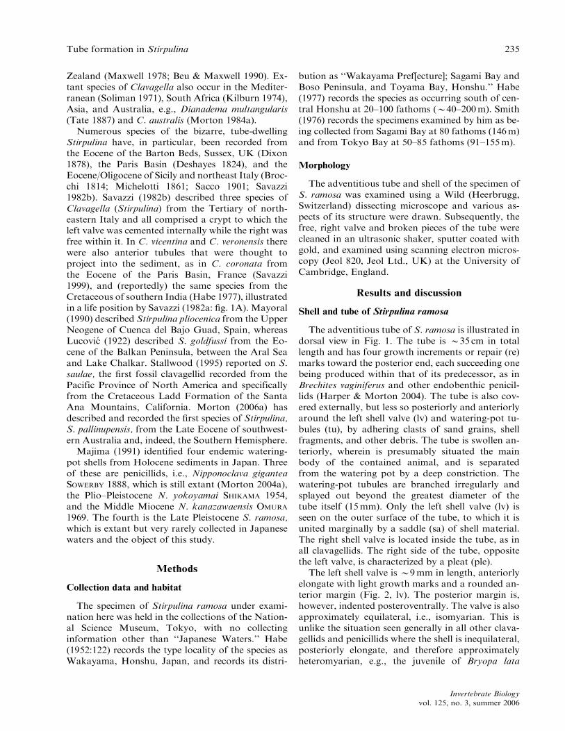
The adventitious tube has been cut through dorso-ventrally and lengthwise so that the internal structureof the left side can be seen (Fig. 6). The left valve (lv)is united with the fabric of the tube marginally (lvm).The adventitious tube overarches the left valve at thedorsal crests to create a cavity (doc) above it. The
Fig. 3. Anterior end of the adventitious tube as seen from the right lateral aspect. For abbreviations see Appendix.
Fig. 4. A. Detail of the external structure of the adventitious tube. B. Posterior end of the adventitious tube as seen from
the posterior aspect. C. A section through the adventitious tube just posterior to the internal lip. For abbreviations see
Appendix.
238 Morton
Invertebrate Biologyvol. 125, no. 3, summer 2006

shell itself has an opisthodetic primary ligament (prl)set upon resilifers. There is a relatively large anterioradductor muscle scar (aa), and the anterior end of thepallial line (pl) arises from this. Amazingly, however,the posterior adductor muscle scar (pa), the pallialsinus (ps), and the greatly thickened ventral and pos-terior sections of the pallial line are located on theinternal surface of the tube—not the shell! Posteriorto the posterior adductor muscle scar is the internallyprojecting lip (li) of the tube that it encircles except atthe right side. The constriction (con) between thetube and the watering pot is internally stained brown.
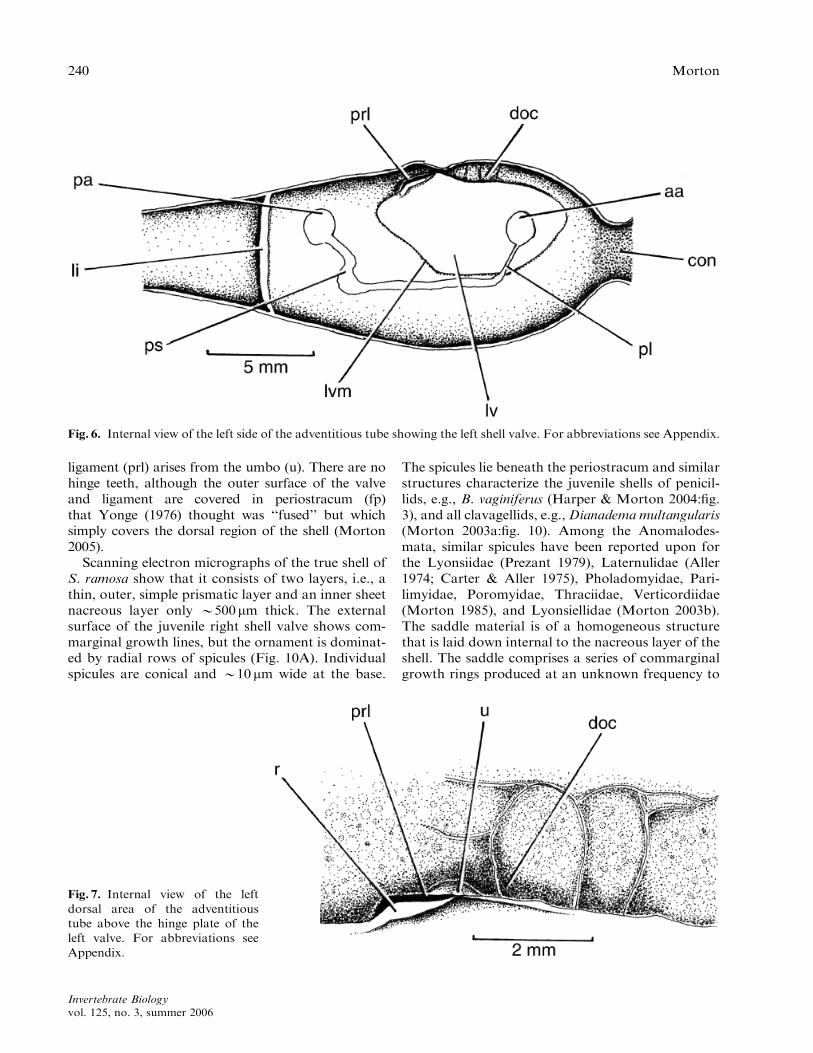
Figure 7 illustrates the dorsal edge of the leftshell valve. From the umbo (u) arises the ligament(prl) set upon a resilifer (r). Because the dorsal crests
overarch the dorsal regions of the left valve, a cavity(doc) is created behind them so that unlike some otherclavagellids, e.g., B. lata (Morton 2005), this compo-nent of the valve margin is not united with the tube.The dorsal cavity is internally configured to match theexternal pattern of anterior and posterior dorsal crestsand adhering clasts. The right shell valve is illustratedto the same scale and in the correct orientation tomatch that of the left in Fig. 8. As noted above, the leftvalve is B9mm in overall length whereas the right isB16mm and has, on its inner surface, the scars of theadductor muscles (aa, pa), a pallial line (pl), a shallowpallial sinus (ps), and, unusually, a shallow pedal gape(pg) sinus. The dorsal margin of the right valve is il-lustrated in Fig. 9. As in the left valve, the primary
Fig. 5. Watering pot as seen from
the anterior aspect. For abbrevia-
tions see Appendix.
Tube formation in Stirpulina 239
Invertebrate Biologyvol. 125, no. 3, summer 2006
ligament (prl) arises from the umbo (u). There are nohinge teeth, although the outer surface of the valveand ligament are covered in periostracum (fp)that Yonge (1976) thought was ‘‘fused’’ but whichsimply covers the dorsal region of the shell (Morton2005).
Scanning electron micrographs of the true shell ofS. ramosa show that it consists of two layers, i.e., athin, outer, simple prismatic layer and an inner sheetnacreous layer only B500mm thick. The externalsurface of the juvenile right shell valve shows com-marginal growth lines, but the ornament is dominat-ed by radial rows of spicules (Fig. 10A). Individualspicules are conical and B10mm wide at the base.
The spicules lie beneath the periostracum and similarstructures characterize the juvenile shells of penicil-lids, e.g., B. vaginiferus (Harper & Morton 2004:fig.3), and all clavagellids, e.g.,Dianadema multangularis(Morton 2003a:fig. 10). Among the Anomalodes-mata, similar spicules have been reported upon forthe Lyonsiidae (Prezant 1979), Laternulidae (Aller1974; Carter & Aller 1975), Pholadomyidae, Pari-limyidae, Poromyidae, Thraciidae, Verticordiidae(Morton 1985), and Lyonsiellidae (Morton 2003b).The saddle material is of a homogeneous structurethat is laid down internal to the nacreous layer of theshell. The saddle comprises a series of commarginalgrowth rings produced at an unknown frequency to
Fig. 6. Internal view of the left side of the adventitious tube showing the left shell valve. For abbreviations see Appendix.
Fig. 7. Internal view of the left
dorsal area of the adventitious
tube above the hinge plate of the
left valve. For abbreviations see
Appendix.
240 Morton
Invertebrate Biologyvol. 125, no. 3, summer 2006

create the unusual structure between shell and tube.The adventitious tube has a single-layered structurecomprising stacks (Taylor et al. 1973) of flat platycrystals (Fig. 10B), except at the posterior, siphonalend where layers are laid down as either growth orrepair increments, between which are held clasts ofsand grains and organic material, as in B. vaginiferus(Harper & Morton 2004).
The internal surface of the watering-pot end of theadventitious tube (Fig. 11) is stained dark brown.The broken wall of the tube (bt) is not uniformlycontinuous as in B. vaginiferus (Harper & Morton2004) because, on the right side, the ventrolateralcomponent (vtl) is folded under the dorsolateral one(dtl) at the position of the external pleat (pl). Theventrolateral component of the tube is, moreover,
formed into a sinusoidal crest within the basal righthalf of the watering pot, accounting for the sinus inthe pedal gape of the right valve pallial line scar atthis location (Fig. 8, pg) but not the left. The pleatalso unites with and likely creates the dorsoventrallyaligned pedal gape (pg). The central (ctu) and lateraltubules (ltu) open into the cavity of the watering pot,presumably effecting communication between the in-terstitial water of the burrow heading and the mantlecavity of the contained bivalve via the pedal gape, asin the penicillids B. vaginiferus and Foegia novae-zelandiae (Morton 2002a, 2004c).
Formation of the tube and the watering pot
Savazzi (1982a:fig. 17.13) illustrated how the wa-tering pots of the fossils S. coronata, S. vicentina, andS. bacillus are formed in a manner different from thatof penicillids, by progressive encasement of the rightvalve and the contained animal inside the tube. Themain fabric of the tube is formed first, presumablyfrom the general outer surface of the mantle, and theleft valve is progressively incorporated into its fabric.At this stage, folds of mantle tissue secrete calciumcarbonate at their leading edges and gradually forminto five elements that eventually coalesce but leave apleat extending from the also enlarged ‘‘watering-pot’’ tubules. Such a process mirrors the situationdescribed for S. coronata by Morton (2006a,b) and
Fig. 8. Internal views of the right
(above) and left (below) shell valves.
For abbreviations see Appendix.
Fig. 9. Internal view of the hinge plate of the right shell
valve. For abbreviations see Appendix.
Tube formation in Stirpulina 241
Invertebrate Biologyvol. 125, no. 3, summer 2006

thus, eventually, a watering pot is produced that isnot a bilaterally symmetrical disc as in penicillids, butis O shaped. This study of S. ramosa refines this viewbecause, for the first time, the tube of a species ofStirpulina can be viewed internally.
Figure 12 presents a diagrammatic scheme basedaround the secretion of the tube, notably the water-ing pot, from not five mantle lobes (Savazzi 1982a)
but two. Against the template of the burrow wall anda film of periostracum that is produced first, a dorsalextension of the mantle lobe (dtl) arising from theanterior and posterior margins of the left mantlemargin progressively extends outward, covering thedorsal surface of the burrow, secreting calcium car-bonate as it grows, and eventually extending onto theright side of the burrow wall. Simultaneously, the
Fig. 10. Scanning electron micro-
scope micrographs of (A) the dorsal
area of the right valve showing the
shell spinules and (B) a broken
surface of the adventitious tube
(scale bars 100 and 10 mm, respec-
tively).
Fig. 11. Internal view of the
watering pot as seen from the
posterior aspect. For abbreviations
see Appendix.
242 Morton
Invertebrate Biologyvol. 125, no. 3, summer 2006

ventral extension of the mantle lobe (vtl) also extendsfrom the ventral mantle margin, secreting calciumcarbonate that it lays down against the ventralburrow wall and finally extending upward to curveinternal to the dorsal, downward-growing mantlecomponent. This forms the pleat (ple) in the rightside of the tube and the watering pot. The ventralmantle lobe also produces the internal border of thepedal gape at the point of internal union of the twosecreting surfaces within the watering pot. The ven-tral mantle lobe secretes the sinusoidal crest that isformed internally on the right side of the base of thewatering pot. Finally, the two secreting surfaces alsocreate the central (ctu) and lateral (ltu) tubules of thewatering pot, possibly around papillae that form ontissues internal to the watering pot, as in B. vagini-ferus (Harper & Morton 2004).
Tube construction: Stirpulina versus Brechites
All known penicillids and clavagellids, including,as herein shown, S. ramosa, possess calcareous spic-
ules on their shells, particularly posteriorly. Suchspicules have been reported upon for a number ofother anomalodesmatans by Harper et al. (2000),most notably representatives of the Lyonsiidae (Prez-ant 1979). Morton (2006a) has argued that the an-cestors of the Clavagellidae and Penicillidae probablyarose from lyonsiid ancestors, albeit at differenttimes, i.e., the Mesozoic and Cenozoic, respectively.The superficial similarity in tube structure betweenStirpulina (Clavagellidae) and the various endo-benthic representatives of the Penicillidae, such asB. vaginiferus, has been responsible for all watering-pot shells being considered by some, most notablySmith (1998), to belong to the same family—theClavagellidae. There is, however, a major distinctionbetween clavagellids and penicillids. Representativesof the Penicillidae all have a shell that is of a relativelyuniform size among all individuals of a given species(Morton 1984b, 2002a, 2004a,b, 2006b). This is be-cause the tube is constructed only once when indi-viduals reach an adult body size even though the shellis of juvenile dimensions (Harper & Morton 2004).
Fig. 12. Diagrammatic illustration
of how the watering pot is secreted.
For abbreviations see Appendix.
Tube formation in Stirpulina 243
Invertebrate Biologyvol. 125, no. 3, summer 2006

Morton (2004a) considered this unusual situation torepresent an example of heterochrony. Harper &Morton (2004) also demonstrated, for B. vaginiferus,that having attained a valve height ofB3mm, a sad-dle area is secreted from the valve margins. Thisbrings the shell height generally to B10mm, but ismuch bigger in Nipponoclava gigantea (Morton2004a). Tube formation commences at this size andbegins with extensive expansion and growth of thefused mantle lobes far beyond the diminutive shellmargin such that the valves are forced apart untilthey are splayed widely (like a butterfly). The rela-tively huge body (in comparison with the tiny shell)contained in a continuous bag of periostracum mustresemble the myoid Panopea as illustrated by Yonge(1971:fig. 23). As the animal grows, the pliable per-iostracal bag is pressed closely against the surround-ing sediments; clasts adhere to it but, importantly, theburrow so formed becomes the template againstwhich the tube is now secreted also, as with the per-iostracum around it, by the greatly inflated mantle.The tube once hardened thus has the general form ofthe sediments that encased it. This has been demon-strated for B. vaginiferus by Morton (2002a), whoshowed that the tube is long, thin, and smooth in in-dividuals inhabiting intertidal sands and short, squat,and distorted in conspecifics in adjacent rocky areas.All authors (Gray 1858b; Lacaze-Duthiers 1883;Lamy 1923; Purchon 1968; Taylor et al. 1973; Smith1978; Savazzi 1982a; Morton 1984b, 2002a,b) agreethat the adventitious tube is constructed but once andin a single episode, and that there is no capacity forfurther expansion of tube diameter or modification ofit (save for repairs) except, importantly, at the pos-terior, siphonal end, which may be either extended orrepaired; such structural features are visible as inter-nally secreted increments. Following damage, how-ever, even within the burrow, the tube can be repairedanywhere (Harper & Morton 2004).
The clavagellid B. lata has been reported to be ableto grow within its crypt (Savazzi 2000). If so, thenthis is the most important piece of evidence that theclavagellid tube is produced in a wholly differentmanner from that of penicillids, as described byHarper &Morton (2004). Such growth is herein dem-onstrated for S. ramosa, but has resulted in the mostunusual situation whereby, because the internal rightvalve continues to grow, the positions of the adduc-tor muscle and pallial line scars also grow outward—as is typical of the Bivalvia. Because the left valve,however, is, earlier in ontogeny, fused marginally tothe adventitious tube and hence cannot grow further,the adductor muscles and pallial line scars becomelocated on the tube beyond the valve margin. Thus,
not only is the mechanism of tube formation inS. ramosa wholly different from that in the penicillidB. vaginiferus (Harper &Morton 2004), so is the con-tinued growth of the right valve beyond that of thefinal size of the left. Because the clavagellid rightvalve is also free within the tube, it can, unlike inpenicillids, be involved in the exchange of supernat-ant and interstitial water with the mantle cavity. Thisis not possible in penicillids, resulting in the adoptionof amazing adaptations, as in Kendrickiana veitchi, tomediate such fluid exchanges (Morton 2004b).
Stirpuliniola: a junior synonym of Stirpulina
This study first demonstrates that the adventitioustube of S. ramosa possesses a pleat down its rightside. Savazzi (2005:fig. 5) also shows that, like S.ramosa, the Eocene fossil S. bacillus had not only aright valve that is significantly larger than the left butalso a groove on the right side of the tube that ex-tends to the anterior pedal gape. The same situationoccurs in the Eocene fossil S. coronata (Morton2006a), and thus all the above species can be regard-ed as congeneric. There was thus no justification forKuroda & Habe (1971) to introduce the name Stir-puliniola for the Japanese species, and this can there-fore be relegated to a junior synonym of Stirpulina.
Comparison of clavagellid genera
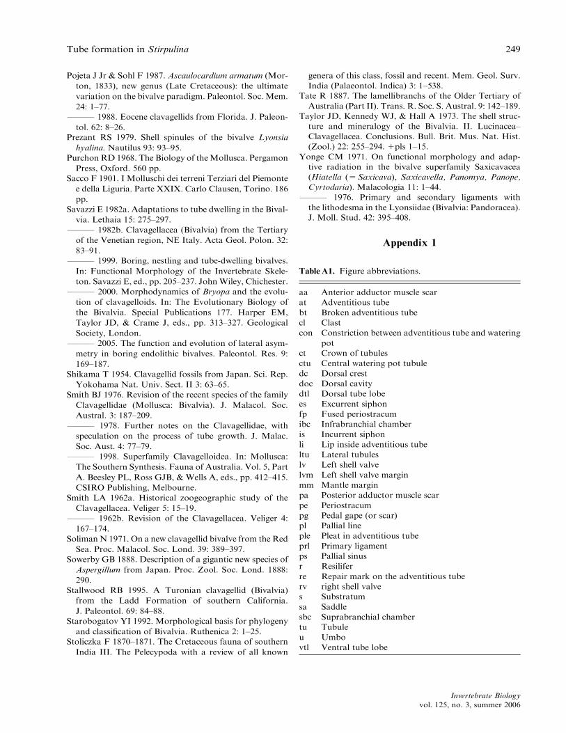
Notwithstanding the above, this study of S. ram-osa finally enables a comparison to be made of thefour genera of extant clavagellids currently recog-nized, i.e., Dacosta, Bryopa, Dianadema, and Stir-pulina. This is only possible in terms of shell andadventitious tube/crypt structure, however, becauseno intact specimens of S. ramosa are extant to studythe internal tissues (Table 1). In representatives of allgenera, only the left valve is fused either to the bur-row wall (Dacosta and Bryopa) or into the fabric ofan adventitious tube (Dianadema and Stirpulina).There is an opisthodetic ligament in Stirpulina,Dianadema, and Bryopa that is lost in the endolithicadult of B. aligamenta (Morton 2005), but an internalamphidetic ligament in Dacosta that is located be-tween chondrophores (Morton 1984a). Similarly, theligament is covered by a two-layered periostracum,but there is no ventral lithodesma. The shell compris-es the same two layers (inner nacre, outer prismatic)and the juveniles of representatives of all genera pos-sess external shell spicules. There is much evidencetherefore to suggest that the four genera are all lo-cated correctly in the Clavagellidae. In terms of theirinternal anatomy, the four genera show similarities
244 Morton
Invertebrate Biologyvol. 125, no. 3, summer 2006

also: all are strongly dimyarian with a thick pallialline, and a deep pallial sinus to accommodate fusedsiphons, but, presumably because of their cryptic life-
styles, there are reduced pedal retractor muscles inboth Dacosta and Dianadema (vestigial posteriorpedal retractors) whereas they are absent in Bryopa
Table 1. Comparison of shell, adventitious tube/crypt, and anatomical characters (where known) of extant genera of the
Clavagellidae (Clavagelloidea).
Character Genus
Dacosta Dianadema Bryopa Stirpulina
Shell Adult only Adult only Adult only Adult only
Valves Left valve cemented
to crypt; right free
internally
Left valve cemented
to crypt; right free
internally
Left valve cemented
to crypt; right free
internally
Left valve cemented
to tube; right free
internally
Spicules Present Present Present Present
Ligament Internal, amphidetic
between
chondrophores
Internal,
opisthodetic
Reduced/lost;
internal
opisthodetic in the
juvenile
Internal,
opisthodetic
Lithodesma Absent Absent Absent Absent
Periostracum Two layers Two layers Two layers Unknown
Microstructure: outer layer Prismatic Prismatic Prismatic Prismatic
Microstructure: inner sheet Nacre Nacre Nacre Nacre
Crypt/adventitious tube Present Present Absent Present
Adventitious tube Posterior extension
of crypt
Cemented to
substratum
Posterior extension
of crypt
An adventitious
tube
Watering pot Absent Absent Absent Present
Anterior tubules Present Present Absent Present
Juvenile:
Ontogeny Normal Normal Unique Normal
Adductor muscles Dimyarian Dimyarian Dimyarian Dimyarian
Pallial sinus Present Present Present Present
Adult:
Adductor muscles Present Present Present Present
Pedal retractor muscles Anterior and
posterior both
present
Anterior present.
Posterior as a
remnant
Absent Probably absent
Pedal disc Absent Absent Absent Unknown
Pericardial proprioreceptor Absent Present Absent Unknown
Ctenidial ciliation Type E Type E Type E Unknown
Ctenidial/labial palp junction Category 3 Category 3 Category 3 Unknown
Pallial fusion Type C Type C Type C Unknown
Fourth pallial aperture Absent Absent Absent Unknown
Pedal gape Present Present Present Unknown
Radial mantle glands Present Present Absent Unknown
Siphonal sense organs Present Present Unknown Unknown
Statocysts Type B3 Absent Unknown Unknown
Tube formation in Stirpulina 245
Invertebrate Biologyvol. 125, no. 3, summer 2006

and possibly too in Stirpulina. Other comparisons ofthe four genera must await discovery of an intactspecimen of S. ramosa, but representatives of thethree other genera all show, as one would expect,anatomical adaptations to their particular lifestyles.
Significance of dorsal tube crests
Some species of penicillid watering-pot shells, mostnoticeably K. veitchi (Morton 2004b), produce solidanterior and posterior calcium carbonate concretionsthat progressively unite to eventually cover andthereby conceal, in this case, the two shell valvesthat are incorporated into the fabric of the adventi-tious tube. The situation is, however, different inS. ramosa in that the concretions are not solid, buthollow, so that the dorsal margin of the left valve isnot, unlike other areas, united into the fabric of thetube. The importance of this is that, because it is notunited dorsally but is within the cavity (doc) createdby the dorsal crests, the right valve can still bemoved, i.e., opened and closed against that of thefixed left valve. The ligament therefore retains its
typically bivalve function to act as the opening thrustagainst the agonistic contraction forces generated bythe adductor muscles (Fig. 13A). As in any typicalbivalve, therefore, water can be inhaled via the in-current siphon (is) and exhaled via the excurrent si-phon (es). In so doing, water passes from theinfrabranchial (ibc) to the suprabranchial chamber(sbc), allowing the filtration, collection, and ingestionof organic particulates by the ctenidia. Such move-ment of water is effected by the pumping action cre-ated by the interplay of adductor muscles (aa/pa) andligament. That is, the free right valve, as the onlymovable solid object in the composition of the ad-ventitious tube complex, acts as a paddle interactingwith the fixed surfaces of the left valve and adventi-tious tube. In the case of S. ramosa, moreover, thesame forces can probably pump water into and outof the burrow heading via the pedal gape (pg) andwatering-pot tubules (ltu), as in B. vaginiferus andFoegia novaezelandiae (Morton 2002a, 2004c).
The significance of the dorsal cavity is illustrated inFig. 13B, which represents a diagrammatic transversesection through the tube, shell valves, and body of
Fig. 13. Diagrammatic transverse sections through the adventitious tubes of (A) Stirpulina ramosa and (B) Dianadema
multangularis. Source adapted from Morton (2003). For abbreviations see Appendix.
246 Morton
Invertebrate Biologyvol. 125, no. 3, summer 2006
D. multangularis (Morton 2003a: fig. 16). In this spe-cies, the dorsal crest is open to form the dorsal crownof tubules (ct) that characterize its apex. Thus, the sit-uation in S. ramosa previses that seen in D. mu-ltangularis, wherein a dorsal cavity created to allowvalve movement in the former adventitious tube-dwell-ing clavagellid has been opened and, as a consequence,allowed adoption of an endobenthic lifestyle in thelatter through greater efficiency in the aeration of itscrypt. This adaptationmay have been taken to an evengreater extreme by the fossil Ascaulocardium armatum(Pojeta & Sohl 1987), wherein the dorsal crown oftubules seen in D. multangularis is hypertrophied toencircle the adventitious tube (Pojeta & Sohl 1987:fig. 6). Significantly, Pojeta & Sohl (1987: fig. 1–6)describe and illustrate a triple junction pleat on theright-hand side of the A. armatum adventitious tube.
Clavagellidae and Penicillidae: convergent evolution
In a recent study of the endolithic B. aligamenta,Morton (2005) argued that this genus and its extantallies, Dacosta, Dianadema, and Stirpulina, whichconstitute the modern Clavagellidae, are distinctlydifferent from the watering-pot shells of the Pen-icillidae, and thus the two families constitute a re-markable example of convergent evolution. This isevidenced in two ways. First, although, unlike theclavagellidsDacosta andBryopa, no representative ofthe Penicillidae has adopted an endolithic lifestyle,both families possess epibenthic, cemented genera,i.e.,Dianadema (Clavagellidae) (Morton 2003a; Oliv-er & Holmes 2004) and Humphreyia (Penicillidae)(Morton 2002b). Second, it is in the adoption of anendobenthic lifestyle by Stirpulina and representa-tives of the Penicillidae—Brechites, Nipponoclava,Kendrickiana, Foegia, and Penicillus (Morton1984b, 2002a, 2004a–c, 2006b)—that the most amaz-ing example of convergent evolution is seen. In un-derstanding such a convergence, however, it is nowclear that the endobenthic lifestyle has not been asuccessful evolutionary development in the Clavagell-idae, only S. ramosa surviving to the present day, butleaving behind a rich array of similar Mesozoic wa-tering-pot fossils virtually worldwide. Conversely,however, there are few fossils of the endobenthicCenozoic Penicillidae, possibly because of the fragil-ity of the adventitious tubes, but these watering-potshells have radiated and evolved into some of themost amazing and bizarre bivalves ever seen.
This study shows that Stirpulina and the endo-benthic penicillids have achieved their endobenthictube-dwelling lifestyles in quite different ways, but anintact specimen of S. ramosa is needed for examina-
tion to try and discover why its Mesozoic ancestorswere so singularly unsuccessful in comparison withthe later evolving (Cenozoic) Penicillidae, especiallyin tropical seas.
Acknowledgments. I am grateful to Dr. Hiroshi Saitoof the National Science Museum, Tokyo, for permissionto examine the specimen of Stirpulina ramosa hereinreported upon and to Dr. E.M. Harper, University ofCambridge, for help in taking the SEM micrographsreproduced in Fig. 10.
References
Aller RC 1974. Prefabrication of shell ornament in the bi-
valve Laternula. Lethaia 7: 43–56.
Appukuttan KK 1974. Rediscovery of Clavagella (Bryopa)
lata (Clavagellidae, Bivalvia) from the Gulf of Mannar,
southeast coast of India. J. Malacol. Soc. Austral. 3:
19–24.
Beu AG & Maxwell PA 1990. Cenozoic Mollusca of New
Zealand. N. Z. Geol. Surv. Paleontol. Bull. 58: 1–518.
Brocchi G 1814. Conchiologica fossile subapennina con
osservazioni geologiche sugli Apennini e sul suolo adia-
cente. Stamperia Reale, Milan. 1012 pp.
Broderip WJ 1834. On Clavagella. Proc. Zool. Soc. Lond.
(1833–1834) 2: 115–117.
Bruguiere M 1789. Encyclopedie Methodique; histoire
naturelle des vers. Vol. 1(XV), 126–130. genus 33.
Panckoucke, Paris.
Carter JG & Aller RC 1975. Calcification of the bivalve
periostracum. Lethaia 8: 315–320.
Deshayes G-P 1824. Description des Coquilles Fossiles des
environs de Paris. Tome Premiere, Conchiferes, Paris.
814 pp.
Dixon F 1878. The Geology of Sussex; or the Geology and
Fossils of the Tertiary and Cretaceous Formations of
Sussex. W.J. Smith, Brighton. 422 pp.
D’Orbigny AD 1843–1847. Terrains Cretaces. Vol. 3.
Lamellibranches. Victor Massin, Paris. 807 pp.
Dunker G 1882. Index Molluscorum Maris Japonici.
Casselis Cattorum. T. Fischer, Stuttgart, Germany. 301 pp.
Forbes E 1846. Report on the fossil invertebrata from
southern India, collected by Mr. Kaye andMr. Cunliffe.
Trans. Geol. Soc. Ser. 2 VII: 97–174. 1unpaginated map
1VII–IX plate.
Gray JE 1847. A list of the genera of recent Mollusca, their
synonyms and types. Proc. Zool. Soc. Lond. 15: 129–219.
FFF 1858a. On the families of Aspergillidae, Gas-
trochaenidae, and Humphreyadae. Proc. Zool. Soc.
Lond. 26: 307–318.
FFF 1858b. On the development of the shell and tube in
Aspergillum. Ann. Mag. Nat. Hist. 1: 423–426.
Habe T 1952. Illustrated Catalogue of Japanese Shells. 18.
Pholadomyidae, Clavagellidae, Pandoridae, Juliidae,
and Condylocardiidae in Japan. Publisher unknown.
FFF 1977. Systematics of Mollusca in Japan. Bivalvia
and Scaphopoda. Hokururyu-kan, Tokyo. 372 pp.
Tube formation in Stirpulina 247
Invertebrate Biologyvol. 125, no. 3, summer 2006
Harper EM & Morton B 2004. Tube construction in
the watering pot shell Brechites vaginiferus (Bivalvia;
Anomalodesmata; Clavagelloidea). Acta Zool. 85:
149–161.
Harper EM, Hide EA, & Morton B 2000. Relationships
between the extant Anomalodesmata: a cladistic test. In:
The Evolutionary Biology of the Bivalvia. Special Pub-
lications 177. Harper EM, Taylor JD, & Crame JA, eds.,
pp. 129–143. Geological Society, London.
Jones DS & Nicol D 1989. Eocene clavagelloids (Mollusca:
Pelecypoda) from Florida: the first documented occur-
rence in the Cenozoic of the western hemisphere. J. Pale-
ontol. 63: 320–323.
Keen M & Smith LA 1969. Superfamily Clavagellacea
d’Orbigny 1844. In: Treatise on Invertebrate Paleontol-
ogy, Part N, Vol. 2: Mollusca 6, Bivalvia. Moore RC,
ed., pp. N857–N859. Geological Society of America &
University of Kansas Press, Lawrence.
Kilburn RN 1974. A new species of Clavagella s. s. (Bival-
via: Clavagellidae) from Natal, South Africa. J. Conch-
yliol. 111: 89–92.
Kuroda T & Habe T 1971. Sea shells of Sagami Bay.
Hosokawa, Tokyo.
Lacaze-Duthiers H 1883. Anatomie de l’Arrosoir (Asp-
ergillum dichotomum, L. Reeve). Arch. Zool. Exp. Gen.
2: 1–68.
LamyE 1923. Les Clavagelles et Arrosoirs de laMer Rouge
(d’apres les materiaux recueillis par le Dr. Jousseaume).
Bull. Mus. Nat. d’ Hist. Nat. Paris. 19: 104–107.
Little C 2005. Mysterious fossils. Palaeontol. Newslett. 59:
66–67.
Lucovic MT 1922. The Eocene molluscan fauna from the
area between the Aral Sea and Lake Chalkar and its im-
portance. Ann. Geol. Penin. Balkan. 8: 22–82.
Majima R 1991. Redescription and mode of occurrence of
Nipponoclava yokoyamai (Shikama 1954), (Clavagelli-
dae: Bivalvia) from the Plio–Pleistocene warm-
water fauna in Japan. Proc. Paleontol. Soc. Jpn. 162:
781–793.
Maxwell PA 1978. Taxonomic and nomenclatural notes on
some New Zealand Cenozoic Mollusca, with descrip-
tions of new taxa. N. Z. J. Zool. 5: 15–46.
Mayoral E 1990. Bivalvia Clavagellacea (Stirpulina,
pliocenica nov. sp.) del Neogeno superior de la Cuenca
del Bajo Guadalquivir. Treb. Mus. Geol. Barcelona 1:
117–134.
Michelotti G 1861. Etudes sur le Miocene Inferieur de
l’Italie septentrionale. Hollandsche Maatschappij der
Wetenschappen. Haarlam. Natuurkundige Verhandelin-
gen. Ser. 2 15: 1–184.
Morton B 1983. The biology and functional morphology of
Eufistulana mumia (Bivalvia: Gastrochaenacea). J. Zool.
Lond. 200: 381–404.
FFF 1984a. The biology and functional morphology
of Clavagella australis (Bivalvia: Anomalodesmata).
J. Zool. Lond. 202: 489–511.
FFF 1984b. Adventitious tube construction in Brechites
vaginiferus (Bivalvia: Anomalodesmata: Clavagellacea)
with an investigation of the juvenile of ‘Humphreyia
strangei’. J. Zool. Lond. 203: 461–484.
FFF 1985. Adaptive radiation in theAnomalodesmata. In:
The Mollusca. Vol. 10. Evolution. Trueman ER & Clark
MR, eds., pp. 405–459. Academic Press, New York.
FFF 2002a. Biology and functional morphology of the
watering pot shell Brechites vaginiferus (Bivalvia: Anoma-
lodesmata: Clavagelloidea). J. Zool. Lond. 257: 545–562.
FFF 2002b. The biology and functional morphology
of Humphreyia strangei (Bivalvia: Anomalodesmata:
Clavagellidae): an Australian cemented watering pot
shell. J. Zool. Lond. 258: 11–25.
FFF 2003a. The biology and functional morphology of
Dianadema gen. nov. multangularis (Tate, 1887) (Bival-
via: Anomalodesmata: Clavagellidae). J. Zool. Lond.
259: 389–401.
FFF 2003b. The biology and functional morphology of
Bentholyonsia teramachii (Bivalvia: Lyonsiellidae): clues
to the origin of predation in the deep water Anoma-
lodesmata. J. Zool. Lond. 261: 363–380.
FFF 2004a. The biology and functional morphology of
Nipponoclava gigantea: clues to the evolution of tube
dwelling in the Penicillidae (Bivalvia: Anomalodesmata:
Clavagelloidea). J. Zool. Lond. 264: 1–15.
FFF 2004b. The biology and functional morphology of
Kendrickiana gen. nov. veitchi (Bivalvia: Anomalodes-
mata: Clavagelloidea) from southern Australia. Invert.
Biol. 123: 244–259.
FFF 2004c. The biology and functional morphology
of Foegia novaezelandiae (Bivalvia: Anomalodesmata:
Clavagelloidea) from Western Australia. Malacologia
46: 37–55.
FFF 2005. Biology and functional morphology of a new
species of endolithic Bryopa (Bivalvia: Anomalodes-
mata: Clavagelloidea) from Japan and a comparison
with fossil species of Stirpulina and other representatives
of the Clavagellidae. Invert. Biol. 124: 202–219.
FFF 2006a. A new species and first record of the endo-
benthic clavagellid Stirpulina (Bivalvia: Anomalodes-
mata) from the Late Eocene of southern Western Aus-
tralia. Alcheringa 30: 101–108.
FFF 2006b. The functional morphology of Penicillus phil-
ippinensis (Anomalodesmata: Clavagelloidea: Penicillidae)
and the evolution of an unique muscular system in the
Bivalvia. Rec. Western Australian Mus. 23: 175–192.
Morton SG 1833. Supplement to the ‘‘Synopsis of the or-
ganic remains of the Ferruginous Sand Formation of the
United States’’. Am. J. Sci. 24: 128–132.
Nicol D 1968. A new Meiocardia (Pelecypoda, Glossidae)
from the Eocene of Florida. Nautilus 81: 89–93.
Oliver PG & Holmes AM 2004. Cryptic bivalves with
descriptions of new species from the Rodrigues lagoon.
J. Nat. Hist. 38: 3175–3227.
Omura K 1969. Fossil Penicillus from Ishikawa and
Kagoshima Prefectures, Japan. Ann. Sci., Kanazawa
Univ. Coll. Lib. Arts. Part 2 6: 25–39.
Owen R 1835. On the anatomy of Clavagella Lam. Trans.
Zool. Soc. Lond. 269–274.
248 Morton
Invertebrate Biologyvol. 125, no. 3, summer 2006
Pojeta J Jr & Sohl F 1987. Ascaulocardium armatum (Mor-
ton, 1833), new genus (Late Cretaceous): the ultimate
variation on the bivalve paradigm. Paleontol. Soc. Mem.
24: 1–77.
FFF 1988. Eocene clavagellids from Florida. J. Paleon-
tol. 62: 8–26.
Prezant RS 1979. Shell spinules of the bivalve Lyonsia
hyalina. Nautilus 93: 93–95.
Purchon RD 1968. The Biology of theMollusca. Pergamon
Press, Oxford. 560 pp.
Sacco F 1901. I Molluschi dei terreni Terziari del Piemonte
e della Liguria. Parte XXIX. Carlo Clausen, Torino. 186
pp.
Savazzi E 1982a. Adaptations to tube dwelling in the Bival-
via. Lethaia 15: 275–297.
FFF 1982b. Clavagellacea (Bivalvia) from the Tertiary
of the Venetian region, NE Italy. Acta Geol. Polon. 32:
83–91.
FFF 1999. Boring, nestling and tube-dwelling bivalves.
In: Functional Morphology of the Invertebrate Skele-
ton. Savazzi E, ed., pp. 205–237. JohnWiley, Chichester.
FFF 2000. Morphodynamics of Bryopa and the evolu-
tion of clavagelloids. In: The Evolutionary Biology of
the Bivalvia. Special Publications 177. Harper EM,
Taylor JD, & Crame J, eds., pp. 313–327. Geological
Society, London.
FFF 2005. The function and evolution of lateral asym-
metry in boring endolithic bivalves. Paleontol. Res. 9:
169–187.
Shikama T 1954. Clavagellid fossils from Japan. Sci. Rep.
Yokohama Nat. Univ. Sect. II 3: 63–65.
Smith BJ 1976. Revision of the recent species of the family
Clavagellidae (Mollusca: Bivalvia). J. Malacol. Soc.
Austral. 3: 187–209.
FFF 1978. Further notes on the Clavagellidae, with
speculation on the process of tube growth. J. Malac.
Soc. Aust. 4: 77–79.
FFF 1998. Superfamily Clavagelloidea. In: Mollusca:
The Southern Synthesis. Fauna of Australia. Vol. 5, Part
A. Beesley PL, Ross GJB, & Wells A, eds., pp. 412–415.
CSIRO Publishing, Melbourne.
Smith LA 1962a. Historical zoogeographic study of the
Clavagellacea. Veliger 5: 15–19.
FFF 1962b. Revision of the Clavagellacea. Veliger 4:
167–174.
SolimanN 1971. On a new clavagellid bivalve from the Red
Sea. Proc. Malacol. Soc. Lond. 39: 389–397.
Sowerby GB 1888. Description of a gigantic new species of
Aspergillum from Japan. Proc. Zool. Soc. Lond. 1888:
290.
Stallwood RB 1995. A Turonian clavagellid (Bivalvia)
from the Ladd Formation of southern California.
J. Paleontol. 69: 84–88.
Starobogatov YI 1992. Morphological basis for phylogeny
and classification of Bivalvia. Ruthenica 2: 1–25.
Stoliczka F 1870–1871. The Cretaceous fauna of southern
India III. The Pelecypoda with a review of all known
genera of this class, fossil and recent. Mem. Geol. Surv.
India (Palaeontol. Indica) 3: 1–538.
Tate R 1887. The lamellibranchs of the Older Tertiary of
Australia (Part II). Trans. R. Soc. S. Austral. 9: 142–189.
Taylor JD, Kennedy WJ, & Hall A 1973. The shell struc-
ture and mineralogy of the Bivalvia. II. Lucinacea–
Clavagellacea. Conclusions. Bull. Brit. Mus. Nat. Hist.
(Zool.) 22: 255–294. 1pls 1–15.
Yonge CM 1971. On functional morphology and adap-
tive radiation in the bivalve superfamily Saxicavacea
(Hiatella (5Saxicava), Saxicavella, Panomya, Panope,
Cyrtodaria). Malacologia 11: 1–44.
FFF 1976. Primary and secondary ligaments with
the lithodesma in the Lyonsiidae (Bivalvia: Pandoracea).
J. Moll. Stud. 42: 395–408.
Appendix 1
TableA1. Figure abbreviations.
aa Anterior adductor muscle scar
at Adventitious tube
bt Broken adventitious tube
cl Clast
con Constriction between adventitious tube and watering
pot
ct Crown of tubules
ctu Central watering pot tubule
dc Dorsal crest
doc Dorsal cavity
dtl Dorsal tube lobe
es Excurrent siphon
fp Fused periostracum
ibc Infrabranchial chamber
is Incurrent siphon
li Lip inside adventitious tube
ltu Lateral tubules
lv Left shell valve
lvm Left shell valve margin
mm Mantle margin
pa Posterior adductor muscle scar
pe Periostracum
pg Pedal gape (or scar)
pl Pallial line
ple Pleat in adventitious tube
prl Primary ligament
ps Pallial sinus
r Resilifer
re Repair mark on the adventitious tube
rv right shell valve
s Substratum
sa Saddle
sbc Suprabranchial chamber
tu Tubule
u Umbo
vtl Ventral tube lobe
Tube formation in Stirpulina 249
Invertebrate Biologyvol. 125, no. 3, summer 2006