structural basis for bivalent smac-mimetics recognition in the iap protein family
TRANSCRIPT

doi:10.1016/j.jmb.2009.04.033 J. Mol. Biol. (2009) 392, 630–644
Available online at www.sciencedirect.com
Structural Basis for Bivalent Smac-MimeticsRecognition in the IAP Protein Family
Federica Cossu1†, Mario Milani1,2†, Eloise Mastrangelo1,2,Patrice Vachette3, Federica Servida4, Daniele Lecis5, Giulia Canevari1,Domenico Delia5, Carmelo Drago6, Vincenzo Rizzo6,Leonardo Manzoni6,7, Pierfausto Seneci6,8, Carlo Scolastico6,8
and Martino Bolognesi1⁎
*Corresponding author. E-mail addr† F.C. and M.M. contributed equaAbbreviations used: IAP, inhibito
DIABLO, second mitochondria-derimotif; SAXS, small-angle X-ray scatPEG MME, polyethylene glycol mon
0022-2836/$ - see front matter © 2009 E
XIAP is an apoptotic regulator protein that binds to the effector caspases -3and -7 through its BIR2 domain, and to initiator caspase-9 through its BIR3domain. Molecular docking studies suggested that Smac-DIABLO mayantagonize XIAP by concurrently targeting both BIR2 and BIR3 domains; onthis basis bivalent Smac-mimetic compounds have been proposed andcharacterized. Here, we report the X-ray crystal structure of XIAP-BIR3domain in complex with a two-headed compound (compound 3) withimproved efficacy relative to its monomeric form. A small-angle X-rayscattering study of XIAP-BIR2BIR3, together with fluorescence polarizationbinding assays and compound 3 cytotoxicity tests on HL60 leukemia cellline are also reported. The crystal structure analysis reveals a network ofinteractions supporting XIAP-BIR3/compound 3 recognition; moreover,analytical gel-filtration chromatography shows that compound 3 forms a1:1 stoichiometric complex with a XIAP protein construct containing bothBIR2 and BIR3 domains. On the basis of the crystal structure and small-angle X-ray scattering, a model of the same BIR2-BIR3 construct bound tocompound 3 is proposed, shedding light on the ability of compound 3 torelieve XIAP inhibitory effects on caspase-9 as well as caspases -3 and -7. Amolecular modeling/docking analysis of compound 3 bound to cIAP1-BIR3domain is presented, considering that Smac-mimetics have been shown tokill tumor cells by inducing cIAP1 and cIAP2 ubiquitination anddegradation. Taken together, the results reported here provide a rationalefor further development of compound 3 as a lead in the design of dimericSmac mimetics for cancer treatment.
© 2009 Elsevier Ltd. All rights reserved.
1Department of BiomolecularSciences and Biotechnology,University of Milano, ViaCeloria 26, I-20133, Milano,Italy2CNR-INFM S3, NationalResearch Center onNanostructure and BioSystemsat Surfaces, Via Campi 213/A,41100-Modena, Italy3Institut de Biochimie et deBiophysique Moléculaire etCellulaire, UMR8619 CNRS,Université Paris-Sud, IFR115,F-91405 Orsay, France4Fondazione Matarelli,Department of MedicalFarmacology, Chemotherapyand Toxicology, University ofMilano, Via Vanvitelli 32,I-20129 Milano, Italy5Istituto Nazionale dei Tumori,Via Venezian 1, I-20133,Milano, Italy6Centro Interdisciplinare Studibio-molecolari e applicazioniIndustriali (CISI), University ofMilano, Via Fantoli 16/15,I-20138, Milano, Italy7CNR–ISTM, Via Fantoli16/15, I-20138, Milano, Italy
ess: [email protected] to this work.r of apoptosis protein; BIR, baculoviral IAP repeat; XIAP, X-linked IAP; Smac-ved activator of caspases - direct IAP binding protein with low pI; IBM, IAP bindingtering; ITC, isothermal titration calorimetry; NSD, normalized spatial discrepancy;omethyl ether.
lsevier Ltd. All rights reserved.
631XIAP/Smac-Mimetics Recognition
8Department of Organic andIndustrial Chemistry,University of Milano, ViaVenezian 21, I-20133, Milano,Italy
Received 10 February 2009;received in revised form15 April 2009;accepted 16 April 2009Available online22 April 2009
Keywords: inhibition of apoptosis; Smac-DIABLO; XIAP; cIAP; pro-apoptoticdrugs
Edited by I. WilsonIntroduction
The apoptotic process involves a cascade of eventsthat inactivate critical survival pathways in multi-cellular organisms.1 Inhibition of apoptosis canprevent physiological cell death, thus contributingto the development and progression of tumormalignancy.2 Apoptosis initiation and executionphases are both dependent on a subset of caspases(cysteine-dependent aspartyl-specific proteases3)that are regulated by a family of inhibitor ofapoptosis proteins (IAPs4). By direct interactionwith initiator and executioner caspases, IAPs canblock cell death in response to diverse stimuli.Therefore, these critical apoptosis regulators havebeen recognized as attractive targets for the devel-opment of innovative therapies in the treatment ofcancer and neurodegenerative diseases.5–7
The IAP proteins contain one to three zinc-bindingbaculoviral IAP repeat (BIR) domains that arerequired for anti-apoptotic activity.8 Most IAPs alsohave a C-terminal RING domain, endowed with E3ubiquitin ligase activity.9,10 The BIR domains host azinc-finger motif and are generally composed of fiveα-helices and a three-stranded β-sheet. Some IAPs,like cIAP1 and cIAP2, contain also a caspase-associated recruitment domain (CARD) locatedbetween the BIR3 domain and the C-terminal RINGdomain.11 cIAP1 and 2 are crucial regulators ofreceptor-mediated apoptosis,12,13 being able to inter-act with tumor necrosis factor receptor (TNFR) andtumor receptor-associated factors (TRAFs) throughthe first two α-helices of their BIR1 domains.14–16Another important member of the IAP family, theX-linked IAP (XIAP), is highly expressed in manyhuman tumor cell lines and in tumor-affectedtissues from patients.17,18 XIAP selectively targetsinitiator caspase-9 through its XIAP-BIR3 domain,19
whereas it inhibits both executioner caspase-320 andcaspase-721 by means of the XIAP-BIR2 domainand, particularly, the domain's N-terminal, knownas the linker region (lk).The inhibitory function of different IAPs is antag-
onized by the second mitochondria-derived activator
of caspases - direct IAP binding protein with low pI(Smac-DIABLO22,23), an elongated α-helical dimericprotein of 40 kDa, released from the mitochondria.Structural and binding studies show that Smac-DIABLO binds to the XIAP-BIR3 domain through aspecific IAP binding motif (IBM), built by its N-terminal tetrapeptide Ala-Val-Pro-Ile (AVPI).24 SmacAVPI competes directly with a similar tetrapeptide(ATPF) of the activated caspase-921 promoting therelease of the protease from the IAP complex. WhenIAPs are over-expressed, Smac-DIABLO may not besufficient to overcome the inhibitory effect on caspases.In such cases, synthetic IBM-like molecules (Smac-mimetics) may be employed to relieve caspasebinding, thereby promoting apoptosis in malignantcells. Along these lines of thought, and in order toavoid intrinsic limitations posed by peptide com-pounds, several laboratories have been activelydesigning Smac peptidomimetics and non-peptidomi-metics with improved binding affinities, proper cell-permeability, in vivo stability and bioavailability,25,26
as potential drug leads for new cancer therapeuticapproaches.Since in silico molecular docking studies suggested
that Smac-DIABLO may bind simultaneously toXIAP-BIR2 and -BIR3 domains,24 bivalent Smac-mimetic compounds, targeting XIAP-BIR2 and -BIR3domains, have been proposed and characterized.27,28
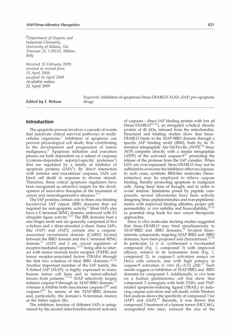
In particular, Li et al. synthesized a two-headedcompound (Fig. 1, compound 3) with improvedefficacy, relative to its monomeric form (Fig. 1,compound 2), in caspase-3 activation assays onHeLa cells extracts, and with high potency incaspase-9 activation in vitro (Ki=0.12 μM).27 Suchresults suggest co-inhibition of XIAP-BIR2 and -BIR3domains by compound 3. Additionally, in vivo testson a human glioblastoma cell line show thatcompound 3 synergizes with both TNFα and TNF-related apoptosis-inducing ligand (TRAIL) in indu-cing caspase activation and cell death, while Westernblot analysis shows the specificity of compound 3 forcIAP1 and cIAP2.27 Recently, it was shown thatcompound 3 treatment of a human tumor (HCC461),xenografted into mice, reduced the size of the
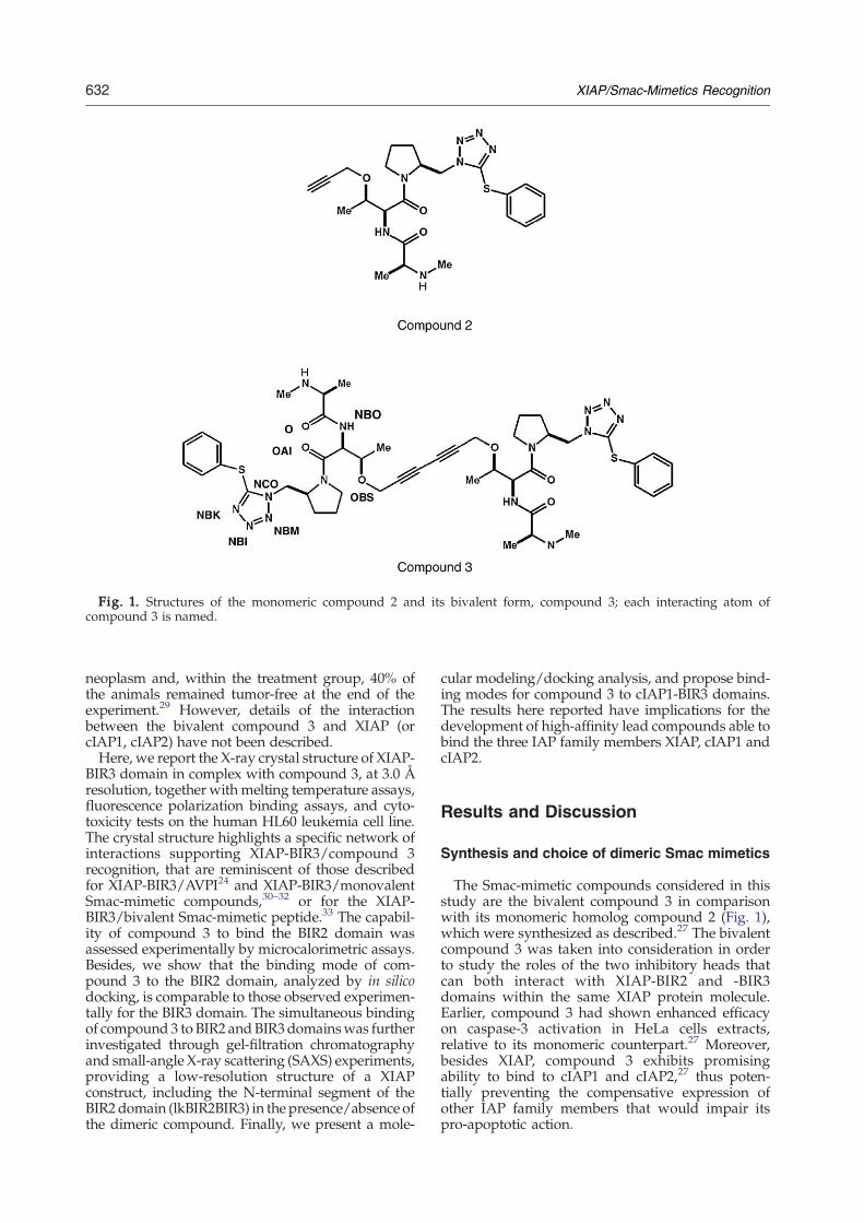
Fig. 1. Structures of the monomeric compound 2 and its bivalent form, compound 3; each interacting atom ofcompound 3 is named.
632 XIAP/Smac-Mimetics Recognition
neoplasm and, within the treatment group, 40% ofthe animals remained tumor-free at the end of theexperiment.29 However, details of the interactionbetween the bivalent compound 3 and XIAP (orcIAP1, cIAP2) have not been described.Here, we report the X-ray crystal structure of XIAP-
BIR3 domain in complex with compound 3, at 3.0 Åresolution, together with melting temperature assays,fluorescence polarization binding assays, and cyto-toxicity tests on the human HL60 leukemia cell line.The crystal structure highlights a specific network ofinteractions supporting XIAP-BIR3/compound 3recognition, that are reminiscent of those describedfor XIAP-BIR3/AVPI24 and XIAP-BIR3/monovalentSmac-mimetic compounds,30–32 or for the XIAP-BIR3/bivalent Smac-mimetic peptide.33 The capabil-ity of compound 3 to bind the BIR2 domain wasassessed experimentally by microcalorimetric assays.Besides, we show that the binding mode of com-pound 3 to the BIR2 domain, analyzed by in silicodocking, is comparable to those observed experimen-tally for the BIR3 domain. The simultaneous bindingof compound 3 to BIR2 andBIR3domainswas furtherinvestigated through gel-filtration chromatographyand small-angle X-ray scattering (SAXS) experiments,providing a low-resolution structure of a XIAPconstruct, including the N-terminal segment of theBIR2 domain (lkBIR2BIR3) in the presence/absence ofthe dimeric compound. Finally, we present a mole-
cular modeling/docking analysis, and propose bind-ing modes for compound 3 to cIAP1-BIR3 domains.The results here reported have implications for thedevelopment of high-affinity lead compounds able tobind the three IAP family members XIAP, cIAP1 andcIAP2.
Results and Discussion
Synthesis and choice of dimeric Smac mimetics
The Smac-mimetic compounds considered in thisstudy are the bivalent compound 3 in comparisonwith its monomeric homolog compound 2 (Fig. 1),which were synthesized as described.27 The bivalentcompound 3 was taken into consideration in orderto study the roles of the two inhibitory heads thatcan both interact with XIAP-BIR2 and -BIR3domains within the same XIAP protein molecule.Earlier, compound 3 had shown enhanced efficacyon caspase-3 activation in HeLa cells extracts,relative to its monomeric counterpart.27 Moreover,besides XIAP, compound 3 exhibits promisingability to bind to cIAP1 and cIAP2,27 thus poten-tially preventing the compensative expression ofother IAP family members that would impair itspro-apoptotic action.

633XIAP/Smac-Mimetics Recognition
Smac-mimetics binding assays
Monomeric compound 2 and dimeric compound 3were tested for their in vitro binding toXIAP-BIR3 and-lkBIR2BIR3, using two reported assay formats.34,35
The Ki values (Table 1) show that compound 3 is abetter inhibitor for both XIAP-BIR3 and -lkBIR2BIR3relative to compound 2. The higher affinity ofcompound 3 (IC50 of 230.8±32.8 nM) for XIAP-BIR3,compared to that displayed by compound 2 (IC50 of387.0±33.5 nM), can be explained by the dimericnature of compound 3. In fact, for a well knownstatistical effect,36 the macroscopic dissociation con-stant of a divalent ligand can be lower by a factor of 2–4 relative to that measurable for the correspondingmonomeric ligand with identical microscopic disso-ciation constant (free energy of interaction). This istrue under the assumption of truly independent sites,which is an ideal situation. The observed ratio (1/1.7)is in keepingwith such an explanation, and suggests amodest destabilizing interaction between the twosites upon binding.The monomeric compound 2 shows an IC50 value
for XIAP-lkBIR2BIR3 similar to that displayed forXIAP-BIR3 (Table 1). Such behavior is expected, sinceBIR3 is the high-affinity binding site for the Smac N-terminal AVPI peptide (Kd∼500 nM), while BIR2 isthe low-affinity binding site (Kd∼10 μM) for the samepeptide.35 Thus, the presence of the BIR2 domain inthe XIAP-lkBIR2BIR3 construct should not affect themeasured IC50 for a monomeric compound. Incontrast, simultaneous binding of compound 3 toXIAP-BIR2 and -BIR3, as shown by gel-filtrationanalysis and the SAXS structure (see below), results ina significant increase in potency (IC50 of 3.3±0.6 nM;Table 1, right).
Microcalorimetric assays
Isothermal titration calorimetry (ITC) can provideaccurate information on the thermodynamic contri-butions of enthalpy and entropy changes to freeenergies of binding, directly measuring the heatexchanged during a biomolecular binding event,providing an estimate of the dissociation constant.ITC experiments run on the protein domains hererevealed a micromolar affinity of the XIAP-BIR2
Table 1. Cytotoxicity and in vitro IC50 values ofcompounds 2 and 3
Cytotoxicity Fluorescence binding assays
IC50 (μM) IC50 (nM)
Compound HL60 XIAP-BIR3 XIAP-lkBIR2BIR3
2 7.00±1.64 387.0±33.5 295.4±61.53 0.07±0.02 230.8±32.8 3.3±0.6
Cytotoxic activity in vivo displayed by compounds 2 and 3 on theHL60 leukemia cancer cell line, determined in three independentexperiments. In vitro IC50 values of compounds 2 and 3 on XIAP-BIR3 and XIAP -lkBIR2BIR3 determined by fluorescence bindingassays in three independent experiments. All data are expressedas mean±SD.
domain for both compound 2 (Kd=9.0±1.6 μM;Supplementary Data Fig. S4A) and compound 3(Kd=3.0±0.6 μM; Supplementary Data Fig. S4B).These results complement the information pro-vided by analytical gel-filtration and SAXS experi-ments, demonstrating the actual binding of BIR2 tocompound 3, proving that the enhanced affinity ofcompound 3 for lkBIR2BIR3 is due to simultaneousbinding of the divalent molecule to two distinctprotein domains.
Cell based inhibition assays
Compounds 2 and 3 were tested for 72 h on HL60leukemia cells. The results given in Table 1 indicatethat the IC50 of the dimeric compound 3 (0.07 μM) is100-fold lower than that of themonomeric compound2 (7 μM), underlining its “drug-like” potential. More-over, it has been reported recently that various humancancer cell lines undergo apoptosis upon treatmentwith compound 3, without requiring exogenous pro-apoptotic stimuli or co-treatment with chemothera-peutic agents.29
Crystal structures of XIAP-BIR3/compound3 complex
Analysis of compound 3 bindingmode to the XIAP-BIR3 domain was addressed through X-ray crystal-lography. XIAP-BIR3/compound 3 3D structure wassolved by the molecular replacement method at 3.0 Åresolution (Rgen=23.1%, Rfree=31.1%, eight BIR3molecules per asymmetric unit; see Materials andMethods). As reported earlier,20,21,24 the XIAP-BIR3domain is composed of five α-helices and a three-stranded β-sheet, hosting a zinc-finger motif (Fig. 2a).Inspection of difference Fourier maps at variousstages of the crystallographic refinement revealedstrong residual electron density located in the IBMgroove (the XIAP-BIR3/AVPI recognition groove),comprised between the β3 strand and the α3 helix(Fig. 2a and b), for all the eightmolecules in the crystalasymmetric unit. One head of compound 3 could bemodeled promptly in such residual densities, andaccordingly refined. Therefore, the refined model iscomposed of eight BIR3 molecules (XIAP residues253±2 through 351±5) and eight compound 3molecules, each of which has an inhibitory headbound to BIR3 and the other devoid of any contact tothe protein. The eight asymmetric unit copies of theXIAP-BIR3/compound 3 complex can be divided intotwo structural groups: group I, subunits A, C, E, andG; and group II, subunits B, D, F, and H. For the fourmolecules in group I it was possible to model the C-terminal α-helix only up to residue 347±1; in group I,the free head of compound 3 is fully disordered in thesolvent. On the other hand, for the moleculesbelonging to group II, the entire C-terminal α-helix(up to residue 357±1) could be modeled, andcompound 3 is entirely defined (two heads) in theelectron density. However, in group II, pairs ofcompound 3-free heads (B and D; F and H) sharecommon locations in the asymmetric unit. The
Fig. 2. (a) A 3D stereo-view of the overall architecture of XIAP BIR3 domain. The five α-helices building BIR3 areshown in purple, the three anti-parallel β-strands are shown in yellow; note the extended C-terminal α5 helix. The corezinc atom is represented as a coral sphere. The difference electron density falls in the IBM groove between the β3 strandand the α3 helix, where one monovalent part of compound 3 binds. (b) Detail of the crystal packing of two BIR3 molecules(B and D) in presence of compound 3: it is clear how the two free heads of compound 3 share the same electron density incorrespondence to a 2-fold axis. (c) A view of the BIR3/compound 3 interaction network. The main residues involved instabilizing interactions with compound 3 are shown in green; nitrogen atoms are shown in blue, and oxygen atoms areshown in red. The compound 3 molecule is in light blue; the main hydrogen bonds linking BIR3 and the compound 3 areshown as broken lines (drawn with CCP4 mg58).
634 XIAP/Smac-Mimetics Recognition
matching of the location shared by two heads isclearly deduced by the shape of their electron density(Fig. 2b). We therefore modeled the superimposing
heads of two ligand pairs with 0.5 occupancy, thusimplying that when one half of compound 3 (e.g. freehead B) occupies the observed position, half of the

Table 2. X-ray data-collection and refinement statisticsfor the BIR3/compound 3 complex
A. Data collectionSpace group P31Unit-cell parametersa=b (Å) 119.1c (Å) 105.6α=β (°) 90γ (°) 120Solvent content (%, v/v) 65.9Molecules/per asymmetric unita.u. 8Resolution (Å) 40.0–3.0Mosaicity (°) 0.9№ of unique reflections 33,440 (4861)Completeness (%) 99.8 (99.9)Redundancy 3.6 (3.7)Rmerge
a (%) 10.1 (64.0)Average I/σ (I) 13.3 (2.2)
B. RefinementR-factorb (%) 23.1Rfree
c (%) 31.1r.m.s.d. from idealBond lengths (Å) 0.011Bond angles (°) 2.042Average protein B-factor (Å2) 73.1Average compound 3 B-factor (Å2) 50.6Ramachandran plotResidues in most favored regions (%) 80.7Residues in additionally allowed regions (%) 19.0Residues in generously allowed regions (%) 0.3
Values in parentheses are for the highest resolution shell.a Rmerge=Σ |I – (I)| / Σ I×100, where I is the intensity of a
reflection and (I) is its average intensity.b R-factor=Σ |Fo – Fc| / Σ |Fo|×100%c Rfree for cross-validation was calculated with 5% of reflections
that were selected at random and were not included in therefinement.
635XIAP/Smac-Mimetics Recognition
other compound 3 (e.g. free head D) is disordered inthe solvent and vice versa. Such peculiar feature can beexplained by the mutual orientation of the B-D and F-H protein dimers in group II. In fact, in each dimer acouple of antiparallel C-terminal α-helices closes theligand in a sort of “hug”, restraining the conforma-tional freedom of the free head that results in theobserved superposition (Fig. 2b).
Table 3
XIAP-BIR3/compound 3(crystal structure)
XIAP-BIR2/co(docking m
Residue Interaction Mean (Å) Residue Interact
Thr 308 N-OAI 3.0 Lys 208 N-OAN-O 3.1 O-NB
Oγ1-OBS 3.4Glu 314 Oɛ1-N 2.8 Asp 214 Oδ2-N
Oɛ2-N 2.8Gln 319 Oɛ1-N 3.4 Glu 219 Oɛ1-N
Oɛ1-OTrp 323 Nɛ1-O 3.1 His 223 Nɛ1-O
Nɛ2-O
Left: Average distances (in Å) between atoms of compound 3 and BIR3observed in the crystal structure. Centre: Compound 3/XIAP-BIR2 inBIR3 interactions from the docking model.
Superposition of the eight BIR3 protein domains(amino acids 253–344) yields r.m.s.d. values of 0.13–0.24 Å among molecules belonging to the samestructural group, and 0.46–0.52 Å when comparingthe two different structural groups. Data collectionand refinement statistics are given in Table 2, togetherwith stereochemical quality of the structure.
Structure and recognition in theXIAP-BIR3/compound 3 complex
Superposition of the eight independent moleculesin the crystal asymmetric unit shows containedconformational variability for the protein residuesinteracting with compound 3. In particular, all theatoms from amino acids falling within 4.0 Å from theligand (292, 297–299, 306–310, 314, 319, and 323–324)have an r.m.s.d. of 0.4±0.1 Å. Comparison of thecrystal structures shows that the protein/ligandinteractions stabilizing the XIAP-BIR3/compound 3complex resemble those observed in the XIAP-BIR3/Smac-DIABLO structure (PDB code 1G73;24; r.m.s.d.0.38Å) and in the XIAP-BIR3/ bivalent Smac-mimeticpeptide (the cyclized (CH3-AKPF)2 peptide; PDB code2VSL33). One of the two heads of compound 3 isbound to the IBM groove (roughly lined by residuesLys297, Thr308-Asp309,Glu314, andTrp323), exchan-ging hydrogen bonds and van der Waals contactswith residues Gly306, Thr308, and Asp309, located inthe β3 strand, with Glu314 and Gln319 belonging totheβ3 –α3 loop, andwithTrp323 andTyr324 in theα3helix (Table 3, left and Fig. 2c). In particular, the N-terminal part of the compound 3 head is bound to theprotein mainly by electrostatic interactions, whilemimicking an antiparallel β-sheet, as shown for theAla1-Val2 N-terminal residues of Smac-DIABLO andfor the Ala1-Lys2 N-terminal residues of the bivalentSmac-mimetic peptide.33 In contrast, the remainingpart of the same head (pyrrolidine, tetrazole andphenyl rings) provides van der Waals interactionsonly. In detail, the N-terminal-bound end ofcompound 3 exchanges a salt bridge with Glu314(2.8±0.2 Å; distance averaged over the eight inde-pendent molecules) and a loose hydrogen bond with
mpound 3odel)
cIAP1-BIR3/compound 3(docking model)
ion Dist. (Å) Residue Interaction Dist. (Å)
I 2.7 Arg 308 N-OAI 3.1O 2.7 O-NBO 2.9
O-OBS 3.02.8 Asp 314 Oδ2-N 2.7
2.8 Glu 319 Oɛ1-N 2.53.63.8 Trp323 Nɛ1-O 3.23.0
amino acids involved in the main protein/ligand interactions, asteractions from the docking model. Right: Compound 3/cIAP1-

636 XIAP/Smac-Mimetics Recognition
Gln319 (3.4±0.4 Å) as observed for XIAP-BIR3/monovalent Smac-mimetic compounds (Smac005,30
Smac010,30 Smac037,31 and compound 2132). In fact,all the bicyclic Smac-mimetic compounds terminalamines lose hydrogen bonds/van der Waals contactswith Gln319, gaining other interactions mainlyinvolving Thr308 (Smac005 and compound 21) andAsp309 (Smac010, Smac037 and compound 21). Thecompound 3 methyl group (CB) is well inserted in ahydrophobic surface depression lined by Leu307and Trp310. The next peptide plane is stabilizedby hydrogen bonds with the side chain of Trp323(3.1±0.2 Å) and the main-chain carbonyl group ofThr308 (3.1±0.3 Å). The carbonyl OAI is hydrogenbonded to the peptide N atom of Thr308 (3.0±0.2 Å)and, partially, with its side chain (3.4±0.4 Å). Thefollowing two rings (pyrrolidine and tetrazole) areinvolved in hydrophobic contacts mainly withGly306, Trp323 (stacking interaction with pyrroli-dine ring), and Tyr324. Finally, the terminal phenylgroup is well located in a hydrophobic dip encircledby the side chains of Leu292, Lys297, and Lys299.When compared to the XIAP-BIR3/AVPI and XIAP-BIR3/bivalent Smac-mimetic peptide complexes,33
the XIAP-BIR3/compound 3 structure shows theconservation of all electrostatic interactions, but theloss of a hydrogen bond to the main-chain carbonylgroup of Gly306.Conversely, the second head of compound 3
(when traceable in density) is not found in contactwith any part of the protein in the crystal structure.
Melting temperature thermal shift assays
Thermal shift assay is an experimental techniquemonitoring fluorescence variations reported by aprotein-bound dye during protein thermal denatura-tion. The method was developed originally for drugdiscovery, to allow rapid identification of proteinligands by screening compound libraries.37 Sincesmall molecules (e.g. an enzyme inhibitor) bound toa protein can affect (often stabilize) its structure, theassay monitors the variation in melting temperature(Tm) induced by ligand binding. Sypro orange, thefluorescent dye used here, binds efficiently to theunfolded protein displaying an increase of fluores-cence intensity during temperature-dependent pro-tein unfolding.36
The Tm values for the protein constructs consideredhere were shifted toward higher temperatures by thebinding of both the monomeric/dimeric Smac-mimetic compounds. Taking the Tm values asindicative of increasing stabilization of the protein/ligand adducts produced, compound 2 was found tostabilize all constructs, with ΔTm values of +10.1degC, +9.0 degC and+14.3 degC inXIAP-BIR2, -BIR3and -lkBIR2BIR3, respectively, while the ΔTm valuesfor compound 3 were +10.3 degC, +10.0 degC and+17.6 degC in XIAP-BIR2, -BIR3 and -lkBIR2BIR3,respectively. Interestingly, the stabilization of XIAP-lkBIR2BIR3 by compound 2 is lower by more than 3degC (ΔTm +14.3 degC) than that measured forcompound 3 (ΔTm +17.6 degC). Such findings
indicate that the stabilization effects of both themonomeric and dimeric Smac mimetics on a singleprotein domain are similar, but that compound 3induces higher stability for the XIAP-lkBIR2BIR3construct. Such an effect may be explained by thesimultaneous binding of compound 3 to the XIAP-BIR2 and -BIR3 domains, resulting in (i) the stabiliza-tion of the two domains, and (ii) a more compactshape of the overall protein structure. The latterstructural effect is supported by the gel-filtrationassays and the SAXS analysis described below.Although the effects on Tm are clear-cut, and mayshow a trend, such assays must be taken only as aqualitative ranking of Smac-mimetics affinity for thethree BIR constructs. Themain result suggested by thethermal shift assays is that all compounds bindeffectively to XIAP-BIR3, and to the XIAP-BIR2domain. (Experimental sigmoid Tm plots, as well asother experimental data given below are reported ingraphical form as Supplementary Data).
Analytical gel-filtration assays
In order to check whether the crystallizationconditions might have prevented the simultaneousinteraction of compound 3 with two XIAP-BIR3domains, we performed analytical gel-filtrationassays using a fixed concentration of protein(33 μM) and an excess of compound 2 or 3 (5 mM).The chromatograms obtained for XIAP-BIR3 in thepresence of compound 3 show a peak at an elutionvolume (Ve) of 10.6 ml, corresponding to the dimericform of the protein. In the presence of compound 2, orin the absence of the Smac-mimetics, a peak at Ve of11.8 ml, corresponding to the monomeric form ispresent in the chromatogram. The absence of adimeric assembly from our crystal structure obtainedin the presence of compound 3 is likely due to thecrystal growth conditions, i.e. higher concentrationsof protein and salt relative to the analytical gel-filtration tests.We investigated the interaction of compound 3with
the XIAP-lkBIR2BIR3 construct. The chromatogramshows a peak shift of Ve from 10.5 ml to 10.8 ml in thepresence of an excess of compound 3 (5 mM),suggesting an actual reduction of the protein volume.A more compact protein moiety is compatible withthe simultaneous binding of compound 3 to the twodomains of the construct, as confirmed by the SAXSdata (see below). Similar results have been reportedrecently for two different bivalent Smac-mimetics: thebivalent cyclic peptide inRef. 33, and the bivalent SM-164 in Ref. 28. In both cases, the presence of a divalentcompound causes a shift of the GF peak towardhigher Ve values, suggesting the simultaneous bind-ing of the bivalent compounds to the BIR2 and theBIR3 domains. Moreover, in both cases, a mutation ofthe BIR2 domain in the IBM groove (E219R) alters thebinding of the divalent compounds, indicating thatthe BIR2 IBM groove is directly involved in theinteraction. In contrast, analytical gel-filtration in thepresence of compound 2 (5 mM) shows that XIAP-lkBIR2BIR3 does not change its shape (Ve 10.5 ml).

637XIAP/Smac-Mimetics Recognition
Virtual docking of compound 3 to XIAP-BIR2
XIAP is well known to inhibit caspase-3 and -7 bymeans of a protein region located N-terminal to theBIR2 domain (the so-called linker region).20,21 How-ever, a recent study showed that additional interac-tions between BIR2 and caspases -3 and -7 involve aregion of the BIR2 domain structurally related to theIBM groove described for the XIAP-BIR3/caspase-9interaction.19 The BIR2 domain IBM groovemay thusstrengthen the binding between XIAP and caspases -3and -7. As a consequence, Smac-mimetics able to bindthe XIAP-BIR2 IBM groove might promote caspase-3and -7 activity in apoptosis. Moreover, compoundsshowing high affinity for other XIAP homologs, suchas cIAPs, would enhance the apoptosis-promotingeffect.An in silico docking approach based on the
program AutoDock438 was used to propose amodel for compound 3 binding to XIAP-BIR2. Inparticular, we performed virtual docking searchesusing the high-resolution crystal structure of XIAP-BIR2 (PDB code 1I3O, subunit E20). The BIR2/Smac-mimetic complex model obtained was subse-quently compared to the crystal structure of XIAP-BIR3/compound 3, considering that XIAP-BIR2and -BIR3 domains display an amino acid sequenceidentity of 41.5%, and their crystal structures havean r.m.s.d. of 0.89 Å (77 Cα pairs). The modelproduced in silico indicated that compound 3roughly binds to the BIR2 domain as observedexperimentally for the XIAP-BIR3 domain. More-over, the estimated binding free energy of com-pound 3 to BIR2 appears to be close to that obtainedfor the XIAP-BIR3 complex model (about –8.6 kcal/mol for XIAP-BIR2/compound 3, and –8.1 kcal/molfor -BIR3/compound 3).Analysis of the ligand/protein interaction network
in the XIAP-BIR2/compound 3 model (Fig. 3)
indicates that some conservatively substituted aminoacids, such as XIAP-BIR2 Lys208, Asn209, Asp214,Glu219, and His223, are predicted to be involved inproductive hydrogen bonding interactions with theSmac-mimetic compound (Table 3, centre).
XIAP-lkBIR2BIR3 SAXS study
The two scattering patterns of XIAP-lkBIR2BIR3solutions in the absence/presence of the compound3 inhibitor, are shown in Fig. 4a. They are compositecurves obtained by combining data recorded at low(small-angle) and higher (wider-angle) concentra-tions, as explained in Materials and Methods. Theyexhibit significant differences, suggesting that com-pound 3 binding causes a global conformationalchange in the protein. Guinier plot analysis of thetwo curves shows a reduction of the radius ofgyration in the presence of the inhibitor, from27.7±0.3 Å to 24.6±0.3 Å. I(0)/c values yield amolecular mass estimate of about 28 kDa and 29 kDafor the apo protein and the complex with theinhibitor, respectively, in good agreement with thevalues of 28,847 Da derived from the protein aminoacid sequence, and of 971 Da for the inhibitor. Thisagreement shows that protein samples at a concen-tration of 1 mg/ml were essentially free of inter-molecular interactions. The distance (or pair)distribution functions p(r) (Fig. 4b) yield values of105±5 Å and 29.1±0.2 Å for the maximal diameterand radius of gyration of the apo protein, respec-tively, to be compared with 95±5 Å and 25.2±0.2 Å,respectively, obtained in the presence of the inhibitor.All these results point to a conformational transitionof XIAP-lkBIR2BIR3 from an extended to a morecompact conformation upon inhibitor binding. Thep(r) profile of the apo protein is broadly spread,with a first peak around 20 Å and a clear shoulderbetween 35 and 60 Å, corresponding predominantly
Fig. 3. Virtual docking modelsfor the XIAP-BIR2/compound 3complex. The main residuesinvolved in stabilizing interactionswith BIR2/compound 3 (labeled)are shown in purple. The com-pound 3 molecule is shown in lightblue; and the protein non-interact-ing residue is shown in orange.Nitrogen atoms are shown in blue,and oxygen atoms are shown in red(drawn with CCP4 mg58).

Fig. 4. XIAP-lkBIR2BIR3 SAXS patterns. The scattering patterns (a) and distance (or pair) distribution functions p(r) (b)of XIAP-lkBIR2BIR3 solutions in the absence/presence of compound 3 are shown in blue and red, respectively. c and d,Most typical SAXS models of XIAP-lkBIR2BIR3 in the absence (c) or in the presence (d) of compound 3 obtained with theprogram Dammin (surfaces are shown in white). (e) XIAP-lkBIR2BIR3/compound 3 model (BIR2 in cyan, BIR3 inmagenta, and compound 3 in orange) superimposed on the most typical dummy residue model obtained with Gasbor(dummy atoms are shown as white spheres). The left-hand and right-hand views are related by a 90° rotation around avertical axis. The figure was realized using Pymol [http://pymol.sourceforge.net/].
638 XIAP/Smac-Mimetics Recognition
to intra- and interdomain distances, respectively,while most distances are found in a narrow range(15–45 Å) in the presence of the inhibitor. This
suggests that the BIR2 and BIR3 structured domainsare well resolved in the apo protein and likelymobile around a flexible linker. Conversely the
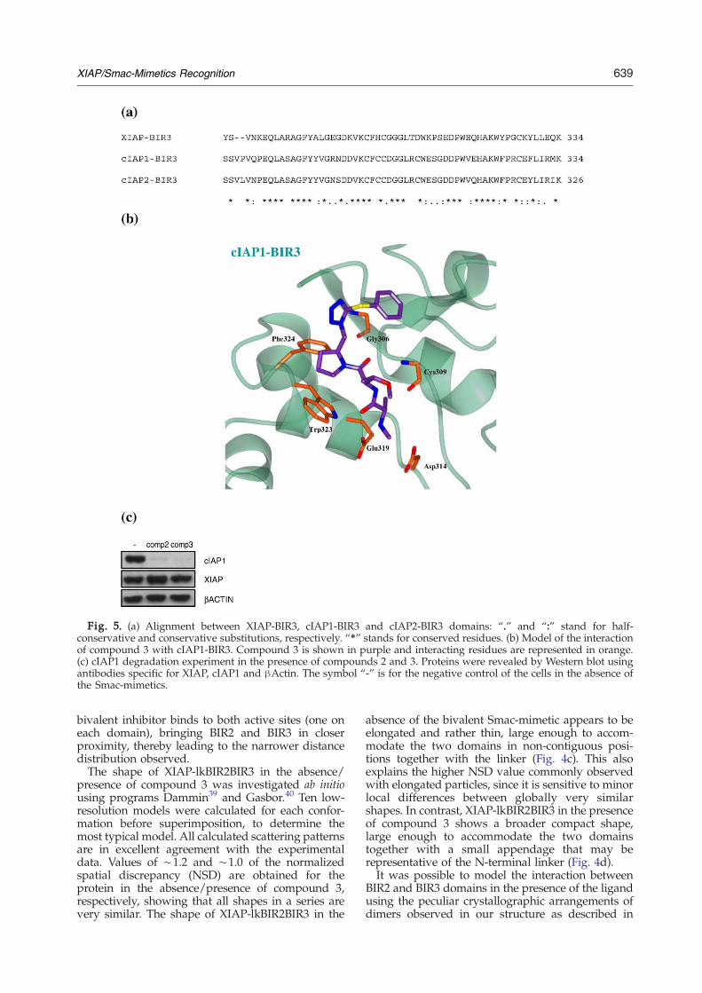
Fig. 5. (a) Alignment between XIAP-BIR3, cIAP1-BIR3 and cIAP2-BIR3 domains: “.” and “:” stand for half-conservative and conservative substitutions, respectively. “⁎” stands for conserved residues. (b) Model of the interactionof compound 3 with cIAP1-BIR3. Compound 3 is shown in purple and interacting residues are represented in orange.(c) cIAP1 degradation experiment in the presence of compounds 2 and 3. Proteins were revealed by Western blot usingantibodies specific for XIAP, cIAP1 and βActin. The symbol “-” is for the negative control of the cells in the absence ofthe Smac-mimetics.
639XIAP/Smac-Mimetics Recognition
bivalent inhibitor binds to both active sites (one oneach domain), bringing BIR2 and BIR3 in closerproximity, thereby leading to the narrower distancedistribution observed.The shape of XIAP-lkBIR2BIR3 in the absence/
presence of compound 3 was investigated ab initiousing programs Dammin39 and Gasbor.40 Ten low-resolution models were calculated for each confor-mation before superimposition, to determine themost typical model. All calculated scattering patternsare in excellent agreement with the experimentaldata. Values of ∼1.2 and ∼1.0 of the normalizedspatial discrepancy (NSD) are obtained for theprotein in the absence/presence of compound 3,respectively, showing that all shapes in a series arevery similar. The shape of XIAP-lkBIR2BIR3 in the
absence of the bivalent Smac-mimetic appears to beelongated and rather thin, large enough to accom-modate the two domains in non-contiguous posi-tions together with the linker (Fig. 4c). This alsoexplains the higher NSD value commonly observedwith elongated particles, since it is sensitive to minorlocal differences between globally very similarshapes. In contrast, XIAP-lkBIR2BIR3 in the presenceof compound 3 shows a broader compact shape,large enough to accommodate the two domainstogether with a small appendage that may berepresentative of the N-terminal linker (Fig. 4d).It was possible to model the interaction between
BIR2 and BIR3 domains in the presence of the ligandusing the peculiar crystallographic arrangements ofdimers observed in our structure as described in

640 XIAP/Smac-Mimetics Recognition
Materials and Methods. Figure 4e shows the modelfor XIAP-lkBIR2BIR3 complexed with compound 3thus obtained, superimposed on the most typicalSAXS dummy residue model for XIAP-lkBIR2BIR3/compound 3. The dummy residue model fits quitesatisfactorily our XIAP-lkBIR2BIR3 model that wasassembled on the basis of independent principles. Itshould be noted that here we do not propose a high-resolution model of XIAP-lkBIR2BIR3 compound 3interaction (a high-resolutionmodel of the interactionwith a different dimeric compound has been reportedrecently33) but just a low-resolution molecular shapethat is in agreement with the SAXS experimentalresults.
Compounds 2 and 3 in cIAP degradation
It has been reported recently that Smac-mimeticsmay kill cancer cells via a mechanism involvingubiquitination and degradation of cIAP1 and cIAP2,resulting in TNFα-mediated cell death.16,29,41 Sincecompound 3 was shown to interact with cIAP1 andcIAP2,13,27 we performed a virtual docking searchanalyzing the possible binding mode(s) of com-pound 3 on the cIAP1-BIR3 domain, whose crystalstructure was published recently (PDB code 3D9U,subunit A42).A superposition of XIAP-BIR3/compound 3 crystal
structure on the cIAP1-BIR3 domain (40 Cα pairs)indicates a r.m.s.d. of 0.58 Å. The two homolog BIR3domains (XIAP-BIR3 and cIAP1-BIR3) show asequence homology of 37% and a good conservationof the residues belonging to their IBMpockets (Fig. 5a,the cIAP1-BIR3 residues number are that of the PDB3D9U). Among all amino acids involved in theinteraction network between XIAP-BIR3 and com-pound 3 observed in the crystal structure, most areconserved (Gly306 and Trp323) or substituted con-servatively (Asp311, Glu319, and Phe324). OnlyArg308 and Cys309 are not conserved within thepocket (Fig. 5a). Nevertheless, the analysis of thepredicted binding mode for compound 3 (Fig. 5b)suggests that the non-conservative substitutionsshould not affect its affinity for cIAP1-BIR3 (thebinding free energy value for cIAP1-BIR3/compound3 complex is –10.5 kcal/mol). The cIAP1-BIR3/compound 3 complex model suggests conservationof crucial interactions that involve the Gly306 andArg308 backbones, the Asp314, Glu319 and Phe324(Table 3, right and Fig. 5b). Compound 3 pyrrolidinering establishes hydrophobic contacts to Trp323 andPhe324. Moreover, since amino acids involved in thecIAP1-BIR3/compound 3 interactions are well con-served in the cIAP2-BIR3 domain (Fig. 5a), the sameinteraction network might stabilize compound 3binding to cIAP2.The experimental approaches here adopted to
characterize compound 3 binding to XIAP-lkBIR2-BIR3 led us to extend our views to cIAP1 and -2members of the family through the simulativeapproaches described. Since the interactions of theSmac-mimetics with cIAP1-BIR3 predicted by thedocking algorithms suggested efficient binding, as an
independent approach, we carried out cIAP1 degra-dation experiments in the presence of both com-pounds 2 and 3 (similar results are shown in Ref. 43),using the MDA-MB231 cell line. In keeping with thehypothesis suggested by our docking results,Westernblot analysis revealed that both the Smac-mimeticcompounds were able to induce degradation of thecIAP1 protein (Fig. 5c).As awhole, our results provide comprehensive new
structural and recognition information on the inter-action of Smac-mimetics with the IBM grooves ofXIAP (BIR2 and BIR3 domains), of cIAP1 and cIAP2.New working grounds for the development of high-affinity lead compounds specifically binding the threemembers of the IAP family are thus available.
Materials and Method
Chemistry
Compounds 2 and 3 were synthesized as described.27
Cloning, expression and purification of human XIAPBIR domains
The cDNA coding for human XIAPwas retro-transcribedfrom a pool of human mRNAs. The sequences coding forregions 241–356 (XIAP-BIR3), 124–356 (XIAP-lkBIR2BIR3)and 140–240 (XIAP-BIR2) were cloned in pET28(b) (Nova-gen), in NheI-BamHI sites. All the plasmids were used totransform Escherichia coli strain BL21(DE3) as described.31
The proteins were stored in 20 mM Tris pH 7.5, 200 mMNaCl, 10 mM DTT.
Fluorescence polarization assays
Fluorescence polarization experiments were performedas described.34,35 Briefly, the experiments were performedin black, flat-bottom 96-well microplates (GREINER BIO-ONE), and fluorescence polarization was measured withan Ultra plate reader (Tecan). For the XIAP-BIR3 construct,a fluorescently labeled Smac peptide (AbuRPF-K(5-Fam)-NH2) (FITC-Smac35) was used at a final concentration of5 nM, added to an assay buffer together with increasingconcentrations of XIAP-BIR3 (0 – 20 μM). For the XIAP-lkBIR2BIR3 construct, a fluorescently labeled dimeric Smacpeptide (Smac-1F34) was used at a final concentration of1 nM, added to the assay buffer together with increasingconcentrations of lkBIR2BIR3 (0 – 2 μM). The final volumein each well was 125 μl, with the assay buffer consisting of100 mM potassium phosphate pH 7.5, 100 μg/ml bovine γ-globulin, 0.02% (w/v) sodium azide. After shaking for15 min, the plate was incubated for 3 h at roomtemperature. Fluorescence polarization was measured atexcitation and emission wavelengths of 485 nm and530 nm, respectively. The equilibrium binding graphswere constructed by plotting millipolarization units (mP)as a function of protein concentration. Data were analyzedusing Prism 4.0 software (Graphpad Software). Compound3 and the monomeric control compound 2 were evaluatedfor their ability to displace the fluorescent probe fromrecombinant protein. Fluorescent probe (5 nM FITC-Smacfor XIAP-BIR3 or 1 nM Smac-1F for XIAP-lkBIR2BIR3), andserial dilutions of the two Smac-mimetics (concentration

641XIAP/Smac-Mimetics Recognition
0.4 nM –4 μM) were added to each well, to a final volumeof 125 μl in the assay buffer described above. After mixingon a shaker for 15 min and incubation for 3 h at roomtemperature, fluorescent polarization was measured withthe Ultra plate reader (Tecan).
Microcalorimetric experiments
The binding affinity of compound 2 and compound 3 tothe XIAP-BIR2 domain were tested with ITC experimentsusing VP-ITC technology. Comparable results wereobtained for the two compounds (monomeric and dimericform) with two separate ITC experiments. The measure-ments were done at 4 °C after 30 consecutive injections ofconstant volumes (10 μl) of compound 2 (concentrated at500 μM) or of compound 3 (concentrated at 300 μM) to theprotein sample (2 ml, concentrated at 28 μM). The heatmeasurements were analyzed using the program Origin7.0 (OriginLab Corporation, One Roundhouse Plaza,Northampton, MA 01060) with a specific package forprocessing microcalorimetric data (Microcal Origin).
In vitro profiling: cytotoxicity
The HL60 human promyelocytic leukemia cell line wasobtained from Interlab Cell Line Collection (ICLC, Genova,Italy). The cell linewas cultured at density of 1×10⁵ cells/mlin RPMI 1640 medium supplemented with 10% (v/v) fetalbovine serum (FBS) at 37 °C in a 5% (v/v) CO2 fullyhumidified atmosphere.The effect of compound 2 and compound 3 on cell
growth was evaluated by means of a colorimetric assay forthe quantification of cell proliferation and viability basedon the cleavage of the WST-8 tetrazolium salt bymitochondrial dehydrogenases in viable cells (Promokine,Germany). The kits utilize the tetrazolium salts WST-8 thatare reduced to water-soluble, orange formazan dyes bydehydrogenases present in viable cells. The absorbance ofthe formazan dye is proportional to the number ofmetabolically active cells. Briefly, at time zero and aftertreatment with Smac-mimetic compounds for 72 h, 10 μl ofWST-8 was added to each of the 96-well culture platescontaining 1×10⁴ cells in 100 μl of complete medium. Afterincubation at 37 °C for 4 h, the absorbance at 450 nm wasmeasured using a microplate reader 1420 VICTOR multi-label counter (EG&G Wallac, Finland). The data wereexpressed as mean percentage of three replicates normal-ized to the untreated control. IC50 values were calculatedas the concentration of compound inhibiting growth by50%, relative to control cultures. The results are summar-ized in Table 1.
cIAP degradation assay
The MDA-MB231 cell line was treated with 5 μM Smac-mimetics or left untreated. After 3 h, cells were harvestedand lysed. Proteins were revealed by Western blot withantibodies specific for XIAP (BD Biosciences), cIAP1(R&DSystems) and βActin (Sigma).
Crystallization
Crystallization trials of XIAP-BIR3 in the presence ofvarious amounts of compound 2 or compound 3 wereperformed at 20 °C using an Oryx-8 crystallization robot(Douglas Instruments, East Garston, UK) in microbatch
plates, that were covered at the end of the experiment with1.5 ml of paraffin oil and 1.5 ml of Al's oil (50:50 mixture ofparaffin oil and silicon oil). Elongated hexagonal prisms ofabout 100×30×30 μm were obtained only for the XIAP-BIR3/compound 3 complex after two weeks using 20%(w/v) polyethylene glycol monomethyl ether (PEG MME)2000, 60 mM sodium acetate trihydrate, pH 4.6, 120 mMammonium sulfate, 400 mM sodium potassium tartratetetrahydrate. For X-ray data collection, crystals wereharvested in a cryoprotectant solution (25% (w/v) PEGMME 2000, 60 mM sodium acetate trihydrate pH 4.6,120 mM ammonium sulfate, 400 mM sodium potassiumtartrate tetrahydrate, 25% (v/v) glycerol) before being flash-cooled in liquid nitrogen. The crystals diffracted to amaximum resolution of 3.0 Å on beamline ID 29 at theEuropean Synchrotron Radiation Facility (ESRF-Grenoble,France).
Structure determination and refinement
The X-ray diffraction data for the XIAP-BIR3/compound 3 complex were indexed (MOSFLM44) andscaled, cutting the resolution to 3.0 Å (SCALA45) in thetrigonal P3 space group. A molecular replacementsearch (PHASER46), using the structure of XIAP-BIR3,from the Smac-DIABLO complex (PDB code 1G7324),from which the C-terminal α-helix had been deleted(thus retaining amino acids 253–347), was used as searchmodel, locating nine XIAP-BIR3 molecules in spacegroup P31 (log-likelihood gain=1081). Rigid bodyrefinement (REFMAC;47 R-factor 44.4%, Rfree 43.8%)and restrained refinement (R-factor 37.6%, Rfree 41.8%)followed by visual inspection of the map (COOT48)showed steric clashes between two of the nine chains,which were therefore omitted from the asymmetric unitmodel (thus retaining seven model molecules, R-factor36.0%, Rfree 39.9%). An additional subunit was deletedbased on refinement and B-factor value considerations(leaving six chains/asymmetric unit, R-factor 35.3%,Rfree 39.0%). Inspection of residual electron density, andthe observation that the asymmetric unit at this stagedisplayed two dimers (interface area 310 Å2) and twoisolated chains, helped locating two additional XIAP-BIR3 chains that, together with the isolated ones, yieldedtwo additional dimeric assemblies, for a total of eightasymmetric unit chains (R-factor 32.3%, Rfree 37.0%).Residual map inspection at this stage showed clearelectron density close to the IBM groove. Such densityallowed us to model compound 3 bound to eachmolecule in the crystal asymmetric unit. Several refine-ment cycles (Refmac5 and Buster49) and manualrebuilding48 resulted in the refined model (R-factor23.0%, Rfree 31.4%) composed of eight BIR3 molecules(amino acids 253±2 through 351±5) and eight com-pound 3 molecules. The stereochemical quality of themodel was checked using the program Procheck.50
Thermal shift assays
To monitor protein unfolding, the fluorescent dye Syproorange was used to monitor the unfolding transition.Using a MiniOpticon Real Time PCR Detection System(Bio-Rad), designed originally for PCR,51 thermal shiftassays were conducted in the presence of the Smac-mimetics. Solutions of 2.4 μl of the purified XIAP-BIRprotein constructs (XIAP-BIR2, -BIR3 and -lkBIR2BIR3)were mixed with 3.5 μl of Sypro orange (Sigma)diluted 60-fold, 19 μl of the protein storage buffer and
642 XIAP/Smac-Mimetics Recognition
0.1 μl of 10 mM Smac-mimetics (compounds 2/3).Distilled water was added in place of the inhibitors forthe control samples. The final concentrations of proteinranged between 0.5 mg/ml and 5 mg/ml; the sampleplates were heated from 25 C to 95 C at a heating rateof 2 degC/min. The fluorescence intensity was mea-sured within the ranges excitation 470–505 nm, emis-sion 540–700 nm.
Analytical gel-filtration experiments
Analytical gel-filtration experiments were done with aSuperdex 75 column (GE Healthcare) coupled to an AKTAPurifier system using 20 mM Tris–HCl pH 7.5, 200 mMNaCl, 10 mM DTT. Recombinant XIAP-BIR3 (residues241–356) was run on the column at a concentration of1 mg/ml either alone or after incubation for 30 min withan excess of compound 3 (5 mM). The recombinant XIAP-BIR3 and -lkBIR2BIR3 (residues 124–356), each at aconcentration of 1 mg/ml, were subsequently run on thecolumn alone or after incubation with 5 mM compound 2as a control.
Molecular modeling
The AutoDock4 package38 was used for dockingcompound 3 to the protein targets, and the PythonMolecule Viewer 1.4.5 was used to analyze the data. TheXIAP-BIR220 (PDB code 1I3O, subunit E) crystal structurewas adopted to produce in silico models of the XIAP-BIR2/compound 3 complex; the cIAP1-BIR3 crystalstructure (PDB code 3D9U, subunit A) was used for thecIAP1-BIR3/compound 3 complex model. A dockinggrid (32×46×44 points; grid step of 0.375 Å) wascentered at Leu207, or Leu307, in XIAP-BIR2 andcIAP1-BIR3, respectively, resulting in a 3416 Å3 box, inwhich the search was performed. Only one active head ofthe inhibitor was used to model the protein/compound 3interactions. During the docking simulation, the proteinmodels were rigidly constrained, whereas 10 rotationsaround single bonds were allowed for compound 3. Thedocking procedure consisted of 100 independent geneticalgorithm (GA) runs.38 The three protein/compound 3docked models displaying the lowest binding free energywere retained for structural analysis.
Model of BIR2-BIR3 in complex with compound 3
The model of XIAP-lkBIR2BIR3 complexed with com-pound 3 was produced by a simple superposition of twohalves of two compound 3 molecules. In particular, rigidtranslation of subunit D so as to superimpose the head ofcompound 3 bound to subunit D to the head of compound3 not bound to subunit B produced a dimeric assembly ofsubunits B and D bridged by one molecule of inhibitor(Fig. 2b). Finally, the crystal structure of BIR2 (PDB 2VM5)was superimposed on BIR3 subunit D, thereby producinga model of the BIR2-BIR3 assembly in the presence ofcompound 3. This model (composed of 215 amino acidsover 250 of the real assembly) was further used incombination with the SAXS results.
Small-angle X-ray scattering
X-ray scattering data were collected at the beamlineSWING of Synchrotron SOLEIL (Gif-sur-Yvette, France).
The data were recorded using a CCD-based detector(AVIEX)with a sample-detector distance of 1.84m, coveringthe range of momentum transfer 0.012bqb0.45 Å-1 (q=4πsinθλ, where 2θ is the scattering angle and λ=1.033 Å thewavelength of the X-rays). XIAP-lkBIR2BIR3 with andwithout the inhibitor compound 3 was studied in 20 mMTris–HCl buffer pH 7.5, 200 mM NaCl, 10 mM DTT atprotein concentrations between 1 mg/ml and 6 mg/ml.Solutions were circulated continuously during data record-ing through the 1.8 mm diameter quartz capillary using theautomatic sample changer (Agilent) at a flow-rate ensuringan irradiation time of ∼1 s. Under these conditions, noradiation damage could be detected in preliminary tests. Allmeasurements were performed at 10 °C. Data wereaveraged after normalization to the intensity of thetransmitted beam before buffer subtraction using theprogram package PRIMUS.52 The forward scattering I(0)and the radius of gyration (Rg) were evaluated using theGuinier approximation.53 The curves of the most dilute,interaction free, and of the most concentrated samples werespliced after scaling to protein concentration to yield acombined, complete scattering pattern. The distance dis-tribution function p(r) corresponds to the distribution ofdistances between any pair of volume elements within oneparticle. It was determined using the indirect Fouriertransform method as implemented in the programGNOM.54 The molecular masses of the solutes wereevaluated by comparison of the forward scattering withthat of a reference 3.9 mg/ml lysozyme solution in 50 mMsodium acetate buffer pH 4.5, 100 mM NaCl.Low-resolution shapes can be determined using the
programDammin,whichdescribes the protein as a compactassembly of identical dummy atoms.39 Typically, 10 modelsare calculated and superimposed using the Damaver suiteof routines.55 They are compared using a measure ofsimilarity called NSD,56 the smaller the NSD value thehigher the similarity. Ab initio models were also producedusing the program Gasbor, which describes the protein as achain ofNdummyresidues,whereN is the actual number ofprotein residues (250 amino acids for XIAP-lkBIR2BIR3).40
In away similar to Dammin, 10modelswere calculated andcompared using NSD values.
Protein Data Bank accession numbers
Atomic coordinates and structure factors have beendeposited with the Protein Data Bank with accessionnumber 3G76.57
Acknowledgements
This studywas supported by grants from the ItalianMinistry of University and Research FIRB Project“Biologia Strutturale” (contract RBLA03B3KC_005, toM.B.). We are grateful to Fondazione AssociazioneRenato Dulbecco for financial support. We thankJ. Pérez and G. David (Synchrotron SOLEIL) forhelp with SAXS measurements. E.M. and M.M.thank the EU/TNA program for travel support toSynchrotron SOLEIL. We are grateful to ElenaCasale (Nerviano Medical Sciences, Italy) forinvaluable help during the microcalorimetric assays.We thank the European Community Research

643XIAP/Smac-Mimetics Recognition
Infrastructure Action under the FP6 “Structuringthe European Research Area” Programme (throughthe Integrated Infrastructure Initiative “IntegratingActivity on Synchrotron and Free Electron LaserScience”) for support.
Supplementary Data
Supplementary data associated with this articlecan be found, in the online version, at doi:10.1016/j.jmb.2009.04.033
References
1. Steller, H. (1995). Mechanisms and genes of cellularsuicide. Science, 267, 1445–1449.
2. Thompson, C. B. (1995). Apoptosis in the pathogenesisand treatment of disease. Science, 267, 1456–1462.
3. Salvesen, G. S. & Abrams, J. M. (2004). Cell death andcancer: an introduction. Oncogene, 23, 2774–2784.
4. Deveraux, Q. L. & Reed, T. C. (1999). IAP familyproteins: suppressors of apoptosis. Gene Dev. 13,239–252.
5. Nicholson, D. W. (2000). From bench to clinic withapoptosis-based therapeutic agents. Nature, 407,810–816.
6. Okouchi, M., Ekshyyan, O., Maracine, M. & Aw, T. Y.(2007). Neuronal apoptosis in neurodegeneration.Antioxid. Redox Signal. 9, 1059–1096.
7. Ponder, B. A. (2001). Cancer genetics. Nature, 411,336–341.
8. Liston, P., Fong, W. G. & Korneluk, R. G. (2003). Theinhibitors of apoptosis: there is more to life than Bcl2.Oncogene, 22, 8568–8580.
9. Salvesen, G. S. & Duckett, C. S. (2002). IAP proteins:blocking the road to death's door.Nature Rev. Mol. CellBiol. 3, 401–4010.
10. Vaux, D. L. & Silke, J. (2005). IAPs, RINGs andubiquitylation. Nat. Rev. Mol. Cell Biol. 6, 287–297.
11. Hofmann, K., Bucher, P. & Tschopp, J. (1997). TheCARD domain: a new apoptotic signalling motif.Trends Biochem. Sci. 22, 155–156.
12. Shu, H. B., Takeuchi, M. & Goeddel, D. V. (1996). Thetumor necrosis factor receptor 2 signal transducersTRAF2 and c-IAP1 are components of the tumornecrosis factor receptor 1 signaling complex. Proc. NatlAcad. Sci. USA, 93, 13973–13978.
13. Wang, C. Y., Mayo, M. W., Korneluk, R. G., Goeddel,D. V. & Baldwin, A. S., Jr (1998). NF-kappaBantiapoptosis: induction of TRAF1 and TRAF2 andc-IAP1 and c-IAP2 to suppress caspase-8 activation.Science, 281, 1680–1683.
14. Rothe, M., Pan, M. G., Henzel, W. J., Ayres, T. M. &Goeddel, D. V. (1995). The TNFR2-TRAF signalingcomplex contains two novel proteins related tobaculoviral inhibitor of apoptosis proteins. Cell, 83,1243–1252.
15. Samuel, T., Welsh, K., Lober, T., Togo, S. H., Zapata,J. M. & Reed, J. C. (2006). Distinct BIR domains ofcIAP1 mediate binding to and ubiquitination of tumornecrosis factor receptor-associated factor 2 and secondmitochondrial activator of caspases. J. Biol. Chem. 281,1080–1090.
16. Varfolomeev, E., Blankenship, J. W., Wayson, S. M.,Fedorova, A. V., Kayagaki, N., Garg, P. et al. (2007).
IAP antagonists induce autoubiquitination of c-IAPs,NF-kappaB activation, and TNFalpha-dependentapoptosis. Cell, 131, 669–681.
17. Tamm, I., Kornblau, S. M., Segall, H., Krajewski, S.,Welsh, K., Kitada, S. et al. (2000). Expression andprognostic significance of IAP-family genes in humancancers and myeloid leukemias. Clin. Cancer Res. 6,1796–1803.
18. Vischioni, B., van der Valk, P., Span, S. W., Kruyt, F. A.,Rodriguez, J. A. & Giaccone, G. (2006). Expression andlocalization of inhibitor of apoptosis proteins innormal human tissues. Hum. Pathol. 37, 78–86.
19. Shiozaki, E. N., Chai, J., Rigotti, D. J., Riedl, S. J., Li, P.,Srinivasula, S. M. et al. (2003). Mechanism of XIAP-mediated inhibition of caspase-9. Mol. Cell, 11,519–527.
20. Riedl, S. J., Renatus, M., Schwarzenbacher, R., Zhou,Q., Sun, C., Fesik, S. W. et al. (2001). Structural basis forthe inhibition of caspase-3 by XIAP. Cell, 104, 791–800.
21. Huang, Y., Park, Y. C., Rich, R. L., Segal, D., Myszka,D. G. & Wu, H. (2001). Structural basis of caspaseinhibition by XIAP: differential roles of the linkerversus the BIR domain. Cell, 104, 781–790.
22. Du, C., Fang, M., Li, Y., Li, L. &Wang, X. (2000). Smac,a mitochondrial protein that promotes cytochrome c-dependent caspase activation by eliminating IAPinhibition. Cell, 102, 33–42.
23. Verhagen, A. M., Ekert, P. G., Pakusch, M., Silke, J.,Connolly, L. M., Reid, G. E. et al. (2000). Identificationof DIABLO, a mammalian protein that promotesapoptosis by binding to and antagonizing IAPproteins. Cell, 102, 43–53.
24. Wu, G., Chai, J., Suber, T. L., Wu, J. W., Du, C., Wang,X. & Shi, Y. (2000). Structural basis of IAP recognitionby Smac/DIABLO. Nature, 408, 1008–1012.
25. Sun, H., Nikolovska-Coleska, Z., Yang, C. Y., Xu, L.,Liu, M., Tomita, Y. et al. (2004). Structure-based designof potent, conformationally constrained Smacmimetics. J. Am. Chem. Soc. 126, 16686–16687.
26. Wist, A. D., Gu, L., Riedl, S. J., Shi, Y. & McLendon,G. L. (2007). Structure-activity based study of theSmac-binding pocket within the BIR3 domain ofXIAP. Bioorg. Med. Chem. 15, 2935–2943.
27. Li, L., Thomas, R. M., Suzuki, H., De Brabander, J. K.,Wang, X. & Harran, P. G. (2004). A small moleculeSmac mimic potentiates TRAIL- and TNFalpha-mediated cell death. Science, 305, 1471–1474.
28. Sun, H., Nikolovska-Coleska, Z., Lu, J., Meagher, J. L.,Yang, C. Y., Qiu, S. et al. (2007). Design, synthesis, andcharacterization of a potent, nonpeptide, cell-perme-able, bivalent Smac mimetic that concurrently targetsboth the BIR2 and BIR3 domains in XIAP. J. Am. Chem.Soc. 129, 15279–15294.
29. Petersen, S. L., Wang, L., Yalcin-Chin, A., Li, L., Peyton,M.,Minna, J. et al. (2007). Autocrine TNFalpha signalingrenders human cancer cells susceptible to Smac-mimetic-induced apoptosis. Cancer Cell, 12, 445–456.
30. Cossu, F., Mastrangelo, E., Milani, M., Sorrentino, G.,Lecis, D., Delia, D. et al. (2009). Designing Smac-mimetics as antagonists of XIAP, cIAP1, and cIAP2.Biochem. Biophys. Res. Commun. 378, 162–167.
31. Mastrangelo, E., Cossu, F., Milani, M., Sorrentino, G.,Lecis, D., Delia, D. et al. (2008). Targeting the X-linkedinhibitor of apoptosis protein through 4-substitutedazabicyclo[5.3.0]alkane smac mimetics. Structure,activity, and recognition principles. J. Mol. Biol. 384,673–689.
32. Sun, H., Stuckey, J. A., Nikolovska-Coleska, Z., Qin,D., Meagher, J. L., Qiu, S. et al. (2008). Structure-based
644 XIAP/Smac-Mimetics Recognition
design, synthesis, evaluation, and crystallographicstudies of conformationally constrained Smacmimetics as inhibitors of the X-linked inhibitor ofapoptosis protein (XIAP). J. Med. Chem. 51, 7169–7180.
33. Nikolovska-Coleska, Z., Meagher, J. L., Jiang, S., Yang,C. Y., Qiu, S., Roller, P. P. et al. (2008). Interaction of acyclic, bivalent smacmimeticwith the x-linked inhibitorof apoptosis protein. Biochemistry, 47, 9811–9824.
34. Nikolovska-Coleska, Z., Meagher, J. L., Jiang, S.,Kawamoto, S. A., Gao, W., Yi, H. et al. (2008). Designand characterization of bivalent Smac-based peptidesas antagonists of XIAP and development and valida-tion of a fluorescence polarization assay for XIAPcontaining both BIR2 and BIR3 domains. Anal.Biochem. 374, 87–98.
35. Nikolovska-Coleska, Z., Wang, R., Fang, X., Pan, H.,Tomita, Y., Li, P. et al. (2004). Development andoptimization of a binding assay for the XIAP BIR3domain using fluorescence polarization. Anal. Biochem.332, 261–273.
36. Cantor, C. R. & Schimmel, P. R. (1980). The behavior ofbiological macromolecules. In Biophysical Chemistry,part III, pp. 850-852, Freeman, W. H. & Co, SanFrancisco, CA.
37. Pantoliano, M. W., Petrella, E. C., Kwasnoski, J. D.,Lobanov, V. S., Myslik, J., Graf, E. et al. (2001). High-density miniaturized thermal shift assays as a generalstrategy for drug discovery. J. Biomol. Screen. 6, 429–440.
38. Morris, G. M., Goodsell, D. S., Halliday, R. S., Huey,R., Hart, W. E., Belew, R. K. & Olson, A. J. (1998).Automated docking using a Lamarckian geneticalgorithm and an empirical binding free energyfunction. J. Comput. Chem. 19, 1639–1662.
39. Svergun, D. I. (1999). Restoring low resolutionstructure of biological macromolecules from solutionscattering using simulated annealing. Biophys. J. 76,2879–2886.
40. Svergun, D. I., Petoukhov, M. V. & Koch, M. H. (2001).Determination of domain structure proteins from X-ray solution scattering. Biophys. J. 80, 2946–2953.
41. Vince, J. E., Wong, W.W., Khan, N., Feltham, R., Chau,D., Ahmed, A. U. et al. (2007). IAP antagonists targetcIAP1 to induce TNFalpha-dependent apoptosis. Cell,131, 682–693.
42. Kulathila, R., Vash, B., Sage, D., Cornell-Kennon, S.,Wright, K., Koehn, J. et al. (2009). The crystal structureof the BIR3 domain from cIAP1 in complex with theN-terminal peptide of SMAC and Caspase-9. ActaCryst. D, 65, 58–66.
43. Wang, L., Du, F. & Wang, X. (2008). TNF-alphainduces two distinct caspase-8 activation pathways.Cell, 133, 693–703.
44. Steller, I, Bolotovsky, R &MG, R. (1997). An algorithmfor automatic indexing of oscillation images usingFourier analysis. J. Appl. Crystallogr. 30, 1036–1040.
45. Collaborative Computational Project, Number 4.(1994). The CCP4 suite: programs for protein crystal-lography. Acta Crystallog. D, 50, 760–763.
46. McCoy, A. J., Grosse-Kunstleve, R. W., Adams, P. D.,Winn, M. D., Storoni, L. C. & Read, R. J. (2007). Phasercrystallographic software. J. Appl. Crystallogr. 40,658–674.
47. Winn, M. D., Isupov, M. N. & Murshudov, G. N.(2001). Use of TLS parameters to model anisotropicdisplacements in macromolecular refinement. ActaCrystallogr. D, 57, 122–133.
48. Emsley, P. & Cowtan, K. (2004). Coot: model-buildingtools for molecular graphics. Acta Crystallogr. D, 60,2126–2132.
49. Roversi, P., Blanc, E., Vonrhein, C., Evans, G. &Bricogne, G. (2000). Modelling prior distributions ofatoms for Macromolecular Refinement and Comple-tion. Acta Crystallogr. D, 56, 1313–1323.
50. Laskowski, R. A., Rullmannn, J. A., MacArthur, M.W.,Kaptein, R. & Thornton, J. M. (1996). AQUA andPROCHECK-NMR: programs for checking the qualityof protein structures solved by NMR. J. Biomol. NMR,8, 477–486.
51. Lo, M. C., Aulabaugh, A., Jin, G., Cowling, R., Bard, J.,Malamas, M. & Ellestad, G. (2004). Evaluation offluorescence-based thermal shift assays for hit identi-fication in drug discovery. Anal. Biochem. 332, 153–159.
52. Konarev, P. V., Volkov, V. V., Sokolova, A. V., Koch, M.H. J. & Svergun, D. I. (2003). PRIMUS: a Windows PC-based system for small-angle scattering data analysis.J. Appl. Crystallogr. 36, 1277–1282.
53. Guinier, A. (1939). Diffraction of X-rays of very smallangles – application to the study of ultramicroscopicphenomenon. Ann. Phys. 12, 161–237.
54. Svergun, D. I. (1992). Determination of the regulariza-tion parameter in indirect-transform methods usingperceptual criteria. J. Appl. Crystallogr. 25, 495–503.
55. Volkov, V. V. & Svergun, D. I. (2003). Uniqueness ofab initio shape determination in small-angle scattering.J. Appl. Crystallogr. 36, 860–864.
56. Kozin, M. B. & Svergun, D. I. (2001). Automatedmatching of high- and low- resolution structuralmodels.J. Appl. Crystallogr. 34, 33–41.
57. Berman, H. M., Westbrook, J., Feng, Z., Gilliland, G.,Bhat, T. N., Weissig, H. et al. (2000). The Protein DataBank. Nucleic Acids Res. 28, 235–242.
58. Potterton, E., McNicholas, S., Krissinel, E., Cowtan, K.& Noble, M. (2002). The CCP4 molecular-graphicsproject. Acta Crystallogr. D, 58, 1955–1957.