streptococcal toxic shock syndrome and peritonitis ... 07/a9.pdf · streptococcal toxic shock...
TRANSCRIPT

J Formos Med Assoc 2002 • Vol 101 • No 7 509
Streptococcal Toxic Shock Syndrome and Peritonitis
(J Formos Med Assoc2002;101:509–13)
Key words:group A Streptococcusperitonitistoxic shock syndrome
Department of Pediatrics, National Taiwan University Hospital, Taipei.Received: 20 December 2001. Revised: 18 February 2002. Accepted: 2 April 2002.Reprint requests and correspondence to: Dr. Li-Min Huang, Department of Pediatrics, National Taiwan University Hospital,7 Chung-Shan South Road, Taipei, Taiwan.
STREPTOCOCCAL TOXIC SHOCK SYNDROME
MANIFESTING AS PERITONITIS IN A CHILD
Tien-Chi Liang, Chun-Yi Lu, Frank Leigh Lu, Ping-Ing Lee, and Li-Min Huang
Group A Streptococcus (GAS) is known to cause infectionssuch as pharyngitis, pneumonia, impetigo, erysipelas,necrotizing fasciitis, and septicemia, and can be compli-cated by rheumatic fever, glomerulonephritis, anderythema nodosum [1]. In 1987, Cone et al described twopatients with severe GAS infections who had clinicalfeatures similar to staphylococcal toxic shock syndrome(TSS). This syndrome was designated streptococcal TSS[1]. Due to the availability of effective antimicrobialagents, improvements in sanitation, or a probable de-crease in the virulence of the microorganism, the fre-quency and severity of these infections has declined [2].However, in the past few years, there appears to have beena change in the spectrum of disease caused by GAS,including more frequent reports of streptococcal TSS.The reemergence of serious infection is probably relatedto the renewed prevalence of more virulent strains andmore susceptible host factors [3].
Here, we report a case of streptococcal TSS with theinitial presentation of peritonitis and shock. A highdegree of suspicion is important to make this diagnosisand to achieve good therapeutic results.
Case Report
A previously healthy 5-year-old girl was referred from a localclinic. She had had intermittent fever with sore throat for 2
Abstract: Streptococcal toxic shock syndrome (TSS) with the initial manifestationof peritonitis is rare. We report the case of a 5-year-old girl who presented withperitonitis and shock. Emergency laparotomy was performed but no perforatedvisceral organ was found. Acute respiratory distress syndrome, impaired renalfunction, and coagulopathy developed later. Group A β-hemolytic Streptococcus (GAS)was isolated from the pleural effusion and the diagnosis of streptococcal TSS wasmade. This association highlights the need for thorough examination and closeobservation in the management of childhood peritonitis.
days. Four episodes of vomiting had been noted before thedevelopment of fever. Dyspnea and tachycardia developedgradually. She complained of abdominal pain with diarrheasince that morning. At the emergency room, her conscious-ness was clear. Physical examination revealed a body tem-perature of 36.7°C, pulse rate of 145 beats per minute, andblood pressure of 58/30 mmHg. Diffuse tenderness andrigidity of the abdominal wall with silent bowel sounds werenoted. Complete blood cell count revealed a leukocyte countof 13,320/µL with 3% metamyelocytes, 28% band form, and59% polymorphonuclear leukocytes. Platelet count was14,200/µL. Chest roentgenography showed a clear lung fieldwith an elevated left hemi-diaphragm. No free air was noted(Fig. 1). Plain abdominal film showed local ileus (Fig. 2).Abdominal echography demonstrated decreased bowel peri-stalsis with some ascites at the Douglas pouch. Peritonitiswith shock was diagnosed.
Fluid challenge, inotropic agent, and antibiotic treatmentwith ticarcillin disodium plus clavulanate potassium, amikacin,and metronidazole were given. Emergency laparotomy wasperformed on the same day. However, no perforated visceralorgan was found, and only about 100 mL yellowish ascites,a hyperemic appendix, and disseminated mesenteric lym-phadenopathy with dilated ileum and colon were noted.Only incidental appendectomy was performed and anascites sample was sent for bacterial culture. The culturedid not yield any pathogen. Hypotension persisted andacute respiratory distress syndrome developed after surgery.Strong inotropic treatment with full doses of dopamine,dobutamine, and norepinephrine were used to normalizeblood pressure. Chest roentgenography 1 day after surgeryshowed bilateral haziness (Fig. 3). Leukocytosis (white

J Formos Med Assoc 2002 • Vol 101 • No 7
T.C. Liang, C.Y. Lu, F.L. Lu, et al
510
Fig. 1. Standing chest roentgenogram shows a clear lung field withelevated left hemi-diaphragm. No free air is evident.
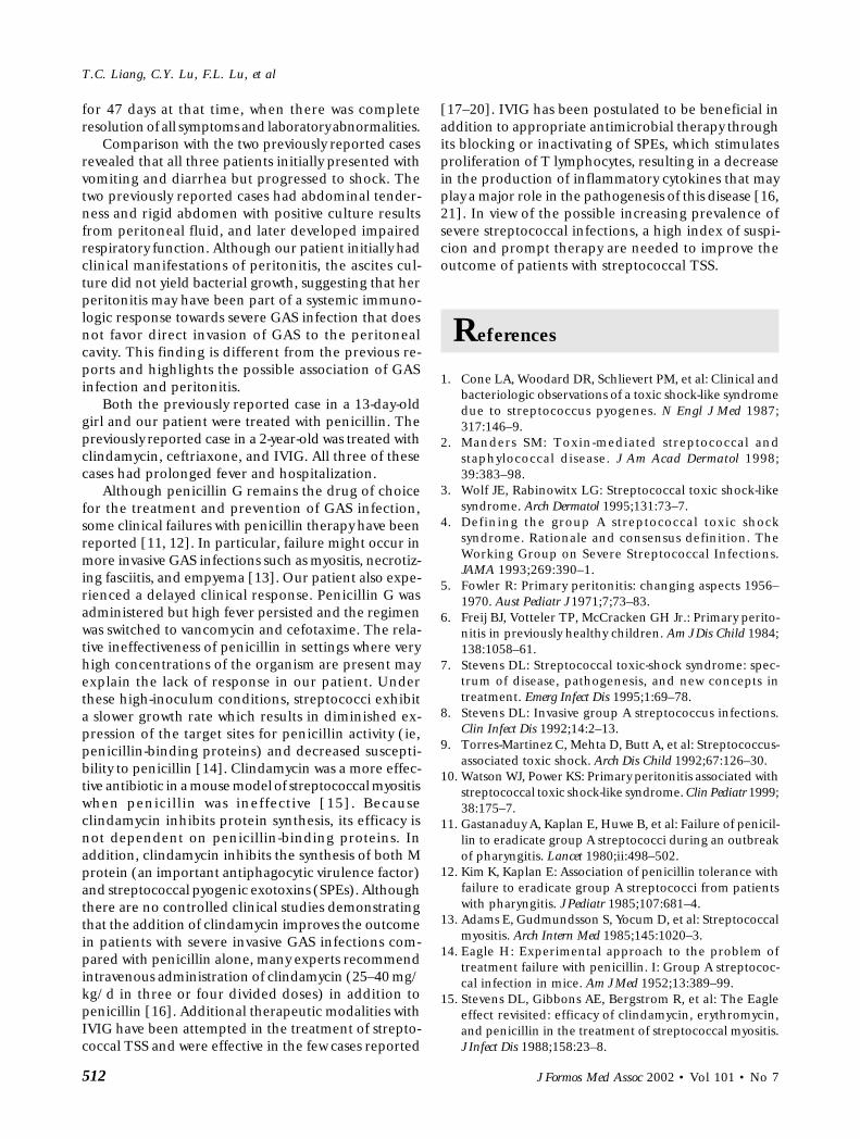
Fig. 3. Supine chest roentgenography shows increased infiltrationsover bilateral lung fields, especially near white-out of left lung field.
blood cell count, 20,600/µL), thrombocytopenia (plateletcount, 67,000/µL), and impaired renal function (blood ureanitrogen/creatine, 63.8/2.1 mg/dL) developed on the day aftersurgery. Chest echography revealed bilateral pleural effusion.Paracentesis from the right pleural cavity yielded 116 mLof turbid, yellowish, sticky fluid with 50,300 leukocytes/mL,70,000 red blood cells/mL, 3.1 g/dL protein, 65 mg/dL glucose,and 19,900 U/L lactate dehydrogenase. Numerous poly-mononuclear cells with gram-positive cocci were seenon Gram’s stain of the fluid, so the antibiotic regimenwas changed to vancomycin, ceftazidime, metronidazole,and azithromycin. Bacterial culture of the pleural effusionyielded GAS that was sensitive to penicillin. Diagnosis ofstreptococcal TSS was confirmed by culture result and clini-cal findings.
Thereafter, penicillin G (370,000 U/kg/d) was adminis-tered for 4 days. However, fever and intermittent abdominalpain persisted. Culture from the tip of the penrose drainplaced during surgery grew methicillin-resistant Staphylo-coccus aureus, and possible secondary infection wassuspected. The final antibiotic regimen used was vancomycinand cefotaxime. Fever subsided gradually thereafter and nofever was noted after the 19th day after surgery. Cefotaximewas discontinued but vancomycin was used for another 10days until the patient’s activity and appetite improved. Shewas discharged after hospitalization for 29 days withoutresidual symptoms.
Fig. 2. Plain abdominal roentgenography reveals local ileus over theleft upper abdomen.

J Formos Med Assoc 2002 • Vol 101 • No 7 511
Streptococcal Toxic Shock Syndrome and Peritonitis
Discussion
Serious infections due to GAS were common in theearly 1900s. After several decades of seemingly decreas-ing virulence, GAS infection has reemerged as a majorsource of morbidity and mortality. It is believed that theability of more virulent strains to produce circulatingtoxins leads to clinical disease [3].
The criteria to define streptococcal TSS include theisolation of GAS from a normally sterile site,hypotension, and involvement of at least two organsystems (renal impairment, coagulopathy, abnormalliver function, acute respiratory distress syndrome,skin rash, or soft tissue necrosis) [4]. Our patientfulfilled the diagnostic criteria of streptococcal TSS.This case is rare in regard to its initial clinicalmanifestations. Although pneumonia and empyemawere later confirmed, the initial clinical picture, in-cluding negative chest roentgenography, directed allattention to a possible abdominal event. Laparotomyrevealed peritonitis and lymphadenopathy. The lungappeared to be the primary affected organ as indicatedby the elevated left hemi-diaphragm on the first chestroentgenogram, which indicated infrapulmonaryeffusion. The actual etiology of the condition becameevident 1 day later and streptococcal empyema withTSS was confirmed.
Primary peritonitis is a diffuse suppurative perito-neal infection without an identifiable source that mayoccur in healthy infants and children as well as inchildren with underlying disease such as immuno-deficiency, cirrhosis, and nephrotic syndrome.Primary peritonitis is not a common pediatric disease.Studies from the early 1900s reported peritonitis in10% of pediatric abdominal emergencies, whereasreviews conducted in the 1960s and 1970s noted amarked decrease in the incidence of primary peritoni-tis [5]. Streptococcus pneumoniae and GAS were the tradi-tional etiologic agents [6]. By the 1970s, the number ofnephrotic children with streptococcal peritonitis haddeclined, and the relative frequency of peritonitiscaused by gram-negative bacilli and staphylococci ap-parently had increased. Primary peritonitis was oftenmisdiagnosed as appendicitis. Reddened intestine andenlarged mesenteric lymph nodes were often noted assurgical findings [6].
Streptococcal TSS occurs in people of all ages; mostdo not necessarily have predisposing underlying dis-eases [7]. Pain involving an extremity, usually abrupt inonset and severe, and fever are common early symptoms.Eighty percent of patients have clinical signs of softtissue infection. Most patients (70%) progress to ne-crotizing fasciitis or myositis and require surgical
debridement, fasciotomy, or amputation [8]. Multisys-tem disease with acute respiratory distress syndromeand renal failure frequently develop, as well as fever,striking leukocytosis, and elevated creatine kinaseconcentrations. Unusual presentations includeendophthalmitis, pneumonia, peritonitis, perihepatitis,and myocarditis [7].
Primary peritonitis associated with streptococcalTSS has rarely been reported. A review of the literaturerevealed only two childhood cases. The first was a 13-day-old infant [9] and the second was a 2-year-old girl[10]. The 13-day-old female patient had a 3-day historyof erythematous rash and diarrhea, vomiting for 24hours, and refusal to feed. She had a distended, rigidabdomen with absent bowel sound and blood pressureof 54/30 mmHg on admission. Initial investigationsshowed leukocytosis, thrombocytopenia, and prolongedprothrombin and partial thromboplastin time. Pro-found shock developed over the next 18 hours. Laparo-tomy was performed and purulent peritoneal fluid wasfound. High-dose penicillin was administered. Bloodand peritoneal cultures grew GAS that was sensitive topenicillin. Mechanical ventilation was required foranother 3 days after the operation and desquamationoccurred for more than 1 week, affecting most of herbody. Spiking fever with a pneumonia patch in theright lower lobe was noted. She was finally dischargedafter 22 days in the hospital after discontinuing antibi-otic therapy 1 day previously. Intermittent fever contin-ued for a further 20 days and she was asymptomatic andthriving on follow-up thereafter.
The previously reported 2-year-old female had haddiarrhea and vomiting for 2 days and 1 day of fever andirritability before admission [10]. She remained febrileand continued to have intermittent abdominal painand diarrhea after admission. On the third day ofhospitalization, distended abdomen and faint perium-bilical erythema developed. Paracentesis yielded cloudy,yellowish fluid with polymorphonuclear leukocytes andabundant intracellular and extracellular gram-positivecocci. Signs of shock developed 5 hours later. Herrespiratory condition deteriorated over the next 24hours and she required intubation. Her leukocyte andplatelet counts fell to 5,200/µL and 82,000/µL,respectively. GAS grew from peritoneal fluid, blood,and urine cultures. She received clindamycin andceftriaxone for a total of 14 days and 400 mg/kg ofintravenous immunoglobulin (IVIG) daily for 5 days.Erythematous macular rash waxed and waned overseveral weeks with intermittent desquamation that even-tually involved her forearms, palms, ankles, and soles aswell as her flanks, periumbilical area, and lowerabdomen. Fever with temperature greater than 39°Cpersisted for another 2 weeks after the discontinuationof antimicrobial therapy. She had been hospitalized
J Formos Med Assoc 2002 • Vol 101 • No 7
T.C. Liang, C.Y. Lu, F.L. Lu, et al
512
for 47 days at that time, when there was completeresolution of all symptoms and laboratory abnormalities.
Comparison with the two previously reported casesrevealed that all three patients initially presented withvomiting and diarrhea but progressed to shock. Thetwo previously reported cases had abdominal tender-ness and rigid abdomen with positive culture resultsfrom peritoneal fluid, and later developed impairedrespiratory function. Although our patient initially hadclinical manifestations of peritonitis, the ascites cul-ture did not yield bacterial growth, suggesting that herperitonitis may have been part of a systemic immuno-logic response towards severe GAS infection that doesnot favor direct invasion of GAS to the peritonealcavity. This finding is different from the previous re-ports and highlights the possible association of GASinfection and peritonitis.
Both the previously reported case in a 13-day-oldgirl and our patient were treated with penicillin. Thepreviously reported case in a 2-year-old was treated withclindamycin, ceftriaxone, and IVIG. All three of thesecases had prolonged fever and hospitalization.
Although penicillin G remains the drug of choicefor the treatment and prevention of GAS infection,some clinical failures with penicillin therapy have beenreported [11, 12]. In particular, failure might occur inmore invasive GAS infections such as myositis, necrotiz-ing fasciitis, and empyema [13]. Our patient also expe-rienced a delayed clinical response. Penicillin G wasadministered but high fever persisted and the regimenwas switched to vancomycin and cefotaxime. The rela-tive ineffectiveness of penicillin in settings where veryhigh concentrations of the organism are present mayexplain the lack of response in our patient. Underthese high-inoculum conditions, streptococci exhibita slower growth rate which results in diminished ex-pression of the target sites for penicillin activity (ie,penicillin-binding proteins) and decreased suscepti-bility to penicillin [14]. Clindamycin was a more effec-tive antibiotic in a mouse model of streptococcal myositiswhen penicillin was ineffective [15]. Becauseclindamycin inhibits protein synthesis, its efficacy isnot dependent on penicillin-binding proteins. Inaddition, clindamycin inhibits the synthesis of both Mprotein (an important antiphagocytic virulence factor)and streptococcal pyogenic exotoxins (SPEs). Althoughthere are no controlled clinical studies demonstratingthat the addition of clindamycin improves the outcomein patients with severe invasive GAS infections com-pared with penicillin alone, many experts recommendintravenous administration of clindamycin (25–40 mg/kg/d in three or four divided doses) in addition topenicillin [16]. Additional therapeutic modalities withIVIG have been attempted in the treatment of strepto-coccal TSS and were effective in the few cases reported
[17–20]. IVIG has been postulated to be beneficial inaddition to appropriate antimicrobial therapy throughits blocking or inactivating of SPEs, which stimulatesproliferation of T lymphocytes, resulting in a decreasein the production of inflammatory cytokines that mayplay a major role in the pathogenesis of this disease [16,21]. In view of the possible increasing prevalence ofsevere streptococcal infections, a high index of suspi-cion and prompt therapy are needed to improve theoutcome of patients with streptococcal TSS.
References
1. Cone LA, Woodard DR, Schlievert PM, et al: Clinical andbacteriologic observations of a toxic shock-like syndromedue to streptococcus pyogenes. N Engl J Med 1987;317:146–9.
2. Manders SM: Toxin-mediated streptococcal andstaphylococcal disease. J Am Acad Dermatol 1998;39:383–98.
3. Wolf JE, Rabinowitx LG: Streptococcal toxic shock-likesyndrome. Arch Dermatol 1995;131:73–7.
4. Defining the group A streptococcal toxic shocksyndrome. Rationale and consensus definition. TheWorking Group on Severe Streptococcal Infections.JAMA 1993;269:390–1.
5. Fowler R: Primary peritonitis: changing aspects 1956–1970. Aust Pediatr J 1971;7;73–83.
6. Freij BJ, Votteler TP, McCracken GH Jr.: Primary perito-nitis in previously healthy children. Am J Dis Child 1984;138:1058–61.
7. Stevens DL: Streptococcal toxic-shock syndrome: spec-trum of disease, pathogenesis, and new concepts intreatment. Emerg Infect Dis 1995;1:69–78.
8. Stevens DL: Invasive group A streptococcus infections.Clin Infect Dis 1992;14:2–13.
9. Torres-Martinez C, Mehta D, Butt A, et al: Streptococcus-associated toxic shock. Arch Dis Child 1992;67:126–30.
10. Watson WJ, Power KS: Primary peritonitis associated withstreptococcal toxic shock-like syndrome. Clin Pediatr 1999;38:175–7.
11. Gastanaduy A, Kaplan E, Huwe B, et al: Failure of penicil-lin to eradicate group A streptococci during an outbreakof pharyngitis. Lancet 1980;ii:498–502.
12. Kim K, Kaplan E: Association of penicillin tolerance withfailure to eradicate group A streptococci from patientswith pharyngitis. J Pediatr 1985;107:681–4.
13. Adams E, Gudmundsson S, Yocum D, et al: Streptococcalmyositis. Arch Intern Med 1985;145:1020–3.
14. Eagle H: Experimental approach to the problem oftreatment failure with penicillin. I: Group A streptococ-cal infection in mice. Am J Med 1952;13:389–99.
15. Stevens DL, Gibbons AE, Bergstrom R, et al: The Eagleeffect revisited: efficacy of clindamycin, erythromycin,and penicillin in the treatment of streptococcal myositis.J Infect Dis 1988;158:23–8.

J Formos Med Assoc 2002 • Vol 101 • No 7 513
Streptococcal Toxic Shock Syndrome and Peritonitis
16. American Academy of Pediatrics. Committee on Infec-tious Diseases: Severe invasive group A streptococcalinfections: a subject review. Pediatrics 1998;101:136–40.
17. Barry W, Hudgins L, Donta ST, et al: Intravenous immu-noglobulin therapy for toxic shock syndrome. JAMA1992;267:3315–6.
18. Yong JM: Necrotising fasciitis. Lancet 1994;343:1427.19. Lamothe F, D’Amico P, Ghosn P, et al: Clinical usefulness
of intravenous human immunoglobulins in invasive group
A streptococcal infections: case report and review. ClinInfect Dis 1995;21:1469–70.
20. Perez CM, Kubak BM, Cryer HG, et al: Adjunctive treat-ment of streptococcal toxic shock syndrome using intra-venous immunoglobulin: case report and review. Am JMed 1997;102:111–3.
21. Stevens DL: Editorial response: Rationale for the use ofintravenous gamma globulin in the treatment of streptococ-cal toxic shock syndrome. Clin Infect Dis 1998;26:639–41.