serum opacity factor, a streptococcal virulence factor ... · causes pharyngitis, tonsilitis,...
TRANSCRIPT

1
Serum Opacity Factor, a Streptococcal Virulence Factor that Binds to Apolipoproteins A-I and A-II and Disrupts High-Density Lipoprotein
Structure* Harry S. Courtney†¶, Yong-Mei Zhang‡, Matthew W. Frank‡, and Charles O. Rock‡
From the †Veterans Affairs Medical Center and Department of Medicine, University of Tennessee Health Science Center, Memphis, Tennessee 38104, and the ‡Department of Infectious Diseases, St. Jude
Children's Research Hospital, Memphis, Tennessee 38105 Running Title: Mechanism of SOF Action
¶Address Correspondence to: Harry S. Courtney, Veterans Affairs Medical Center, Research Service (151), 1030 Jefferson Avenue, Memphis, TN 38104. Tel: 901-523-8990 ext 7548; FAX: 901-577-7273; Email: [email protected].
*This work was supported by research funds from the Department of Veterans Affairs (H.S.C.), National Institutes of Health grant GM 34496 (C.O.R.), Cancer Center (CORE) support grant CA 21765, and the American Lebanese Syrian Associated Charities. The costs of publication of this article were defrayed in part by the payment of page charges. This article must therefore be hereby marked “advertisement” in accordance with 18 U.S.C. Section 1734 solely to indicate this fact.
1Abbreviations used are: SOF, serum opacity factor; rSOF, recombinant opacification domain of SOF; HDL, high-density lipoprotein; LDL, low-density lipoprotein; VLDL, very-low density lipoprotein; DCIC, 3,4-dichloroisocoumarin
Serum opacity factor (SOF) is a virulence determinant of group A streptococci that opacifies mammalian sera. We analyzed the specificity and mechanism of the opacity reaction using a recombinant form of the N-terminal opacification domain of SOF, rSOF. Our data indicate that rSOF is neither a protease nor a lipase, but rather it is the binding of rSOF to high density lipoprotein (HDL) that triggers the opacity reaction. rSOF did not opacify plasma from apoAI–/– mice or purified low- or very low-density lipoproteins, but readily opacified HDL. rSOF binding to HDL was characterized by two high affinity binding sites, it bound to ApoAI (Kd = 6 nM) and ApoAII (Kd = 30 nM), and both ApoAI and ApoAII blocked the binding of rSOF to HDL. Electron microscopic examination and biochemical analyses of HDL treated with rSOF revealed the formation of lipid droplets devoid of apolipoproteins. Thus, SOF interacts with HDL in human blood by binding to ApoAI and ApoAII and causing the release of HDL’s lipid cargo, which coalesces to form lipid droplets, resulting in opacification. The disruption of HDL may attenuate its anti-inflammatory functions and contribute to the pathogenesis of group A streptococcal infections.
SOF was discovered in 1938 as a substance produced by the group A streptococcus, Streptococcus pyogenes, that turns mammalian serum opaque (1). It is expressed by approximately half of the clinical isolates of S. pyogenes (2), an important human pathogen that causes pharyngitis, tonsilitis, impetigo, necrotizing fasciitis, and toxic shock syndrome. SOF is a large protein with a Mr of ~110 kDa that is found in both culture supernatants and attached to the surface of streptococci via its cell-wall anchoring motif LPASG. The sof genes from different serotypes of S. pyogenes have approximately 60% homology between their deduced amino acid sequences (3-5). The N-terminal two-thirds of SOF are composed of alternating variable and conserved regions. The C-terminus of SOF is highly conserved and contains a repeating peptide domain that binds to fibronectin (3,4,6,7) and fibrinogen (8), and mediates streptococcal adhesion to host cells (9). The binding of fibronectin is an important function for group A streptococci as indicated by the findings that these bacteria express at least ten different fibronectin-binding proteins (10) that have roles in adhesion and invasion (11). The opacification domain of SOF is contained within amino acid residues 148 to 843 (Fig. 1) and is clearly distinct from the fibronectin-binding domain, as the deletion of the fibronectin-binding domain had no effect on the
http://www.jbc.org/cgi/doi/10.1074/jbc.M512538200The latest version is at JBC Papers in Press. Published on January 3, 2006 as Manuscript M512538200
by guest on April 26, 2019
http://ww
w.jbc.org/
Dow
nloaded from

2
opacity reaction of SOF (3-6). Insertional inactivation of sof reduced the virulence of S. pyogenes in a mouse model of infection (3) and reduced the growth of S. pyogenes in human blood (12), and immunization of mice with SOF provided protection against infections by S. pyogenes (13), identifying SOF as a virulence determinant and a vaccine candidate.
Various catalytic activities have been proposed to account for the opacity reaction of SOF, including those as a lipoproteinase (14), a cholesterol esterase (15), and more recently as an apolipoproteinase that degrades apolipoprotein A-I (ApoAI), the major protein in high density lipoprotein particles (HDL) (16). HDL functions primarily in the reverse cholesterol pathway by removing cholesterol from peripheral tissues and transporting it to the liver (17). In addition, HDL plays a major role in controlling atherogenesis and inflammation due to infections. HDL neutralizes the pro-inflammatory activities of lipopolysaccharide from Gram-negative bacteria and lipoteichoic acid from Gram-positive bacteria (18-21). It is thought that HDL has a prominent role in controlling sepsis and shock because HDL attenuates the cytokine response, reduces the rate of mortality in animal sepsis models, and the concentration of HDL decreases in septic patients (20). Thus, bacterial factors that disrupt HDL structure would diminish its ability to modulate host responses to bacterial infections and contribute to the pathogenesis of infections. This report describes the use of recombinant SOF (rSOF; Fig. 1) to investigate its mechanism of interaction with serum lipoproteins that gives rise to the serum opacification phenomenon first described in 1938.
EXPERIMENTAL PROCEDURES Materials—Purified human very low-density
lipoproteins (VLDL), low-density lipoproteins (LDL), HDL, apolipoprotein A-I (ApoAI), and apolipoprotein A-II (ApoAII) were purchased from Calbiochem (San Diego, CA). A polyhistidine tagged, truncated form of sof2 encoding amino acids 38-843 was cloned and expressed in E. coli (rSOF) and purified by metal affinity chromatography as previously described (3).
Opacification Assays—The capacity of SOF to opacify horse and human serum was determined
by adding 50 µl of rSOF at various concentrations to 2 ml of serum and recording the absorbance at 405 nm at timed intervals. The human lipoproteins, HDL, VLDL, and LDL were diluted in 0.05 M Tris-HCl, 0.15 M NaCl, pH 7.2 to ~2 mg/ml and 200 µl were added to microtiter wells. Ten µl of rSOF were added to the wells to obtain 0, 0.1, 1.0, 10 µg/ml final concentrations. The microtiter plate was incubated at 37°C and the absorbance at 405 nm recorded at timed intervals.
Plasma from the mouse strain B6.129P2-Apoa1tm1Unc/J was obtained from The Jackson Laboratory (Bar Harbor, ME). These mice have a targeted mutation in apoAI and do not express ApoAI in their plasma. Control plasma was collected from C5BL/6J mice. Ten µl of buffer or rSOF (1 µg/ml final concentration) were added to 200 µl of plasma from these mice and incubated for 3 hours at 37°C and the absorbance at 405 nm recorded.
Absorption of Lipoproteins—Human sera were absorbed with PHM-L Liposorb beads according to the manufacturers directions (Calbiochem, San Diego, CA). The absorbed sera and unabsorbed sera were reacted with rSOF (1 µg/ml) or buffer overnight at 37°C. Two hundred µl were added to microtiter wells in quadruplicate and the absorbance at 405 nm recorded. Absorbed and unabsorbed sera reacted with buffer served as blanks.
Assays for Inhibitors of SOF—Stock concentrations of inhibitors were prepared in dimethyl sulfoxide (DMSO) or Tris-saline as required for solubility. Inhibition assays consisted of the addition of 100 µl of inhibitor to 1.8 ml of horse serum followed by addition of 100 µl of rSOF to obtain a final concentration of 1 µg/ml. The mixtures were incubated at 37°C for 3 hours and then the absorbance at 405 nm was measured. Blank controls consisted of Tris-saline or DMSO without inhibitors. Percent of control was determined by the formula (A405 with test agent/A405 with Tris-saline) × 100. Inhibitors were purchased from Calbiochem except for the cocktail of protease inhibitors which was obtained from Sigma (St. Louis, MO).
Assays to determine if 3,4-dichloroisocoumarin (DCIC) targets SOF or HDL were performed by preincubating rSOF or horse serum with concentrations of DCIC ranging from 22 µM to 2.2 mM for 3 hours at 37°C. Controls
by guest on April 26, 2019
http://ww
w.jbc.org/
Dow
nloaded from

3
included rSOF or serum pretreated with DMSO or Tris-saline. After pretreatment, the mixtures were dialyzed overnight against Tris-saline using a membrane with a molecular weight sizing of 50 kDa. DCIC-treated rSOF was then added to fresh horse serum and non-treated rSOF was added to DCIC-treated horse serum. In each case, the final concentration of rSOF was 1 µg/ml. The mixtures were incubated overnight at 37°C and the absorbance at 405 nm was recorded. Percentage of maximum opacification was calculated by the formula (A405 of pretreated sample/A405 of control) x 100.
Lipid Analysis—Lipids were extracted using chloroform:methanol as described by Bligh and Dyer (22). Lipid class distribution was determined by thin-layer chromatography on thin rods developed with hexane:ethylether:acetic acid (80/20/0.5, v/v) for the neutral lipids and chloroform:methanol:acetic acid: water (50/25/8/3, v/v) for phospholipid separation. The individual classes were quantitated using the Iatroscan MK-6 thin-layer chromatograph equipped with a flame ionization detector, and classes were identified by comparison with standards.
AlphaScreen Binding Assays—The AlphaScreen technique for measuring biological interactions has been described in detail (23). It measures energy transfer between a donor bead (streptavidin coated) and acceptor bead (nickel coated) that have been bound to biotinylated and histidine-tagged compounds, respectively. When the two components interact, excitation energy is transferred from the donor to the acceptor bead resulting in the emission of high amplitude fluorescence, or the AlphaScreen signal. In our experiments, rSOF was engineered with a polyhistidine tag and the lipoproteins, VLDL, LDL, and HDL, as well as the purified apolipoproteins AI and AII were biotinylated using the EZ-Link NHS-Sulfo-Biotin kit from Pierce Biotechnology Inc. using the methods recommended by the manufacturer. The parameters of the binding assay for HDL-rSOF interactions were 100 nM rSOF and concentrations of biotinylated HDL ranging from 0 to 0.09 µg/ml. Concentrations of biotinylated VLDL and LDL ranged between 0 to 2.5 µg/ml in binding assays with rSOF. Binding assays between rSOF and purified apolipoproteins used 100 nM rSOF and concentrations of biotinylated ApoAI and ApoAII
ranging between 0 and 0.05 µM. For the competitive inhibition experiments, 100 nM rSOF, 0.2 µg/ml biotinylated HDL, and concentrations of unlabeled ApoAI and ApoAII ranging between 0 and 0.8 µM were used. The biotinylated compounds (and unlabeled compounds when present) were mixed with rSOF for 30 min followed by the addition of streptavidin donor beads and nickel chelate acceptor beads (50 µg/ml final concentration for each). The reaction mixtures were then incubated for 1 hour before being read by the Fusion Universal Microplate Analyzer with excitation at 680 nm and emission at 600 nm. The binding constants were calculated using non-linear equations for either one site Y=Bmax*X/(Kd+X) or two site (Y=Bmax1*X/(Kd1+X) + Bmax2*X/(Kd2+X)) binding where Bmax is the maximal binding and Kd is the concentration of ligand required to reach half-maximal binding.
Ultracentrifugation of HDL—Untreated HDL or HDL treated overnight with 1 µg/ml of rSOF were centrifuged at 35,000 rpm for 16 hr at 15°C in a Beckman TL-100 ultracentrifuge. The top layer (200 µl) of opaque material and the bottom fraction (200 µl) were collected and the absorbance at 405 nm was measured. These materials were subjected to 14% SDS-PAGE and stained with Coomassie Brilliant Blue R250. The lipid composition of the layers was determined as described above. Untreated HDL served as a control.
Western Blots—The top and bottom layers generated by the ultracentrifugation experiments described above were subjected to SDS-PAGE and transferred to nitrocellulose. The membrane was blocked with bovine serum albumin and incubated with a 1:500 dilution of rabbit anti-rSOF serum. The membrane was washed and incubated with a 1:7,500 dilution of peroxidase-conjugated goat anti-rabbit IgG. After washing, immunoreactive bands were detected using ECL western blot reagents (Amersham).
RESULTS
Lipoprotein Specificity of rSOF—We cloned, expressed, and purified a recombinant, polyhistidine-tagged, truncated form of SOF in which the fibronectin-binding domain was deleted (rSOF) (Fig. 1). Previous studies show that this
by guest on April 26, 2019
http://ww
w.jbc.org/
Dow
nloaded from

4
construct retains full ability to opacify serum and that the fibronectin-binding domain and the opacification domain of SOF are functionally independent (3). The opacity reaction of rSOF was time and dose dependent with maximal activity occurring at approximately 1 µg/ml (Fig. 2A-B). The opacity reaction appeared to decrease at high concentrations of rSOF. However, high concentrations of rSOF did not alter the total amount of lipid released, but rather reduced the size of the lipid particles (Fig. 2D-F). The opacification assay, which measures the absorbance at 405 nm, relies on light scattering and is dependent upon particle size. Thus, as particle size decreases so will the absorbance.
To investigate the potential role of lipoproteins in the opacity reaction of rSOF, sera from two donors were absorbed with Liposorb beads to remove lipoproteins. This treatment reduced the opacity reaction of rSOF by 99% (A405 unabsorbed sera = 0.932 ± 0.20, A405 absorbed sera = 0.007 ± 0.011), indicating that one or more of the lipoproteins were the target of rSOF. To resolve which of the lipoproteins is the target, purified lipoproteins were incubated with rSOF overnight at 37°C, and the opacification quantified by measuring the absorbance at 405nm (Fig. 2C). There was little or no increase in the absorbance when rSOF was added to LDL or VLDL, but there was a dramatic increase in the absorbance of HDL treated with rSOF. These results show that the opacification activity of SOF selectively targets HDL.
Additional support for a role of HDL in these macroscopic reactions came from the finding that plasma from mice that do not express ApoAI was not opacified by rSOF (A405 = 0.035), whereas the plasma from control mice became opaque when treated with rSOF (A405 = 0.807). Thus, the opacity reaction of plasma from ApoAI-negative mice was less than 5% of the response of wild type plasma to rSOF. In addition to the absence of ApoAI, the apoAI–/– mice have dramatically reduced levels of HDL, reduced levels of ApoAII, and ApoC, but have increased levels of ApoE and ApoAIV (24,25). The inability of rSOF to opacify plasma devoid of ApoAI supported the conclusion that HDL was the SOF target.
rSOF Catalytic Activity—It was proposed that SOF is an esterase that would potentially be capable of degrading the lipid components of the
HDL particle (26). Therefore, the lipid compositions of untreated and rSOF-treated HDL were compared to determine if there was any evidence for lipid degradation following rSOF treatment (Fig. 3A). The lipids of HDL mainly consists of cholesterol esters, cholesterol, phosphatidylcholine and a trace of triglycerides (27). If there was a significant decrease in the amount of one of the lipids, then there should also be a concomitant increase in its degradation products. For example, phospholipase hydrolysis of the phospholipids would increase the concentrations of lysophospholipids and free fatty acids. We incubated HDL with 10 µg/ml rSOF for 18 h in triplicate and the amount of phosphatidylcholine in the control without rSOF was 396 ± 60 µg/assay compared to 415 ± 57 µg/assay in the SOF-treated samples. Similarly, cholesterol ester was 428 ± 53 µg/assay in the control and 387 ± 73 µg/assay in the 10 µg/ml SOF-treated HDL. We also performed the experiment using 100 µg/ml rSOF and found the phosphatidylcholine levels were 414 ± 51 µg/assay and the cholesterol ester were 355 ± 81 µg/assay after an 18 h incubation. An example of the chromatographic analysis is shown in Figs. 3A and 3B illustrating that incubation of HDL with 100 µg/ml rSOF for 18 h did not result in a significant increase in either lysophosphatidylcholine or free fatty acids. These assays performed in the presence of very high concentrations of rSOF strongly argue against a lipase-like activity for the protein. In addition, a panel of lipase or phospholipase inhibitors did not block the serum opacity reaction (Table 1). Thus, rSOF does not alter the lipid composition of HDL and is not a lipase, phospholipase, or esterase.
It has also been suggested that SOF is an aspartic protease that induces opacification of serum by cleavage of ApoAI of HDL (16). This possibility was examined by incubating rSOF with HDL overnight followed by gel electrophoresis to detect fragmentation or degradation of the ApoAI and ApoAII protein components. The results demonstrated that 10 µg/ml of rSOF (ten times the concentration required for optimal opacification) did not produce fragmentation of either ApoAI or ApoAII (Fig. 3C). Various protease inhibitors were also tested for their ability to block the opacity reaction of rSOF (Table I). The finding
by guest on April 26, 2019
http://ww
w.jbc.org/
Dow
nloaded from

5
that a variety of protease inhibitors, including a cocktail of protease inhibitors containing the aspartic protease inhibitor pepstatin A, had no inhibitory effect on the opacity reaction of rSOF supports the conclusion that rSOF is not a protease.
Interestingly, one inhibitor, DCIC, did block 77% of the serum opacity reaction. In the experiments listed in Table I, the inhibitors were added to a mixture of rSOF and serum. Thus, it was not clear if DCIC was targeting rSOF directly or inhibiting a component associated with HDL that is activated by rSOF in some manner. For example, phospholipase A2 is associated with HDL (28). To distinguish between these possibilities, various concentrations of DCIC were added to serum or to rSOF and incubated for three hours. DCIC has a half-life of 20 minutes, and less than 1% of DCIC remained active after 3 hours. The residual DCIC was removed by dialysis and the DCIC-treated samples were analyzed for activity (Fig. 4A). DCIC-treated rSOF did not opacify serum, whereas DCIC-treated serum was readily opacified by rSOF. These data indicate that the inhibitory effect of DCIC was due to its interaction with rSOF.
The finding that DCIC blocked the serum opacity reaction of rSOF but other inhibitors with similar specificity failed to block this reaction raised a question concerning the mechanism of inhibition of rSOF by DCIC. DCIC inhibits phospholipase and serine protease activity by irreversible modification of the serine active site (29); however, DCIC also alters amino acids outside of the active site (30). Thus, DCIC may modify a number of amino acid residues of rSOF and interfere with its binding to HDL. To test this hypothesis, rSOF was treated with DCIC and its binding to HDL measured. DCIC-treated rSOF failed to bind to HDL (Fig. 4B), suggesting that DCIC did not interfere with an enzyme activity of rSOF, but inhibited the opacity reaction of rSOF by blocking the interaction of rSOF with HDL. This concept was supported by the findings that none of the other phospholipase or serine protease inhibitors had any effect on the opacity reaction of rSOF and that rSOF did not alter the lipids or proteins of HDL. These data indicated that rSOF is neither a protease nor a lipase and suggested that the binding of rSOF to HDL was required to initiate the opacity reaction.
Binding of rSOF to HDL, ApoAI and ApoAII—Quantitative binding assays indicated that rSOF bound to HDL with high affinity (Fig. 5A). These data fit best to a two site binding model (R2 = 0.997) with apparent Kds for rSOF of 0.3 ± 0.1 ng/ml and 27 ± 3 ng/ml (Fig. 5A). rSOF also bound to LDL and VLDL (Fig. 5B). These binding data best fit a single site binding equation (R2 = 0.996) with apparent Kds for rSOF of 0.2 ± 0.25 µg/ml and 0.15 ± 0.03 µg/ml for LDL and VLDL, respectively. It is likely that the high affinity binding sites in HDL are found on one or more of its protein components, because the lipids of HDL are also found in other lipoproteins, whereas its major proteins are distinct. Competitive inhibition experiments demonstrated that both ApoAI and ApoAII blocked the binding of rSOF to HDL (Fig. 6A). Furthermore, rSOF bound to purified ApoAI (Kd 6 nM) and to purified ApoAII (Kd 30 nM) (Fig. 6B and 6C). These data indicated that both ApoAI and ApoAII were the receptors for rSOF on HDL particles.
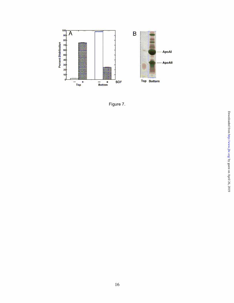
Morphological Changes in HDL—Electron micrographs showed that treatment of HDL with rSOF induced the formation of very large, lipid-like particles that appeared to be the result of the fusion of smaller lipid particles (Fig. 2E). If so, then the density of these particles should be quite different from untreated HDL and should migrate differently during centrifugation. Therefore, HDL was treated with 1 µg/ml of rSOF, subjected to ultracentrifugation, and the top and bottom layers were collected and analyzed. In the untreated HDL samples, the lipids and apolipoproteins remained together and sedimented to the bottom of the gradient (Fig. 7A). In the rSOF-treated HDL samples, an opaque layer formed at the surface of the gradient corresponding to approximately 75% of the lipids in the sample (Fig. 7A). The apolipoproteins along with 25% of the lipid were found in the bottom layer and only trace amounts of proteins in the top layer (Fig. 7A and 7B). Lipid analyses of the top opaque layer indicated that its lipid profile was the same as untreated HDL, providing additional evidence that rSOF did not alter the lipid composition of HDL (data not shown). These findings suggested that rSOF induced changes in HDL that resulted in the formation of lipid particles that were essentially free of apolipoproteins and it is these protein-
by guest on April 26, 2019
http://ww
w.jbc.org/
Dow
nloaded from

6
deficient particles responsible for turning serum opaque due to their insolubility in aqueous media.
Western blot analysis using anti-rSOF serum indicated that rSOF was associated with both the apolipoprotein fraction (50%) and the lipid-rich top layer (50%) (not shown). This finding verified the association of rSOF with apolipoproteins but also suggests that rSOF may interact to some extent with lipids. The amount of rSOF in this experiment (1 µg/ml) was 10-times higher than the amount required for opacification (Fig. 2) and was sufficient to saturate the high-affinity apolipoprotein binding sites (Fig. 5).
DISCUSSION
This work establishes that SOF binds to the apolipoprotein components of HDL to trigger the macroscopic serum opacity reaction. Only HDL among the lipoproteins produced the opacity reaction and rSOF did not opacify plasma from ApoAI-deficient mice. Our results showing that lack of hydrolysis of either lipids or proteins in HDL particles treated with rSOF differs from previous studies and suggested that hydrolytic enzymatic mechanisms did not account for the serum opacification reaction of SOF (14-16). Although DCIC is a hydrolase inhibitor that blocked the opacity reaction of rSOF, this inhibitor is not specific for hydrolase active sites and covalently modifies residues in other regions of proteins (30). Our data show that DCIC-modified rSOF cannot bind to HDL (Fig. 4).
rSOF bound to purified ApoAI and ApoAII and these polypeptides blocked the binding of rSOF to HDL leading to the conclusion that the SOF opacification domain interacts with HDL through ApoAI/ApoAII. ApoAI subunits span the surface of a mature particle and HDL assembly is mediated by interactions between lipids and the hydrophobic face of the carboxy-terminal amphipathic α-helix of ApoAI (27,31). There is a large conformational change that occurs when ApoAI interacts with lipids and is incorporated into an HDL particle, reflecting a transition from 55% α-helical to 75% α-helical content (27). We
propose that the binding of rSOF to the apolipoproteins of HDL induces a conformational change that reduces their ability to maintain interactions with the lipids, resulting in the disruption of the particle and release of lipids that fuse to form large, lipid particles that turn serum opaque. Although rSOF interacted with LDL and VLDL particles, this interaction was not accompanied by the dissolution of their structure or the formation of coalesced lipid droplets. The association of rSOF with the protein-deficient lipid particles suggests that rSOF binds to lipids. This property may account for the low-affinity binding of rSOF to LDL and VLDL; however, the mechanism of rSOF interaction with these lipoproteins remains to be clarified.
It is becoming increasingly apparent that bacterial infections alter the levels and structures of plasma lipoproteins. HDL is an anti-inflammatory and anti-atherogenic factor that neutralizes the pro-inflammatory responses caused by lipopolysaccharide, lipoteichoic acid, and acute phase response proteins such as C-reactive protein (20,32). Infections and shock can trigger events that result not only in the loss of the anti-inflammatory properties of HDL, but also in the conversion of HDL to a pro-inflammatory form (33). Exogenous HDL has been used effectively to combat sepsis and shock in animals (20) and ablate inflammatory responses in humans (34), illustrating its importance in controlling these events. SOF is an established virulence factor in S. pyogenes (3,12,13), and although the importance of its carboxy-terminal fibronectin-binding properties cannot be dismissed, the opacification domain of SOF disrupts HDL structure and may contribute to the pathogenesis of streptococcal infections by attenuating the anti-inflammatory functions of intact HDL.
Acknowledgements—We thank Yi Li for the excellent technical assistance and Loretta Hatmaker for preparing the electron micrographs.
REFERENCES
1. Ward, H. K., and Rudd, G. V. (1938) Aust J Exp Biol Med Sci 16, 181-192
by guest on April 26, 2019
http://ww
w.jbc.org/
Dow
nloaded from
7
2. O'Brien, K. L., Beall, B., Barrett, N. L., Cieslak, P. R., Reingold, A., Farley, M. M., Danila, R., Zell, E. R., Facklam, R., Schwartz, B., and Schuchat, A. (2002) Clin Infect Dis 35, 268-276
3. Courtney, H. S., Hasty, D. L., Li, Y., Chiang, H. C., Thacker, J. L., and Dale, J. B. (1999) Mol Microbiol 32, 89-98
4. Rakonjac, J. V., Robbins, J. C., and Fischetti, V. A. (1995) Infect Immun 63, 622-631 5. Kreikemeyer, B., Talay, S. R., and Chhatwal, G. S. (1995) Mol Microbiol 17, 137-145 6. Kreikemeyer, B., Martin, D. R., and Chhatwal, G. S. (1999) FEMS Microbiol Lett 178, 305-311 7. Jeng, A., Sakota, V., Li, Z., Datta, V., Beall, B., and Nizet, V. (2003) J Bacteriol 185, 1208-1217 8. Courtney, H. S., Dale, J. B., and Hasty, D. L. (2002) Curr Microbiol 44, 236-240 9. Oehmcke, S., Podbielski, A., and Kreikemeyer, B. (2004) Infect Immun 72, 4302-4308 10. Courtney, H. S., Hasty, D. L., and Dale, J. B. (2002) Ann Med 34, 77-87 11. Courtney, H. S., and Podbielski, A. (2004) Group A Streptococcal Invasion of Host Cells. In:
Lamont, R. J. (ed). Bacterial Invasion of Host Cells, Cambridge University Press, Cambridge 12. Courtney, H. S., Hasty, D. L., and Dale, J. B. (2005) Mol Microbiol In press 13. Courtney, H. S., Hasty, D. L., and Dale, J. B. (2003) Infect Immun 71, 5097-5103 14. Krumwiede, E. (1954) J Exp Med 100, 629-638 15. Rowen, R., and Martin, J. (1963) Biochemica et Biophysica Acta 70, 396-405 16. Saravani, G. A., and Martin, D. R. (1990) FEMS Microbiol Lett 56, 35-39 17. Fielding, P. E., and Fielding, C. J. (1996) Dynamics of lipoprotein transport in the human
circulatory system. In: Vance, D. E., and Vance, J. E. (eds). Biochemistry of Lipids, Lipoproteins and Membranes, Elsevier, Amsterdam
18. Parker, T. S., Levine, D. M., Chang, J. C., Laxer, J., Coffin, C. C., and Rubin, A. L. (1995) Infect Immun 63, 253-258
19. Grunfeld, C., Marshall, M., Shigenaga, J. K., Moser, A. H., Tobias, P., and Feingold, K. R. (1999) J Lipid Res 40, 245-252
20. Wu, A., Hinds, C. J., and Thiemermann, C. (2004) Shock 21, 210-221 21. Levine, D. M., Parker, T. S., Donnelly, T. M., Walsh, A., and Rubin, A. L. (1993) Proc Natl Acad
Sci U S A 90, 12040-12044 22. Bligh, E. G., and Dyer, W. J. (1959) Can J Biochem Physiol 37, 911-917 23. Zhang, Y. M., Wu, B., Zheng, J., and Rock, C. O. (2003) J Biol Chem 278, 52935-52943 24. Williamson, R., Lee, D., Hagaman, J., and Maeda, N. (1992) Proc Natl Acad Sci U S A 89, 7134-
7138 25. Li, H., Reddick, R. L., and Maeda, N. (1993) Arterioscler Thromb 13, 1814-1821 26. Rowen, R. (1961) Journal of Experimental Medicine 114, 807-823 27. Lund-Katz, S., Liu, L., Thuahnai, S. T., and Phillips, M. C. (2003) Front Biosci 8, d1044-1054 28. Petrovic, N., Grove, C., Langton, P. E., Misso, N. L., and Thompson, P. J. (2001) J Lipid Res 42,
1706-1713 29. Harper, J. W., and Powers, J. C. (1985) Biochemistry 24, 7200-7213 30. Rusbridge, N. M., and Beynon, R. J. (1990) FEBS Lett 268, 133-136 31. Rye, K. A., Clay, M. A., and Barter, P. J. (1999) Atherosclerosis 145, 227-238 32. Wadham, C., Albanese, N., Roberts, J., Wang, L., Bagley, C. J., Gamble, J. R., Rye, K. A.,
Barter, P. J., Vadas, M. A., and Xia, P. (2004) Circulation 109, 2116-2122 33. Khovidhunkit, W., Kim, M. S., Memon, R. A., Shigenaga, J. K., Moser, A. H., Feingold, K. R.,
and Grunfeld, C. (2004) J Lipid Res 45, 1169-1196 34. Pajkrt, D., Doran, J. E., Koster, F., Lerch, P. G., Arnet, B., van der Poll, T., ten Cate, J. W., and
van Deventer, S. J. (1996) J Exp Med 184, 1601-1608
by guest on April 26, 2019
http://ww
w.jbc.org/
Dow
nloaded from
8
FIGURE LEGENDS
FIG. 1. The serum opacity reaction and domain structure of SOF and the recombinant protein, rSOF. Panel A, the macroscopic serum opacity reaction of rSOF. Human serum was incubated overnight with either buffer (left tube) or 1 µg/ml rSOF (right tube). Panel B, model of SOF. The location and assignment of the functional domains in SOF are based on the findings of Rakonjac et al. (4), Kreikemeyer et al. (5), and Courtney et al. (3). The recombinant form of SOF (rSOF) used in this study encompassed residues 38-843 and was constructed with a polyhistidine amino-terminal tag to facilitate purification and HDL interaction studies. Relevant protein domains are labeled; Fn, fibronectin-binding repeats; LPASG anchor, the region responsible for anchoring SOF to the bacterial cell wall.
FIG. 2. The lipoprotein specificity for the opacity reaction of purified rSOF. Panel A, the time-dependent opacification of serum was determined by adding either 1 µg/ml rSOF (closed circles) or 0.1 µg/ml rSOF (open circles) to serum and recording the A405 at timed intervals. Panel B, the concentration-dependent opacification of serum by rSOF. Serum was treated for 3 hours with the indicated concentrations of rSOF and the absorbance of the solutions determined at 405 nm. Panel C, the lipoprotein specificity of rSOF was determined by incubating rSOF (1 µg/ml) with purified human lipoproteins for 3 hours and recording the absorbance at 405 nm. Panels D-F, Purified human HDL was incubated overnight at 37°C with buffer (Panel D), 1 µg/ml rSOF (Panel E), or 10 µg/ml rSOF (Panel F). The samples were stained with 2% phosphotungstic acid and electron micrographs taken at 30,000× magnification.
FIG. 3. rSOF does not hydrolyze the components of HDL. The lipid profile of HDL treated for 18 h with 100 µg/ml rSOF was determined by lipid extraction, thin-layer chromatography and quantitation with the Iatroscan. This instrument employs a flame-ionization detector to measure the mass of lipids separated on a thin-layer rod. The position of free fatty acids and lysophosphatidylcholine are indicated. Panel A, a representative example of the separation of the HDL phospholipids following treatment with 100 µg/ml SOF. Phosphatidylcholine (PtdCho) did not reveal significant degradation to lysophosphatidylcholine (LysoPC), which comprised 1.6% of the total choline phospholipids. The small sphingomyelin (SM) peak is also noted. Other minor lipids, like phosphatidylinositol and phosphatidylethanolamine migrate further up the rod and are not shown. Panel B, the separation of the neutral lipid species showed primarily cholesterol and cholesterol ester. There was no accumulation of free fatty acids (FA), which was 0.6% of the cholesterol ester peak in both samples, indicating that there was no hydrolysis of either cholesterol ester or phosphatidylcholine following treatment with 100 µg/ml rSOF. Panel C, the effect of rSOF on the protein profile of HDL was determined by SDS gel electrophoretic analysis of HDL treated with 0, 1, and 10 µg/ml of rSOF as detailed in “Experimental Procedures.”
FIG. 4. DCIC inhibition of serum opacification and rSOF binding to HDL. Panel A, DCIC targets rSOF. Various concentrations of DCIC were added to serum or to rSOF and incubated for 3 hours at 37°C and samples dialyzed. DCIC-treated rSOF (open circles) was added to untreated serum and DCIC-treated serum (closed circles) was added to untreated rSOF. The concentration of rSOF was 1 µg/ml in each case. The samples were incubated for 3 hours and the absorbance at 405 nm was recorded. Note that DCIC treatment of rSOF prevented the opacity reaction but DCIC treatment of serum did not block the opacity reaction. Panel B, DCIC blocks binding of rSOF to HDL. rSOF was pretreated with DCIC (open box) or DMSO (closed box) as described above and its binding to HDL quantified using an AlphaScreen binding assay as described under “Experimental Procedures.” Assays contained 100 nM rSOF and 0.08 µg/ml HDL, and assays were performed in triplicate and the standard deviation is shown.
FIG. 5. Interaction of rSOF with HDL, LDL and VLDL. Panel A, binding of rSOF to various concentrations of HDL. Panel B, binding of rSOF to LDL and VLDL. The binding of the various concentrations of biotinylated HDL, LDL, and VLDL to 100 nM rSOF was determined using the AlphaScreen binding assays and data analysis as described under “Experimental Procedures.” Assays were performed in triplicate and the standard deviation is shown.
by guest on April 26, 2019
http://ww
w.jbc.org/
Dow
nloaded from
9
FIG. 6. Binding of rSOF to ApoAI and ApoAII. Panel A, various concentrations of ApoAI (closed circles) and ApoAII (open circles) were tested for their ability to inhibit the binding of rSOF to HDL. Panel B, the binding of biotinylated ApoAI to rSOF. Panel C, binding of biotinylated ApoAII to rSOF. AlphaScreen binding assays and data analysis were performed as described under “Experimental Procedures.” Assays were performed in triplicate and the standard deviation is shown.
FIG. 7. Analysis of lipid particles induced by rSOF. Purified human HDL was treated with buffer or 1 µg/ml of rSOF overnight at 37°C. The rSOF-treated and untreated HDL samples were centrifuged at 35,000 rpm for 16 hrs at 15°C in a Beckman TL-100 ultracentrifuge. Untreated HDL (protein plus lipid) sediments to the bottom of the tube under these conditions. In rSOF-treated HDL, virtually all of the material causing opacification rose to the top during centrifugation. Panel A, the top and bottom layers were collected and lipid content determined as described under “Experimental Procedures.” Lipid assays were performed in triplicate and the standard deviation is shown. Panel B, the protein content of top and bottom layers obtained from rSOF-treated HDL is shown by Coomassie blue staining of SDS-polyacrylamide gels.
by guest on April 26, 2019
http://ww
w.jbc.org/
Dow
nloaded from

10
Table 1. The Effect of Enzyme Inhibitors on Opacification of Serum by rSOF
_____________________________________________________________________________________ Serum Opacity Reaction Test Agent Concentration Targeted enzymes (Percent of Control)# Tris-HCL 0.1 M n/a (control) 100 DMSO 5% n/a (solvent control) 95 EGTA 10 mM Ca-dependent enzymes 106 Cocktail protease inhibitorsa - various proteases 109 DCIC 4.0 mM serine proteases, PLA2 23 AEBSF 5.0 mM serine proteases 102 Benzamidine 5.0 mM serine proteases 97 EACA 4.0 mM serine proteases 93 Soybean trypsin inhibitor 1 mg/ml serine proteases 102 Carboxypeptidase inhibitor 2 mg/ml carboxypeptidases 90 OBAA 1.2 mM PLA2 96 Quinacrine 0.4 mM PLA2 104 D609 0.9 mM PLC 95 Neomycin 1.1 mM PLC, PLD 109 HELSS 0.9 mM Ca independent PLA2 93 # Inhibitors were dissolved in Tris-saline or DMSO (OBAA, D609, HELSS) and added to horse serum, followed by the addition of SOF to obtain a concentration of 1 µg/ml. The absorbance at 405 nm was measured after 3 hours of incubation and the percent of control calculated as described in Experimental Procedures. a - cocktail of inhibitors containing aminoethyl benzenesulfonyl fluoride, pepstatin A, E-64, bestatin, leupeptin, and aprotinin. Abbreviations: DMSO - dimethyl sulfoxide, DCIC - 3,4-dichloroisocoumarin, PLA – phospholipase A, PLC – phospholipase C, PLD – phospholipase D, AEBSF - 4-(2-aminoethyl)benzenesulphonyl fluoride, EACA – ε-amino-n-caproic acid, OBAA - 3-[(4-octadecyl)benzoyl]acrylic acid, HELSS - (E)-6-(bromomethylene)tetrahydro-3-(1-naphthalenyl)-2H-pyran-2-one.
by guest on April 26, 2019
http://ww
w.jbc.org/
Dow
nloaded from

11
Figure 1.
Figure 2.
by guest on April 26, 2019
http://ww
w.jbc.org/
Dow
nloaded from


Harry S. Courtney, Yong-Mei Zhang, Matthew W. Frank and Charles O. RockA-I and A-II and disrupts high-density lipoprotein structure
Serum opacity factor, a streptococcal virulence factor that binds to apolipoproteins
published online January 3, 2006J. Biol. Chem.
10.1074/jbc.M512538200Access the most updated version of this article at doi:
Alerts:
When a correction for this article is posted•
When this article is cited•
to choose from all of JBC's e-mail alertsClick here
by guest on April 26, 2019
http://ww
w.jbc.org/
Dow
nloaded from