strategic committee and study group on … · p.zza s. onofrio 4 00165 roma tel. 06/68592508 fax...
TRANSCRIPT
STRATEGIC COMMITTEE AND STUDY GROUP ON IMMUNODEFICIENCIES ITALIAN ASSOCIATION OF PAEDIATRIC HAEMATOLOGY AND ONCOLOGY
ATAXIA TELANGIECTASIA Recommendations for diagnosis and treatment
Final version June 2007

Coordinator AIEOP Strategic Committee and Study Group on Immunodeficiencies:
A. Plebani Paediatric Clinic Brescia
Scientific Committee: A.G. Ugazio (Rome) I. Quinti (Rome) D. De Mattia (Bari) F. Locatelli (Pavia) L.D. Notarangelo (Brescia) A. Pession (Bologna) MC. Pietrogrande (Milan) C. Pignata (Naples) P. Rossi (Rome) PA. Tovo (Turin) C. Azzari (Florence) M. Aricò (Palermo)
Head: Document preparation:
M. Fiorilli (Rome) M. Fiorilli (Rome) L. Chessa (Rome) V. Leuzzi (Rome) M. Duse (Rome) A. Plebani (Brescia) A. Soresina (Brescia)
Data Review Committee:
M. Fiorilli (Rome) A. Soresina (Brescia) R. Rondelli (Bologna)
Data Collection-Management-Statistical Analysis:
AIEOP-FONOP Operative Centre c/o Sant’Orsola-Malpighi Hospital Via Massarenti 11 (pad. 13) 40138 Bologna
2

AIEOP SCSGI PARTICIPATING CENTRES
0901 ANCONA Clinica Pediatrica
Ospedale dei Bambini “G. Salesi” Via F. Corridoni 11 60123 ANCONA Tel.071/5962360 Fax 071/36363 e-mail: [email protected]
Prof. Coppa Prof. P.Pierani
1308
BARI
Dipart. Biomedicina dell’Età Evolutiva Clinica Pediatrica I P.zza G. Cesare 11 70124 BARI Tel. 080/5478973 - 5542867 Fax 080/5592290 e-mail: [email protected]@bioetaev.uniba.it
Prof. D. De Mattia Dr. B. Martire
1307
BARI
Clinica Pediatrica III Università di Bari P.zza Giulio Cesare 11 70124 BARI Tel. 080/5426802 Fax 080/5478911 e-mail: [email protected]
Prof. L. Armenio Dr. F. Cardinale
1306 BARI Dip.di Scienze Biomediche e Oncologia umana Sez. Medicina Interna Policlinico P.zza G. Cesare 11 70124 BARI Tel. 080/5478828-862 Fax 080/5478820 e-mail: [email protected]
Prof. F. Dammacco Prof. G. Ranieri
0603 BOLOGNA Clinica Pediatrica Via Massarenti 11 40138 BOLOGNA Tel. 051/6364678 Fax 051/6364679 e-mail: [email protected]@orsola-malpighi.med.unibo.it
Prof. M. Masi Dr. A. Miniaci
0605 BOLOGNA Div. Pediatria Ospedale “Maggiore” Largo Nigrisoli, 2 40133 BOLOGNA Tel. 051/6478564
Prof. G. Ambrosioni
3
fax 051/6478949
0305 BRESCIA Clinica Pediatrica Spedali Civili P.le Spedali Civili, 1 25123 BRESCIA Tel. 030/3995700 - 3995715 Fax 030/3388099 e-mail: [email protected]@[email protected]
Prof. A.Plebani Prof.L.D.Notarangelo Dr. A. Soresina
BRESCIA Servizio di Immunologia Clinica Spedali Civili P.le Spedali Civili 1 25123 BRESCIA Tel. 030/3995486 e-mail: [email protected]
Prof.R. Cattaneo Dr. P. Airò
1602 CAGLIARI Centro TMO Ospedale Regionale Microcitemie Clin. Pediatrica Univ. Cagliari Via Jenner 09121 CAGLIARI Tel. 070/6095512 Fax 070/6095694 e-mail: [email protected]
Prof. Cao Dr. F. Cossu
1603 CAGLIARI Allergologia e Immunol. Clinica Policlinico Universitario Via S.Giorgio 12 09124 CAGLIARI Tel.070/60286240 Fax 070/60286212 e-mail: [email protected]
Prof. S. Del Giacco Prof. P. Manconi
1901 CAMPOBASSO Div. Pediatrica Ospedale Cardarelli ASL3 Centromolise Campobasso Località Tappino 86100 Campobasso Tel. 0874/409272 Fax 0874/409273
Dr. I. Evangelista
1401 CATANZARO U.O. Ematologia ed Oncologia Pediatrica Az. Osp.“Pugliese-Ciaccio” Viale Pio X 88100 CATANZARO Tel. 0961/883069-205 Fax 0961/883250 e-mail [email protected]
Dr. S. Magro Dr. S. Morgione
4

1404 CATANZARO U.O. di Pediatria Univ. Studi di Catanzaro Ospedale Pugliese Viale Pio X 88100 CATANZARO Tel. 0961/ 883007 Fax 0961/883489/727305 e-mail [email protected]@tin.it
Prof. P. Strisciuglio Dr. E. Anastasio
1502 CATANIA Div. Ematologia-Oncologia Pediatrica Clinica Pediatrica Università Catania Via Santa Sofia 78 95123 CATANIA Tel. 095/3782536 3782490 Fax 095/222532 e-mail: [email protected] [email protected]
Prof. G. Schillirò Dr. A. Sciotto
1003 CHIETI Cattedra di Medicina Interna, Immun.Clinica e Reumatologia pal. SEBI, Univ G. d'Annunzio Via dei Vestini 66013 Chieti scalo (CH) tel 0871-3556706 e-mail: [email protected]
Prof. R. Paganelli
0312 COMO Divisione Pediatria Azienda Osped. “Sant’Anna” Via Napoleone 60 22100 COMO Tel. 031/5855353 Fax 031/5855948 e-mail: [email protected]
Dott. M. Sticca
1403 COSENZA U.O. Pediatria Ospedale "Annunziata" Via Migliori 1 87100 Cosenza tel. 0984/681343 Fax 0984/681315 e-mail: [email protected] [email protected]
Dr. D. Sperlì Dr. L. Carpino
5

0701
FLORENCE
Dipart. di Pediatria Ospedale “A. Meyer” Via L. Giordano, 13 50132 FIRENZE Tel. 055/5662542 Fax 055/570380 e-mail: [email protected] [email protected]
Prof.ssa G. Bernini Dr. C. Azzari
FLORENCE
Dipartimento di Biomedicina SOD Immunoallergologia Az Opsedaliero-Universitaria Careggi Firenze SOD Immunologia e Terapie Cellulari Az. Opsedaliero-Universitaria Careggi Viale Morgagni 85 50134 FIRENZE Tel- 055-4296426 - 4296495 Fax 055 7947425 Tel Day Hospital 055 7947421 e-mail: [email protected]
Prof. E. Maggi Prof. S. Romagnani Dr. A. Matucci Dr. A. Vultaggio
0202 GENOA Seconda Divis. Pediatria Istituto G. Gaslini P.zza G. Gaslini 5 16147 GENOVA Tel. 010/5636793 FAX 010/5636211 e-mail: [email protected]@ospedale-gaslini.ge.it
Dr. E. Castagnola Dr. M. Gattorno
L’AQUILA Clinica Pediatrica Università degli studi dell’Aquila L’AQUILA Tel. 0862/312029 Fax 0862/312029
Prof. G.Nigro
LECCE Unità Operativa di Pediatria - U.T.I.N. Az. Osp. "Card. G. Panico" Via San Pio X n.4 73039 Tricase (LE) Tel.: 0833/544104 TeleFax:0833/543561 E-Mail: [email protected]
Dr. G. Presta Dr. A. Civino
6

0315 MANTUA Pediatria Ospedale Poma Via Albertoni 1 46100 MANTOVA Tel. 0376/201454 Fax 0376/201772 e-mail: [email protected]
Dr. G. Gambaretto Dr. S. Fasoli
1504 MESSINA Genetica e Immunologia Pediatrica Az. “G.Martino” Via Consolare Valeria Gazzi 98100 MESSINA Tel. 090/2213114 e-mail: [email protected]
Prof. C. Salpietro
0314 MILAN Clinica Pediatrica II Università di Milano Via Commenda 9 20122 MILANO Tel. 02/55032496 Fax 02/50320210 e-mail: [email protected]@policlinico.mi.it
Prof. M C. Pietrogrande Dr. R M. Delle Piane Dr. Panisi
0316 MILAN Ist. Clinici Perfezionamento Div. Medicina Generale P.zza San Barnaba 8 20123 MILANO Tel. 02/57992672 FAX 02/57992659
Dr. G. Cambiaghi
0317 MILAN Dip. Medicina e Chirurgia Università di Milano Pol San Marco Corso Europa 7 24040 ZINGONIA-OSIO SOTTO Tel. 035/886308 FAX 035/886308 e-mail: [email protected]
Prof. M. Pietrogrande
0318 MILAN Palazzo DIBIT Istituto San Raffaele Via Olgettina 58 MILANO Tel. 02/26434875 - 26434669 Fax 02/26434668 E-mail: [email protected] [email protected]
Prof. M G. Roncarolo Dr. A. Aiuti
0302 MONZA Clinica Pediatrica Ospedale “S. Gerardo” Via Donizetti 106 20052 MONZA Tel. 039/2333513
Prof. G. Masera Prof. A. Biondi Dr. A. Sala
7
Fax 039/2301646 e-mail: [email protected]
1207 NAPLES Unità Specialistica di Immunologia Dipart. di Pediatria Univ. Studi di Napoli “Federico II” Via Pansini 5 80131 NAPOLI Tel. 081/7464340 Fax 081/5451278 e-mail: [email protected]
Prof. C. Pignata
1203 NAPLES Divisione di Pediatria-Ematologia Ospedale “Pausilipon” Via Posillipo 226 80123 NAPOLI Tel. 081/2205410 Fax 081/2205418 e-mail: [email protected]
Prof. V. Poggi Dr. G. Menna
1208 NAPOLI I Div. Med. Pediatrica Ospedale Santobono Via M. Fiore 6 80100 NAPOLI Tel. 081/2205636 - 5584058 Fax 081/2205608
Dr. R. Di Nardo
1209 NAPLES Pediatria Ospedale S. Leonardo ASL NA5 Via Castellammare di Stabia 80054 GRAGNANO (NA) Tel. 081/8711782 Fax 081/8729341 e-mail: [email protected]
Dr. A. D’Apuzzo
1210 NAPLES I Div. Pediatria Osp. SS. Annunziata Via Egiziaca A Forcella 80139 NAPOLI Tel. 081/2542504– 2600 Fax 081/2542635 e-mail: [email protected]
Dr. A. Pelliccia
1204 NAPLES II Pediatria Ospedale Annunziata ASLNA1 Tel. 081/2542544-634 Fax 081/2542635
Dott. A. Correra
1211 NAPLES Centro per la diagnosi e cura delle Immunodeficienze Primitive Immunologia e Allergologia Clinica
Prof. G. Marone Dr. G. Spadaro
8

Univ. Studi di Napoli “Federico II” Via Pansini 5 80131 NAPOLI Tel. 081/7462261 FAX 081/2203998 e-mail: [email protected]
0401 PADUA Clinica Oncoematol. Pediatrica Università di Padova Via Giustiniani 3 35128 PADOVA Tel. 049/8218003 FAX 049/8213510 e-mail: [email protected]; [email protected]@[email protected]
Prof. M. Carli Prof. L. Zanesco Prof. G. Basso Dr. C. Putti
0410 PADUA Dip. Medicina Clinica e Sperim. Immunologia Clinica Via Giustiniani 2 35128 PADOVA Tel. 049/8756523 FAX 049/8754179 e-mail: [email protected]
Prof. G. Semenzato Prof. C. Agostini
1505 PALERMO U.O. Clinica Pediatrica Via Benedettini 1 90100 PALERMO Tel. 091/6666038 – 6249 Fax 091/421630 e-mail: [email protected]
Prof. G M. Amato
1501 PALERMO Oncoematologia Pediatrica Via Benedettini 1 90100 PALERMO Tel. 091/6666130-015 Fax 091/6666001 e-mail: [email protected]
Dr. M.Aricò Dr. A.Trizzino
0601 PARMA Oncoematologia Pediatrica Dip. di Pediatria Az. Ospedaliera di Parma Via A. Gramsci 14 43100 PARMA Tel. 0521/702222/702210 Fax 0521/702360 e-mail: [email protected]@ao.pr.it
Dr. G.C. Izzi Dr. P. Bertolini
0303 PAVIA Oncoematologia Pediatrica IRCCS Policlinico San Matteo P.le Golgi, 2
Prof. F. Locatelli Dr. M. Zecca
9

27100 – Pavia Tel.: 0382/502607 Fax: 0382/501251 e-mail: [email protected]
0319 PAVIA Clinica Pediatrica Policlinico “S.Matteo” P.le Golgi 2 27100 PAVIA Tel. 0382/502770-557-629 Fax 0382/527976 e-mail: [email protected]@smatteo.pv.it
Prof. G. Rondini Dr. G.L. Marseglia Prof. R. Maccario Dr. G. Bossi
0903 PESARO U.O. Pediatria Neonatologia Az. Ospedaliera San Salvatore P.le Cinelli 4 61100 PESARO Tel. 0721/362310 Fax 0721/362311 e-mail: [email protected]@ospedalesansalvatore.it
Dott. L. Felici
0703 PISA Clinica Pediatrica III Via Roma 66 56100 PISA Tel. 050/992840-2222 Fax 050/888622 e-mail: [email protected]@clp.med.unipi.it
Dr. C. Favre Dr. R. Consolini
0607 RIMINI Divisione Pediatria Ospedale “Infermi” Via Settembrini 11 47900 RIMINI Tel. 0541/705210 Fax 0541/705360 [email protected]
Prof. V. Vecchi Dr. P. Sacchini Dr. G. Rinaldi
1110 ROME Div.ne di Immunoinfettivologia Ospedale Bambino Gesù P.zza S. Onofrio 4 00165 ROMA Tel. 06/68592508 Fax 06/68592508 e-mail: [email protected] [email protected]@opbg.net
Prof. A.G. Ugazio Prof. P. Rossi Dr. S.Livadiotti Dr. C. Cacrini
1107 ROME Clinica Pediatrica Università Cattolica Sacro Cuore Largo Gemelli 8 00135 ROMA
Prof. A. Stabile
10
Tel. 06/30514348-4290 Fax 06/3051343 e-mail: [email protected]
1108 ROME Ist. Clinica Pediatrica Università “La Sapienza” Viale Regina Elena 325 00163 ROMA Tel. 06/4404994 e-mail: [email protected]
Prof. M. Duse Dr. M. Iacobini
1109 ROME Dipart. Medicina Clinica Università “La Sapienza” Viale dell’Università 37 00186 ROMA Tel. 06/49972007 Fax 06/4463877 e-mail: [email protected]
Prof. I. Quinti Dr. V. Guazzi
1111 ROME Centro Interdisciplinare Pediatria Policlinico Tor Vergata Univ. Tor Vergata Viale Oxford 81 00133 ROMA tel.06/20900736 fax 06/20900530 e-mail: [email protected]
Prof. P. Rossi Prof. V. Moschese
1212 SALERNO Pediatria A.O.R.N. “S.Giovanni di Dio E. Ruggi d’Aragona” Via S. Leonardo 84100 SALERNO tel.089/200486 fax 089200496 e-mail: [email protected]@hotmail.com
Dr. F. Cecere
0702 SIENA Dipartimento di Pediatria Università degli studi di Siena V.le Bracci 16 53100 SIENA tel. 0577/263415 fax 0577/263415 e-mail: [email protected]
Prof. G. Morgese Dr. A. Acquaviva
0408 TREVISO Divisione Pediatrica
Ospedale Regionale Treviso Via Ospedale 7 31100 TREVISO Tel. 0422/322266 Fax 0422/322232 e-mail: [email protected]
Dott. G. De Zan Dr. S. Strafella
11

0501 TRIESTE U.O. Emato-oncologia Pediatrica
Ospedale Infantile “Burlo Garofolo” Via dell’Istria 65/I 34137 TRIESTE Tel. 040/3785342 Fax 040/3785494 e-mail: [email protected] [email protected]
Prof. P. Tamaro Dr. M. Rabusin
0105 TURIN Dip.to Scienze Pediatriche e Dell’Adolescenza Ospedale Infantile Regina Margherita Piazza Polonia 94 10126 TORINO Tel. 011/3135798 Fax 011/ 3135015 e-mail: [email protected] [email protected]
Prof. P.A. Tovo Dr. S. Martino
0309 VARESE Clinica Pediatrica Ospedale “Filippo Del Ponte” P.zza Biroldi 1 21100 VARESE Tel. 0332/285300- 299247 Fax 0332/235904 [email protected]
Prof. L. Nespoli Dr. M. Marinoni
0405 VENICE Dip.to Oncologia ed Ematologia Oncologica Ospedale P.F. Calvi Largo S. Giorgio 2 NOALE (VE) Tel. 041/5896221 Fax 041/5896259 e-mail: [email protected]
Prof. A. Porcellini
0409 VERONA Centro Fibrosi Cistica Ospedale Civile di Verona P.le Stefani 1 37126 VERONA Tel. 045/8123740 FAX 045/8122042 e-mail: [email protected]
Dr. G.A. Cazzola
12
INDEX AIMS page 14 1. INTRODUCTION page 15 1.1 Epidemiology 1.2 Genetics and pathophysiology of AT 1.3 Clinical and immunological phenotype 1.3.1 Neurological manifestations 1.3.2 Cutaneous manifestations 1.3.3 Immunological manifestations 1.3.4 Other clinical manifestations 1.3.5 Cancer risk and radiosensitivity 1.4 Biological markers 1.5 Differential diagnosis 2. DIAGNOSTIC PROTOCOL page 26 2.1 Inclusion criteria 2.2 Certain diagnosis 2.3 Tests at onset and during follow-up 3. TREATMENT RECOMMENDATIONS page 29 3.1 Recommendations for the management of immunological problems and infection 3.2 Recommendations for the management of neurological problems 3.3 Recommendations for the management of respiratory problems 3.4 Recommendations for the management of other problems (diet/ gastroenterological problems, ENT, etc.) 3.5 Recommendations for radioprotection 3.6 Attenuated chemotherapy 3.7 New therapeutic approaches 4. PREVENTION page 33 Prenatal diagnosis and disease carrier status 5. REFERENCES page 36 6. ATTACHMENTS page 40
13
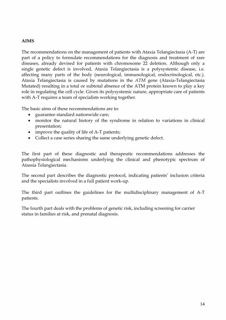
AIMS The recommendations on the management of patients with Ataxia Telangiectasia (A-T) are part of a policy to formulate recommendations for the diagnosis and treatment of rare diseases, already devised for patients with chromosome 22 deletion. Although only a single genetic defect is involved, Ataxia Telangiectasia is a polysystemic disease, i.e. affecting many parts of the body (neurological, immunological, endocrinological, etc.). Ataxia Telangiectasia is caused by mutations in the ATM gene (Ataxia-Telangiectasia Mutated) resulting in a total or subtotal absence of the ATM protein known to play a key role in regulating the cell cycle. Given its polysystemic nature, appropriate care of patients with A-T requires a team of specialists working together. The basic aims of these recommendations are to:
• guarantee standard nationwide care; • monitor the natural history of the syndrome in relation to variations in clinical
presentation; • improve the quality of life of A-T patients; • Collect a case series sharing the same underlying genetic defect.
The first part of these diagnostic and therapeutic recommendations addresses the pathophysiological mechanisms underlying the clinical and phenotypic spectrum of Atassia Telangiectasia.
The second part describes the diagnostic protocol, indicating patients’ inclusion criteria and the specialists involved in a full patient work-up. The third part outlines the guidelines for the multidisciplinary management of A-T patients.
The fourth part deals with the problems of genetic risk, including screening for carrier status in families at risk, and prenatal diagnosis.
14

1. INTRODUCTION Ataxia Telangiectasia (A-T) is a multisystem disease inherited in an autosomal recessive fashion and characterized by cerebellar ataxia, telangiectasia in the skin and eyes, immunodeficiency, radiosensitivity and an increased incidence of tumours both in A-T patients and their relatives (Boder, 1985). A-T cells are hypersensitive to ionizing radiation and radiomimetic substances due to failure to activate the cell cycle checkpoints after treatment with these agents (Taylor et al., 1975). The hypersensitivity to ionizing radiation was disclosed for the first time in an A-T patient exposed to radiotherapy at conventional doses that proved fatal (Gotoff, 1967). The gene responsible for the disease, ATM (A-T mutated), belongs to a family of genes well preserved on the evolutionary scale that regulates the cell cycle checkpoints and programmed cell death (Savitsky et al., 1995; Delia et al., 2003; Shiloh, 2003). Cerebellar ataxia first appears around the first year of life at the toddler stage with frequent falls and a wide-based ataxic gait. A characteristic truncal ataxia may already be apparent at six months of age with anteroposterior and lateral oscillations. The ataxia is progressive and most A-T patients will be using a wheelchair by puberty. It is caused by a degeneration of Purkinje cells which appear reduced in number and markedly pycnotic at cytological examination. In rare cases children do not develop ataxia until puberty or present choreoathetosis at onset. The other neurological hallmark of A-T is oculomotor apraxia varying in onset but present in almost all patients after two years of age. Other common neurological signs encountered in A-T patients include restricted eye movements (strabismus, nystagmus) and slurred speech (dysarthria) (progressively worsening to the point that patients can no longer speak coherently, giving the impression of being mentally retarded, whereas this is very rare in A-T), abnormally low muscle tone (hypotonus) and sometimes epilepsy. Prominent blood vessels (telangiectasias) are the second hallmark of the disease, appearing in most A-T patients between two and eight years of age (average age 72 months) (Harding 1988). Immune problems varying in severity are present in around 60% of A-T patients and involve both humoral and cell-mediated immune responses (Chun and Gatti, 2004; Gatti, 1982; Gatti, 1991). Around 30% of A-T patients do not have immunodeficiency so that the absence of immune problems should not preclude a diagnosis of Ataxia Telangiectasia. The clinical diagnosis of Ataxia Telangiectasia may be difficult before the characteristic skin and eye telangiectasias become evident. Discovery of the ATM gene, whose mutations are responsible for the disease, has paved the way to more accurate laboratory diagnosis. The classic A-T phenotype is caused by mutations in both alleles of the gene that truncate or destabilize the protein product. The ATM gene is located on chromosome 11q22.3 and encodes a protein that plays a key role in regulating the cell cycle and in repairing double-stranded DNA (Savitsky et al., 1995; Delia et al., 2003; Shiloh, 2003). Molecular tests will readily distinguish the clinical A-T phenotype from that of other recessive cerebellar ataxias like Friedreich’s ataxia, Ataxia telangiectasia-like disorder, oculomotor apraxia types 1 and 2, and Nijmegen breakage syndrome. Conversely, some atypical patients with mild or minimal signs of disease (late onset mild slowly progressive ataxia or late onset spinal muscular atrophy) may now be classified as A-T on the basis of absent ATM protein or mutations in the encoding gene (Chun and Gatti, 2004) (see Table 1). Table 1 – Clinical and cellular signs of A-T, AT-LD, NBS, AOA1, AOA2
15

Disease A-T AT-LD NBS AOA1 AOA2
Mutated gene ATM Mre11 NBS1 APTX SETX Chromosome 11q23 11q21 8q21 9q13 9p34 Clinical phenotype Age at onset 2-6 years 2-6 years 1-4 years 2-6 years 11-22
years Progressive cerebellar ataxia + + - + + Oculomotor apraxia + + - + + CNS abnormalities - - + - - Neuronal degeneration + + - + ? Predisposition to cancer + - + - - Immunodeficiency + - + - - Telangiectasia + - - - - Elevated alphafetoprotein + - - - + Hypoalbuminaemia - - - + - Hypercholesterolaemia - - - + - Cell phenotype Radiosensitivity + + + - - Chromosome fragility + + + - - Radioresistant DNA synthesis
+ + + - -
1.1 Epidemiology The disease is found in all populations at varying frequency depending on the different rates of consanguinity and the clinical capacity to differentiate A-T from similar diseases. The incidence in the USA population is estimated at one in 40,000 live births (Swift et al., 1986). The carrier rate is between 0.5 and 2.0% among the general population. On the basis of consanguinity and applying Dahlberg’s formula, an epidemiological survey of 72 Italian A-T families registered with the Italian Registry for Ataxia (RIAT) extrapolated a theoretical disease frequency of one in every 7090 conceptions and a healthy carrier rate from 1.69 to 3.43% of the population (Chessa et al., 1994). The average survival of A-T patients is 19 – 25 years, with a wide range of variability (Crawford 2006). Life expectancy is irrespective of the severity of neurological manifestations. Death is caused by recurrent bronchopulmonary infections associated with a state of cachexia, or cancer in 10-15% of cases. The life expectancy of A-T patients has improved radically in the last 20 years due to the general improvement in living conditions and treatments, and to the identification of patients with “variant” forms of disease. Nowadays many patients live to the age of 25 years and some even survive until 40 or 50 years (Doerk et al., 2004). Respiratory insufficiency, with or without identifiable infections, is the major cause of morbidity and mortality in long-term A-T survivors.
1.2 Genetics and pathophysiology of A-T
16

The mutated gene in patients with Ataxia Telangiectasia was mapped in 1988 on the long arm of chromosome 11 in the 11q22-23 region (Gatti et al., 1988). However, it was only thanks to the participation of many groups in an international consortium in 1995 that the gene region was restricted to the 500kb interval (Foroud et al., 1991; Gatti et al., 1994; Lange et al., 1995) and the gene identified by positional cloning and called ATM (Ataxia-Telangiectasia Mutated) (Savitsky et al., 1995). ATM is a very large gene extending for more than 150 kilobases of genomic DNA and comprising 66 exons, 62 of which are encoding. The transcript consists of 9168 nucleotides and encodes for the ATM protein containing 3056 aminoacids and belonging to the family of phosphatidylinositol 3-kinases (PI3-K). The ATM protein plays a key role in controlling the cell cycle and repairing DNA double-strand breaks (Lavin and Shiloh, 1997; Shiloh, 2003). ATM acts by recognizing and facilitating the repair of a subcategory of double-strand breaks (DSBs) or a form of damage like oxidative stress that is converted into DSB in the DNA. Recognition probably leads to the activation of cell cycle checkpoints. ATM is present in the nucleus as an inactive dimer and is activated in response to DSBs due to autophosphorylation of serin 1981 that causes dimer dissociation in the active monomer form able to phosphorylate many target proteins (Bakkenist and Kastan, 2003). Errors in the repair of DSBs, the most lethal form of DNA damage, give rise to small deletions or insertions at the lesion site that may result in chromosomal translocations and genomic instability and finally cancer. Progressive neurodegeneration could be the outcome of a defect in controlling the cell cycle in post-mitotic cells (Yang and Herrup, 2005), a theory also supported by evidence of a mitotic checkpoint defect in A-T cells after irradiation (Takagi et al., 1998). From the mutational standpoint, patients are usually compound heterozygous for two distinct mutations inherited from their parents, but patients homozygous for the same mutation are not uncommon, especially among populations with a high rate of consanguinity as in Italy. More than 400 different mutations have been identified to date spread throughout the ATM gene with no evidence of mutational hot spots (http://chromium.liacs.nl/lovd/). Most mutations give rise to a highly unstable truncated protein that produces a null phenotype in around 85% of cases. In many countries, including Italy, recurrent mutations have been identified whose carriers share a common haplotype indicating a founder effect (Gilad et al., 1996; Ejima and Sasaki, 1998; Laake et al., 1998; Stankovic et al., 1998; Telatar et al., 1998). The haplotype serves to identify carriers, especially when the mutation has not been identified. Mutations have been identified in more than 90% of Italian patients. Most mutations are truncated as shown by the absence of the ATM protein in patients’ cells. In addition, recurrent mutations have been identified the commonest being mutation 7517 del(4) in exon 53 found in patients with classic A-T and A-T variants. 1.3 Clinical and immunological phenotype 1.3.1 Neurological manifestations A-T is a neurodegenerative disease characterized by the onset and slow progression of different neurological deficits. Phenotypic variability is largely the result of the timing of emergence of different symptoms and the speed of disease progression as the key symptoms of the illness are usually all present in adult patients (Chun and Gatti, 2004).
17

Cerebellar ataxia is the earliest and most prominent neurological symptoms of A-T and almost always the reason for the first specialist consultation. Early clinical signs. Most children with A-T appear normal at birth and have the same psychomotor development as their peers until the time of walking. However, disease onset is associated with a prolonged phase of postural uncertainty with frequent truncal oscillations and falls often noted by parents as the first cause for alarm in an otherwise normal child. On close questioning the parents will recall the early walking stage noting delayed postural motor development, described as clumsiness, decreased muscle tone or instability while sitting. Although they do not resolve, these symptoms may sometimes appear to improve between three and seven years of age, delaying the first specialist consultation. In other cases, symptoms persist leading the parents or paediatrician to make a neurological referral which at this stage may often prove inconclusive or misleading from a diagnostic standpoint (Chun and Gatti, 2004; Perlman et al., 2003; Miller, 2004). This is because the early symptoms of truncal ataxia in the first year of life are sometimes construed as excessive instability of the trunk and head while sitting and standing, proximal hypotonia, slowness in using the arms and handling objects (Leuzzi et al., 1993; Sedgwik and Boder, 1972). These signs may be extremely subtle and in some cases not present at such an early age. Walking and posture. Frank cerebellar ataxia is usually noted at the toddler age between 16 and 18 months. Despite broad interindividual variations, the ataxia presents hallmarks highly suggestive of A-T. Walking is unsteady primarily because of a lack of synergy between trunk and limbs already evident on standing. Slow truncal oscillations require ongoing adaptations of the limbs or trigger other compensation mechanisms with frequent adjustments of the head position. Walking tends to involve sudden uncoordinated movements: half of the body is projected forwards while the corresponding limb is swung our in an adduction movement, crossing the walking line and excessively reducing the support base at the time of load-bearing. Irrespective of the actual ataxia, bradykinesia and hypokinesia are typical features of A-T ataxia. Despite these difficulties, A-T children are able to adopt a strategy to compensate and correct their unsteady walking for a long time. Fast walking is one way of compensating because less balance is needed for quick movements (Crawford, 1998; Crawford et al., 2000). As time passes, the unexpected amplitude of each step is exacerbated by the irregular walking pace. Some patients will present dystonic foot postures (equino-varus-supinated) balancing on the forefoot in the load-bearing phase. Lastly, worsening bradykinesia and hypokinesia added to restricted eye movements linked to the onset of ocular dyspraxia (see below) make walking increasingly strenuous and ultimately no longer possible. Unsteady posture also presents characteristic features in A-T. The trunk swings from one extreme to the other, often to the point of losing control of body posture. Patients will sometimes adopt awkward unnatural postures before a corrective reaction occurs which in turn is excessive and a cause of further destabilization. With time and disease progression many patients present rapid segmental or focal movements of the trunk in the form of adventitious postural-induced jerks, but they may also attempt late uncoordinated postural adjustments. Head instability shares the same features, often with prolonged changes in position from one extreme to the other, in extreme flexion or extension before a
18

correction or even overcorrection. Head movements are influenced early on by the appearance of ocular dyspraxia (see below). From two years of age other signs of cerebellar dysfunction appear including dysarthria, loss of facial expression with reduced muscle tone and salivation, and a generalized decrease of muscle tone. The ataxia progresses slowly but inexorably and by 12 years of age most children with AT are unable to walk unaided (Perlman et al., 2003). Whereas this event occurs with some degree of interindividual variability, the progressive worsening of motor coordination is apparent from the first years of life. Ataxia of the upper limbs, intentional tremor and segmental myoclonus appear later making drawing and writing difficult and then impossible after the age of ten. Brain MRI is often normal in the first two years of life but subsequently (but not invariably) discloses signs suggestive of cerebellar atrophy. Extrapyramidal signs. Involuntary movements are common in A-T. Choreoathetosis is present in around 90% of patients and sometimes prevails over the signs of ataxia. Segmental myoclonus may affect both the extremities and the trunk (Miller 2004). From the first years of life children present rapid choreiform movements of the hands and feet in performing intentional tasks hands which often go unrecognized. Later still, intentional movements of the arms become imprecise and uncoordinated as a result of cerebellar dysmetria, and ballistic movements also become ataxic with unsteady posture and action tremor. Dystonia (in the form of progressive athetosis of the fingers) is commonly encountered in children and adolescents, while early torsion dystonia involving the head and limbs has been reported in some cases with involvement of the proximal muscles with a flexed trunk posture developing as the dystonia worsens (Bodensteiner et al., 1980). During the second decade of life the movement disorders worsen and severe postural tremor, intentional tremor, dysmetria, multifocal myoclonus, tic-like movements and chorea are common.
Bulbar functions and facial expression. All A-T patients have difficulty speaking (Crawford 1998). Most A-T children are dysarthric and dysprosodic. After the age of five years the dysarthria tends to worsen and speech becomes slow and words articulated with the emphasis typical of cerebellar dysarthrias involving motility of the lips, tongue and palate. Another later but constant findings is difficulty chewing and swallowing, responsible for “ab ingestis”, often secondary, bronchopulmonary infections. The time required to feed increases markedly as the disease progresses and inadvertent aspiration of food becomes dramatically more important in the second decade of life. Increasing difficulty feeding is accompanied by weight loss and slow height gain. After the first years of life, the facial expression of A-T children diminishes and at school age their mask-like expression may mimic mental retardation which is usually absent. This impression is however dispelled by a broad pleasing smile or wink, but these too subside during the second decade of life becoming less expressive. Oculomotor deficits. Due to their early onset, the oculomotors deficits of A-T are particularly suggestive and reflect involvement of cerebellar and extrapyramidal functions caused by the functional impairment of the flocculus (Crawford, 1998). They are present in virtually
19

all A-T patients and often herald the onset of telangiectasia, thereby constituting an important diagnostic sign. With head fixed, intentional oculomotor movements start after a prolonged latency and are continuous, but unlike ophthalmoplegia, will be completed if a sufficiently long time is allowed. When the head is turned rapidly towards a peripheral stimulus the eyes deviate tonically in the opposite direction then slowly follow the direction of the head. Ocular movements are smooth and normal in amplitude when the head is mobilized passively, whereas the impaired conjugate motility of the eyes mainly apparent with intentional movements. Optokinetic nystagmus is absent. Often evident in the early years of life, strabismus tends to disappear. The oculomotor disorder in A-T impairs the neural mechanisms responsible for stabilizing the image on the retina and hence implicated in maintaining and inhibiting fixation. Gaze inhibition in particular is a prerequisite for implementing a change in gaze (Lewis et al 1999; Miller 2004). The progression of these deficits makes reading and writing increasingly difficult and ultimately no longer possible. Peripheral neuropathy and myelopathy. Whereas muscle strength and osteotendinous reflexes are normal in A-T children in the first decade of life, signs of peripheral neuropathy will appear at around the age of ten years (disappearance of O-T reflexes, loss of vibratory sensitivity and leg position sense). However a frank Romberg sign is never observed. Neurophysiopathological and neuropathological studies have disclosed demyelination and astrocyte proliferation of the dorsal vertebrae and degeneration of the anterior horn neurons. Peripheral nerve studies have demonstrated a primarily axonal polyneuropathy (Stankovic et al., 1998). However, the clawfoot or horsefoot encountered in A-T children often has a dystonic rather than peripheral origin. Although it is similar to the clawfoot seen in other neuropathies (Friedreich’s ataxia, Charcot-Marie-Tooth, etc.), the foot deformity in A-T is often accentuated with walking, is not associated with a rigid arcus plantaris, forefoot atrophy or hallux flexus, and is often encountered early on when the O-T reflexes are not yet evoked. A significant number of older A-T patients develop a neurogenic amyotrophy caused by atrophy of the anterior horn motorneurons and dorsal nerve root ganglia of the spinal cord. The signs of atrophy mainly affect the hands and feet. Cognitive development. Cognitive functions are also spared in adulthood although they may be difficult to assess due to the severe dysarthria (Gatti et al., 1991; Miller, 2004). Most A-T patents have an IQ within or slightly above normal range, while no cases of IQ below 50 have been reported (Gatti et al., 1991). There is no evidence of cognitive deterioration in the first decade of life, and development appears to stabilize. In general verbal IQ in adult A-T patients is lower than in normal subjects (Mostofsky et al., 2000). A short-term memory deficit has been reported in the few older A-T patients in the third decade of life (Gatti et al., 1991). 1.3.2 Cutaneous manifestations Telangiectasias are the second hallmark of A-T and occur in most but not all patients (ataxia sine telangiectasia). Telangiectasias consist in prominent blood vessels in the whites of the eyes, occasionally present at birth yet in other A-T patients may not develop until the teenage years. Telangiectasias typically appear in the conjunctiva where they start in the conjunctival fornix and then increase to cover the entire conjunctiva. Other areas
20

typically affected include the flexor regions of the limbs and neck, the retroauricular region, eyelids and more seldom other body regions. Telangiectasias may be absent in some rare cases (Willems et al., 1993). There is anecdotal evidence of angiectasias arising in the wall of the bladder or in the lungs after radiotherapy or treatment with chemotherapeutic agents like vincristine (Chun and Gatti, 2004). Other characteristic cutaneous features are progeric changes including early grey hair, reduced skin elasticity, poikilodermy, café-au-lait spots, hypertrichosis and senile keratosis (Cohen, 1984). Acanthosis nigricans and eczema may also appear (Cohen et al., 1984). All these manifestations progressively increase with age. Skin granulomas may appear in rare cases similar to those more commonly encountered in common variable immunodeficiency (CVID) (Mitra, 2005). 1.3.3 Immunological manifestations Around 60% of A-T patients present varying degrees of immune deficiency, while 30% do not have immune problems (Woods et al., 1992). Hence the lack of an immune deficit does not preclude a diagnosis of A-T. The immune deficit in A-T consists in impaired antibody and T-cell responses (Gatti, 1991). The impaired antibody responses are associated with a reduced number of B cells and abnormal immunoglobulins. Around two thirds of patients present a marked reduction or absence of IgA and IgE, commonly associated with a deficit of subclasses IgG2 and IgG4. Some patients have a serum immunoglobulin profile compatible with a hyper-IgM phenotype. Unlike the idiopathic IgA deficit, A-T patients seldom have allergic reactions to the IgA contained in human gammaglobin preparations for i.m. or i.v. use. This is probably due to the frequent concurrence of IgE deficit in A-T patients as opposed to the high incidence of IgE-mediated manifestations in idiopathic IgA deficit. Oligoclonal immunoglobulins accompanying episodes of lymphocyte proliferation (lymphadenopathy, hepatosplenomegaly) are commonly found in A-T patients. A broad range of T-cell abnormalities are present in A-T (Fiorilli, 1982) including lymphopenia, a reduced response to mitogens and impaired T-cytokine production (Paganelli, 1984). The T-cell defect may be due to a reduced thymopoiesis resulting in the generation of a limited T-cell reservoir (Giovannetti, 2002). Determination of lymphocyte subpopulations shows a marked decrease of subpopulations CD3+ and CD4+ with normal or slightly raised values of CD8+. The CD4+/CD8+ ratio is often inverted. There is also a depletion of D45RA+ lymphocytes, so-called T-virgin cells, preferentially localized in the lymph nodes. There is often a relative increase in TCR γ/δ-bearing with respect α/β)-bearing T cells or those with compound hybrid TCR genes, i.e. part γ and part β, 50-100 times more common than in normal subjects. As a result there is a reduced generation of αβ T cells (Carbonari, 1990) whereas individual T cell function is spared (Pashankar, 2006). The clinical manifestations of immune deficiency in A-T are similar to those characteristic of antibody defects: recurrent infections of the upper and lower airways and gastrointestinal infections/infestations. Encapsulated bacteria are the most commonly isolated, mainly Haemophilus influenzae, Streptococcus pneumoniae and Pseudomonas aeruginosa. There is a markedly impaired response to pneumococcal polysaccharides (Sanal, 1999), but combined immunization with the PCV7 and PPV23 vaccines appears to trigger a significant anti-pneumococcal response in A-T patients (Stray-Pedersen, 2005). Although many patients present a cell-mediated immune defect, viral and fungal infections, pneumocystis and intracellular bacterial infections are uncommon in A-T
21

(Nowak-Wegrzy, 2004). In addition, vaccinations with attenuated viruses do not appear to give rise to complications in A-T patients (Nowak-Wegrzy, 2004). Infections remain one of the main causes of death in A-T despite prompt aggressive treatments. As in patients with agammaglobulinaemia or cystic fibrosis, the chronic evolution and recurrent respiratory infections frequently give rise to pulmonary fibrosis and respiratory insufficiency. Although the severity of the neurological defect is not correlated to overall survival (Crawford, 2006), the neurological problems limit the possibility of delaying lung damage by respiratory physiotherapy and facilitate respiratory infections from aspiration and stagnation. 1.3.4 Other clinical manifestations More than half of A-T patients have glucose intolerance, insulin resistance and hyperglycaemia. Most patients present a somatic hypoevolutism: height and weight are usually below the third percentile in adolescence, mainly in patients with recurrent or chronic respiratory infections. No hormone deficits responsible for delayed body growth have been found. The absence or delay in pubertal body changes may be associated with atrophy of the gonads and later insulin-resistant diabetes (Crawford, 1998). Female A-T patients often have a late start of menstruation and delayed development of secondary sexual characteristics. The ovaries are sometimes absent or hypoplastic and patients are therefore sterile. By contrast, spermatogenesis and ejaculation were reported as normal in the few male A-T patients examined, and sexual reproduction has been noted in the rare A-T subjects surviving to adulthood (Stankovic et al., 1998). Around half of A-T patients have elevated levels of liver enzymes like alkaline phosphatase and transanimases. Fatty infiltration of the portal liver cells has been described in some biopsies and is an occasional finding of little clinical significance. Variable levels of biological markers of the disease have also been noted (ATM protein levels, serum alphafetoprotein, radiosensitivity (see Chun and Gatti, 2004; see below – diagnosis). 1.3.5 Cancer risk and radiosensitivity A-T patients have an increased incidence of tumours which are the second commonest cause of death. One in three A-T individuals will develop a tumour in the course of their lives and cancer may occasionally arise even before the diagnosis of A-T is suspected. Tumours occurring before the age of 20 years are leukaemia or lymphomas (namely T-cell lymphomas) in 85% of cases, with an increased incidence of 70 and 250 fold respectively. Acute T-cell leukaemia and T-cell lymphomas are more common in childhood, while T-cell prolymphocytic leukaemia (T-PLL) accounts for 10% of T-cell tumours thereafter. B-cell tumours encountered in older A-T patients are leukaemias and lymphomas which have no cytogenetic or clinical specificity and are present in the same tumours in the general population. After the age of 20 years solid (mainly epithelial) tumours are more common and include dysgerminomas, brain tumours (astrocytomas, medulloblastomas and gliomas), gastric, pancreatic and hepatic carcinomas, and retinoblastoma. Cases of rhinopharyngeal carcinoma have also been reported. The frequency of these tumours is slightly higher than in the general population, whereas solid gynaecological, ovarian and
22

uterine tumours are more common in A-T patients than in women of the same age. All these tumours should be treated with attenuated protocols (see chemotherapy). Some A-T patients with cancer treated with radiotherapy presented an acute and chronic reaction of irradiated healthy tissues, with erythema, skin desquamation and fibrosis. Like resistance to radiotherapy, radiosensitivity is caused by the mutated ATM gene due to dysfunction of the protein implicated in the chain of reactions to which the AT protein belongs, some of which, like IGF-IR, EGF and Chk2, are responsible for radioresistance (Mamon et al., 2003; Takai et al., 2002; Yu et al., 2003; Sartor, 2003). Tumour occurrence varies even among A-T patients in the same family, some of whom will develop multiple primary tumours, whereas others do not develop any. The link between A-T and the increased incidence of lymphoid and non-lymphoid tumours is due to the role of the ATM protein in protecting against DNA double-strand breaks (DSBs). The ATM defect abolishes the cell cycle checkpoints following a DSB, with inappropriate progression of the cycle and accumulation of damage until cell death (Taylor 2007). ATM-correlated tumorigenesis results from both an increased frequency of rearrangements of the chromosomes involving the loci of immunoglobulins or T-cell receptors on the one hand and the oncogenes on the other, and genetic events linked to the role ATM plays together with other tumour susceptibility alleles (BRCA, p53, etc.) (Ahmed 2006). 1.4 Biological markers Among the analytes currently examined in the laboratory, serum α-fetoprotein (AFP) is the most reliable, being elevated in around 95% of A-T patients. Clinically A-T patients with normal AFP levels at molecular analysis either have an ATM gene mutation (rare, defined as ataxia without telangiectasia [A(-T)]) or an APTX gene mutation (common, AOA1). We think serum AFP determination is compulsory for the differential diagnosis of A-T. The karyotypic mutation consists in an increased rate of spontaneous chromatid ruptures and the presence of rearrangements involving chromosomes 7 and 14 in the loci for the heavy chains of immunoglobulins and T-cell receptors. Before molecular tests became available, radiosensitivity testing was used for the purposes of diagnosis. These have now lost their importance following observations that some A-T patients have reduced or almost normal sensitivity to ionising radiation, while non A-T individuals have radiosensitivity levels similar to those of A-T patients. In addition, radiosensitivity testing is arduous and requires a great deal of experience. Research laboratories performing radiosensitivity testing follow specific protocols developed over the years for both normal and pathological reference values such as the colony surviving assay (CSA), radioresistant DNA synthesis (RDS) and chromosome fragility tests performed using ionising radiation or radiomimetic compounds like bleomycin and neocarzinostatin. Other laboratory tests, largely used for research purposes include cytofluorimetry to measure the G1/G2 ratio in the cell cycle, and the neutral and alkaline comet assay to determine the severity of DNA damage. The most reliable molecular marker for A-T diagnosis is universally considered to be the determination of ATM protein in stabilized lymphoblastoid cell lines from patients with a clinical diagnosis of A-T. Protein levels are reduced (less than 20%) or absent in 99% of
23

patients with ATM gene mutations. Protein determination takes four to six weeks for stabilization of the lymphoblastoid line and another week for Western blotting analysis. Mutation analysis is done by different methods to identify the possible different types of mutation: DHPLC on encoded exons, followed by direct sequencing of the region with the abnormal chromatographic pattern; transcript sequencing; multiple ligation of probe amplification (MLPA). 1.5 Differential diagnosis The autosomal recessive ataxias form heterogeneous groups of diseases sharing other characteristic neurological and clinical disorders in addition to cerebellar ataxia (Di Donato et al., 2001; Koenig, 2003) (see table). A-T itself may present with different clinical signs, sometimes making it difficult to establish certain diagnosis on the basis of clinical features alone. In these situations biochemical-molecular tests are very helpful for the purposes of establishing a final diagnosis of A-T forms hitherto classified as “A-T variants” according to clinical and laboratory findings (Ying and Decouteau, 1981; Fiorilli et al., 1985; Taylor et al., 1987; Chessa et al., 1992). ATM gene mutations have also been identified in some of these patients (McConville et al., 1996; Gilad et al., 1998) thereby confirming the diagnosis of A-T. Mutations of the Mre11 gene, a DSB repair gene belonging to the MNR complex, have been identified in patients with the clinical and laboratory features of A-T but lacking telangiectasias (A-T-like disorder) (Stewart et al., 1999; Pitts et al., 2001; Delia et al., 2004; Petrini, 1999). In addition, mutations of the APTX gene have been found in patients with neurological features very similar to A-T but with normal α-fetoprotein and lacking multisystem involvement, chromosome instability and increased sensitivity to ionising radiation, and defined as ataxia oculomotor apraxia (AOA). All patients with these “A-T variants” have been investigated for the A-T phenotype at cell level (radiosensitivity) and by molecular (DHPLC and sequencing) and biochemical tests (Western blot). The discovery of mutations in the Mre11 gene in patients with ataxia telangiectasia-like disorder (ATLD) and mutations in the APTX gene in patients with con AOA1, presenting the same neurological phenotype and age at onset as A-T patients, led to the production of a panel of antibodies against proteins Mre11, NBS1, Rad50 and APTX. This panel allows a first level differential diagnosis between pathological forms similar to A-T such as ataxia telangiectasia-like disorder, Nijmegen breakage syndrome and ataxia oculomotor apraxia types 1 and 2. As a rule of thumb, if α-fetoprotein levels are normal in a patient with A-T-like neurological features, serum albumin and cholesterol should be measured for a differential diagnosis with AOA1. Thereafter it would be useful to contact the central laboratories responsible for molecular diagnosis (see below).
24

2. DIAGNOSTIC PROTOCOL 2.1 Inclusion criteria Male and female persons of any age presenting the following clinical and laboratory features will be eligible for enrolment:
- ataxia /choreoathetosis and/or - oculomotor apraxia - with or without telangiectasias
and - elevated levels of serum α-fetoprotein
A registration form (Form 1.01) and a diagnosis form (Form 30.01) will be filled in for patients meeting these inclusion criteria. Annual follow-up forms (Form 30.02) will then be filled in and sent to the AIEOP/FONOP Centre in Bologna. All subjects meeting the inclusion criteria will be enrolled in the Protocol.
2.2 Certain diagnosis Certain diagnosis of A-T can only be established by:
- analysis of ATM protein expression (with evidence of reduced or absent protein expression)
or - analysis of ATM gene mutation (with identification of the mutation)
2.3 Sending samples
On request of a centre in the network, the Rome Centre or Turin Centre will undertake analysis of levels of the ATM protein and correlated proteins and analysis of the ATM gene mutation for certain diagnosis of A-T. Before sending blood samples specific informed consent must be obtained from each family (see attachments) and kept by the centre in the network for each of its patient enrolled in the Protocol.
After making arrangements by telephone, the following must be sent for these tests: - a 10-20 cc blood sample in heparin sodium The samples must be sent at room temperature to one of the following laboratories: ROME: Prof. Luciana Chessa
25

U.O.D. Genetica Medica A.O. S. Andrea Via di Grottarossa 1039 00189 Roma, Italy Tel/fax: (39).06.33775258
[email protected] TURIN : Dr. Alfredo Brusco Dipartimento di Genetica
Università di Torino Via Santena 19 10126 Torino, Italy Tel. (39) 011.6334480 FAX (39) 011.6705668 e-mail: [email protected]
- Samples must be accompanied by n° 1 National Health Service request form duly
filled in (date of sampling, patient details with place and date of birth, place of residence, health card number, tax code number, reason: molecular analysis for rare diseases: RD040).
- Samples must also be accompanied by Form AT/A duly filled in and and sent to the charge of the Coordinating Centre via TRACO 10 service which guarantees delivery of samples by 10 a.m. on the following day.
- Samples must be sent from Monday to Wednesday each week. - The results of the test for protein levels will be notified within two to four
months. - The outcome of mutation analysis will be notified within six to eight months. 2.3 Tests to be performed at diagnosis and during follow-up: At diagnosis: - α-fetoprotein - Haemochrome - Lymphocyte subpopulations (CD3, CD4, CD8, CD19) - T-cell proliferation to mitogens (PHA) (optional) - IgG, IgA, IgM, IgE - IgG subclasses (optional) - Autoantibodies (ANA, ASMA, AMA, APCA) (optional) - Glycaemia - Cholesterol - Albuminaemia - SGOT, SGPT, gGT - Karyotype on 50 metaphases to search for clonal aberrations - Brain MRI - Neurological assessment - Disability scale: Vineland Index
Barthel Index (from 16 years)
26

- Spirometry (if the test can be performed) During follow-up: Every 6 months: - Neurological and neurorehabilitation assessment Every 12 months: - α-fetoprotein - Haemochrome - Lymphocyte subpopulations (CD) - T-cell proliferation to mitogens (PHA) (optional) - IgG, IgA, IgM, IgE - IgG subclasses (optional) - Disability scale: Vineland Index
Barthel Index (from 16 years) - Spirometry (if the test can be performed) - Sputum culture Tests to be performed on clinical indication: - Abdominal ultrasound scan and/or MRI (for suspected lymphoma or other tumours) - Brain MRI - EMG and ENG* - Echocardiography - Holter ECG - Blood gas analysis *EMG and ENG (from the age of six years; before in case of emergence of clinical signs suugestive of peripheral involvement)
27

3. THERAPEUTIC RECOMMENDATIONS The complexity of the different diseases sharing the same genetic mutation, and the heterogeneous clinical manifestations currently make it difficult to provide uniform therapeutic recommendations for all patients. The most appropriate treatment strategies must be implemented depending on the prevalence of a particular problem or a series of different problems. The following recommendations are listed for the management of different clinical problems.
3.1 Recommendations for the management of immunological problems and infections Prophylaxis against P. carinii. Prophylaxis with co-trimoxazole must be considered in patients with a significant CD4+ deficiency. In these cases reference can be made to the recommendations suggested for immunodepressed children: prophylaxis should be started if CD4+ cells are < 750/mm3 in children aged < 12 months; < 500/mm3 in children aged between one and five years; < 200/mm3 in children aged > five years. It should be emphasized that PCP infection is extremely rare and no controlled studies are currently available on the topic. Vaccinations. For AT patients with a mild immune impairment there are no contraindications to administration of vaccines belonging to the vaccine calendar in use in Italy. Live attenuated vaccines can be administered if the proliferative response to mitogens is normal and the absolute number of CD4+ lymphocyte is > 500/ mm3. If these parameters are not met vaccination should be postponed. Administration of purified polysaccharide-protein conjugate vaccines (tetanus, diphtheria, pertussis, hepatitis anti-H. influenzae, anti-influenza, anti-pneumococcus) is recommended in all subjects as they are not harmful and trigger an antibody response depending on the intensity of the immune deficiency. In particular, systematic use of active immune prophylaxis with conjugate vaccines is recommended: anti-pneumococcus; anti-meningococcus; anti–H. Influenzae. The vaccine schedule should be based on the patient’s age and the vaccine characteristics.
- Conjugate anti-pneumococcal vaccination is recommended: for children under one year of age, two administrations one month apart and a third dose in the second year of life; for children over one year, two administrations with at least a two month interval in between the doses.
- Conjugate anti-meningococcal vaccination is recommended: for children under one year of age, three administrations at one month intervals; for children over one year, a single administration. - Conjugate anti–H. influenzae vaccination is recommended: for children under one year of age, three administrations (according to the vaccine calendar in use in Italy); for children over one year, a single administration.
Antibiotic prophylaxis. AT patients may have an increased susceptibility to infections due in part to the immune defect and in part to “mechanical” problems (pneumonia ab ingestis cause by impaired swallowing; diminished chest expansion and difficulty walking due to neurological problems).
28

In any case prompt aggressive antibiotic therapy is recommended to control acute episodes of infection. In case of recurrence antibiotic prophylaxis can be implemented for a duration to be established for each patient by the treating physician. Immunoglobulin replacement therapy The humoral immune defect is usually mild and does not require immunoglobulin replacement therapy unless the patient is clinically symptomatic, namely with a high frequency of airway infections. Of course this treatment is indicated in forms of A-T: - with severe hypogammablobulinaemia; - with a severe T-cell deficiency similar to that seen in SCID. Therapy should be administered and monitored according to the guidelines provided in the Recommendations for XLA and CVID. 3.2 Recommendations for the management of neurological and neurorehabilitation problems A-T is a slowly progressive neurodegenerative disease leading to loss of major adaptive functions, namely neuromotor functions, in the space of a few years due to loss of many neurological mechanisms involved in postural control, fine coordination tasks and visual exploration of the environment. As in other CNS disorders, the reduced motor autonomy and ultimately the inability to walk unaided mark an accelerated disease progression because they reflect not only disease evolution, but also the ensuing cascade of effects in terms of residual neurological skills and the patient’s general condition. Like all degenerative conditions, lacking an aetiological treatment the aim of symptomatic therapy (rehabilitation and drug management) is to postpone the loss of basic neuromotor skills for as long as possible or create the conditions for patients to recover neuromotor function or compensate so as to enhance adaptation and autonomy. Each neurological function is sustained by a scaffolding of mutually harmonized skills and automatisms. A functional loss appears when these elements deteriorate beyond the point of internal substitution by the system. Dwindling function can sometimes be postponed by means of aids acting as simplified replacements of components of functional organization. At other times these same aids may allow relearning via alternative original routes. Dedicated rehabilitation measures should be evaluated early on for A-T children to combat critical periods in disease evolution. Certain critical stages mark disease evolution and require specific rehabilitative interventions:
a) The emergence of truncal ataxia as a factor limiting neuromotor development (second year of life);
b) Supervening oculomotor apraxia (third to fourth year of life) having a cumulative effect on the preceding difficulty undermining postural stability, stability of reference points in the visual environment, quality of visual exploration and implicit representation of the environment;
c) Progressive motor slowing (bradykinesia and hypokinesia) making the patient’s adjustments to adapt to changes in environmental conditions increasingly late and
29

ineffective (fifth to sixth year of life) and lastly the loss of the ability to stand and walk unaided (< ten years);
d) Difficulties in communication and social interaction linked to loss of coherent speech and writing skills (sixth to ninth year of life).
Multidisciplinary intervention must be planned for each of these stages and symptoms bearing in mind the specific needs of the individual patient. Intervention should aim at: a) the use of motor functions affected by the disease in a meaningful setting enhancing the single anti-gravitational and coordination skills constituting their essential elements; b) an individual search for compensation strategies and mechanisms; c) the development and adaptation of aids to replace individual skills or functional modules no longer subject to recovery. Lastly, many A-T patients present neurological symptoms that may benefit from specific drugs (postural tremor, parkinsonian signs, hypokinesia/brachykinesia, choreic or choreo-athetosic dyskinesias, etc.) that should be prescribed adjusting the dose in relation to the individual patient and the symptom to be targeted by the treatment.
3.3 Recommendations for the management of respiratory problems
Considering mainly the likelihood of “mechanical” problems (pneumonia ab ingestis due to impaired swallowing; diminished chest expansion and difficulty walking due to neurological problems), it is important to assess the need to devise a programme of respiratory physiokinesitherapy tailored to the individual patient.
3.4 Recommendations for the management of other problems (diet/ gastroenterological problems, ENT, etc.)
The appropriate specialists should be involved in the management of specific problems such as gastroenterological, otolaryngological conditions, etc. 3.5 Recommendations for radioprotection The radiosensitivity of A-T patients emerged dramatically for the first time in 1968 with a fatal reaction to antitumoral radiotherapy (Morgan 1968). For this reason, treatment with ionising radiation and radiomimetic drugs is absolutely contraindicated in A-T patients. A-T patients presenting tumours must be treated using attenuated chemotherapy protocols. Although accurate epidemiological information is not currently available on the risk of developing tumours related to exposure of A-T patients to radiodiagnostic procedures, diagnostic ionising radiation (standard x-ray, CT scans, scintigraphy) must only be used when strictly necessary. Whenever possible, radiodiagnostic techniques should be replaced by ultrasound or magnetic resonance imaging. Heterozygous carriers of ATM mutations have an in vitro radiosensitivity in between that of normal subjects and homozygous A-T individuals (Neubauer 2002). However, heterozygous A-T carriers do not appear to have an increased sensitivity to radiotherapy (Bremer, 2003) and hence can be treated using standard protocols. 3.6 Attenuated chemotherapy
30

Leukaemia and lymphomas can be treated and effective chemotherapy protocols are available for use in A-T patients. However, as noted above the radiosensitivity of A-T patients is an absolute contraindication to treatment with ionising radiation and radiomimetic drugs. In case of tumours attenuated chemotherapy protocols must be instituted in agreement with the AIEOP and adult Haematology Centres. 3.7 Drugs trials currently underway The well-established evidence that A-T cells are exposed to continuous oxidative stress suggested the possibility of treating the ATM mutation with exogenous antioxidants: experimental trials are currently underway.
31

4. PREVENTION 4.1 Prenatal diagnosis and identifying disease carrier status Identifying ATM gene mutations is important not only to understand the molecular mechanisms underlying the disease, but also for prenatal diagnosis and genetic counselling. Prenatal diagnosis was performed long before the advent of molecular analysis by radiosensitivity testing of foetal cells. This approach often led to false negative results (personal observation). More recently, linkage analysis has been successfully used in patients whose mutations had not been identified. Direct molecular prenatal diagnosis offers 100% accuracy rate but is only possible when the familial mutations are known (Chessa et al., 1999). Molecular segregation. Identifying carriers among A-T family members has always been a focal point of research both for the possible increased predisposition to cancer and cardiovascular diseases and for the need to offer in-depth genetic counselling. The increased sensitivity to ionising radiation also affects heterozygous carriers, albeit to a lesser extent than A-T patients, but the accuracy of identifying health carriers by this method does not exceed 85% [45], making it unsuitable for diagnostic purposes. Identification of the ATM gene and its mutations in an affected inidividual in an A-T family has fostered molecular segregation studies to disclose which siblings are heterozygous carriers in addition to the parents. In the rare cases in which the underlying mutations cannot be identified, haplotype analysis with microsatellite markers will yield a result with a confidence interval in excess of 98%. Predispostion to cancer. The predisposition of healthy carriers of ATM mutations to develop tumours remains controversial and is the topic of debate among disease experts. Michael Swift’s pioneering studies provided epidemiological evidence of a significant increase in breast cancer among the close relatives of A-T patients (Swift et al., 1987). Data collected in Italy indicated a greatly increased risk of stomach cancer in A-T families with respect to the general population (Chessa and Fiorilli, 1993). Even the molecular identification of A-T heterozygous carriers has failed to clarify the issue fully despite attempts to form a consortium of researchers worldwide to reach a significant number of A-T families. Renwick et al. (2006), studying 443 families with breast cancer and 521 controls, recently demonstrated that ATM gene mutations give heterozygous carriers a 2.37 relative risk of developing cancer. ATM is thus a minor factor of genetic susceptibility to breast cancer. 4.2 Sending samples
At the request of centres in the network, the Rome Centre or the Turin Centre will undertake analysis of the ATM gene mutation to identify carrier status in the parents and close relatives of A-T patients. If the proband’s mutations cannot be identified, molecular analysis can be performed on family members by reconstruction of the disease haplotype with informative markers. Before sending blood samples specific informed consent must be obtained from each family and kept by the centre in the network for each of its patient enrolled in the Protocol.
After making arrangements by telephone, the following must be sent for these tests:
32

- a 20 cc blood sample in heparin sodium or - a 7 cc blood sample in EDTA The samples must be sent at room temperature to one of the following laboratories: ROME: Prof. Luciana Chessa U.O.D. Genetica Medica A.O. S. Andrea Via di Grottarossa 1039 00189 Roma, Italy Tel/fax: (39).06.33775258
[email protected] TURIN: Dr. Alfredo Brusco Dipartimento di Genetica
Università di Torino Via Santena 19 10126 Torino, Italy Tel. (39) 011.6334480 FAX (39) 011.6705668 e-mail: [email protected]
- Samples must be accompanied by n° 1 National Health Service request form duly
filled in (date of sampling, patient details with place and date of birth, place of residence, health card number, tax code number, reason: molecular analysis for rare diseases: RD040).
- Samples must also be accompanied by Form AT/A duly filled in and sent to the charge of the Coordinating Centre via TRACO 10 service which guarantees delivery of samples by 10 a.m. on the following day.
- Samples must be sent from Monday to Wednesday each week. - The test results will be notified within two to four months.
33

REFERENCES Ahmed M, Rahman N. ATM and breast cancer susceptibility. Oncogene 25, 5906–5911, 2006 Aicardi J, Barbosa C, Anderman E, Anderman F, Morcos R, Ghanem K, Fukuyama Y, Awaya Y, Moe P. Ataxia-Ocular Motor Apraxia: a syndrome mimicking Ataxia-Telangiectasia. Ann Neurol 24, 497-502, 1988 Bakkenist CJ, Kastan MB. DNA damage activates ATM through intermolecular autophosphorylation and dimer dissociation. Nature 421, 499-506, 2003 Barbot C, Coutinho P, Chorao R, Ferreira C, Barros J, Fineza I, Dias K, Monteiro JP, Guimaraes A, Mendonca P, Moreira MC, Sequeiros G. Recessive Ataxia with Ocular Apraxia. Arch Neurol 58, 201-205, 2001 Boder E. Ataxia telangiectasia: an overview. In Gatti RA, Swift M eds: "Ataxia-Telangiectasia: Genetics, Neuropathology and Immunology of a Degenerative Disease of Childhhod". Alan R Liss, pp 1-63, 1985 Bremer M, Klopper K, Yamini P, Bendix-Waltes R, Dorkb T, Karstensa JH. Clinical radiosensitivity in breast cancer patients carrying pathogenic ATM gene mutations: no observation of increased radiation-induced acute or late effects. Radiother Oncol 69, 155–160, 2003 Carbonari M, Cherchi M, Paganelli R, Giannini G, Galli E, Gaetano C, Papetti C, Fiorilli M. Relative increase of T cells expressing the gamma/delta rather than the alpha/beta receptor in ataxia-telangiectasia. N Engl J Med 322, 73–76, 1990 Chessa L, Lisa A, Fiorani O, Zei G. Ataxia Telangiectasia in Italy: genetic analysis. Int. J. Rad. Biol. 66, S31-S33, 1994 Chessa L, Fiorilli M. Epidemiology of Ataxia-Telangiectasia in Italy. in: NATO ASI Series, vol. H77 "Ataxia-Telangiectasia", R.A. Gatti and R.B. Painter eds., 191-197, 1993 Chessa L, Petrinelli P, Antonelli A, Fiorilli M, Elli R, Marcucci L, Federico A, Gandini E. Heterogeneity in Ataxia Telengiectasia: classical phenotype associated with low cellular radiosensitivity. Am. J. Med. Genet. 42, 741-746, 1992 Cohen LE, Tanner DJ, Scaefer HG, Levis WR. Common and uncommon cutaneous finding in patients with ataxia telangiectasia. J Am Acad Dermatol 10, 431–438, 1984 Crawford TO, Skolasky RL, Fernandez R, Rosquist KJ, Lederman HM. Survival probability in ataxia telangiectasia. Arch Dis Child 91, 610-611, 2006 Delia D, Fontanella E, Ferrario C, Chessa L, Mizutani S. DNA damage-induced cell cycle-phase regulation of p53 and p21waf21 in normal and ATM-defective cells. Oncogene 22, 7866-7869, 2003 Delia D, Piane M, Buscemi G, Savio C, Palmeri S, Lulli P, Calessi L, Fontanella E, Chessa L. Mre11 mutations and impaired ATM-dependent responses in an Italian family with Ataxia Telangiectasia Like Disorder (ATLD). Hum Mol Genet 13, 2155-2163, 2004 Di Donato S, Gellera C, Mariotti C. The complex clinical and genetic classification of inherited ataxias. II. Autosomal recessive ataxias. Neurol Sci. 22, 219-228, 2001 Fiorilli M, Antonelli A, Russo G, Crescenzi M, Carbonari M, Petrinelli P. Variant of ataxia-telangiectasia with low-level radiosensitivity. Hum Genet 70, 274-277, 1985 Fiorilli M, Businco L, Pandolfi F, Paganelli R, Russo G, Aiuti F. Heterogeneity of immunological abnormalities in ataxia-telangiectasia. J Clin Immunol 3, 135–41, 1983 Fiorilli M, Carbonari M, Crescenzi M, Russo G, Aiuti F. T-cell receptor genes and ataxia-telangiectasia. Nature 313, 186, 1985 Gatti RA, Boder E, Vinters HV, Sparkes RS, Norman A, Lange K. Ataxia-telangiectasia: an interdisciplinary approach to pathogenesis. Medicine 70, 99-117, 1991
34

Geoffroy-Perez B, Janin N, Ossian K, Lauge A, Stoppa-Lyonnet D, Andrieu N. Variation in breast cancer risk of heterozygotes for ataxia-telangiectasia according to environmental factors. Int J Cancer 99, 619–23, 2002 Gilad S, Chessa L, Khosravi R, Russel P, Galanty Y, Piane M, Gatti RA, Jorgensen TJ, Shiloh Y, Bar-Shira A. Genotype-phenotype relationships in Ataxia-Telangiectasia (A-T) and A-T variants. Am J Hum Genet 62, 551-561, 1998 Giovanetti A, Mazzetta F, Caprini E, Aiuti A, Marziali M, Pierdominici M, Cossarizza A, Chessa L, Quinti I, Russo G, Fiorilli M. The T cell receptor Vβ repertoire is restricted in ataxia-telangiectasia by skewed usage of variable genes, decreased thymic output and peripheral T cell expansion. Blood 100, 4082-4089, 2002 Gotoff SP, Amirmokri E, Liebner EJ: Ataxia-telangiectasia. Neoplasia, untoward response to x-irradiation, and tuberous sclerosis. Am J Dis Child 114, 617-625, 1967 McConville CM, Stankovic T, Byrd PJ, McGuire GM, Yao QY, Lennox GG, Taylor MR. Mutations associated with variant phenotypes in ataxia-telangiectasia. Am J Hum Genet 59, 320-330, 1996 Mitra A, Pollock B, Gooi J, Darling JC, Boon A, Newton-Bishop JA. Cutaneous granulomas associated with primary immunodeficiency disorders. Brit J Dermatol 2005;153:194–199 Moreira MC, Barbot C, Tachi N, Kozuka N, Mendonca P, Barros J, Coutinho P, Sequeiros J, Koenig M. Homozygosity mapping of Portuguese and Japanese forms of ataxia-oculomotor apraxia to 9p13, and evidence for genetic heterogeneity. Am J Hum Genet. 68, 501-508, 2001a Moreira MC, Barbot C, Tachi N, Kozuka N, Uchida E, Gibson T, Mendonca P, Costa M, Barros J, Yanagisawa T, Watanabe M, Ikeda Y, Aoki M, Nagata T, Coutinho P, Sequeiros J, Koenig M. The gene mutated in ataxia-ocular apraxia 1 encodes the new HIT/Zn-finger protein aprataxin. Nat Genet 29, 189-193, 2001b Moreira MC, Klur S, Watanabe M, Nemeth AH, Le Ber I, Moniz JC, Tranchant C, Aubourg P, Tazir M, Schols L, Pandolfo M, Schulz JB, Pouget J, Calvas P, Shizuka-Ikeda M, Shoji M, Tanaka M, Izatt L, Shaw CE, M'Zahem A, Dunne E, Bomont P, Benhassine T, Bouslam N, Stevanin G, Brice A, Guimaraes J, Mendonca P, Barbot C, Coutinho P, Sequeiros J, Durr A, Warter JM, Koenig M. Senataxin, the ortholog of a yeast RNA helicase, is mutant in ataxia-ocular apraxia 2. Nat Genet 36, 225-257, 2004 Nemeth AH, Bochukova E, Dunne E, Huson SM, Elston J, Hannan MA, Jackson M, Chapman CJ, Taylor AM. Autosomal recessive cerebellar ataxia with oculomotor apraxia (ataxia-telangiectasia-like syndrome) is linked to chromosome 9q34. Am J Hum Genet 67, 1320-1326, 2000 Neubauer S, Arutyunyan R, Stumm M, Dork T, Bendix R, Bremer M, Varon R, Sauer R, Gebhart E. Radiosensitivity of ataxia telangiectasia and Nijmegen breakage syndrome homo- and hetero-zygotes as determined by three-colour-FISH chromosome painting. Radiat Res 157, 312-321, 2002 Nowak R. Discovery of AT gene sparks biomedical research bonanza. Science 23, 1700-1701, 1995 Nowak-Wegrzyn A, Crawford TO, Winkelstein JA, Carson KA, Lederman HM. Immunodeficiency and infections in ataxia-telangiectasia. J Pediatr 144, 505– 511, 2004 Paganelli R, Capobianchi MR, Matricardi PM, Cioe L, Seminara R, Dianzani F. Defective interferon-gamma production in ataxia-telangiectasia. Clin Immunol Immunopathol 32, 387– 391, 1984 Pashankar F, Singhal V, Akabogu I, Gatti RA, Goldman FD. Intact T cell responses in ataxia telangiectasia. Clin Immunol 120, 156-162, 2006
35

Petrini JHJ. The mammalian Mre11-Rad50-Nbs1 protein complex: integration of functions in the cellular DNA-damage response. Am J Hum Genet 64, 1264-1269, 1999 Pitts SA, Kullar HS, Stankovic T, Stewart GS, Last JIK, Bedenham T, Armstrong SJ, Piane M, Chessa L, Taylor AMR, Byrd PJ. hMRE11: genomic structure and a null mutation identified in a transcript protected from nonsense-mediated mRNA decay. Hum Molec Genet 10, 1155-1162, 2001 Rasio D, Negrini M, Croce CM. Genomic organization of the ATM locus involved in ataxia-telangiectasia. Cancer Res 55, 6053-6057, 1995 Reliene R, Schiestl RH. Antioxidants suppress lymphoma and increase longevity in Atm-deficient mice. J Nutr 137, 229S-232S, 2007 Renwick A, Thompson D, Seal S, Kelly P, Chagtai T, Ahmed M, North B, Jayatilake H, Barfoot R, Spanova K, McGuffog L, Evans DG, Eccles D, Easton DF, Stratton MR, The Breast Cancer Susceptibility Collaboration (UK), Rahman N. ATM mutations that cause ataxia-telangiectasia are breast cancer susceptibility alleles. Nature Genetics 38, 873 – 875, 2006 Sanal O, Ersoy F, Yel L, Tezcan I, Metin A, Ozyurek H, Gariboglu S, Fikrig S, Berkel AI, Rijkers GT, Zegers BJ. Impaired IgG antibody production to pneumococcal polysaccharides in patients with ataxia-telangiectasia. J Clin Immunol 19, 326–334, 1999 Savitsky K, Bar-Shira A, Gilad S, Rotman G, Ziv Y, Vanagaite L, Tagle DA, Smith S, Uziel T, Sfez S, Ashkenazi M, Pecker I, Fridman M, Harnik R, Patanjali SR, Simmons, Clines GA, Sartiel A, Gatti RA, Chessa L, Sanal O, Lavin MF, Jaspers NGJ, Taylor AMR, Arlett CF, Miki T, Weissman SM, Lovett M, Collins FS, Shiloh Y. A single Ataxia Telangiectasia gene with a product similar to PI-3 kinase. Science 268, 1749-1753, 1995 Shiloh Y. ATM and related protein kinases: safeguarding genome integrity. Nat Rev Cancer 3, 155-168, 2003 Shimazaki H, Takiyama Y, Sakoe K, Ikeguchi K, Niijima K, Kaneko J, Namekawa M, Ogawa T, Date H, Tsuji S, Nakano I, Nishizawa M. Early-onset ataxia with ocular motor apraxia and hypoalbuminemia: the aprataxin gene mutation. Neurology 59, 590-595, 2002 Stewart GS, Maser RS, Stankovic T, Bressan DA, Kaplan MI, Jaspers NJ, Raams A, Byrd PJ, Petrini JH, Taylor AM. The DNA double-strand break repair gene hMre11 is mutated in individuals with an ataxia-telangiectasia-like-disorder. Cell 99, 477-486, 1999 Stray-Pedersen A, Aaberge IS, Früh A, Abrahamsen TG. Pneumococcal conjugate vaccine followed by pneumococcal polysaccharide vaccine; immunogenicity in patients with ataxia-telangiectasia. Clin Exp Immunol 140, 507–516, 2005 Swift M, Morrell D, Massey RB, Chase CL. Incidence of cancer in 161 families affected by ataxia-telangiectasia. N Engl J Med 325, 1831–1836, 1991 Swift M., Reitnauer P.J., Morrell D., Chase C.L. Breast and other cancers in families with ataxia telangiectasia. New Engl J Med 316, 1289-1294, 1987 Taylor AMR, Harnden DG, Arlett CF, Harcourt SA, Lehmann AR, Stevens S, Bridges BA. Ataxia-telangiectasia: a human mutation with abnormal radiation sensitivity. Nature 258, 427-429, 1975 Taylor AMR, Byrd PJ. Molecular pathology of ataxia telangiectasia. J Clin Pathol 58, 1009–1015, 2005 Tubani L, Donato G, Piercacciante A, Baratta L, Fioprentini A, Fiorilli M. Autonomic dysfunction in patients with Ataxia-Telangiectasia. Clin Neurophysiol 117, 1630–1635, 2006 Woods CG, Taylor AM. Ataxia telangiectasia in the British Isles: the clinical and laboratory features of 70 affected individuals. Q J Med 82, 169–179, 1992
36
Ying KL, Decoteau WE. Cytogenetic anomalies in a patient with ataxia, immune deficiency, and high alpha-fetoprotein in the absence of telangiectasia. Cancer Genet Cytog 4, 311-317, 1981
37
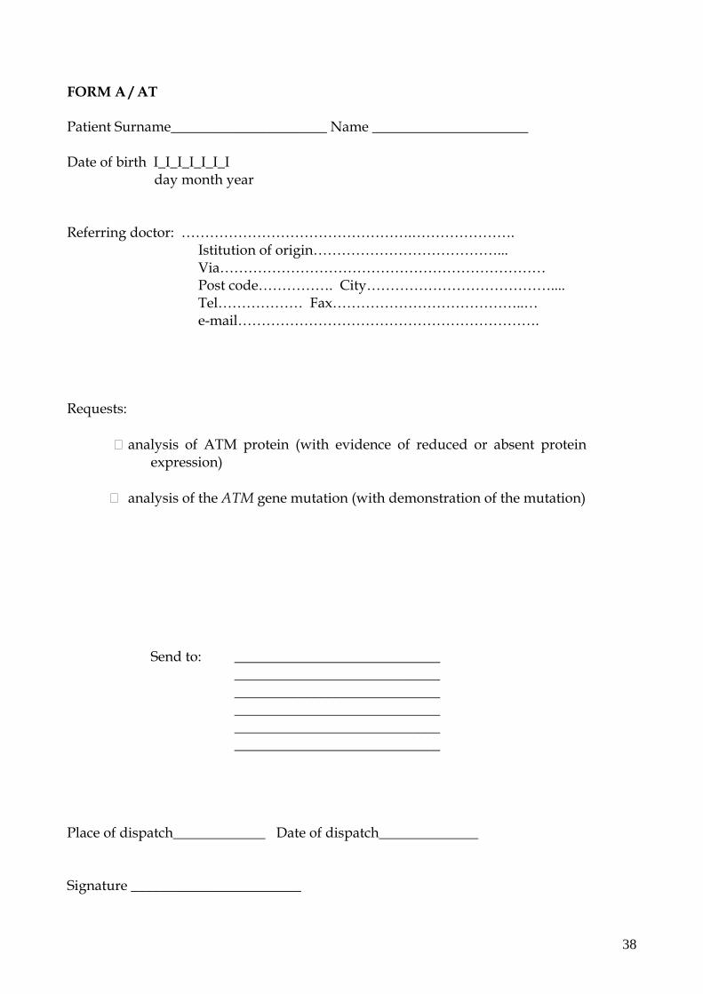
FORM A / AT Patient Surname______________________ Name ______________________ Date of birth I_I_I_I_I_I_I day month year Referring doctor: ………………………………………….…………………. Istitution of origin…………………………………... Via…………………………………………………………… Post code……………. City………………………………….... Tel……………… Fax…………………………………..… e-mail………………………………………………………. Requests:
analysis of ATM protein (with evidence of reduced or absent protein expression)
analysis of the ATM gene mutation (with demonstration of the mutation)
Send to: _____________________________ _____________________________ _____________________________ _____________________________ _____________________________ _____________________________
Place of dispatch_____________ Date of dispatch______________ Signature ________________________
38

Form B / AT Informed consent to intravenous/subcutaneous immunoglobulin treatment of
minors
I/We the undersigned…………………………………parent/s of…………………………….. born in ………………………………( ) on……….………. have been informed by Dr./Prof………………………………… that the clinical conditions of my/our daughter/son require treatment with immunoglobulins and that such treatment is not completely free of risks (including the transmission of hepatitis, etc.) I/we have fully understood what has been explained to me/us by Dr./Prof……………………………. both in terms of clinical conditions and the risks linked to the treatment and those which could arise by not receiving the treatment. I/we therefore consent / do not consent (*) to my/our daughter/son receiving at this institution the immunoglobulin treatment required throughout his/her disease course. (*) delete as appropriate. Date…………………….. Signature…………………………………………. Withdrawal of informed consent I/We the undersigned parent/s of ………………………………. born in ……………….……… ( ) on ………..withdraw my/our consent to immunoglobulin treatment signed by me/us on ……………. date…………………….. Signature………………………………………….
Informed consent to intravenous immunoglobulin treatment of adults I, the undersigned…………………………………..born in…………………( ) on……………. Have been informed by Dr./Prof………………………………… that my clinical condition requires treatment with immunoglobulins and that such treatment is not completely free of risks (including the transmission of hepatitis, etc.) I have fully understood what has been explained to me by Dr./Prof……………………………. both in terms of clinical conditions and the risks linked to the treatment and those which could arise by not receiving the treatment. I therefore consent/do not consent to receive at this institution the immunoglobulin treatment required throughout my disease course. (*)delete as appropriate. Date…………………….. Signature…………………………………………. Withdrawal of informed consent I the undersigned ………………………………. born in ……………….……… ( ) on ………..withdraw my consent to immunoglobulin treatment signed by me on …………….
Date…………………….. Signature………………………………………….
39

Form C / AT Patient’s Surname: __________________ Name _____________ Date of birth I_I_I_I_I_I_I day month year Referring physician : ………………………………………….…………………. Institution…………………………………... Address …………………………………………………………… post code……………. City………………………………….... Tel……………… Fax…………………………………..… e-mail………………………………………………………. Surname_____________________ Name ____________________________ Family relationship with the patient ___________________________________ Requests:
analysis of ATM protein (with evidence of reduced or absent protein expression)
analysis of the ATM gene mutation (with demonstration of the mutation)
Send to: _____________________________ _____________________________ _____________________________ _____________________________ _____________________________ _____________________________
Place of dispatch_____________ Date of dispatch______________ Signature ________________________
40