straightforward selection of broadly neutralizing single-domain
TRANSCRIPT

Straightforward Selection of Broadly Neutralizing Single-DomainAntibodies Targeting the Conserved CD4 and Coreceptor BindingSites of HIV-1 gp120
Julie Matz,a,b,c,d Pascal Kessler,e Jérôme Bouchet,f,g Olivier Combes,e Oscar Henrique Pereira Ramos,e Francis Barin,h Daniel Baty,a,b,c,d
Loïc Martin,e Serge Benichou,f,g Patrick Chamesa,b,c,d
Inserm,U1068, CRCM, Marseille, Francea; Institut Paoli-Calmettes, Marseille, Franceb; Aix-Marseille University, Marseille, Francec; CNRS, UMR7258, CRCM, Marseille, Franced;CEA, iBiTecS, SIMOPRO, Gif-sur-Yvette, Francee; Institut Cochin, CNRS UMR8104, Université Paris Descartes, Paris, Francef; Inserm U1016, Paris, Franceg; Université FrançoisRabelais, Inserm UMR 966, Tours, Franceh
Few broadly neutralizing antibodies targeting determinants of the HIV-1 surface envelope glycoprotein (gp120) involved in se-quential binding to host CD4 and chemokine receptors have been characterized. While these epitopes show low diversity amongvarious isolates, HIV-1 employs many strategies to evade humoral immune response toward these sensitive sites, including acarbohydrate shield, low accessibility to these buried cavities, and conformational masking. Using trimeric gp140, free or boundto a CD4 mimic, as immunogens in llamas, we selected a panel of broadly neutralizing single-domain antibodies (sdAbs) thatbind to either the CD4 or the coreceptor binding site (CD4BS and CoRBS, respectively). When analyzed as monomers or ashomo- or heteromultimers, the best sdAb candidates could not only neutralize viruses carrying subtype B envelopes, corre-sponding to the Env molecule used for immunization and selection, but were also efficient in neutralizing a broad panel of enve-lopes from subtypes A, C, G, CRF01_AE, and CRF02_AG, including tier 3 viruses. Interestingly, sdAb multimers exhibited abroader neutralizing activity spectrum than the parental sdAb monomers. The extreme stability and high recombinant produc-tion yield combined with their broad neutralization capacity make these sdAbs new potential microbicide candidates for HIV-1transmission prevention.
Neutralizing antibodies (NAbs) are a natural defense mecha-nism against virus infections and are the basis of efficient
vaccines (1, 2). In the case of HIV-1, NAbs target the viral enve-lope, a trimeric complex constituted by the noncovalent associa-tion of surface gp120 and transmembrane gp41 glycoproteins (3).This complex is responsible for interacting with the primary re-ceptor, CD4, and then with a chemokine coreceptor, CCR5 orCXCR4, expressed at the surface of HIV-1 target cells (4). Thesurface gp120 glycoprotein elicits both neutralizing and nonneu-tralizing antibodies during natural infection. Antibodies that lackneutralizing activity are often directed against the gp120 regionsthat are occluded on the trimer but exposed upon shedding. Incontrast, anti-HIV-1 NAbs bind to the functional envelope glyco-protein complex and typically recognize conserved or variableepitopes near the receptor-binding regions.
HIV-1 has evolved many strategies to evade the host humoralimmune response, including high sequence variability, protectionof sensitive epitopes by a shield of carbohydrate moieties, andconformational and entropic masking (5, 6). Consequently, theneutralizing antibody response during HIV-1 infection is weakand narrow, and only a few monoclonal antibodies with broadneutralization breadth, including among others b12, VRC01,PG16, X5, and 17b, have been isolated (6, 7). The binding mode ofb12 and VRC01 has been carefully analyzed, and they were shownto bind to the CD4-binding site (CD4BS) of gp120, mainly usingtheir heavy chain variable region (VH) domain to reach the cavityof gp120 involved in recognition of CD4 (8, 9). Other neutralizingantibodies, such as X5 or 17b, bind gp120 epitopes unveiled by theconformational change induced by CD4 binding and involved ininteraction with the coreceptor (10, 11). While these coreceptorbinding site (CoRBS) epitopes are buried until the conforma-
tional change happens, they become accessible to such antibodies(CD4-induced [CD4i] antibodies) after CD4 binding to gp120.However, Labrijn et al. have shown that conventional Ig antibod-ies face steric constraints on access to these epitopes due to theclose proximity of the viral and cellular membranes, which leavesa very narrow space (10). They have also shown that small anti-body fragments derived from CD4i antibodies, such as the 25-kDasingle-chain variable fragment (scFv), can reach their epitope andblock the infection event more efficiently than the correspondingfull-length parental antibody and better even than the corre-sponding Fab fragment.
Single-domain antibody fragments (sdAbs), derived fromcamelid antibodies naturally devoid of light chains, are small frag-ments of 13 kDa, i.e., 1/12 the size of conventional antibodies andhalf the size of scFv. Because their antigen binding site is consti-tuted by a single VH domain, they usually bind to cavities at thesurface of their antigen, often inserting a protruding CDR3 hyper-variable loop within the cavity (12). Nonetheless, these fragmentsnot only bind their antigens with low nanomolar affinities but areextremely stable, are very efficiently produced in Escherichia coli,
Received 23 February 2012 Accepted 25 October 2012
Published ahead of print 14 November 2012
Address correspondence to Patrick Chames, [email protected], or SergeBenichou, [email protected].
Supplemental material for this article may be found at http://dx.doi.org/10.1128/JVI.00461-12.
Copyright © 2013, American Society for Microbiology. All Rights Reserved.
doi:10.1128/JVI.00461-12
January 2013 Volume 87 Number 2 Journal of Virology p. 1137–1149 jvi.asm.org 1137
on January 30, 2018 by guesthttp://jvi.asm
.org/D
ownloaded from

and present a large degree of homology with the VH3 subset ofhuman VH genes. Thus, because of their small size and their ten-dency to bind cavities, they have been proposed as good neutral-izing antibody candidates. If CD4BS sdAbs with good neutralizingcapacities have already been isolated (13, 14), to our knowledge,no CoRBS sdAb has yet been discovered to date.
Using the scorpion toxin scyllatoxin as a small disulfide-stabi-lized structural scaffold, we have developed a series of small CD4mimics that present optimal interaction with gp120 and bind toviral particles and diverse HIV-1 envelopes with CD4-like affinity.Interestingly, they possess CD4 functional properties, includingthe ability to unmask gp120 conserved neutralization epitopesthat are cryptic on the unbound glycoprotein (15–19). In the pres-ent work, to increase the chance of selecting neutralizing sdAbstargeting the CD4 and coreceptor binding sites of gp120, we haveimmunized llamas with gp140 (trimeric version of gp120 boundto the gp41 ectodomain), either free or cross-linked to a CD4mimic (16). The last version was aimed to enhance CD4i epitopeexposure. Various selection strategies have led to the isolation ofseveral CD4BS and CoRBS single-domain antibodies with broadneutralization properties.
MATERIALS AND METHODSPreparation of recombinant Env constructs and CD4 mimics. For sim-plicity, gp120 and gp140 stemming from the SF162 strain are referred to asgp120 and gp140. Glycoproteins from other strains are explicitly identi-fied (for instance, gp120YU2).
The Freestyle 293 expression system (Life Technologies, Invitrogen)was used to generate transient cells expressing the various gp120,gp120YU2, and gp120CN54 monomers or the gp140 and gp140YU2 tri-meric form, using Freemax as the transfection reagent. The various enve-lopes were purified as previously described by miniCD4 affinity chroma-tography (17). They were then used as such or cross-linked to miniCD4derivative M64U1-SH through disulfide bond formation as describedpreviously (16). Wild-type gp140YU2, gp140YU2 D368R (having achange from Asp to Arg at position 368), gp140YU2 I420R, and thegp140YU2 I423M N425K G431E triple mutant were kindly provided byRichard Wyatt (Department of Immunology and Microbial Science, TheScripps Research Institute, La Jolla, CA).
The miniCD4 derivatives M48U1 and M64U1-SH were synthesizedand purified by reverse-phase chromatography as described earlier (16)and characterized by mass spectrometry.
For the fluorescence polarization assay, we obtained in the same way aspecific miniCD4, M64-Fluo, which was coupled to fluorescein at theεNH2 of Lys11 through a short polyethylene glycol (PEG) linker obtainedfrom 8-amino-3,6-dioxaoctanoic acid.
The M64 sequence is TpaNLKWCQKRCKSLGLLGRCAdPTFCACV-NH2, where Tpa stands for 3-mercaptopropanoyl (-COCH2CH2SH,equivalent to an amineless cysteinyl residue) and dP for D-proline.
Immunization and library construction. Two llamas (Lama glama)were immunized with either free gp140 or purified cross-linked gp140-S-S-M64U1 complex. Each llama received 4 injections of 250 �g of antigenwith Freund’s incomplete adjuvant at 3-week intervals. Two weeks afterthe last immunization, 400 ml of blood was collected per llama. TotalRNA was isolated from peripheral blood lymphocytes. IgG RNA se-quences were reverse transcribed with reverse transcriptase using primer3=CH2-2 (GGTACGTGCTGTTGAACTGTTCC). VH and VHH (variabledomains of camelid heavy-chain antibodies) genes of IgG were amplifiedby PCR using primer 3=CH2-2 and a mix of 5=VH1/3/4-Sfi (20). After gelpurification of fragments corresponding to VHH genes, amplificationswere carried out by PCR using primers 3=VHH-Not (described in refer-ence 20) and a mix of 5=VH1/3/4 Sfi. These genes were subcloned usingSfiI and NotI restriction sites.
Selection and screening by phage display. Libraries were rescued us-ing KM13 helper phage to carry out selections by phage display.
First, basic selections were performed with free gp120 or gp120-S-S-M64U1 immobilized on Epoxy Dynabeads via monoclonal antibody(MAb) D7324 (Aalto Bio Reagents, Dublin, Ireland), and all phage-sdAbswere eluted by trypsin treatment (1 h at room temperature, trypsin at 1mg/ml). In some cases, a competitive elution was performed usingminiCD4 M48U1 (21), followed by a trypsin treatment. Selections werealso performed using the trimeric gp140 or gp140-S-S-M64U1 bounddirectly to Epoxy Dynabeads and using trypsin elution.
Two last selections were designed to obtain broad-spectrum sdAbs.The first rounds were done using free gp120YU2 or gp120-S-S-M64U1,and the second rounds were performed using free gp120CN54 andgp120YU2-S-S-M64U1, respectively, with trypsin elution.
Selected clones were sequenced to identify distinct sdAbs.Construction of multivalent proteins. Genes for homotrivalent con-
structs of two sdAbs of interest, JM2 and JM4, with (G4S)7 linkers (denoted as“x” in construct names) between each domain were obtained from Geneartand subcloned into phagemid pHEN. To obtain the coding sequence of two-domain versions (JM2x2, JM2x3, JM2x4, JM2x5, JM4x2, JM4x3, JM4x4, andJM4x5), genes of sdAbs JM2, JM3, JM4, and JM5 were amplified by PCRusing primers Trim2For (AGTGGTGGCGGAGGTAGCGCTAGCGAGGTGCAGCTGGTGGAG) and Trim3Rev (TGAGATGAGTTTTTGTTCTGCGGCCGCTGAGGAGACGGTGACCTG). PCR products were clonedinto the digested trivalent plasmid by using the InFusion system (Clon-tech). To create the remaining bivalent-protein genes (JM3x2, JM3x3,JM3x4, JM3x5, JM5x2, JM5x3, JM5x4, and JM5x5), PCR amplificationswere performed using primers Trim1For (CTCGCGGCCCAGCCGGCCATGGCCGAGGTGCAGCTG) and Trim1Rev (CCGCTGCCACCTCCCCCCAGGCCTGAGGAGACAGTGACCTG) to amplify the JM3 and JM5genes, which were substituted for the first sdAb gene of previous con-structs using the InFusion system (the cloning scheme is illustrated in Fig.S1 in the supplemental material) (22). All constructs were checked bysequencing.
Production and purification of sdAbs and multivalent proteins. AllsdAbs were produced in E. coli BL21(DE3). Production was done by seed-ing 400 ml of 2TY-Amp-50x5052 (autoinductive medium) (23). The cul-ture was incubated first for 3 h at 37°C and then overnight at 30°C. Allmultivalent proteins were produced in E. coli BL21(DE3) by seeding 400ml of 2TY-Amp, and production was induced by the addition of 0.1 mMisopropyl-�-D-thiogalactopyranoside (IPTG) when the optical density at600 nm (OD600) reached 0.4 to 0.7.
For purification, bacteria were lysed using BugBuster protein extrac-tion reagent (Novagen) containing lysozyme and Benzonase. Superna-tants were applied to Talon beads (Clontech), and elution of 6His-sdAbsand multivalent proteins was done by the addition of imidazole. Lastly,elution buffer was changed for phosphate-buffered saline (PBS) usingVivaspin with a 5-kDa molecular-mass cutoff (GE HealthCare Life Sci-ence). sdAbs were stocked in PBS at �20°C. Most sdAbs could be pro-duced with yields in the range of 10 to 100 mg/liter culture, while yieldswere about 1 mg/liter culture for multivalent proteins. Multivalent con-structs that were not easily expressed were not studied further.
Characterization of binding to different subtypes of gp120 andgp140. All steps to characterize binding to the different subtypes of gp120and gp140 were performed at room temperature with mixing, except forbead or plate coating. Washes were carried out between each step.
For enzyme-linked immunosorbent assay (ELISA) of gp120, Epoxybeads were coated (Dynabead, Invitrogen) with MAb D7324, with 8 � 105
beads per well being coated with 0.2 �g of MAb D7324 per well. Coatingwas carried out for 48 h at 4°C on a wheel. Beads were blocked for 1 h with2% milk-PBS (MPBS). gp120 was added at 10 nM for 2 h, and beads weredispensed into a preblocked 96-well plate. sdAbs were added at 10 �g/mlfor 1 h. Bound sdAbs were detected using horseradish peroxidase (HRP)-coupled anti-c-myc antibody (clone 9E10) that was incubated for 1 h, andstaining was performed using ABTS [2,2=-azinobis(3-ethylbenzthiazo-
Matz et al.
1138 jvi.asm.org Journal of Virology
on January 30, 2018 by guesthttp://jvi.asm
.org/D
ownloaded from

linesulfonic acid)]-based solution. The optical density was followed at450 nm.
For ELISA of gp140, the same protocol was used, with 0.1 �g of gp140being used to directly coat 8 � 105 Epoxy beads per well for 48 h at 4°C ona wheel. gp120 and gp140 were used free or covalently cross-linked toM64U1-SH.
Cells. TZM-bl reporter cells (HeLa CD4� CXCR4� CCR5� carryingthe luciferase gene under the control of the HIV-1 long terminal repeat[LTR]) (24) and human embryonic kidney 293T cells were grown inDMEM (Dulbecco’s modified Eagle’s medium) supplemented with 10%fetal calf serum (FCS) and antibiotic/antifungal at 37°C under 5% CO2.
Single-round pseudovirion production and neutralization assay.293T cells were seeded in T75 flasks at a density of 3 � 106 cells/flask andtransfected 24 h later by the calcium phosphate precipitation method witha DNA mix containing the HIV-1 packaging plasmid (pCMV�R8.2) (25),the HIV-1 transducing vector containing the tat gene (pHIvec2-GFP)(26), and a plasmid encoding the HIV-1 envelope glycoprotein (or vesic-ular stomatitis virus G glycoprotein [VSV-G] as control) at concentra-tions of 8, 8, and 2 �g/T75 flask, respectively. Medium was removed 6 hafter transfection, and 10 ml of complete medium was added. Seventy-twohours after transfection, supernatants containing pseudovirions were col-lected, spun to remove cell debris, filtered through 0.45-�m-pore-sizefilters, and stored at �80°C until use in neutralization experiments. Levelsof virus production were measured by HIV-1 Cap24 quantification inELISA (Innogenetics). To determine the TCID50 (50% tissue culture in-fective dose), 5-fold dilutions were carried out in 96-well plates with once-frozen cryovial of each pseudovirion production. Then, 104 TZM-bl cells/well were added and plates were incubated at 37°C for 48 h. After cell lysisusing luciferase cell culture lysis reagent (Promega), luciferase activity wasmeasured using a luciferase assay kit (Promega).
Plasmids encoding HIV-1 envelope glycoproteins (pSVIII-93BR029.2, -92UG975.0, -93BR019.4, -92BR025.9, -91US005.11,-92RW020.5, -92UG037.8, -92HT593.1, -93MW965.26, -92UG024.2,-92UG021.16, -93TH966.8, and -93TH976.17) were obtained from theNIH AIDS Research and Reference Reagent Program. pSVIIIENVHxBC2, pEnv 89.6, pEnv YU2, and pEnv VSV-G plasmids have beendescribed previously (27).
For the neutralization assay, pseudovirions (100 TCID50) were prein-cubated in 96-well plates with monovalent or multivalent sdAbs at 10 �Mand 5 �M, respectively. Plates were incubated for 1 h at 37°C. Then, 104
TZM-bl cells/well were added and plates were incubated at 37°C for 48 h.Luciferase activity was measured as described above. The 50% inhibitoryconcentrations (IC50s) were determined for sdAbs able to inhibit virusentry into TZM-bl cells. In the same manner, virions (100 TCID50) werepreincubated in 96-well plates with different concentrations of sdAbs.Plates were incubated for 1 h at 37°C. Then, 104 cells/well were added andplates were incubated at 37°C for 48 h. Luciferase activity was measured asdescribed above. IC50s were calculated using Prism software (GraphPadSoftware, Inc., San Diego, CA).
Neutralization assay on resistant primary isolates. The virus panelincluded two T-cell-line-adapted (TCLA) strains highly sensitive to neu-tralization (tier 1A; NL4.3 and MN) and nine primary isolates selected fortheir high (tier 1B), moderate (tier 2), or low (tier 3) sensitivity to neu-tralization. Four primary isolates (BX08, BIG, FRO, and KON) of twodifferent clades (B and CRF02_AG) were reported in previous studies(28–30). We added five primary isolates, including four viruses(94UG103, 92BR020, 93IN905, and 92TH021, of clades A, B, C, andCRF01_AE, respectively) identified as indicators for cross-clade neutral-ization (31) and one moderately resistant virus (92RW020, clade A) (31).This virus panel included viruses that were resistant to almost all thebroadly neutralizing human monoclonal antibodies that we tested (2G12,b12, 2F5, 4E10, PG9, and PG16). Monoclonal antibodies b12, 2G12, 4E10,and 2F5 were provided by Polymun Scientific (Vienna, Austria). PG9 andPG16 were kindly provided by P. Poignard (The Scripps Research Insti-tute, La Jolla, CA) through the Neutralizing Antibody Consortium of the
International AIDS Vaccine Initiative (IAVI, New York, NY). The virusstocks for the neutralization assays were prepared by passaging the strainsonly once or twice on phytohemagglutinin-stimulated peripheral bloodmononuclear cells (PBMCs) from HIV-negative healthy blood donors.The same stock of each isolate was used for the entire study. Infectioustiters were determined by infection of 1 � 104 TZM-bl cells with 100 �l ofserial 5-fold dilutions of the viral stocks in quadruplicate in the presence of30 �g/ml of DEAE-dextran. Infection levels were determined after 48 h,using the Bright Glo luciferase assay (Promega) and a Centro LB 960luminometer (Berthold Technologies) to measure luciferase activity incell lysates. Results with relative light unit (RLU) values of �2.5 times thatof the negative control (cells alone) were considered positive.
Aliquots of 50 �l of the dilution corresponding to 100 TCID50 of eachvirus stock were incubated for 1 h at 37°C with 11 �l of 3-fold serialdilutions of each antibody, starting at 50 �g/ml for all the reagents exceptPG9 and PG16, for which the starting dilution was 10 �g/ml. The virus-serum mixture was then used to infect 10,000 TZM-bl cells in a 96-wellmicroplate in the presence of 30 �g/ml DEAE-dextran. Infection levelswere determined after 48 h through determination of the luciferase activ-ity in cell lysates, as described above. IC50s are expressed as the meanamounts required to decrease the relative light units (RLU) by 50%. Theresults are expressed as the mean values of the assays performed in dupli-cate.
Competition between sdAbs using sdAb and phage-sdAb formats.All steps except the coating were performed at room temperature withmixing. Washes were carried out between each step except before theaddition of phage-sdAbs.
Epoxy beads were coated with MAb D7324 as described above. Beadswere blocked for 1 h with 2% MPBS. Free gp120 or gp120-S-S-M64U1 wasincubated at 10 nM for 2 h at room temperature. Serial dilutions from 10�M to 0.01 nM sdAbs were added. After 1 h, subsaturating concentrationsof phage-sdAbs were added. After 1 h of incubation followed by washes,HRP-coupled M13 antibody was added for 1 h and staining was doneusing ABTS-based solution. The OD450 was measured.
Characterization of sdAbs by surface plasmon resonance (SPR). Ex-periments were conducted at 25°C with a 30 �l/min flow rate in HBS(HEPES-buffered saline, 3 mM EDTA, 0.05% Biacore surfactant, pH 7.4)with a Biacore 3000 instrument (Biacore AB, Uppsala, Sweden). sdAbswere immobilized at a level of 1,000 to 2,000 response units (RU) by usingthe amine coupling kit [N-hydroxysuccinimide–1-ethyl-3-(3-dimethyl-aminopropyl) carbodiimide (NHS-EDC)] provided by the manufacturer.gp120 and gp140 of different subtypes were tested complexed or not withminiCD4 (notably M48U1) or soluble CD4 (sCD4) to determine the sdAbspecificities. CD4i antibodies X5 and 17b and CD4BS antibody b12 wereused in competition assays using ranges of dilution from 250 nM to 0.37nM. Calculations were done using the BiaEval 3.2 software furnished withthe Biacore instrument.
Characterization of sdAbs JM2, JM3, JM4, JM5, and JM7 by fluores-cence polarization. Competitive fluorescence polarization was assessed aspreviously described by Stricher et al. (32), using M64-Fluo instead offluorescein-CD4M33. Briefly, competition assays were performed in trip-licate by mixing 7 �l of serial sdAb dilutions (or M48U1), 7 �l of M64-Fluo (2 nM final concentration), and 7 �l of gp120 (8 nM final concen-tration). The fluorescence polarization was determined after 40 min ofequilibration time. Results were expressed as percentages of the results ofcontrol experiments, in the absence of competitor.
Cell surface CCR5 chemokine receptor binding assays. The effects ofsdAbs binding to gp120 on the interaction of the latter with its cellularcoreceptor CCR5 were investigated by flow cytometry (FACSCalibur; BDBiosciences) using adherent CHO-K1 cells overexpressing CCR5 (33).Briefly, 1 �M gp120 was preincubated or not with 3 molar equivalents(eq) of sCD4 (Progenic, Tarrytown, NY) or 5 eq miniCD4 M48U1 for 1 hat room temperature. The mixtures were then diluted to 2 nM gp120 withPBS-bovine serum albumin (BSA) 0.5%, pH 7.4. For competition pur-poses, 100 nM each given sdAb was added. One hundred microliters of
Selection of Broadly Neutralizing Anti-HIV-1 Env sdAbs
January 2013 Volume 87 Number 2 jvi.asm.org 1139
on January 30, 2018 by guesthttp://jvi.asm
.org/D
ownloaded from

this solution was mixed with 2 � 105 CCR5� cells. After 2 h of incubationat 4°C, cells were washed with PBS-BSA 0.5% and incubated with MAbD7324 (1:1,000). A phycoerythrin-tagged antibody (1:500) directedagainst MAb D7324 further detected envelope glycoprotein binding toCCR5 coreceptor.
Epitope mapping by competitive ELISA using mutant Env proteins.Shortly, Maxisorp 96-well plates were coated with sdAbs (500 ng/well, 2 hat room temperature). Next, wells were saturated with PBS containing 3%BSA (overnight, 4°C) and washed (25 mM Tris-HCl, 50 mM NaCl, Tween0.1%, pH 7.4), and serial dilutions of wild-type gp140YU2 trimer or theD368R, I420R, or I423M N425K G431E triple mutant (34) were added inthe absence (for JM2-coated wells) or presence (for JM3-, JM4-, JM5-,and JM7-coated wells) of miniCD4 M48U1 at 10 molar eq. After 2 h ofincubation at room temperature, wells were washed, and bound gp140proteins were detected by sequential recognition using D7324 antibody(1:1,000) and donkey anti-sheep IgG HRP-coupled antibody (1:5,000)(Jackson ImmunoResearch). Plates were read at 450 nm 20 min after theaddition of 3,3=,5,5=-tetramethylbenzidine (TMB).
Determination of affinity by SPR. Experiments were conducted asdescribed above but using an Rmax of 200 RU for the sdAb immobiliza-tion. Dilutions of Env (from 200 nM to 0.4 nM) were tested to determinethe affinity of sdAbs JM2, JM4, and JM7. Calculations were performedusing the BiaEval 3.2 software.
RESULTSSelection of anti-Env sdAbs. To increase the chance of isolatingsingle-domain antibodies (sdAbs) targeting various envelopeepitopes, two llamas were immunized with four injections of ei-ther the trimeric gp140 (SF162 HIV-1 strain) or the same trimericform covalently cross-linked to a CD4 mimic (gp140-S-S-M64U1) to favor the selection of CD4i sdAb binders. The VHHgenes from immunized animals were then amplified from PBMCsand were cloned into a phagemid vector. Electroporation yieldedaround 108 independent clones for each library. Various selectionstrategies were then performed to increase the chance of isolatingsdAbs against a large number of relevant epitopes. Antigens wereimmobilized via the interaction with MAb D7324, targeting theC-terminal end of gp120, and basic strategies included selectionson gp120 monomer, either free or cross-linked to a CD4 mimic tounveil CD4i epitopes. A similar approach was performed using thetrimeric free gp140 and the cross-linked complex gp140-S-S-M64U1. After two rounds of selection, positive clones were iden-tified by phage ELISA using the antigen used for selection in aprimary screening. Positive clones were sequenced (Fig. 1), andrepresentative clones of each family were produced and purified assoluble sdAbs to be further characterized by a secondary screeningusing a panel of antigens.
The results of binding analysis of the selected sdAbs to differentEnv and Env-S-S-M64U1 antigens are reported in Fig. 2. Based ontheir capacity to bind to various conformations of gp120 andgp140, four types of anti-Env sdAbs could be defined. (i) ClonesJM1 and JM2 behaved as CD4BS binders, as they could bind tofree gp120 and gp140 but not to the same envelopes cross-linkedto the CD4 mimic (Fig. 2A). JM2 yielded a higher response andwas therefore selected for further analysis. (ii) Clones JM3, JM4,and JM5 bound to free or miniCD4-cross-linked gp120 and gp140(Fig. 2B), suggesting that they do not share their epitope with JM2.In this assay, JM5 displayed a tendency toward a better affinity forcross-linked Env, suggesting a CD4i behavior. (iii) Clone JM7behaved more drastically, since it could only bind to cross-linkedgp120- or gp140-S-S-M64U1 complexes. (iv) Finally, clones JM8and JM9 yielded no signal against any gp120 form but yielded high
signals on free and cross-linked trimeric gp140 (Fig. 2C). Interest-ingly, these two last clones were only retrieved during selectionsperformed on trimeric gp140. They were thus considered gp140specific. None of the sdAbs were found positive on miniCD4 byELISA (data not shown).
Neutralizing activity of anti-Env sdAbs. A single-round infec-tion assay was first set up to determine the capability of thesemonovalent sdAbs to block HIV-1 infection of a panel of pseu-dotyped virions carrying various envelope glycoproteins fromsubtypes A, B, C, G, or CRF01_AE. sdAbs were compared to thewell-described broadly neutralizing antibody b12. Virus particlespseudotyped with VSV-G were used as a specificity control in thisneutralization assay. Interestingly, a first screening performed us-ing 10 �M each sdAb revealed some neutralization properties formost selected sdAbs, except JM7 (Fig. 3). As expected, sdAbs wereable to efficiently neutralize pseudovirions carrying X4- and R5-tropic envelopes of subtype B, since immunizations and selectionswere performed with subtype B or subtype B=/C envelopes. Moreinterestingly, sdAbs JM3, JM4, and to a lesser extent JM5 couldneutralize most tested pseudovirions, including those pseu-dotyped with subtypes A, C, G, and CRF01_AE envelopes. Deter-mination of the IC50s, which ranged between 0.2 and 40 �g/ml,confirmed these observations (Table 1). In comparison, the IC50smeasured for the b12 monoclonal antibody ranged between 0.005and 1.5 �g/ml, but its neutralizing activity was restricted to sub-type B envelopes at these concentrations.
To further define the neutralization ability of these sdAbs, as-says using primary viruses were performed. This virus panel in-cluded two T-cell-line-adapted (TCLA) strains highly sensitive toneutralization (tier 1A; NL4.3 and MN) and nine primary isolatesselected for their high (tier 1B), moderate (tier 2), or low (tier 3)sensitivity to neutralization. As shown in Table 2, JM2 and JM5were able to neutralize 1 and 2 of these 11 viruses, respectively, and
FIG 1 Sequences of anti-Env sdAbs. Selected clones were sequenced andaligned following the IMGT (international ImMunoGeneTics informationsystem) numbering. Four families were created in accordance with homologyin the complementarity-determining region (CDR). Three unique clone fam-ilies were selected. The total number of times each clone was retrieved is indi-cated as frequency.
Matz et al.
1140 jvi.asm.org Journal of Virology
on January 30, 2018 by guesthttp://jvi.asm
.org/D
ownloaded from

JM3 neutralized 5 of them, outperforming well-known broadlyneutralizing antibodies such as 2G12, b12, 2F5, 4E10, and PG9.JM4 could neutralize 7 of these 11 viruses, thereby matching oroutperforming all broadly neutralizing antibodies tested in thisassay in terms of neutralization breadth. The fact that the sdAbs,except JM3, were not efficient at neutralizing the sensitive MNstrain might suggest a specific mode of interaction with or a par-ticular accessibility to the targeted epitopes.
Characterization of the anti-Env sdAb epitopes. Several ex-perimental approaches were designed to better characterize theepitopes targeted by these anti-Env sdAbs. We first determinedwhether some of these Env binders recognized overlappingepitopes by using competitive ELISA between purified sdAbs andphage-sdAbs for binding to gp140 (Fig. 4A) or gp140-S-S-M64U1(Fig. 4B). As expected, all sdAbs competed with themselves.
In the absence of competitor, phage-sdAb JM2 (phage-JM2)yielded a nearly saturating signal on free Env but could still bind tothe miniCD4-cross-linked Env, albeit at a reduced level. This issurprising since the purified sdAb JM2 only led to a very faintsignal on the same antigen (Fig. 2). This result could be explainedby the presence of a small percentage of free Env in the complexedform in conjunction with the very high sensitivity of the phageELISA format (each phage particle being detected by several doz-ens of anti-M13 HRP-labeled antibodies bound to the phage cap-
sid). In the presence of competitors on free Env, phage-JM2 bind-ing was clearly hampered by the presence of MAb b12 but not byMAb X5 (Fig. 4A), corroborating the CD4BS nature of this sdAb.Of note, the residual binding of phage-JM2 on miniCD4-cross-linked Env was also efficiently competed by b12, confirming thehypothesis regarding the presence of residual free Env in this prep-aration.
In the absence of competitor, phage-JM3, -JM4, and -JM5yielded higher signals on miniCD4-cross-linked Env than on freeEnv (Fig. 4B versus A), suggesting a CD4i behavior. Moreover,binding of phage-JM7 happened only on the cross-linked form(Fig. 4B). As expected, JM3, JM4, and JM5 behaved similarly andcompeted each other on both miniCD4-cross-linked and free Env,indicating that they bind to overlapping epitopes. JM7 could alsocompete with phage-JM3, -JM4, and -JM5 but only on the cross-linked form, as expected since it cannot bind free Env. Phage-JM8bound to both forms of Env and did not compete with othersdAbs.
More surprisingly, phage-JM2 binding to free Env was affectedby the presence of sdAbs JM3 and JM4 but not by CD4i MAb X5(35). Similarly, binding of phage-JM3, -JM4, and -JM5 to free Envwas also hampered by JM2 or the CD4BS MAb b12 (Fig. 4A). Theinfluence of sdAb JM2 was not observed on cross-linked Env (Fig.4B), logically, since this sdAb can hardly bind to this form (Fig. 2).
FIG 2 sdAb binding on different conformations and strains of HIV-1 Env, followed by ELISA. sdAbs binding to gp120 (bound via the D7324 antibody to Epoxybeads) or to gp140 (directly immobilized on Epoxy beads) were detected via a peroxidase-labeled anti-c-myc Ab. Histograms correspond to the three mainfamilies of sdAbs selected. (A) JM1 and JM2 clones bind preferentially to the free form of Env. (B) JM3, JM4, and JM5 bind both Env forms, while JM7 only bindsto the cross-linked Env. (C) JM8 and JM9 bind exclusively to the gp140 trimeric form of Env. Shown are the results of one experiment representative of at leastthree. Error bars represent standard deviations of the means.
Selection of Broadly Neutralizing Anti-HIV-1 Env sdAbs
January 2013 Volume 87 Number 2 jvi.asm.org 1141
on January 30, 2018 by guesthttp://jvi.asm
.org/D
ownloaded from

These results suggest that JM2 binding may stabilize an Env con-formation that prevents recognition by phage-JM3, -JM4, and-JM5, and vice versa. Alternatively, the orientation of the variousantibodies and large phage-VHH particles with respect to gp140may engender steric clashes. Together, these data support a com-mon recognition of CD4i epitopes for JM3, JM4, and JM5 andtargeting of the Env CD4BS for JM2.
Surface plasmon resonance (SPR) analyses using high-immo-bilization-density chips were used to confirm ELISA data and toroughly estimate the binding profiles of the neutralizing anti-EnvsdAbs against various Env antigens (Fig. 5). As expected for aCD4BS antibody, JM2 could clearly bind to the uncomplexedform of gp120 but not to its complexed form (Fig. 5A). On mo-nomeric gp120 (Fig. 5B), JM3, JM4, and JM5, displaying a CD4iprofile by ELISA, yielded higher signals with complexed gp120than with uncomplexed gp120, and similar results were observedwith the trimeric form of Env (gp140) (Fig. 5C), validating a CD4ibehavior. Of note, and in contrast to JM4 and JM5 or the well-
defined CD4i MAb X5 used as a control (11), JM3 did not bindmore efficiently to gp120 complexed to a soluble recombinantCD4 molecule (sCD4) than to free gp120. This observation mightbe related to steric clashes between JM3 and CD4, which do nothappen with the smaller miniCD4. As observed in ELISA (Fig. 2),JM7 was found exclusively positive on the gp120-miniCD4 com-plex and did not bind at all to free gp120. However, unlike otherCD4i sdAbs or MAb X5, JM7 was also found negative on gp120complexed to sCD4. Since we checked by fluorescence polariza-tion that this sdAb was unable to bind directly to the CD4 mimicconstructs (data not shown), this observation suggests that JM7may recognize an epitope specifically formed by the association ofgp120 and the CD4 mimic. Another possible explanation wouldbe that JM7 can only bind gp120 in the CD4-bound state, which ismimicked here by the miniCD4, and that the orientation of JM7allows the presence of a small miniCD4 (3 kDa) but clashes withthe much bigger sCD4 molecule (about 20 kDa). Nevertheless,this observation explains why this sdAb was unable to neutralize
FIG 3 sdAb neutralization ability toward different pseudovirion strains. Each sdAb was tested in a single-round neutralization assay as described in Materials andMethods, using TZM-bl cells as target cells. Pseudovirions (100 TCID50) carrying various envelope glycoproteins from different HIV-1 subtypes were preincu-bated with purified sdAbs at 10 �M in 96-well plates for 1 h at 37°C. TZM-bl cells were then added, and luciferase activity was measured 48 h later. Values areexpressed as percentage of neutralization relative to the luciferase activity measured in the absence of sdAb. Shown are the results of one experiment represen-tative of at least three. Error bars represent standard deviations of the means.
TABLE 1 IC50s of monovalent and multivalent sdAbs on pseudovirions carrying various HIV-1 envelope glycoproteins
Virusb Subtype
IC50 in TZM-bl cells (�g/ml)a
JM2 JM3 JM4 JM5 JM8 JM9 JM2x2 JM2x4 JM2x5 JM3x3 JM3x5 JM4x2 JM4x3 JM4x5 JM5x2 JM5x3 JM5x5 JM4x4x4 b12
92RW020.5 A X 4.4 23.3 X ● ● X 1.5* 0.8 27.5 ● X ● X 1.6 18.9 ● 48* ●92UG037.8 A ● ● ● ● X X 40.8 ● ● 46.5 ● ● ● ● 47.3 35.2 ● X ●HxBC2 B 0.2 9.9 0.7 ● ● ● 0.2 0.6 3.7 16.1 ● 12.9 21.4 54.0 15.1 134.4 ● 73.8 0.00589.6 B X 0.7 0.5 2.6 X ● X ● ● 1.6 11.0 ● 7.0 0.8 29.5 4.8 19.0 8.3 0.14YU2 B 30 8.7 6.6 3.2 ● ● 28.5 92.8* X 16.7 85.1* 96* 60.8* 36.4* 10.9 20.3 5.9* 17* 1.5392HT593.1 B 5 1.4 1.2 1.1 ● ● 18.8 49.6* 15.4* 8.0 X 10.3 32* 12.2 19.3 12.4 X X 1.0491US005.11 B ● 3.6 4.2 3.8 X ● 57.3 108.9* ● 14.7 ● ● 27.4* 32* 32.0 16.3 54.1* 48* 1.1492BR025.9 C 11.4 8.8 9.7 ● ● ● X X X 13.6 ● 99.6* 32* 46.2* 36.2 42.2 ● 96* ●93TH966.8 A/E ● 2.3 X ● X X 42.6 X ● 27.3 ● ● 45.1* ● ● 37.9 ● 59 ●93TH976.17 A/E ● ● ● ● X X 36.8 ● ● 83.8 ● ● ● ● 90.9 20.8 ● 168.96 ●92UG975.10 G 43.8 80.6* ● ● X X 33.3 ● ● 57.9 ● ● ● ● 75.0 134.4 ● X ●
a ●, no neutralization was observed at the highest tested concentration (150 �g/ml); X, IC50 could not be calculated but some neutralization was observed; shaded cell, IC50
determined as 50% of inhibitor effect; *, complete neutralization was not observed at the highest tested concentration of 150 �g/ml.b 92RW020.5, 92UG037.8, and 92BR025.9 are tier 1; 89.6 and YU2 are tier 2.
Matz et al.
1142 jvi.asm.org Journal of Virology
on January 30, 2018 by guesthttp://jvi.asm
.org/D
ownloaded from

all the tested viruses (Fig. 3), despite behavior quite similar to thatof sdAbs JM3, JM4, and JM5 (Fig. 4).
Characterization of the CD4BS sdAb JM2. Since the ELISAand SPR results suggested that JM2 was targeting the gp120CD4BS, we confirmed this hypothesis by performing fluorescencepolarization experiments on gp120, using a fluorescent miniCD4probe (M64-Fluo) and sdAbs including JM2 or the CD4 mimicM48U1 as competitors. This approach relies on the fact that asmall molecule (such as miniCD4) rotates fast in solution andexhibits low fluorescence polarization whereas a large molecule(such as gp120-miniCD4 complex) exhibits a higher fluorescencepolarization because of its slower motion under the same condi-tions. Thus, changes in fluorescence polarization can reflect theassociation or dissociation between molecules of interest, in ourcase, M64-Fluo and gp120. As shown in Fig. 6A, JM2, as well asM48U1, efficiently competed with the M64-Fluo probe for gp120
binding, thereby confirming that JM2 is a CD4BS ligand. M48U1outperformed JM2, as it possesses a subnanomolar affinity forgp120 (21). This experiment was also conducted with JM3, JM4,JM5, and JM7, and no competition was observed.
Since MAb b12 is a well-characterized CD4BS antibody, com-petition experiments were also performed by SPR using this anti-body. As shown in Fig. 6B, MAb b12 competed with sdAb JM2 forbinding to unliganded gp120, thus confirming that this sdAb be-haves as a CD4BS binder.
Finally, when analyzed by flow cytometry on CCR5-express-ing cells, JM2 was also shown to block gp120 binding to CCR5,likely through stabilization of gp120 in a conformation whichdoes not allow CCR5 binding (Fig. 7A). Furthermore, the sub-sequent addition of sCD4 or M48U1 could not restore CCR5binding, suggesting that the CD4 binding site is indeed blockedby JM2.
TABLE 2 IC50s of monovalent sdAbs on viruses with various levels of sensitivity to neutralizationa
Virus Clade Tier
IC50 in TZM-bl cells (�g/ml)b
JM2 JM3 JM4 JM5 2G12 b12 2F5 4E10 PG9 PG16
94UG103 A 2 �50 12.30 13.40 �50 �50 �50 �50 �50 1.78 �0.1292RW020 A 2 �50 �50 �50 �50 4.5 �50 �50 �50 0.14 0.87NL4.3 B 1A 1.45 1.43 0.02 29.60 NT NT NT NT NT NTMN B 1A �50 4.66 �50 �50 �0.12 �0.12 �0.12 �0.12 �0.12 �0.12BX08 B 1B �50 �50 14.45 �50 NT NT NT NT NT NTBIG B 2/3 �50 �50 30.00 �50 43.53 2.33 �50 �50 �10 �0.12FRO B 2/3 �50 29.60 28.30 44.40 �50 �50 26.36 �50 �10 �1092BR020 B 2 �50 �50 �50 �50 �50 �50 �50 �50 �10 �1093IN905 C 2 �50 �50 41.80 �50 �50 �50 �50 �50 3.81 �1092TH021 CRF01_AE 2 �50 �50 �50 �50 �50 �50 �50 �50 �10 �10KON CRF02_AG 2/3 �50 45.23 22.62 �50 �50 �50 �50 �50 �0.12 �0.12a Viruses used were two T-cell-line-adapted strains highly sensitive to neutralization (tier 1A; NL4.3 and MN) and nine primary isolates selected for their high (tier 1B), moderate(tier 2) or low (tier 3) sensitivity to neutralization.b Starting dilutions were 50 �g/ml for all antibodies except PG9 and PG16, for which the starting dilution was 10 �g/ml. NT, not tested. Shading highlights neutralizing effects.
FIG 4 Competition between sdAbs and phage-sdAbs on gp140 or gp140-S-S-M64U1 followed by ELISA. Amounts of 10 �g/ml of sdAbs, X5, and b12 were addedto immobilized Env as indicated in Materials and Methods. After 1 h, phage-sdAbs were added at subsaturating concentration. Bound phage-sdAbs were revealedby using peroxidase-labeled anti-M13 antibody. (A) Competition was done on free gp140. (B) Competition was done on gp140-S-S-M64U1. Data from oneexperiment representative of three independent experiments are shown. Error bars represent standard deviations of the means of triplicates.
Selection of Broadly Neutralizing Anti-HIV-1 Env sdAbs
January 2013 Volume 87 Number 2 jvi.asm.org 1143
on January 30, 2018 by guesthttp://jvi.asm
.org/D
ownloaded from

Characterization of the CD4i sdAbs JM3, JM4, JM5, and JM7.Since our previous ELISA and SPR results suggested that severalanti-Env sdAbs, such as JM3, JM4, JM5, and JM7, were targetingthe CoRBS unveiled by a conformational change due to CD4binding (i.e., CD4i epitopes), we used flow cytometry to checkwhether these sdAbs could block the interaction between gp120-
sCD4 or gp120-M48U1 complexes and the CCR5 coreceptor ex-pressed on cells. Figure 7A shows that CD4i sdAbs JM3, JM4, andJM5 could efficiently block the binding of gp120 to CCR5 under allconditions. This experiment also confirmed that JM7 only binds tothe Env-M48U1 complex. Indeed, JM7 did not block gp120 orgp120-sCD4 binding to CCR5, but it could bind to gp120 and abolishits binding to the CCR5� cells in the presence of M48U1. As expected,no gp120 binding was observed by flow cytometry using untrans-fected CHO cells as negative control (data not shown).
FIG 5 sdAb binding to complexed or uncomplexed Env followed by SPR. sdAbs were immobilized on sensor chip CM5. Using a Biacore 3000 instrument, Envwas injected at various concentrations on different channels. Uncomplexed gp120 and gp120-plus-M48U1 complex were run over immobilized JM2 (A) orimmobilized JM3, JM4, JM5, and JM7, with MAb X5 as a reference (B). Uncomplexed gp140 and gp140-plus-M48U1 complex were run over immobilized JM4,JM5, and JM7 (C). Data from one experiment representative of three independent experiments are shown. RU, response units.
FIG 6 Characterization of CD4BS sdAb. (A) Curves obtained by fluorescencepolarization using a fluorescent miniCD4, M64-Fluo, as probe in the presenceof various concentrations of sdAbs and miniCD4 M48U1. (B) Competitionexperiment performed by SPR using various concentrations of MAb b12mixed with gp120 and injected onto a chip coated with sdAb JM2. Binding ofEnv on sdAbs is expressed as the percentage of binding compared to that of thecontrol (absence of competitor). Data from one experiment representative ofthree independent experiments are shown.
FIG 7 Characterization of CD4i sdAbs. (A) Binding of gp120, gp120-sCD4 com-plex, and gp120-M48U1 (mCD4) complex to CCR5-expressing cells in the pres-ence of various sdAbs was followed by flow cytometry. gp120 binding to CCR5 wasdetected by using D7324 anti-gp120 MAb and a secondary antibody labeled with aphycoerythrin moiety. All results are normalized to the binding of free gp120 in theabsence of sdAb. (B) Results of SPR competition experiments. gp120-plus-M48U1 complex was run over immobilized sdAbs JM4 and JM7 in the presence ofvarious concentrations of CD4i MAbs 17b and X5. Results are expressed as thepercentage of binding obtained in the absence of competing MAb.
Matz et al.
1144 jvi.asm.org Journal of Virology
on January 30, 2018 by guesthttp://jvi.asm
.org/D
ownloaded from

Additional SPR experiments could demonstrate that JM4 andJM7 competed efficiently with the well-characterized CD4i MAbsX5 and 17b (35, 36) for binding to CD4i epitopes (Fig. 7B). Alto-gether, these results clearly confirm that JM3, JM4, JM5, and JM7recognize conformational epitopes shared with MAbs X5 and 17band involved in the interaction of gp120 with the HIV-1 CCR5coreceptor.
Epitope mapping by ELISA using mutant Env proteins. Well-known Env mutants were used to confirm our previous results.ELISAs were performed using (i) gp140YU2 D368R, which affectsthe binding of most CD4BS antibodies, (ii) gp140YU2 I420R,which affects the binding of most CoRBS antibodies, and (iii)gp140YU2 I423M N425K G431E, a triple mutant which exhibitsboth reduced sCD4 and antibody 17b binding (Fig. 8) (34, 37). In
FIG 8 sdAb epitope mapping using Env mutants in ELISA. Each sdAb was assessed for its ability to bind wild-type Env, CD4BS D368R mutant, CoRBS I420Rmutant, and triple I423M N425K G431E mutant, all except JM2 in the presence of miniCD4 M48U1. Results are expressed as percentage of binding comparedto that of wild-type gp140.
Selection of Broadly Neutralizing Anti-HIV-1 Env sdAbs
January 2013 Volume 87 Number 2 jvi.asm.org 1145
on January 30, 2018 by guesthttp://jvi.asm
.org/D
ownloaded from
this assay, JM2 could bind to the I420R and I423M N425K G431Egp140YU2 mutants but not to the D368R mutant, fully confirm-ing its CD4BS binding pattern. JM3 and JM5 presented a lesserability to bind to the Env mutants than to wild-type Env, but JM5did not bind to the I420R mutant, confirming its CoRBS bindingpattern. JM7 bound equally well to wild-type and I420R mutatedEnv but showed a decreased affinity for the D368R mutant and thetriple mutant, confirming the original binding pattern of JM7.Surprisingly, JM4 binding was not significantly affected by any ofthe mutations. One explanation may be that JM4 binds to anepitope located near the CoRBS but differing from those recog-nized by 17b and other CoRBS antibodies.
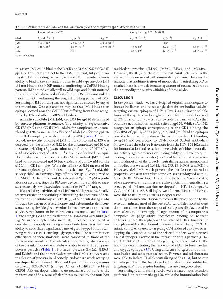
Affinities of sdAbs JM2, JM4, and JM7 for gp120 determinedby surface plasmon resonance. The affinity of representativeCD4BS (JM2) and CD4i (JM4) sdAbs for complexed or uncom-plexed gp120, as well as the affinity of sdAb JM7 for the gp120/miniCD4 complex, were determined by SPR (Table 3). As ex-pected, no specific binding of JM2 to the complexed gp120 wasdetected, but the affinity of JM2 for the uncomplexed gp120 wasmeasured, yielding a ka (association rate) of 1.4 � 104 M�1 s�1, akd (dissociation rate) of 6.0 � 10�4 s�1, and a calculated KD (equi-librium dissociation constant) of 43 nM. In contrast, JM7 did notbind to uncomplexed gp120 but yielded a KD of 0.6 nM for theg120/miniCD4 complex. While the affinity of JM4 measured forthe uncomplexed gp120 resulted in a calculated KD of 3 nM, thissdAb yielded an extremely high affinity for gp120 complexed tothe M48U1 CD4 mimic, and the calculated KD of 32 pM is prob-ably not accurate, since the BIAcore device is not suitable to mea-sure extremely low dissociation rates in the 10�6 s�1 range.
Neutralizing activities of multivalent sdAb proteins. Finally,we investigated the possibility of increasing the spectrum of neu-tralization and inhibitory activity (IC50) of our neutralizing sdAbsthrough the design of several homo- and heteromultivalent con-structs using flexible glycine/serine linkers between monovalentsdAbs. Seven homo- or heterobivalent constructs, listed in Table1, and a single JM4 homotrivalent sdAb (JM4x4x4) were built (seeFig. S1 in the supplemental material), produced, and tested asdescribed previously in a single-round infection assay for theirability to neutralize a significant panel of pseudotyped virions car-rying various HIV-1 envelope glycoproteins. The neutralizationefficiencies of these molecules were compared to those of themonovalent parental sdAb molecules. Importantly, whereas noneof the parental monovalent sdAbs was able to neutralize all pseu-dovirus particles (Table 1), 3 bivalent proteins (JM2x2, JM3x3,and JM5x3) and the homotrivalent protein (JM4x4x4) were ableto at least partly neutralize all tested pseudovirus particles carryingenvelopes from different HIV-1 subtypes. For example, virionsdisplaying 92UG037.8 (subtype A) or 93TH976.17 (subtypeCRF01_AE) envelopes, which were neutralized by none of themonovalent sdAbs, were efficiently neutralized by the four best
multivalent proteins (JM2x2, JM3x3, JM5x3, and JM4x4x4).However, the IC50s of these multivalent constructs were in therange of those measured with monovalent proteins. These resultsindicate that multimerization of monovalent neutralizing sdAbsresulted here in a much broader spectrum of neutralization butdid not modify the relative affinities of these sdAbs.
DISCUSSION
In the present study, we have designed original immunogens toimmunize llamas and select single-domain antibodies (sdAbs)targeting various epitopes of HIV-1 Env. Using trimeric solubleforms of the gp140 envelope glycoprotein for immunization andgp120 for selection, we were able to isolate a panel of sdAbs thatbound to neutralization-sensitive sites of gp120. While sdAb JM2recognizes an epitope corresponding to the CD4 binding site(CD4BS) of gp120, sdAbs JM3, JM4, and JM5 bind to epitopesunveiled by the conformational change induced by CD4 bindingto gp120 and correspond to CD4-induced (CD4i) antibodies.Since we used the subtype B envelope from the HIV-1 SF162 strainfor immunization and selection, these sdAbs exhibited neutraliz-ing activity against viruses expressing subtype B envelopes, in-cluding primary viral isolates (tier 2 and tier 2/3) that were resis-tant to almost all of the broadly neutralizing human monoclonalantibodies that we tested (2G12, b12, 2F5, 4E10, PG9, and PG16).However, sdAb JM3, which presents the broadest neutralizationproperties, can also neutralize some viruses pseudotyped with A,G, or CRF01_AE envelopes. In addition, the best sdAb candidates,when expressed as multimers, are also efficient in neutralizing abroad panel of viruses carrying envelopes from HIV-1 subtypes A,C, G, and CRF01_AE. Strikingly, two of them, JM3x3 and JM5x3,were able to neutralize all virus subtypes tested.
Using a nonspecific elution to recover the phage bound to theselection antigen, most of the best sdAb candidates isolated weredominant clones from the output of basic phage display-based invitro selection. Interestingly, a large amount of this output wascomposed of phage-sdAbs specifically binding to relevantepitopes. Indeed, these phage-sdAbs included CD4BS binders butalso phage-sdAbs that bound preferentially to the gp120/CD4mimic complex, therefore targeting CD4-induced epitopes over-lapping the CoRBS. Most of the selected binders were directedagainst epitopes involved in the interaction with a partner (CD4,and CXCR4 or CCR5). This finding is in good agreement with theliterature demonstrating the tendency of sdAbs to bind cavitiesand cryptic epitopes (38). Using different strategies for both im-munization and subsequent selection, Forsman and collaboratorswere able to isolate CD4BS-neutralizing sdAbs (13), but to ourknowledge, this is the first time that single-domain antibodiestargeting HIV-1 coreceptor binding sites are being described.
Surprisingly, all blocking sdAbs were isolated from selectionperformed on monomeric gp120, while the immunization had
TABLE 3 Affinities of JM2, JM4, and JM7 on uncomplexed or complexed gp120 determined by SPR
sdAb
Uncomplexed gp120 Complexed gp120�M48U1
ka (M�1 s�1) kd (s�1) KD (M) ka (M�1 s�1) kd (s�1) KD (M)
JM2 1.4 � 104 6.0 � 10�4 4.3 � 10�8 NBa
JM4 3.0 � 104 8.9 � 10�5 2.9 � 10�9 1.2 � 105 3.9 � 10�6 3.2 � 10�11
JM7 NB 4.3 � 105 2.7 � 10�4 6.4 � 10�10
a NB, no binding.
Matz et al.
1146 jvi.asm.org Journal of Virology
on January 30, 2018 by guesthttp://jvi.asm
.org/D
ownloaded from

been performed using trimeric Env (gp140). When selectionswere performed on gp140, sdAbs that bound specifically to thetrimeric conformation and showing no binding activity towardthe monomeric gp120 were preferentially selected (sdAbs JM8and JM9). However, these clones could not efficiently block theinfection process in the neutralization assay. It might be that se-lections on trimeric Env more closely mimic the natural immuni-zation process on entire virus and mainly lead to nonneutralizinggp120 binders, as can be seen in patients, due to the many tricksthat HIV-1 has evolved to escape from humoral response.
sdAb JM2 clearly competes with CD4 mimics or MAb b12 forrecognition of the CD4 binding site on gp120 or gp140, and itsbinding, logically, is abolished by the D368R mutation. With anaverage affinity of 40 nM, it could efficiently block infection of avariety of virus subtypes. JM2 also hampered the binding of CD4iphage-JM3, -JM4, and -JM5 (Fig. 4A). Reciprocally, JM2 dis-played on phage is partially hindered by the binding of JM3 andJM4 and, to a lesser extent, JM5. These results suggest that CD4-induced epitopes recognized by JM3, JM4, and JM5 might be lo-cated closer to the CD4BS than conventional CD4i epitopes,thereby leading to steric hindrances between CD4BS binders andCoRBS sdAbs, as has already been described (39). However, CD4iMAbs 17b and X5 are still competing with JM3, JM4, and JM5,implying the recognition of proximate epitopes. JM2 might alsostabilize a gp120 conformation that is not well recognized byCoRBS sdAbs JM3, JM4, and JM5, as can be observed by flowcytometry for the binding of gp120 to CCR5 in the presence ofJM2. Of note, an earlier study showed significant differences in thestructure of CD4-gp120 core complex compared to that of b12-gp120 stabilized core complex (9). Interestingly, the main differ-ences occurred on the bridging sheet, precisely in the CD4i bind-ing area. Thus, JM2 seems to stabilize such an alternative gp120conformation. Nevertheless, a definitive answer will only be ob-tained by crystal structure studies.
JM7 is intriguing since this sdAb is able to bind with subnano-molar affinity to the gp120/miniCD4 complex but binds neither togp120 nor to the gp120/sCD4 complex or the CD4 mimic alone.This could be explained through binding to a bimolecular epitopecreated by the association of miniCD4 and gp120, as observedearlier with MAb 21c, which shares its epitope between gp120 andCD4 (40). An alternative explanation might be that JM7 and sCD4cannot bind simultaneously to gp120 due to steric hindrance.However, we could demonstrate a competition between JM7 andJM3, JM4, and JM5, as well as CD4i MAb X5, suggesting that theseantibodies still at least partly share their epitopes.
The availability of several sdAbs directed against various neu-tralizing epitopes of gp120 opens the possibility of creating mul-tivalent sdAbs. Hultberg and collaborators have recently demon-strated the possibility of significantly increasing (up to 4,000-fold)the neutralization potency of sdAbs directed against the trimericEnv proteins of several viruses (H5N1 influenza virus, rabies virus,and Rous sarcoma virus) by designing bivalent and trivalent con-structs of neutralizing sdAbs (22). This is probably explained by anavidity effect, multivalent sdAbs being able to bind severalprotomers of the single trimeric Env. They also demonstrated thepotential of creating biparatopic constructs by linking two sdAbstargeting different epitopes, yielding molecules with IC50s in thepicomolar range and displaying an increased cross-subtype neu-tralization activity. We therefore used the same approach to gen-erate homo- and heteromultivalent constructs. Unfortunately, no
significant improvement of the neutralizing potency of these mul-tivalent proteins in terms of IC50s was observed compared to thoseof the parental monovalent sdAbs. However, several multivalentsdAbs could neutralize a broader spectrum of pseudotyped virusparticles. While no monovalent sdAb could neutralize the wholebatch of pseudovirions tested, four multivalent proteins were ableto neutralize pseudovirions carrying envelopes from HIV-1 sub-types A, B, C, G, and CRF01_AE. The JM2x2 homobivalent sdAbshowed a better activity than the monovalent JM2 sdAb, probablydue to an avidity effect. Interestingly, the JM4x4x4 homotrivalentsdAb neutralized a broader spectrum of pseudovirions than JM4but with equivalent or higher IC50s. Impressively, JM3x3 was ableto neutralize all pseudovirions tested. The position of the flexiblelinker and the conformation of the resulting molecules also seemto be of importance. Indeed, JM5x2 greatly outperformed JM2x5,and a similar difference was observed between JM5x3 and JM3x5.These differences might also be explained by the angle of bindingof some of these sdAbs that might be more or less tolerant to thepresence of a linker at their N terminus, located near the antigenbinding site.
In conclusion, using an original immunogen and phage-dis-play protocol, we have been able to select a panel of single-domainantibodies targeting the binding sites of CD4 and coreceptors ongp120. These sdAbs are capable of neutralizing a broad spectrumof HIV-1 subtypes, including tier 3 primary isolates. Since theseantibody fragments are extremely stable and are easy to produceand purify at high levels, they could have a great potential in de-veloping effective prevention strategies against HIV-1 transmis-sion, such as in the development of potent microbicides (41).
ACKNOWLEDGMENTS
The following reagents were obtained through the AIDS Research andReference Reagent Program, Division of AIDS, NIAID, NIH: pSVIII-92BR025.9, pSVIII-92RW020.5, pSVIII-92UG975.10, and pSVIII-93BR019.4 from Feng Gao and Beatrice Hahn and pSVIII-91US005.11,pSVIII-92HT593.1, pSVIII-92UG037.8, pSVIII-93TH976.17, and pS-VIII-93TH966.8 from Beatrice Hahn. We acknowledge D. P. Burton, P.Parren, D. Katinger, I. Srivastava, P. Kwong, and the National Institute forBiological Standards and Control Centre for AIDS Reagents (supportedby the European Union Programme EVA contract QLKZ-CT-1999-00609) and the United Kingdom Medical Research Council for the vari-ous antibodies (b12, 17b, and X5) and Env plasmids. We also thank M.Parmentier (University of Brussels, Belgium) for the CHO-K1 CCR5� cellline and S. Brunet and A. Chaillon for their help in neutralization assays.
This work was supported by Inserm, CNRS, Université Paris-Des-cartes, and grants from the Agence Nationale de Recherche sur le SIDA(ANRS) (S.B., D.B., and L.M.). J.M. was supported by a fellowship fromANRS.
REFERENCES1. Stamatatos L, Morris L, Burton DR, Mascola JR. 2009. Neutralizing
antibodies generated during natural HIV-1 infection: good news for anHIV-1 vaccine? Nat. Med. 15:866 – 870.
2. Walker BD, Burton DR. 2008. Toward an AIDS vaccine. Science 320:760 –764.
3. Wyatt R, Kwong PD, Desjardins E, Sweet RW, Robinson J, Hendrick-son WA, Sodroski JG. 1998. The antigenic structure of the HIV gp120envelope glycoprotein. Nature 393:705–711.
4. Liu J, Bartesaghi A, Borgnia MJ, Sapiro G, Subramaniam S. 2008.Molecular architecture of native HIV-1 gp120 trimers. Nature 455:109 –113.
5. Chen L, Kwon YD, Zhou T, Wu X, O’Dell S, Cavacini L, Hessell AJ,Pancera M, Tang M, Xu L, Yang ZY, Zhang MY, Arthos J, Burton DR,Dimitrov DS, Nabel GJ, Posner MR, Sodroski J, Wyatt R, Mascola JR,
Selection of Broadly Neutralizing Anti-HIV-1 Env sdAbs
January 2013 Volume 87 Number 2 jvi.asm.org 1147
on January 30, 2018 by guesthttp://jvi.asm
.org/D
ownloaded from

Kwong PD. 2009. Structural basis of immune evasion at the site of CD4attachment on HIV-1 gp120. Science 326:1123–1127.
6. Gonzalez N, Alvarez A, Alcami J. 2010. Broadly neutralizing antibodiesand their significance for HIV-1 vaccines. Curr. HIV Res. 8:602– 612.
7. Walker LM, Phogat SK, Chan-Hui PY, Wagner D, Phung P, Goss JL,Wrin T, Simek MD, Fling S, Mitcham JL, Lehrman JK, Priddy FH,Olsen OA, Frey SM, Hammond PW, Kaminsky S, Zamb T, Moyle M,Koff WC, Poignard P, Burton DR. 2009. Broad and potent neutralizingantibodies from an African donor reveal a new HIV-1 vaccine target. Sci-ence 326:285–289.
8. Zhou T, Georgiev I, Wu X, Yang ZY, Dai K, Finzi A, Kwon YD, ScheidJF, Shi W, Xu L, Yang Y, Zhu J, Nussenzweig MC, Sodroski J, ShapiroL, Nabel GJ, Mascola JR, Kwong PD. 2010. Structural basis for broad andpotent neutralization of HIV-1 by antibody VRC01. Science 329:811– 817.
9. Zhou T, Xu L, Dey B, Hessell AJ, Van Ryk D, Xiang SH, Yang X, ZhangMY, Zwick MB, Arthos J, Burton DR, Dimitrov DS, Sodroski J, WyattR, Nabel GJ, Kwong PD. 2007. Structural definition of a conservedneutralization epitope on HIV-1 gp120. Nature 445:732–737.
10. Labrijn AF, Poignard P, Raja A, Zwick MB, Delgado K, Franti M, BinleyJ, Vivona V, Grundner C, Huang CC, Venturi M, Petropoulos CJ, WrinT, Dimitrov DS, Robinson J, Kwong PD, Wyatt RT, Sodroski J, BurtonDR. 2003. Access of antibody molecules to the conserved coreceptor bind-ing site on glycoprotein gp120 is sterically restricted on primary humanimmunodeficiency virus type 1. J. Virol. 77:10557–10565.
11. Moulard M, Phogat SK, Shu Y, Labrijn AF, Xiao X, Binley JM, ZhangMY, Sidorov IA, Broder CC, Robinson J, Parren PW, Burton DR,Dimitrov DS. 2002. Broadly cross-reactive HIV-1-neutralizing humanmonoclonal Fab selected for binding to gp120-CD4-CCR5 complexes.Proc. Natl. Acad. Sci. U. S. A. 99:6913– 6918.
12. Van Bockstaele F, Holz JB, Revets H. 2009. The development of nano-bodies for therapeutic applications. Curr. Opin. Investig. Drugs 10:1212–1224.
13. Forsman A, Beirnaert E, Aasa-Chapman MM, Hoorelbeke B, Hijazi K,Koh W, Tack V, Szynol A, Kelly C, McKnight A, Verrips T, de HaardH, Weiss RA. 2008. Llama antibody fragments with cross-subtype HIV-1neutralizing properties and high affinity for HIV-1 gp120. J. Virol. 82:12069 –12081.
14. Koh WW, Steffensen S, Gonzalez M, Hoorelbeke B, Gorlani A, SzynolA, Forsman A, Aasa-Chapman MM, de Haard H, Verrips T, Weiss RA.2010. Generation of a family-specific phage library of llama single chainantibody fragments that neutralize HIV-1. J. Biol. Chem. 285:19116 –19124.
15. Dey AK, Burke B, Sun Y, Sirokman K, Nandi A, Hartog K, Lian Y,Geonnotti AR, Montefiori D, Franti M, Martin G, Carfi A, Kessler P,Martin L, Srivastava IK, Barnett SW. 2012. Elicitation of neutralizingantibodies directed against CD4-induced epitope(s) using a CD4 mimeticcross-linked to a HIV-1 envelope glycoprotein. PLoS One 7:e30233. doi:10.1371/journal.pone.0030233.
16. Martin G, Burke B, Thai R, Dey AK, Combes O, Ramos OH, Heyd B,Geonnotti AR, Montefiori DC, Kan E, Lian Y, Sun Y, Abache T, UlmerJB, Madaoui H, Guerois R, Barnett SW, Srivastava IK, Kessler P,Martin L. 2011. Stabilization of HIV-1 envelope in the CD4-bound con-formation through specific cross-linking of a CD4 mimetic. J. Biol. Chem.286:21706 –21716.
17. Martin G, Sun Y, Heyd B, Combes O, Ulmer JB, Descours A, BarnettSW, Srivastava IK, Martin L. 2008. A simple one-step method for thepreparation of HIV-1 envelope glycoprotein immunogens based on a CD4mimic peptide. Virology 381:241–250.
18. Martin L, Stricher F, Misse D, Sironi F, Pugniere M, Barthe P, Prado-Gotor R, Freulon I, Magne X, Roumestand C, Menez A, Lusso P, VeasF, Vita C. 2003. Rational design of a CD4 mimic that inhibits HIV-1 entryand exposes cryptic neutralization epitopes. Nat. Biotechnol. 21:71–76.
19. Moscoso CG, Sun Y, Poon S, Xing L, Kan E, Martin L, Green D, Lin F,Vahlne AG, Barnett S, Srivastava I, Cheng RH. 2011. Quaternary struc-tures of HIV Env immunogen exhibit conformational vicissitudes andinterface diminution elicited by ligand binding. Proc. Natl. Acad. Sci.U. S. A. 108:6091– 6096.
20. Behar G, Chames P, Teulon I, Cornillon A, Alshoukr F, Roquet F,Pugniere M, Teillaud JL, Gruaz-Guyon A, Pelegrin A, Baty D. 2009.Llama single-domain antibodies directed against nonconventionalepitopes of tumor-associated carcinoembryonic antigen absent from non-specific cross-reacting antigen. FEBS J. 276:3881–3893.
21. Van Herrewege Y, Morellato L, Descours A, Aerts L, Michiels J, Heyn-
drickx L, Martin L, Vanham G. 2008. CD4 mimetic miniproteins: potentanti-HIV compounds with promising activity as microbicides. J. Antimi-crob. Chemother. 61:818 – 826.
22. Hultberg A, Temperton NJ, Rosseels V, Koenders M, Gonzalez-PajueloM, Schepens B, Ibanez LI, Vanlandschoot P, Schillemans J, SaundersM, Weiss RA, Saelens X, Melero JA, Verrips CT, Van Gucht S, de HaardHJ. 2011. Llama-derived single domain antibodies to build multivalent,superpotent and broadened neutralizing anti-viral molecules. PLoS One6:e17665. doi:10.1371/journal.pone.0017665.
23. Studier FW. 2005. Protein production by auto-induction in high densityshaking cultures. Protein Expr. Purif. 41:207–234.
24. Wei X, Decker JM, Wang S, Hui H, Kappes JC, Wu X, Salazar-GonzalezJF, Salazar MG, Kilby JM, Saag MS, Komarova NL, Nowak MA, HahnBH, Kwong PD, Shaw GM. 2003. Antibody neutralization and escape byHIV-1. Nature 422:307–312.
25. Naldini L, Blomer U, Gallay P, Ory D, Mulligan R, Gage FH, VermaIM, Trono D. 1996. In vivo gene delivery and stable transduction ofnondividing cells by a lentiviral vector. Science 272:263–267.
26. Hofmann W, Schubert D, LaBonte J, Munson L, Gibson S, Scammell J,Ferrigno P, Sodroski J. 1999. Species-specific, postentry barriers to pri-mate immunodeficiency virus infection. J. Virol. 73:10020 –10028.
27. Laguette N, Benichou S, Basmaciogullari S. 2009. Human immunode-ficiency virus type 1 Nef incorporation into virions does not increase in-fectivity. J. Virol. 83:1093–1104.
28. Barin F, Brunet S, Brand D, Moog C, Peyre R, Damond F, Charneau P,Barre-Sinoussi F. 2004. Interclade neutralization and enhancement ofhuman immunodeficiency virus type 1 identified by an assay using HeLacells expressing both CD4 receptor and CXCR4/CCR5 coreceptors. J. In-fect. Dis. 189:322–327.
29. Chaillon A, Wack T, Braibant M, Mandelbrot L, Blanche S, WarszawskiJ, Barin F. 2012. The breadth and titer of maternal HIV-1-specific heter-ologous neutralizing antibodies are not associated with a lower rate ofmother-to-child transmission of HIV-1. J. Virol. 86:10540 –10546.
30. Dreja H, O’Sullivan E, Pade C, Greene KM, Gao H, Aubin K, Hand J,Isaksen A, D’Souza C, Leber W, Montefiori D, Seaman MS, AndersonJ, Orkin C, McKnight A. 2010. Neutralization activity in a geographicallydiverse East London cohort of human immunodeficiency virus type 1-in-fected patients: clade C infection results in a stronger and broader hu-moral immune response than clade B infection. J. Gen. Virol. 91:2794 –2803.
31. Simek MD, Rida W, Priddy FH, Pung P, Carrow E, Laufer DS, LehrmanJK, Boaz M, Tarragona-Fiol T, Miiro G, Birungi J, Pozniak A, McPheeDA, Manigart O, Karita E, Inwoley A, Jaoko W, Dehovitz J, Bekker LG,Pitisuttithum P, Paris R, Walker LM, Poignard P, Wrin T, Fast PE,Burton DR, Koff WC. 2009. Human immunodeficiency virus type 1 eliteneutralizers: individuals with broad and potent neutralizing activity iden-tified by using a high-throughput neutralization assay together with ananalytical selection algorithm. J. Virol. 83:7337–7348.
32. Stricher F, Martin L, Barthe P, Pogenberg V, Mechulam A, Menez A,Roumestand C, Veas F, Royer C, Vita C. 2005. A high-throughputfluorescence polarization assay specific to the CD4 binding site of HIV-1glycoproteins based on a fluorescein-labelled CD4 mimic. Biochem. J.390:29 –39.
33. Samson M, Labbe O, Mollereau C, Vassart G, Parmentier M. 1996.Molecular cloning and functional expression of a new human CC-chemokine receptor gene. Biochemistry 35:3362–3367.
34. Feng Y, McKee K, Tran K, O’Dell S, Schmidt SD, Phogat A, Forsell MN,Karlsson Hedestam GB, Mascola JR, Wyatt RT. 2012. Biochemicallydefined HIV-1 envelope glycoprotein variant immunogens display differ-ential binding and neutralizing specificities to the CD4-binding site. J.Biol. Chem. 287:5673–5686.
35. Huang CC, Tang M, Zhang MY, Majeed S, Montabana E, Stanfield RL,Dimitrov DS, Korber B, Sodroski J, Wilson IA, Wyatt R, Kwong PD.2005. Structure of a V3-containing HIV-1 gp120 core. Science 310:1025–1028.
36. Kwong PD, Wyatt R, Robinson J, Sweet RW, Sodroski J, HendricksonWA. 1998. Structure of an HIV gp120 envelope glycoprotein in complexwith the CD4 receptor and a neutralizing human antibody. Nature 393:648 – 659.
37. Shrivastava IH, Wendel K, Lalonde JM. 2012. Spontaneous rearrange-ment of the beta20/beta21 strands in simulations of unliganded HIV-1glycoprotein, gp120. Biochemistry 51:7783–7793.
38. De Genst E, Silence K, Decanniere K, Conrath K, Loris R, Kinne J,
Matz et al.
1148 jvi.asm.org Journal of Virology
on January 30, 2018 by guesthttp://jvi.asm
.org/D
ownloaded from

Muyldermans S, Wyns L. 2006. Molecular basis for the preferential cleftrecognition by dromedary heavy-chain antibodies. Proc. Natl. Acad. Sci.U. S. A. 103:4586 – 4591.
39. Moore JP, Sodroski J. 1996. Antibody cross-competition analysis of thehuman immunodeficiency virus type 1 gp120 exterior envelope glycopro-tein. J. Virol. 70:1863–1872.
40. Diskin R, Marcovecchio PM, Bjorkman PJ. 2010. Structure of a clade C
HIV-1 gp120 bound to CD4 and CD4-induced antibody reveals anti-CD4polyreactivity. Nat. Struct. Mol. Biol. 17:608 – 613.
41. Gorlani A, Brouwers J, McConville C, van der Bijl P, Malcolm K,Augustijns P, Quigley AF, Weiss R, De Haard H, Verrips T. 2012.Llama antibody fragments have good potential for application as HIVtype 1 topical microbicides. AIDS Res. Hum. Retroviruses 28:198 –205.
Selection of Broadly Neutralizing Anti-HIV-1 Env sdAbs
January 2013 Volume 87 Number 2 jvi.asm.org 1149
on January 30, 2018 by guesthttp://jvi.asm
.org/D
ownloaded from