stemi education for emergency department · cad is the most common type of heart ... statin or...
TRANSCRIPT

STEMI Education for Emergency Department

Purpose
To standardize emergency department management of ST elevation myocardial infarction (STEMI) across the province by reinforcing the STEMI reperfusion targets and best practices to improve patient outcomes.

CCN History and Mandate CCN has a solid history of advising the Ministry of Health and Long-Term Care (MOHLTC), Local Health
Integration Networks (LHINs), hospitals and care providers to improve the quality, efficiency, accessibility and equity of cardiac services for patients across Ontario. Currently, CCN’s mandate has expanded to include the management of vascular and more recently stroke services for patients and providers in Ontario.
CCN supports the system by :
System Capacity Planning, Performance Measurement and Management
Developing and Implementing Best Practice Standards from Evidence and Research:
Cardiac and Vascular Surgical Procedures
Heart Rhythm and Heart Failure Management
Interventional Cardiology, STEMI
Stroke and Hypertension Management
Prevention, Rehabilitation, & Palliative Care
Evaluation of Emerging Technologies
Managing the Cardiac and Vascular Registries which is a robust information source of patient clinical data to inform stakeholders
| 3

STEMI Care in Ontario
In Ontario, approximately 8,000 patients per year experience an ST-segment elevation myocardial infarction (STEMI)
Today, 16 Primary PCI hospitals with STEMI programs are located across Ontario
Approximately 40% of STEMI patients self transport to ED
Restoration of blood flow in the coronary artery is achieved through one of the following best practice reperfusion modalities:
1. Primary percutaneous coronary intervention (pPCI)
2. Pharmacoinvasive Intervention (Fibrinolytic therapy plus PCI in 24 hrs)
Reperfusion requires timely diagnosis, transportation and treatment
The goal of the ED STEMI protocol is to ensure a baseline standard of best practice care for all STEMI patients to receive timely access to reperfusion therapy regardless of where they present
| 4
Objectives
Upon completion of this education the learner will be able to:
Name the typical and atypical signs and symptoms of acute coronary syndrome (ACS)
Describe the importance of acquiring a rapid ECG in the early recognition and treatment of a patient with STEMI
Describe the criteria for system activation for managing of a “STEMI patient” Describe the standardized process to follow for STEMI management in the ED
Describe the importance of communication and transfer of accountability between care providers

Coronary Artery Disease (CAD)

Coronary Artery Disease

CAD: Definition
CAD is the most common type of heart disease. It is the leading cause of death in North America in both men and women
Atherosclerosis: buildup of a material called ‘plaque’ on their inner walls
CAD occurs when the coronary arteries become hardened and narrowed

CAD : Consequence

CAD: Development

What Does a STEMI Look Like?
Normal ECG STEMI
STEMI
Post PCI intervention

Coronary Artery Disease (CAD) : Types of Treatment
Surgical: Coronary
artery bypass graft
(CABG)
Non-Surgical : PCI or
Angioplasty
Drugs

Medical Treatment
Drugs:
Aspirin
Antiplatelet (clopidogrel,ticagrelor,prasugrel)
Beta blocker
Ace inhibitor
Statin or lipid-lowering drugs

PCI Treatment
Percutaneous coronary intervention
(PCI or angioplasty)PCI with Stent

Coronary Artery Bypass Graft (CABG)
A surgical procedure to treat the blocked or narrowed arteries by creating a ‘bypass’ for the blocked portion of the coronary artery using either a vein (e.g., saphenous vein) or artery (e.g., left internal mammary artery, radial artery).

Your Role
As a healthcare provider YOU have the opportunity to make a difference in the lives of many; you play an important role in the success of a hospital STEMI program.
Goal:
ED staff to use the information in this presentation to deliver care based on best practice guidelines:
Early recognition of STEMI ACS patients and initiate timely evaluation and intervention
Coordinate practices between paramedic services and PCI hospital
Promote and encourage quality improvement process at your hospital to improve STEMI performance

ED STEMI Protocol

| 18
ED STEMI Protocol Key Messages
It is recognized that across Ontario, there are well established regional and LHIN wide STEMI protocols, transfer agreements and partnerships with ED’s, paramedic services and PCI hospitals
These robust networks serve as a hub and spoke model and will continue in their current form
The ED STEMI initiative builds on the current system to support the networks and close the gaps
The initiative reinforces ED STEMI care best practices by:
1. Reinforcing early identification and decision making (triage, ECG and reperfusion decision) through provincial education, provision of tools and templates
2. Identifying ED’s without a current relationship to their closest PCI hospital and supporting them to develop protocols and networks
3. Reinforcing reperfusion strategies by designating an ED’s reperfusion strategy for non PCI hospitals as either primary PCI (< 60 mins drive time ) or pharmacoinvasive ( > 60 mins drive time )
4. Developing and implementing provincial STEMI performance targets

| 19
ED STEMI Management – Reperfusion Targets
Chest Pain Early Identification
• Triage & perform STAT ECG (Door to ECG ≤ 10MIN)• If negative, repeat ECG q10 mins x 3• Symptoms ≤ 12 hours – STEMI Diagnosis • If Symptoms ≥ 12 hrs consult Cardiologist or Internal Medicine
Primary PCI OR Pharmacoinvasive StrategyReperfusion Decision
For Primary PCI
•Door in Door Out Time (DIDO) ≤ 30 mins•Door to Balloon Time (D2B) ≤ 120 mins
Pharmacoinvasive
• Door to Needle Time (D2N) ≤ 30 mins
• Transfer to PCI hospital ≤24 hrs

Ontario ED STEMI Algorithm

Signs and Symptoms of a Heart Attack
• Chest pain is the most common symptom, but some people don’t experience this…. women are less likely to experience chest pain than men

Confidential – Not for Distribution | 22
Rapid Triage is the Key
Early identification appropriate triage early revascularization
Rapid triage of patients complaining of chest pain
Initiate the STEMI protocol
Acquire ECG
Activating the STEMI response team
(e.g., CODE STEMI, STEMI Alert)
Initiate reperfusion strategy
Primary PCI
Pharmacoinvasive

Early ECG: Target ≤ 10 Minutes
Rapid ECG Early diagnosis Early activation of STEMI response team
Acquiring the ECG within 10 minutes of ED arrival is the first step of STEMI recognition and treatment

STEMI Recognition Essentials of the 12 Lead ECG
Activate STEMI Protocol Immediately
Early activation of a “STEMI response team” is essential to achieving the defined reperfusion target for your hospital:
Primary PCI = door in door out (DIDO) ≤30 minutes
Pharmacoinvasive strategy = fibrinolytic therapy followed by PCI within 24 hrs
Contact the PCI hospital using the hotline; the 24/7 telephone number or paging system that is available for activation
Initiate immediate transfer

Provide Key Transfer of Care Information
Transfer of care and communication to the Cardiologist or Interventional Cardiologist at PCI hospital helps in the management of the STEMI
Time of symptom onset
Qualifying ECG
If ROSC state time
Hemodynamic status
History of AMI/PCI/CABG
Medications given and procedures
Copy of ECG

ED Quick Reference Pocket Card
This STEMI pocket card can be used as a quick reference tool for the management of STEMI along
with the key clinical information exchange between ED staff and the cardiologist at crucial points in
transfers of care

Confidential – Not for Distribution | 28
Geographical Partnership Maps
Purpose:
To identify PCI and non-PCI hospital partnerships (each hospital will receive)
Reinforce the reperfusion strategies by designating an ED’s reperfusion strategy for non-PCI hospital as either:
primary PCI (< 60 mins drive time ) or;
pharmacoinvasive ( > 60 mins drive time )
Provide contact information to promote opportunities for greater communication and mentorships

PCI and Non-PCI Partnership Mapping with Defined Reperfusion Strategy
ED LHIN
Non PCI Hospital Partnership with Hamilton Health Sciences (HHS)
Drive Mins to HHS
Reperfusion Strategy:
HNHB St. Joseph's Healthcare (Hamilton)
5 Primary PCI
HNHB Hamilton Health Sciences - West End Uc
7 Primary PCI
HNHB Hamilton Health Sciences - Juravinski
8 Primary PCI
HNHB St Joseph's Healthcare -Community Health Centre
13 Primary PCI
HNHB Joseph Brant Memorial Hospital
16 Primary PCI
HNHB Hamilton Health Sciences - West Lincoln
25 Primary PCI
MH Halton Healthcare Services - Oakville-Trafalgar
28 Primary PCI
HNHB Brantford General Hospital
32 Primary PCI
HNHB West Haldimand General Hospital
39 Primary PCI
HNHB Brant Community Healthcare System -Willet
40 Primary PCI
HNHB Niagara Health System -St. Catharines
41 Primary PCI
HNHB Niagara Health System -Greater Niagara
50 Primary PCI
HNHB Haldimand War Memorial Hospital
52 Primary PCI
HNHB Niagara Health System -Welland
60 Primary PCI
HNHB Norfolk General Hospital 64 Pharmacoinvasive
HNHB Niagara Health System -Douglas Memorial
65 Pharmacoinvasive
HNHB Niagara Health System -Port Colborne
70 Pharmacoinvasive
Primary PCI: Target transfer to HHS < 60 min
Pharmacoinvasive: Target transfer to HHS < 24 hr
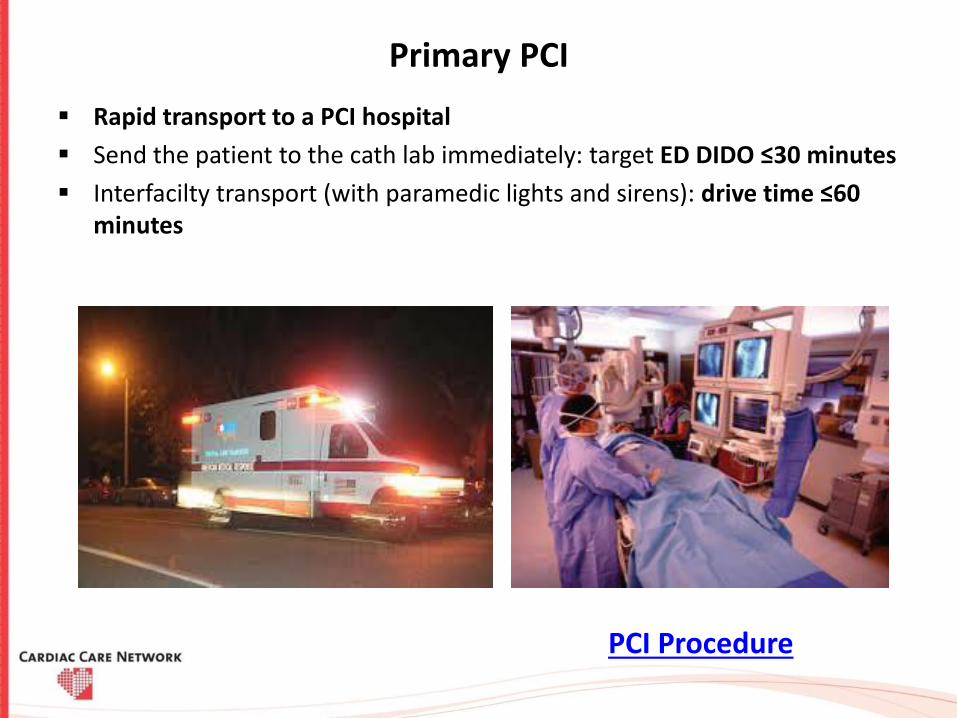
Primary PCI
Rapid transport to a PCI hospital
Send the patient to the cath lab immediately: target ED DIDO ≤30 minutes
Interfacilty transport (with paramedic lights and sirens): drive time ≤60 minutes
PCI Procedure

Pharmacoinvasive
Administer fibrinolytic reperfusion therapy: target ≤30 minutes of ED arrival
Arrange immediate transport to PCI hospital in parallel to fibrinolytic administration i.e., “drip and ship”
Notify PCI hospital immediately
Transfer to PCI hospital: target ≤ 24 hours of ED arrival

Appendix

STEMI Case Scenarios

Case 1: Primary PCI
55 year old woman walks in to a non-PCI ED within 2 hours of chest pain onset
ED protocol:
ECG is acquired within 10 minutes which shows inferior ST elevation
Assessed, given aspirin 162 mg chewable po
PCI hospital is 30 minutes away, there is an established STEMI protocol between paramedic services and the PCI hospital for rapid transfer
STEMI activation by ED as per local process informs the interventional cardiologist (IC) at the PCI hospital and paramedic services
Antiplatelet therapy is administered
Anticoagulation is given as established by the PCI hospital
DIDO is <30 minutes
Suggested antiplatelet therapies: Ticagrelor 180mg PO (preferred) or Clopidogrel 600mg PO (alternate)
Suggested anticoagulation therapies:
According to PCI hospital within network (each non-PCI hospital should be linked to a PCI hospital)

Inferior STEMI
Inferior myocardial infarctions (MI) account for 40-50% of all MIs
Generally have a more favourable prognosis than anterior MI
Up to 40% of patients with an inferior STEMI will have a concomitant right ventricular infarction. These patients may develop severe hypotension in response to nitrates and generally have a worse prognosis
Up to 20% of patients with inferior STEMI will develop significant bradycardia due to second- or third-degree AV block. These patients have an increased in-hospital mortality
Inferior STEMI may also be associated with posterior infarction, which confers a worse prognosis due to increased area of myocardium at risk

How to Recognise an Inferior STEMI
ST elevation in leads II, III and aVF
Progressive development of Q waves in II, III and aVF
Reciprocal ST depression in aVL (± lead I)
ST elevation in lead III > lead II
Presence of reciprocal ST depression in lead I
Signs of right ventricular infarction: STE in V1 and V4R

Example 1: Inferior STEMI Interpretation
What do you see?
Hyperacute (peaked) T waves in II, III and aVF with relative loss of R wave height Early ST elevation and Q-wave formation in lead III Reciprocal ST depression and T wave inversion in aVL ST elevation in lead III > lead II suggests an RCA occlusion; the subtle ST elevation in
V4R would be consistent with this

Example 2: Inferior STEMI Interpretation
What do you see?
ST elevation in II, III and aVF Q-wave formation in III and aVF Reciprocal ST depression and T wave inversion in aVL ST elevation in lead II = lead III and absent reciprocal change in lead I (isoelectric ST
segment) suggest a circumflex artery occlusion

Inferior STEMI Interpretation
What do you see?
Marked ST elevation in II, III and aVF with early Q-wave formation Reciprocal changes in aVL ST elevation in lead III > II with reciprocal change present in lead I and ST elevation
in V1-2 suggests RCA occlusion with associated RV infarction This patient should have right-sided leads to confirm this

Case 2: Primary PCI with Fibrinolytic Absolute or Relative Contraindications
76 year old man walks in to a non-PCI ED 7 hours after symptom onset
ED protocol:
ECG acquisition is done within 10 minutes of arrival
Immediate assessment, given aspirin 162mg chewable po
ECG shows a large anterior STEMI
He has a contraindication to fibrinolytic therapy and is in heart failure (placed on BIPAP)
Emergent transport is arranged
Nearest PCI site is 2 hrs away, IC at PCI hospital is contacted immediately for discussion
Patient is stabilized for transport and transferred for primary PCI
Suggested antiplatelet therapies: Ticagrelor 180mg PO (preferred) or Clopidogrel 600mg PO (alternate)
Suggested anticoagulation therapies:
According to PCI hospital within network (each non-PCI hospital should be linked to a PCI hospital)

Anterior STEMI
Clinical Relevance:
Anterior STEMI results from occlusion of the left anterior descending artery (LAD).
Anterior myocardial infarction carries the worst prognosis of all infarct locations, mostly due to larger infarct size
How to Recognise Anterior STEMI:
ST segment elevation with Q wave formation in the precordial leads (V1-6) ±the high lateral leads (I and aVL)
Reciprocal ST depression in the inferior leads (mainly III and aVF)
Infarct patterns are named according to the leads with maximal ST elevation:
Septal = V1-2
Anterior = V2-5
Anteroseptal = V1-4
Anterolateral = V3-6, I + aVL
Extensive anterior/anterolateral = V1-6, I + aVL

Anterior STEMI
What do you see?
ST elevation is maximal in the anteroseptal leads (V1-4) Q waves are present in the septal leads (V1-2) There is also some subtle STE in I, aVL and V5, with reciprocal ST depression in lead III There are hyperacute (peaked ) T waves in V2-4 These features indicate a hyperacute anteroseptal STEMI

Anterior STEMI
What do you see?
Hyperacute T-waves in V2-6 (most marked in V2 and V3) with loss of R wave height The rhythm is sinus with 1st degree AV block There are premature atrial complexes (beat 4 on the rhythm strip) There are multifocal ventricular ectopy (PVCs of two different types) indicating
an “irritable” myocardium at risk of ventricular fibrillation

Anterior STEMI
What do you see?
ST elevation in V1-6 plus I and aVL (most marked in V2-4). Minimal reciprocal ST depression in III and aVF Q waves in V1-2, reduced R wave height (a Q-wave equivalent) in V3-4 There is a premature ventricular complex (PVC) with “R on T’ phenomenon at the end of
the ECG; this puts the patient at risk for ventricular arrhythmias

Anterior STEMI
What do you see?
Massive ST elevation with “tombstone” morphology is present throughout the precordial (V1-6) and high lateral leads (I, aVL)
This pattern is seen in proximal LAD occlusion and indicates a large territory infarction with a poor LV ejection fraction and high likelihood of cardiogenic shock and death

Case 3: Fibrinolytic Administration :Pharmacoinvasive Treatment
45 year old man presents to non-PCI hospital via paramedic services as the nearest PCI hospital is 3 hours away
ED protocol:
Paramedic qualifying ECG shows a lateral ST elevation
Paramedic services notify the non PCI hospital of STEMI patient enroute, there are no contraindications to fibrinolytic therapy
Fibrinolysis is administered with a door to needle (D2N) time of ≤30 minutes
The appropriate antiplatelet and anticoagulation (as per agreement with PCI hospital)
Interventional cardiologist (IC) at the PCI site is contacted and informed a fibrinolytic has been administered along with the transfer of care information

Case 3 continued…
Immediate transfer arrangements are made with a CTAS 2 assignment to PCI hospital within 24 hrs for Pharmacoinvasive PCI
Provide continuous monitoring and repeat the ECG at 60 minutes and 90 minutes to assess clinical reperfusion if not yet transported
If patient shows evidence of failed reperfusion, arrange emergent transfer with a CTAS 1 assignment and inform IC at PCI hospital
Suggested antiplatelet therapies: Clopidogrel 300 mg PO for patients < 75 years old or Clopidogrel 75 mg PO for patients >=75 years old; Ticagrelor is contraindicated
Suggested anticoagulation therapies:
According to PCI hospital within network (each non-PCI hospital should be linked to a PCI hospital)

Lateral STEMI
How to Recognise a Lateral STEMI
ST elevation in the lateral leads (I, aVL, V5-6).
Reciprocal ST depression in the inferior leads (III and aVF)
Patterns of lateral infarction:
Anterolateral STEMI due to LAD occlusion
Inferior-posterior-lateral STEMI due to left circumflex (LCx) occlusion
Isolated lateral infarction due to occlusion of smaller branch arteries such as the diagonal 1 (D1), obtuse marginal (OM) or ramus intermedius (IM)

High Lateral STEMI
What do you see? ST elevation is present in the high lateral leads ,I and aVL
There is also subtle ST elevation with hyperacute T waves in V5-6.
There is reciprocal ST depression in the inferior leads ,III and aVF with associated ST depression in V1-3 (which could represent anterior ischemia or reciprocal change)
This pattern is consistent with an acute infarction of the lateral wall of the left ventricle (high lateral STEMI)

High Lateral MI
What do you see?
ST elevation is present in the high lateral leads, I and aVL There is reciprocal ST depression in the inferior leads, III and aVF QS waves in the anteroseptal leads (V1-4) with poor R wave progression,
indicates prior anteroseptal infarction This pattern suggests proximal LAD disease with an acute occlusion of the
first diagonal branch (D1)

Anterolateral STEMI
What do you see? There is early ST elevation with hyperacute T waves in the anteroseptal leads ,V1-4 There is also subtle ST elevation in the high lateral leads, I and aVL The presence of reciprocal ST depression in the inferior leads ,III and aVF makes the
lateral ST elevation more obvious This ECG represents the early stages of a large anterolateral infarction The combination of ST elevation in the precordial and high lateral leads is indicative of
proximal LAD occlusion

Inferolateral STEMI
What do you see? There is ST elevation in the inferior (II, III, aVF) and lateral (I, V5-6) leads The precordial ST elevation extends out as far as V4, however the maximal STE is in V6 ST depression in V1-3 is suggestive of associated posterior infarction (the R/S ratio > 1 in V2
is consistent with this) This is an acute inferolateral STEMI with probable posterior extension This constellation of ECG abnormalities is typically produced by occlusion of the proximal circumflex artery

Confidential – Not for Distribution | 53
15-Lead ECG Clinical significance
It is estimated that up to 50% of inferior MIs have right ventricular
and/or posterior involvement due to the RCA blood supply
Right ventricular MIs are preload dependent for cardiac output
Nitrates can further reduce preload (use with caution)
Hypotension associated with RVI responds well to IV fluid bolus
Are rarely seen without inferior MI

Confidential – Not for Distribution | 54
15-Lead ECG
When should it be done?
Any patient with an inferior STEMI
ST depression seen in V1-V3 (reciprocal changes of posterior MI)
12-lead ECG does not view right ventricle or posterior wall

Confidential – Not for Distribution | 55
Acquiring a 15-Lead ECG
Placement of V4R
Acquire 12 lead ECG
Move V4 to V4R (midclavicular, 5th intercostal space on the right side) opposite to normal V4 placement on left

Confidential – Not for Distribution | 56
15-Lead ECG: V8, V9
Process for Acquiring a 15 Lead ECG
Placement of V8, V9
V5 becomes V8
V6 becomes V9
Move V5 to V8 location (midclavicular, 5th intercostal space)
Move V6 to V9 location (between V8 and the spine
Acquire a second 12 lead
Re-label:
V4R, V8, V9

Confidential – Not for Distribution | 57
Left Bundle Branch Blocks (LBBB)

Confidential – Not for Distribution | 58
Left Bundle Branch Blocks (BBB)
LBBB produce ECG changes that can imitate or conceal the ECG changes that are associated with Acute Coronary Syndromes (ACS)
When a BBB is present on the 12 lead ECG try to obtain a copy of an old 12 lead ECG if possible in order to determine if the BBB is old or new
Causes:
Aortic stenosis Ischaemic heart disease Hypertension Dilated cardiomyopathy Anterior MI Primary degenerative disease (fibrosis) of
the conducting system (Lenegre disease) Hyperkalaemia Digoxin toxicity

Confidential – Not for Distribution | 59
Recognition of a LBBB
Wide QRS
(greater then 0.12 seconds or 3 small squares)
Supraventricular rhythm
If both of the above criteria are met suspect a BBB

Confidential – Not for Distribution | 60
Examples of LBBBExample 1:
Example 2:

Confidential – Not for Distribution | 61
Conditions that can Mimic or Conceal STEMI
Ventricular rhythms:
Idioventricular rhythms
Ventricular tachycardia
Premature ventricular complexes
Other conditions:
Ventricular paced rhythms
Left ventricular hypertrophy
Benign early repolarization
Pericarditis
Hyperkalemia