static-content.springer.com10.1007... · web viewhave synthesized chitosan-stabilized cu-nps...
TRANSCRIPT
Carboxymethylated chitosan-stabilized Copper nanoparticles: A promise to contribute a potent antifungal and antibacterial agentSangeeta Tantubay1, Sourav K. Mukhopadhyay2, Himani Kalita1, Suraj Konar1, Satyahari Dey2, Amita Pathak1* and Panchanan Pramanik1*
[1] Sangeeta Tantubay, Himani Kalita, Suraj Konar, Amita Pathak, Panchanan PramanikDepartment of Chemistry, Indian Institute of Technology Kharagpur, Kharagpur 721302, West Bengal, India[2] Sourav K. Mukhopadhyay, Satyahari DeyDepartment of Biotechnology, Indian Institute of Technology Kharagpur, Kharagpur 721302, West Bengal, India*Corresponding authors
Panchanan Pramanik (Corresponding author)Email: [email protected] [email protected].: +91-9434016995
Amita Pathak (Corresponding author)Email: [email protected].: +91-3222-283312Fax: +91-3222-282252
Sangeeta Tantubay (First author)Email: [email protected]
1

No. Contents Page No.1 Comparison with some recent reports on
Cu-NPs/nanocomposites synthesis (Section 1).3-4
2 Required chemicals for synthesis of CMC-stabilized Cu-NPs and chemical characterization of CMC-
stabilized Cu-NPs (Section 2).
4-5
3 MTT assay (Section 3). 5
4 Comparative table of antimicrobial activities of Cu-NPs/nanocomposites, copper salts, and commercially
available antibiotics (Table S1).
6
5 HD, PDI, ZP, and λmax values of CMC-stabilized Cu-NPs at various pHs (Table S2).
7
6 Hydrodynamic Size distribution curves of CMC-stabilized Cu-NPs at various pHs (3-12) (Figure S1).
8
7 Zeta Potential distribution curves of CMC-stabilized Cu-NPs in the pH range of 3 to7 (Figure S2).
9
8 Zeta Potential distribution curves of CMC-stabilized Cu-NPs in the pH range of 8 to 12 (Figure S3).
10
9 References 11-12
2
Section 1. Comparison with some recent reports on Cu-NPs/nanocomposites synthesis.
1a. “A simple robust method for synthesis of metallic copper nanoparticles of high antibacterial potency against E. coli” (Chatterjee et al. 2012)
Summary: This work reports the synthesis of gelatin-stabilized Cu-NPs via aqueous based chemical reduction of copper salt at room temperature in the synthesis period of ~ 6-8 h. Particles’ average size range is ~ 60-80 nm & they have stability ~ one month. These Cu-NPs exhibit good antibacterial activity.
Differences/Advantages: Microwave-assisted synthesis of CMC-Cu-NPs occurs within very short time span (~ 2 minutes). About 4-15 nm sized CMC-Cu-NPs are stable up to two months. This has been found that our synthesized NPs have good antibacterial and antifungal activities without significant toxicity towards mammalian cells. However, neither antifungal property nor the evaluation of cytotoxicity of Cu-NPs has been reported in the work of Chatterjee et al.
1b. “Synthesis, characterization, and antimicrobial properties of copper nanoparticles” (Usman et al. 2013)
Summary: Usman et al. have synthesized chitosan-stabilized Cu-NPs through chemical means, where use of antioxidant (ascorbic acid) and reaction under refluxing condition at 120 °C are accepted. There is good control of particle size (2-350 nm) depending on concentration of chitosan. They have studied antifungal and antibacterial activities of Cu-NPs.
Differences/Advantages: Our method is based on different synthetic approach. Chitosan, the stabilizing agent, suffers from lack of solubility in the biological pH, i.e., 7.2-7.4. Usman et al. have not demonstrated about the stability of dispersed form of NPs in aqueous medium at different pHs including biological pH, which is very much important for studying biological activities. There is no experiment related with cytotoxicity study which is important regarding the potential health risks of Cu-NPs. In our method, surface stabilization of Cu-NPs with CMC (a water soluble derivative of chitosan) overcomes the aqueous stability/dispersibility problem of Cu-NPs. Besides, we have done the in vitro cytotoxicity study of CMC-Cu-NPs.
1c. “Synthesis and antimicrobial activity of monodisperse copper nanoparticles” (Kruk et al. 2015)
Summary: This work describes the synthesis of sodium dodecyl sulfate-stabilized Cu-NPs having particle size range of ~ 50-70 nm via chemical reduction of copper salt in aqu. medium at room temperature in the synthesis period of ~ 2 h. Synthesized Cu-NPs exhibit good antibacterial & antifungal activities.
3
Differences/Advantages: Very less time-consuming preparation, smaller particle size, use of more bio-compatible stabilizing agent (SDS is more toxic than CMC) distinguish our work.
1d. “Nanomaterial with High Antimicrobial Efficacy-Copper/Polyaniline Nanocomposite” (Bogdanovic et al. 2015)
Summary: The authors report a new route to preparation of Copper/Polyaniline Nanocomposite through in situ polymerization at room temperature in the synthesis period of ~ 20 h. These nanocomposites have potent antibacterial and antifungal activities.
Differences/Advantages: Apart from notable procedural differences, the above method does not control surface oxidation of Cu-NPs. In addition, Bogdanovic et al. have not discussed about the pH/pH range where Cu-NPs form stable colloidal form(s). Toxicity of Cu-NPs to mammalian cells has not been studied also.
Section 2. Required chemicals for synthesis of CMC-stabilized Cu-NPs and chemical characterization of CMC-stabilized Cu-NPs.
Chemicals: The chemicals used in the experiments were copper(II)sulfate pentahydrate (CuSO4∙5H2O, 98.5 %; Qualigens Fine Chemicals, Mumbai), chitosan (low molecular weight, 75‒85 % de-acetylated; Sigma-Aldrich), sodium hydroxide pellets (97 %; Merck, Mumbai), chloroacetic acid (minimum acidimetric assay: 99 %; Loba Chemie Pvt. Ltd. Mumbai), 2-propanol (GC assay: 99 %; Merck, Mumbai), hydrazine (N2H4, 99‒100 %; Merck, Mumbai), and ethanol (100 %; Merck, Mumbai ). All the chemicals were used as received without further purification. Water used throughout the experiments was Milli-Q water.
Chemical Characterization of CMC-stabilized Cu-NPs: Phase analysis and crystallinity of the prepared CMC-stabilized Cu-NPs were investigated by X-Ray Diffractometer (XRD) (Phillips: XPERT-PRO) employing CuKα radiation (λ = 1.5425 Å) over 2θ range of 5°‒95° at a scan rate of 1.00266 °/min and with a sampling interval of 0.016711 (30 mA and 40 kV). The functional group analyses of CMC and CMC-stabilized Cu-NPs were executed through Fourier Transformed Infrared (FT-IR) spectroscopy (Perkin-Elmer Spectrum: RXI instrument) within the scan range of 4000‒400 cm-1. Particle size and shape of CMC-stabilized Cu-NPs were obtained using High-Resolution Transmission Electron Microscope (HRTEM) (JEOL, Japan: JEM-2100) at an accelerating voltage of 200 kV. For HRTEM observation, the sample was prepared by dispersing CMC-stabilized Cu-NPs in water (40 µg/mL) and dropping 5 μL of the dispersion on the surface of a 300-mesh carbon coated copper grid (Allied Scientific Product, USA). Surface morphology of the powder sample was characterized by Field Emission Scanning Electron Microscope (FESEM) (Carl Zeiss SMT AG, Germany: SUPRA 40) with an accelerating voltage
4
of 5 kV. Thermal properties of the nanoparticles in the temperature range of 35‒800 ºC under a heating rate of 5 °C/min in both argon and oxygen environments were investigated by Thermogravimetric-Differential Thermal Analysis (TG-DTA) instrument (Perkin-Elmer: Pyris Diamond) using alumina crucibles. Hydrodynamic radii of CMC-stabilized Cu-NPs were obtained through Dynamic Light Scattering (DLS) measurements in a particle size analyzer (BROOKHAVEN INSTRUMENTS CORPORATION: 90 Plus Particle Size Analyzer) using a laser light (λ = 660 nm) scattered at 90°. Zeta potentials were measured using Zetasizer instrument (Malvern, UK: Zetasizer-4) by taking the aqueous dispersions in disposable folded capillary cells. UV-Vis absorption spectra were recorded on an UV/Vis spectrophotometer (Shimadzu: UV-2450) by placing the dispersions in 1 cm quartz cuvettes. X-ray Photoelectron Spectroscopy (XPS) analysis of CMC-stabilized Cu-NPs was performed at 1 × 10⁻¹⁰ Torr pressure on a PHI 5000 Versa Probe II (ULVAC-PHI, INC, Japan) system using a micro focused (100 µm, 25 W, 15 kV) monochromatic Al Kα source (hν = 1486.6 eV), a hemispherical analyzer, and a multichannel detector.
Section 3. MTT assay.
Cell line: L929 (normal mouse fibroblast cell line) were obtained from National Centre for Cell Science (NCCS) Pune, India and was maintained in DMEM medium supplemented with 10% fetal bovine serum at 37 ºC in a 5% CO2 incubator.Cytotoxicity assay: Cytotoxicity of CMC-Cu-NPs in vitro was determined by conventional MTT assay (Mosmann 1983) using normal cell line, L929. Briefly, cells in exponential growth phase were trypsinised and seeded in 96 well culture plates at a density of 1 × 10 5 cells/well. After 12h of cell seeding, cells were treated with CMC-Cu-NPs in different concentrations (3.9-0.24 µg mL-1).Then the plate was further incubated for 24 h at 37 ºC in CO2 incubator. Cell viability was measured spectrophotometrically by measuring absorbance at 570 nm using a micro plate reader (Bio-Rad 550). Cell viability was calculated using the formula;
% Viability = (Test absorbance /Control absorbance)×100
5
Table S1. Comparative table of antimicrobial activities of Cu-NPs/nanocomposites, copper salts, and commercially available antibiotics.
Microorganisms Type of Antimicrobial agent Antimicrobial Activity (µg/mL)
E.coliATCC 25922
Cu-NPs adsorbed mullite powder (Particle Size range ~ 2-5 nm &10-20 nm) (Bagchi et al. 2012)
63.5 (Antibacterial)
E.coli ATCC 25922
Cu-NPs (Size range: 6.86–16.53 nm) (Ruparelia et al. 2008) 140( Antibacterial)
E.coli ATCC 25922
Alginate-stabilized Cu-NPs (Size range: 5.4–7.5 nm) (Diaz-Visurraga et al. 2012)
4( Antibacterial)
E.coli ATCC 25922
Cu-NPs (Size ~ 60 nm)(Effenberger et al. 2014)
> 600( Antibacterial)
E.coli ATCC 25922
Poly-l-lysine-modified reduced graphene oxide stabilized Cu-NPs (Size range: 10–50 nm) (Ouyang et al. 2013)
48.9( Antibacterial)
E.coli ATCC 25922
Bovine serum albumin– Copper Nanocomposites (Average diameters of embedded Cu-NPs: 28 ± 12 nm )(Rastogi and Arunachalam 2013)
50( Antibacterial)
E.coli K12
Gelatin–stabilized Cu-NPs (Average size range: 60–80 nm) (Chatterjee et al. 2012)
3( Antibacterial)
E.coli Iodine–stabilized Cu-NPs Chitosan composite (Average size: 8±4 nm)(Mallick et al. 2012)
21.5( Antibacterial)
E.coli Commercial Cu-NPs (Size ~ 100 nm) (Yoon et al. 2007)
> 60( Antibacterial)
E.coli KACC 10005
Copper Salt [Cu(NO3)2 2.5 H2O] (Veerapandian et al. 2012) 128( Antibacterial)
E.coli ATCC 25922
Copper Salt[CuSO4. 5 H2O] (Effenberger et al. 2014) 200( Antibacterial)
E.coli KACC 10005
Kanamycin (Standard antibiotic) (Veerapandian et al. 2012) 128( Antibacterial)
E.coli F220
Amoxicillin (Known antibiotic) (Li et al. 2005) ~ 525( Antibacterial)
E.coli ATCC 25922
CMC-stabilized Cu-NPs (in this study) 3.9( Antibacterial)
C. tropicalis ATCC 750
Fluconazole (Known antifungal drug) (Law et al. 1996) 4.0( Antifungal)
C. tropicalis NCIM 3110
Voriconazole, Anidulafungin, Amphotericin B (Known antifungal drugs) (Mandal et al. 2014)
1.562, 0.39, 0.19( Antifungal)
C. albicans ATCC 10231
Cu-NPs (Size range: 50–70 nm) (Kruk et al. 2015) 3.75( Antifungal)
C. tropicalis NCIM 3110
CMC-stabilized Cu-NPs (in this study) 3.9( Antifungal)
6
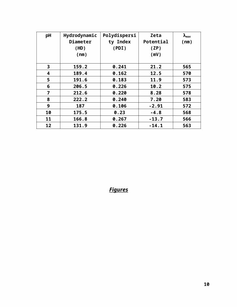
Table S2. HD, PDI, ZP , and λ max values of CMC-stabilized Cu-NPs at various
pHs.
Figures
7
pH Hydrodynamic Diameter (HD)
(nm)
Polydispersity Index (PDI)
Zeta Potential (ZP)(mV)
λmax
(nm)
3 159.2 0.241 21.2 5654 189.4 0.162 12.5 5705 191.6 0.183 11.9 5736 206.5 0.226 10.2 5757 212.6 0.220 8.28 5788 222.2 0.240 7.20 5839 187 0.106 -2.91 57210 175.5 0.23 -4.8 56811 166.8 0.267 -13.7 56612 131.9 0.226 -14.1 563

(A) (B)
(C) (D)
(E) (F)
(G) (H)
(I) (J)
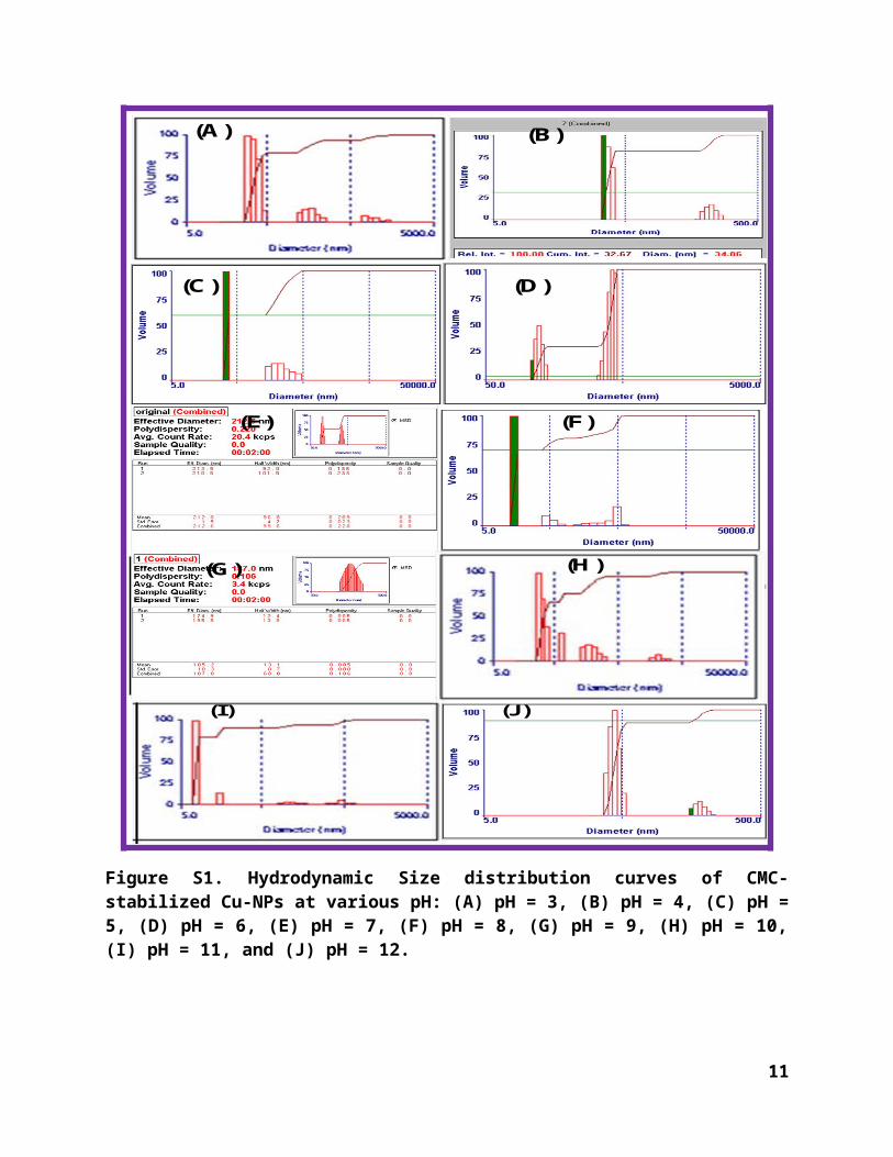
Figure S1. Hydrodynamic Size distribution curves of CMC-stabilized Cu-NPs at various pH: (A) pH = 3, (B) pH = 4, (C) pH = 5, (D) pH = 6, (E) pH = 7, (F) pH = 8, (G) pH = 9, (H) pH = 10, (I) pH = 11, and (J) pH = 12.
8

(A)
(B)
(C)
(D)
(E)
Figure S2. Zeta Potential distribution curves of CMC-stabilized Cu-NPs in the pH range of 3 to7: (A) pH = 3, (B) pH = 4, (C) pH = 5, (D) pH = 6, and (E) pH = 7.
9
(J)
(F)
(G)
(H)
(I)
Figure S3. Zeta Potential distribution curves of CMC-stabilized Cu-NPs in the pH range of 8 to 12: (F) pH = 8, (G) pH = 9, (H) pH = 10, (I) pH = 11, and (J) pH = 12.
References
10
Bagchi B, Dey S, Bhandary S, Das S, Bhattacharya A, Basu R, Nandy P (2012) Antimicrobial efficacy and biocompatibility study of copper nanoparticle adsorbed mullite aggregates. Mat Sci Eng C-Mater 32:1897-1905. doi:10.1016/j.msec.2012.05.011
Bogdanovic U, Vodnik V, Mitric M, Dimitrijevic S, Skapin SD, Zunic V, Budimir M, Stoiljkovic M (2015) Nanomaterial with high antimicrobial efficacy-copper/polyaniline nanocomposite. ACS Appl Mater Interfaces 7:1955-1966. doi:10.1021/am507746m
Chatterjee AK, Sarkar RK, Chattopadhyay AP, Aich P, Chakraborty R, Basu T (2012) A simple robust method for synthesis of metallic copper nanoparticles of high antibacterial potency against E. coli. Nanotechnology 23. doi:10.1088/0957-4484/23/8/085103
Diaz-Visurraga J, Daza C, Pozo C, Becerra A, von Plessing C, Garcia A (2012) Study on antibacterial alginate-stabilized copper nanoparticles by FT-IR and 2D-IR correlation spectroscopy. Int J Nanomed 7:3597-3612. doi:10.2147/Ijn.S32648
Effenberger FB, Sulca MA, Machini MT, Couto RA, Kiyohara PK, Machado G, Rossi LM (2014) Copper nanoparticles synthesized by thermal decomposition in liquid phase: the influence of capping ligands on the synthesis and bactericidal activity. J Nanopart Res 16. doi:10.1007/S11051-014-2588-7
Kruk T, Szczepanowicz K, Stefanska J, Socha RP, Warszynski P (2015) Synthesis and antimicrobial activity of monodisperse copper nanoparticles. Colloids and surfaces B, Biointerfaces 128C:17-22. doi:10.1016/j.colsurfb.2015.02.009
Law D, Moore CB, Joseph LA, Keaney MGL, Denning DW (1996) High incidence of antifungal drug resistance in Candida tropicalis. Int J Antimicrob Ag 7:241-245.
doi:10.1016/S0924-8579(96)00328-7Li P, Li J, Wu CZ, Wu QS, Li J (2005) Synergistic antibacterial effects of beta-lactam antibiotic
combined with silver nanoparticles. Nanotechnology 16:1912-1917. doi:10.1088/0957-4484/16/9/082Mallick S, Sharma S, Banerjee M, Ghosh SS, Chattopadhyay A, Paul A (2012) Iodine-Stabilized
Cu Nanoparticle Chitosan Composite for Antibacterial Applications. Acs Appl Mater Inter 4:1313-1323. doi:10.1021/Am201586w
Mandal SM, Mahata D, Migliolo L, Parekh A, Addy PS, Mandal M, Basak A (2014) Glucose Directly Promotes Antifungal Resistance in the Fungal Pathogen, Candida spp. The Journal of biological chemistry 289:25468-25473. doi:10.1074/jbc.C114.571778
Mosmann T (1983) Rapid Colorimetric Assay for Cellular Growth and Survival - Application to Proliferation and Cyto-Toxicity Assays. J Immunol Methods 65:55-63.
doi:10.1016/0022-1759(83)90303-4Ouyang Y, Cai X, Shi QS, Liu LL, Wan DL, Tan SZ, Ouyang YS (2013) Poly-L-lysine-modified
reduced graphene oxide stabilizes the copper nanoparticles with higher water-solubility and long-term additively antibacterial activity. Colloid Surface B 107:107-114.
doi:10.1016/j.colsurfb.2013.01.073Rastogi L, Arunachalam J (2013) Synthesis and characterization of bovine serum albumin-
copper nanocomposites for antibacterial applications. Colloid Surface B 108:134-141. doi:10.1016/j.colsurfb.2013.02.031
Ruparelia JP, Chatteriee AK, Duttagupta SP, Mukherji S (2008) Strain specificity in antimicrobial activity of silver and copper nanoparticles. Acta Biomater 4:707-716.
doi: 10.1016/j.actbio.2007.11.006
11

Usman MS, El Zowalaty ME, Shameli K, Zainuddin N, Salama M, Ibrahim NA (2013) Synthesis, characterization, and antimicrobial properties of copper nanoparticles. Int J Nanomed 8:4467-4478. doi:10.2147/Ijn.S50837
Veerapandian M, Sadhasivam S, Choi J, Yun K (2012) Glucosamine functionalized copper nanoparticles: Preparation, characterization and enhancement of anti-bacterial activity by ultraviolet irradiation. Chem Eng J 209:558-567. doi:10.1016/j.cej.2012.08.054
Yoon KY, Byeon JH, Park JH, Hwang J (2007) Susceptibility constants of Escherichia coli and Bacillus subtilis to silver and copper nanoparticles. Sci Total Environ 373:572-575.
doi:10.1016/j.scitotenv.2006.11.007
12