srs for optic pathways and skull base meningiomas · clival meningioma. • 1 carotid artery...
TRANSCRIPT

SRS for Optic Pathways and
Skull Base MeningiomasAlfredo Conti
Alma Mater Studiorum
University of Bologna
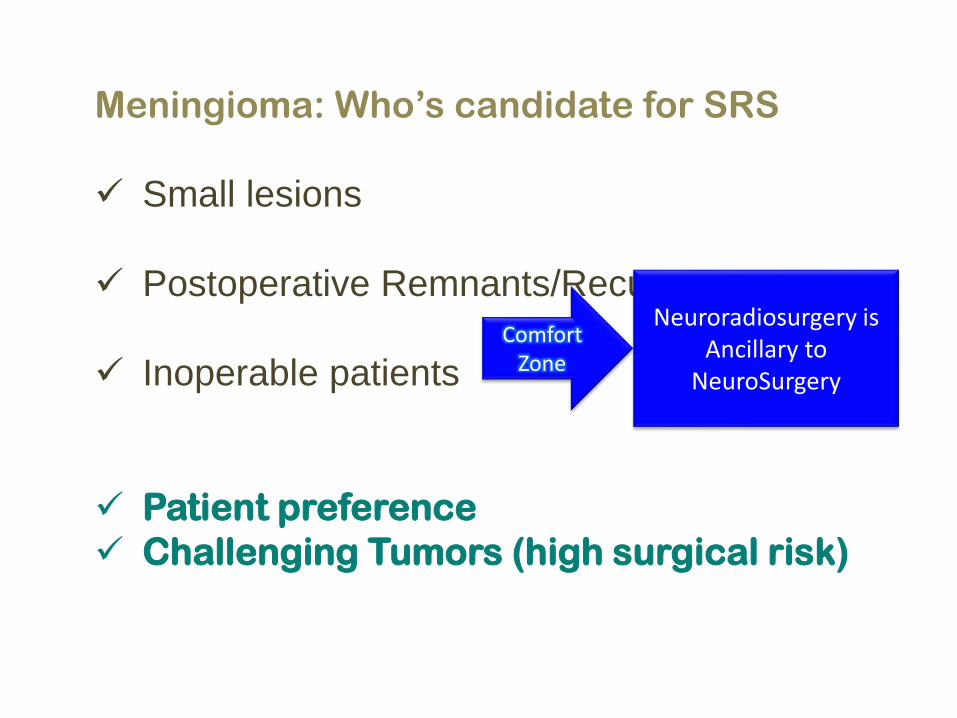
Meningioma: Who’s candidate for SRS
✓ Small lesions
✓ Postoperative Remnants/Recurrences
✓ Inoperable patients
✓ Patient preference
✓ Challenging Tumors (high surgical risk)
Comfort Zone
Neuroradiosurgery is Ancillary to
NeuroSurgery

Can We Offer Radiations as an
Alternative Upfront Treatment?

Some unspeakable truths!
• Meningiomas are sensitive to radiations
• High-precision conventionally fractionated radiotherapy
provides actuarial control rates at 360 months close to
70% with limited toxicity
• High-conformality radiotherapy (single or multisession
radiosurgery) seems to provide similar results being
applicable to virtually any patient

Lesion <30 mm
Lesions <15 cc
Lesions >2-3 mm optic nerves
Lesions >2-3 mm brainstem
Radiation Dose by Tumor Size that will have a 1% (Kjellberg data) or 3%
(integrated logistic formula) risk of radiation necrosis
Diameter (mm) Volume (cc) Gy (1% K) Gy (3% ILF)

Frameless Radiosurgery and
“multisession” or
“hypofractionated” radiosurgery
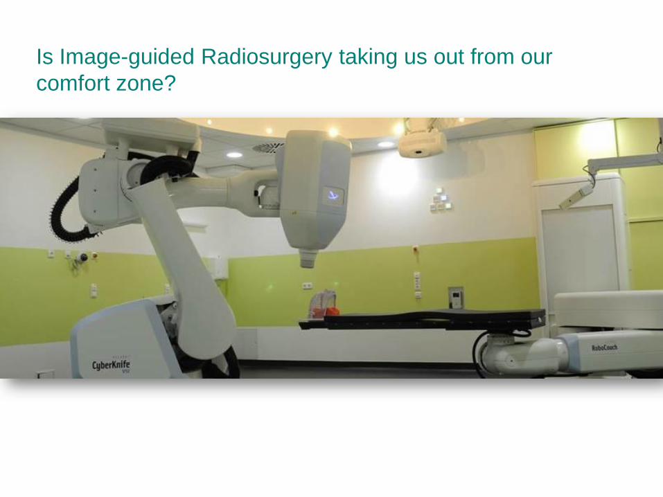
Is Image-guided Radiosurgery taking us out from our
comfort zone?

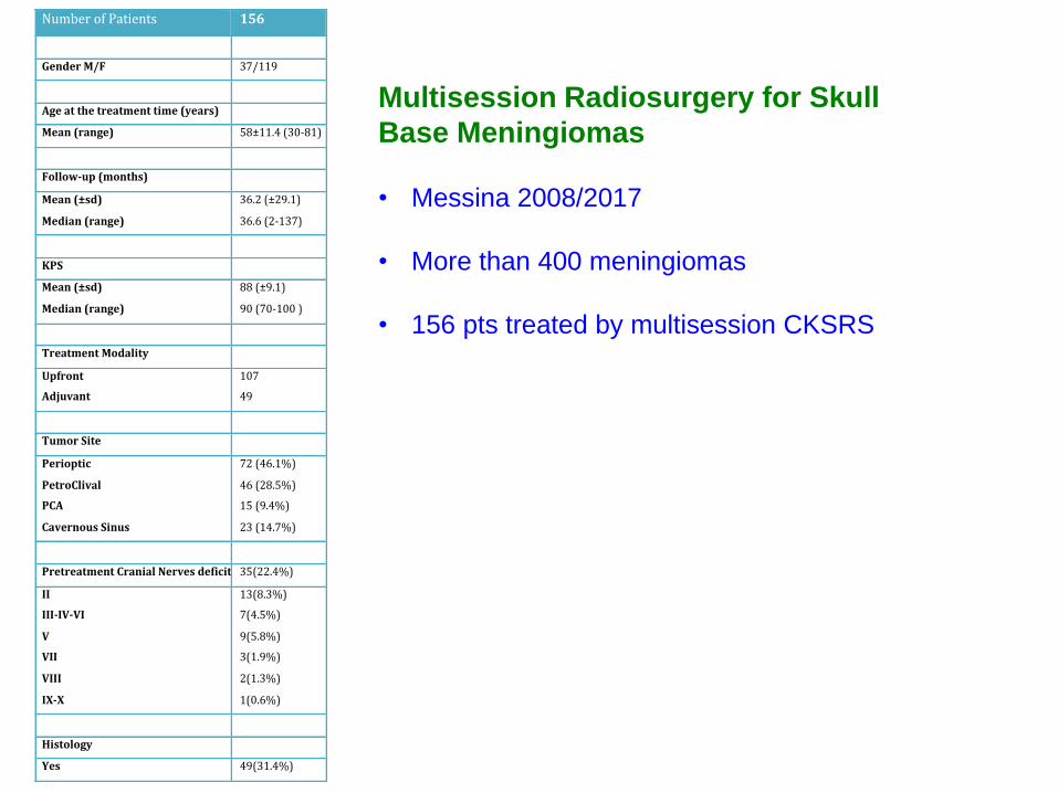
NumberofPatients 156
GenderM/F 37/119
Ageatthetreatmenttime(years)
Mean(range) 58±11.4(30-81)
Follow-up(months)
Mean(±sd)
Median(range)
36.2(±29.1)
36.6(2-137)
KPS
Mean(±sd)
Median(range)
88(±9.1)
90(70-100)
TreatmentModality
Upfront
Adjuvant
107
49
TumorSite
Perioptic
PetroClival
PCA
CavernousSinus
72(46.1%)
46(28.5%)
15(9.4%)
23(14.7%)
PretreatmentCranialNervesdeficit35(22.4%)
II
III-IV-VI
V
VII
VIII
IX-X
13(8.3%)
7(4.5%)
9(5.8%)
3(1.9%)
2(1.3%)
1(0.6%)
Histology
Yes 49(31.4%)
Multisession Radiosurgery for Skull
Base Meningiomas
• Messina 2008/2017
• More than 400 meningiomas
• 156 pts treated by multisession CKSRS
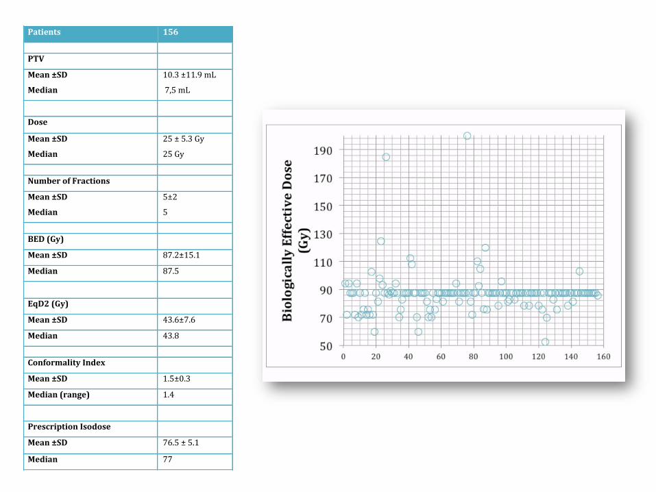
Patients 156
PTV
Mean±SD
Median
10.3±11.9mL
7,5mL
Dose
Mean±SD
Median
25±5.3Gy
25Gy
NumberofFractions
Mean±SD
Median
5±2
5
BED(Gy)
Mean±SD 87.2±15.1
Median 87.5
EqD2(Gy)
Mean±SD 43.6±7.6
Median 43.8
ConformalityIndex
Mean±SD 1.5±0.3
Median(range) 1.4
PrescriptionIsodose
Mean±SD 76.5±5.1
Median 77

25 Gy in 5 fractions

August 2007 August 2017
25 Gy in 5 fractions BED= 87.5 Gy

25 Gy in 5 fractions
BED 87.5

Local Control


2014 2017

2014 2017


BBefore BAfter

12 months 24 months flair
Treatment
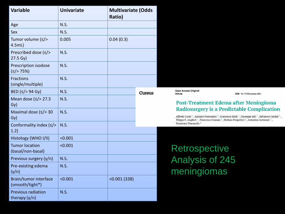
Variable Univariate Multivariate (OddsRatio)
Age N.S.
Sex N.S.
Tumor volume (≤/> 4.5mL)
0.005 0.04 (0.3)
Prescribed dose (≤/> 27.5 Gy)
N.S.
Prescription isodose (≤/> 75%)
N.S.
Fractions (single/multiple)
N.S.
BED (≤/> 94 Gy) N.S.
Mean dose (≤/> 27.5 Gy)
N.S.
Maximal dose (≤/> 30 Gy)
N.S.
Conformality index (≤/> 1.2)
N.S.
Histology (WHO I/II) <0.001
Tumor location (basal/non-basal)
<0.001
Previous surgery (y/n) N.S.
Pre-existing edema (y/n)
N.S.
Brain/tumor interface (smooth/tight*)
<0.001 <0.001 (338)
Previous radiation therapy (y/n)
N.S.
Retrospective
Analysis of 245
meningiomas

Normofractionated stereotactic radiotherapy versus CyberKnife-based hypofractionation in
skull base meningioma: A German and Italian pooled cohort analysis
Conti Alfredo1,2, Senger Carolin3,4, Acker Güliz2,3,5, Kluge Anne3,4, Pontoriero Antonino6; Cacciola
Alberto6; Pergolizzi Stefano6; Germanò Antonino1, Badakhshi Harun6, Kufeld Markus3, Meinert
Franziska2,3, Nguyen Phuong2,3, Loebel Franziska2,3, Vajkoczy Peter2,3, Budach Volker3,4, Kaul David4
CKhFSRT nFSRT p-value
CKCCharité 49 0
CKCMessina 156 0
RTCharité 0 136
Age[y],median(range) 57(27-86) 58(20-84)
Totaldose[Gy],median(range) 25(5-61) 59.4(32.4-63)
Singledose[Gy],median(range) 5(2.67-8) 1.8(1.8-2.8)
Medianfollow-up 32.5(2-135) 41.5(1-232)
GTV[ml],mean±sd 10.1±11.9 25.1±31.2 <.001
Definitiveradiotherapyatfirstdiagnosis
135(65.9%) 57(41.9%)
Adjuvantradiotherapyatfirstdiagnosis
16(7.8%) 34(25%)
Definitiveradiotherapyatrelapse 54(26.3%) 45(33.1%)
In Press

341 Skull Base Meningiomas
CK-hFSRT group local control (LC):
99.4% at 12 months, 96.8% at 3 years and 80.3% at 10 years
nFSRT group LC:
100% at 12 months, 99% at 3 years and 79.1% at 10 years
No significant difference
CK-hFSRT overall 16 patients (7.8%)
• CTCAE II° trigeminal neuralgia was the most common
neuropathy (6.3%).
• The residual cranial deficits included one case of mild
(CTCAE I°) visual disturbance in a patient with a clival
lesion and one mild (CTCAE I°) case of mild third and
sixth cranial nerve deficits in a patient with a spheno-petro-
clival meningioma.
• 1 carotid artery occlusion was reported in a patient with a large
spheno-petro-clival meningioma encasing the carotid artery, that
caused a transient facial nerve (CTCAE III°) deficit
• 1 acute edema with papilloedema
Complications

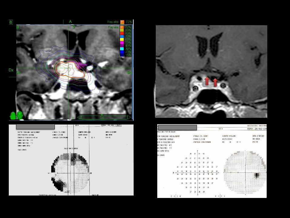
RADIOINDUCED
CAROTID
OCCLUSION?

In the nFSRT 12 patients (8.8%)
4 patients (2.9%) developed mild (CTCAE ) and 1 patient (0.7%) developed
moderate (CTCAE II) optical pathway toxicity.
4 patients (2.9%) developed mild (CTCAE I) and 1 patient (0.7%) showed
moderate (CTCAE II) hearing impairment.
1 patient (0.7%) showed mild and 2 patients (1.47%) showed moderate
(CTCAE II°) trigeminal neuralgia.
4 patients (2.9%) developed mild (CTCAE I°) and 1 patient (0.7%)
developed moderate (CTCAE II°) cranial nerve sensory deficits.
1 case (0.7%) of an acute grade III brain edema was seen and 2 cases
(1.5%) of severe (CTCAE III) vascular stenosis were seen. 1 patient (0.7%)
developed new seizures (CTCAE II).
Complications


Multisession radiosurgery for perioptic
meningiomas: medium-to-long term
results from a CyberKnife cooperative
study
• Patients with an anterior or a medium skull base meningioma
lying within 2mm from the optic nerves and chiasm
• Untreatable by single session RS due to large volume and
proximity to the anterior optic pathways
• 5x5-fraction multisession RS
• At least 36 months follow-up
•Marcello Marchetti•Alfredo Conti•Giancarlo Beltramo•Valentina Pinzi•Antonio Pontoriero•Irene Tramacere•Carolin Senger•Stefano Pergolizzi•Laura Fariselli
May 2019

RESULTS
• 167 patients were included in the analysis.
• The median follow-up period was 51 months (range 36-
129 months)
• Progression-free survival at 3, 5 and 8 years were,
respectively, 98%, 94% and 90%.
• The visual worsening rate was 3.6%.
• The 42% of the patients with a pre-treatment visual
deficit experienced improvement in vision


Dotatoc PET-MRI for PTV defintion


Conclusions
Meningiomas response to high-precision radiotherapy is
very satisfactory
LC at 10 years is >80% with normofractionation and
hypofractionation
Complications 8-9% with hFRT/nFRT
Limited Major complications (<1%)
Limited risk of RION and only when sight is
compromised before treatment