spontaneous csf rhinorrhea arising from the middle cranial fossa
TRANSCRIPT

820
Spontaneous CSF Rhinorrhea Arising from the Middle Cranial Fossa: CT Demonstration Andrew E. Yeates,1 Bennett Blumenkopf,2 Burton P. Drayer,1 Robert H. Wilkins,2 Dennis Osborne,1 and E. Ralph Heinz1
When patients present with spontaneous cerebrospinal fluid (CSF) rhinorrhea the primary diagnostic goal is localizing the site of leakage so that an appropriate approach for surgical repair can be selected. Metrizamide computed tomography (CT) has become the preferred method for investigating CSF rhinorrhea for several reasons [1-4]. CT provides superb bone detail so that fractures or areas of bony discontinuity can be identified. If the patient is actively leaking at the time of the examination, opacified CSF can often be visualized directly as it passes through the fistula, providing the sine qua non for localization. CT also allows examination of the paranasal sinuses for evidence of air-fluid levels and abnormalities that may be associated with CSF rhinorrhea, such as hydrocephalus or empty sella.
The most common sites of spontaneous CSF leak occur in the anterior fossa , sella, and parasellar regions, allowing communication with the paranasal sinuses or nasal fossa. The petro us bone is also a potential source of rhinorrhea via CSF carried by way of the eustachian tube [5] . We report a case of CSF rhinorrhea arising from a relatively uncommon site, the middle cranial fossa, accurately demonstrated by metrizamide CT and confirmed surgically. Bone/dura/arachnoid defects at the floor of the middle fossa formed a fistula between the subarachnoid space and the pterygoid recess of the sphenoid sinus. Although such fistulae have been described [6-9] , they have not been demonstrated by metrizamide CT. Our case emphasizes the importance of examining the full lateral extent of the sphenoid sinus and appreciating the morphology of the middle cranial fossa floor when studying patients with CSF rhinorrhea. The CT findings also lend credence to the hypothesis that middle fossa fistulae can be formed by spontaneous rupture of congenital "pits" at the skull base rather than as the result of previous trauma [8, 9].
Case Report
A 45-year-old woman who had been well noted clear fluid leaking from the left nostril 3 weeks before admission. She subsequently
Received May 25 . 1983; accepted June 16, 1983.
developed headaches radiating from the vertex to the occiput. On admission she denied any history of trauma, visual loss, or endocrine symptoms. Clear watery fluid dripped from her left nostril , and the flow increased when she bent forward . Aside from the patient's obesity, physical and neurologic examinations were normal. Chemical analysis of the fluid was compatible with CSF.
Metrizamide CT was performed with 5 ml of 220 mg Ijml metrizamide injected into the lumbar thecal sac. The patient was tilted 60° head-down for 2 min and was then transferred prone and flat to the CT scanning table. The patient was actively leaking during the CT examination. Contiguous sections were obtained in a direct coronal plane from the frontal sinus to the dorsum sellae using 1.5 mm collimation , 120 kVp, 320 mA, and 9.6 sec scan duration. Images were magnified by a factor of 1.5 and were reconstructed using a hard-bone-target algorithm that improves spatial resolution and edge sharpness when examining bone-air or bone-soft tissue interfaces. The images were photographed at window settings of 250 Hand 4000 H, respectively, for soft tissue and bone detail.
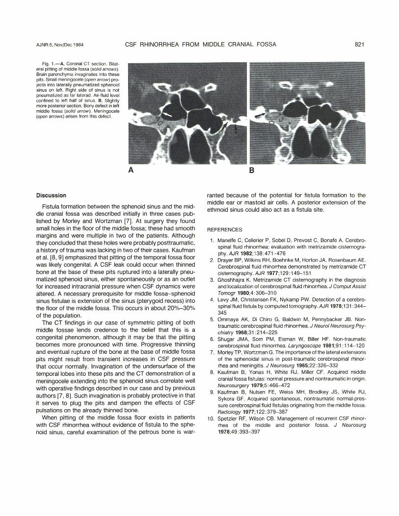
Metrizamide CT showed an air-metrizamide level confined to the left half of the sphenoid sinus (fig. 1). Pneumatization of the sphenoid sinus extended far laterally on the left so that the floor of the middle fossa also formed its roof. The floors of both middle fossae had an irregular, pitted appearance, and on the left there were areas of extreme bony thinning with frank bony defects. Opacified CSF could be identified passing through one of these defects, filling a saclike structure that protruded into the sphenoid sinus, compatible with a small meningocele. On both sides many of the pitted areas appeared to contain tissue from the undersurface of the temporal lobes. The patient had a capacious sella that was partly filled with CSF, compatible with incompetence of the diaphragma sellae; however, there was no evidence of CSF leak from this site . There was no ventricular enlargement.
A subtemporal exposure of the left middle fossa was performed through a left temporal craniectomy. On elevation of the temporal lobe, four small depressions were evident in the middle fossa floor. There were holes in the dura at these sites through which brain protruded. The temporal lobe was carefully dissected away from the middle fossa floor, exposing the anteromedial aspect of the fossa and revealing two other depressions with perforations in the dura. Each hole was plugged with a small piece of temporal muscle and oversewn with small grafts of temporal fascia. The patient did well postoperatively; there was no recurrence of rhinorrhea over 3 months of follow-up.
1 Department of Radiology, Box 3808, Duke University Medical Center, Durham, NC 27710. Address reprint requests to A. E. Yeates. 2 Department of Surgery (Neuroradiology), Duke Universi ty Medical Center, Durham, NC 27710.
AJNR 5:820-821, November/December 1984 0195-6108/84/0506-0820 $00.00 © American Roentgen Ray Society
AJNR :5, Nov/Dec 1984 CSF RHINORRHEA FROM MIDDLE CRANIAL FOSSA 821
Fig. 1.-A, Coronal CT section . Bilateral pitting of middle fossa (solid arrows) . Brain parenchyma invaginates into these pits. Small meningocele (open arrow) projects into laterally pneumatized sphenoid sinus on left . Right side of sinus is not pneumatized as far laterad . Air-fluid level confined to left half of sinus. B, Slightly more posterior section. Bony defect in left middle fossa (solid arrow). Meningocele (open arrows) arises from this defect.
Discussion
A
Fistula formation between the sphenoid sinus and the middle cranial fossa was described initially in three cases published by Morley and Wortzman [7]. At surgery they found small holes in the floor of the middle fossa; these had smooth margins and were multiple in two of the patients. Although they concluded that these holes were probably posttraumatic, a history of trauma was lacking in two of their cases. Kaufman et al. [8, 9] emphasized that pitting of the temporal fossa floor was likely congenital. A CSF leak could occur when thinned bone at the base of these pits ruptured into a laterally pneumatized sphenoid sinus, either spontaneously or as an outlet for increased intracranial pressure when CSF dynamics were altered. A necessary prerequisite for middle fossa-sphenoid sinus fistulae is extension of the sinus (pterygoid recess) into the floor of the middle fossa. This occurs in about 20%-30% of the population.
The CT findings in our case of symmetriC pitting of both middle fossae lends credence to the belief that this is a congenital phenomenon, although it may be that the pitting becomes more pronounced with time. Progressive thinning and eventual rupture of the bone at the base of middle fossa pits might result from transient increases in CSF pressure that occur normally. Invagination of the undersurface of the temporal lobes into these pits and the CT demonstration of a meningocele extending into the sphenoid sinus correlate well with operative findings described in our case and by previous authors [7, 8]. Such invagination is probably protective in that it serves to plug the pits and dampen the effects of CSF pulsations on the already thinned bone.
When pitting of the middle fossa floor exists in patients with CSF rhinorrhea without evidence of fistula to the sphenoid sinus, careful examination of the petrous bone is war-
B
ranted because of the potential for fistula formation to the middle ear or mastoid air cells. A posterior extension of the ethmoid sinus could also act as a fistula site.
REFERENCES
1. Manelfe C, Cellerier P, Sobel D, Prevost C, Bonafe A. Cerebrospinal fluid rhinorrhea: evaluation with metrizamide cisternography. AJR 1982;138:471-476
2. Drayer BP, Wilkins RH, Boehnke M, Horton JA, Rosenbaum AE . Cerebrospinal fluid rhinorrhea demonstrated by metrizamide CT cisternography. AJR 1977;129 :149-151
3. Ghoshhajra K. Metrizamide CT cisternography in the diagnosis and localization of cerebrospinal fluid rhinorrhea. J Comput Assist Tomogr 1980;4:306-310
4. Levy JM, Christensen FK, Nykamp PW. Detection of a cerebrospinal fluid fistula by computed tomography. AJR 1978;131 : 344-345
5. Ommaya AK, Oi Chiro G, Baldwin M, Pennybacker JB. Nontraumatic cereb,ospinal fluid rhinorrhea. J Neurol Neurosurg Psychiatry 1968;31 : 214-225
6. Shugar JMA, Som PM , Eisman W, Biller HF. Non-traumatic cerebrospinal fluid rhinorrhea. Laryngoscope 1981 ;91 : 114-120
7. Morley TP, Wortzman G. The importance of the lateral extensions of the sphenoidal sinus in post-traumatic cerebrospinal rhinorrhea and meningitis . J Neurosurg 1965;22 :326- 332
8. Kaufman B, Yonas H, White RJ, Miller CF. Acquired middle cranial fossa fistulas: normal pressure and nontraumatic in origin . Neurosurgery 1979;5 :466-472
9. Kaufman B, Nulsen FE, Weiss MH , Brodkey JS, White RJ , Sykora GF. Acquired spontaneous, nontraumatic normal-pressure cerebrospinal fluid fistulas originating from the middle fossa. Radiology 1977;122 :379-387
10. Spetzler RF, Wilson CB. Management of recurrent CSF rhinorrhea of the middle and posterior fossa. J Neurosurg 1978;49 :393-397