(spinal arachnoiditis) - multi-systems...
TRANSCRIPT

..J;. tl!j~.,".",1;,;01,!. ...
(Spinal Arachnoiditis)Its Diagnosis and Treatment by
Spinal Cord Stimulation:;~~~ ~1'. ..' ..
.~ .
;;~ ~.::J~ ;'.1i: ;~~::
~::i:~~~"'....
CHRISTIAN DE LA PORTE and JEAN SIEGFRIED
--,
From 1973 to 1981, 94 patients suffering from low-back pain, with orwithout spread into the lower extremities. were candidates for thera-peutic spinal cord stimulation. The etiology of pain in all cases waslumbosacral spinal fibrosis due to multiple myelographies and surgicalinterventions on the lumbar spine. The long-term results. based on afour-year follow-up. reveal a 60% subjective improvement of pain. a40% substantial reduction of medication. and a 26% increase inworking capacity. The concept of spinal arachnoiditis is reviewed andthe term lumbosacral spinal fibrosis proposed. The treatment of thischronic painful and disabling disease is discussed. [Key words: low-back pain syndrome. arachnoiditis spinalis. epidural arachnoiditis.lumbosacral spinal fibrosis, postoperative herniated disc. myelog-raphy, spinal cord stimulation)
.~'!1,. j..
. '. .\
'rs~~';:
f 1~ :',J
HE LOW BACK is one of the most commonlocations of chronic pain and so-calledlumbosacral arachnoiditis: Although in-
. .. - frequently seen in its initial stages. this:tOD"'dition. in addition to the other intraspinal. ex-~ding non-tumorous lesions. has taken a place that~~ot be neglected. Most patients will be evaluated~ one or more myelographies. Surgery will be per-~~ed frequ'~ntly for ruptured lumbar disc.:, -Each Year. 50.000 Amer~cans undergo surgery for
~bar herniated nucleus pulpusis. As far as pain relief:: concerned. the failure rate is as high as 40%.48
healy41 estimates that 75% of compensation pay-.~nts go to these unimproved patients. who represent
of the compensation population. Burton48 esti--11'Iates the accumulated cost of the failed low-back sur-
:~.s~ndrome to be up to $75.000 per patient beforeISslon to the department.
Repetitive back surgery is the unfortunate conse-quence of persistent pain, although the improvementfrom additional operations is very little. McCracken)1from the Workmen's Compensation Board of Ontario,Canada, advised the Ohio Industrial Commission thatno patient is cured by a second low-back operation,20% are improved, 20% are. made worse, and 60% areessentially unchanged. With additional operations, theoutcome worsens and after four c,~rations, 5% areimproved and 50% are made worse. The same authorstates that for "lost-time industrial back il\iuries," thesurgical rate in Ontario in 1977 was 2.65%. For 1978,38.225 "lost-time disability back cases" were regis-tered.
The first report of arachnoiditis as a pathologicalprocess was by Spiller et al in 1903.52 The first de-scription of arachnoiditis spinalis was made by lloiendelin 1908.)~ who named it "meningitis serosa spinalis,"Afterwards. other terms were used for the same entity:Horsley, in 1909 called it "chronic spinal meningi-tis"~I; Stookey. in 1927 called it '.adhesive spinalarachnoiditis"S); while Elkington, in 19)6 called it
~ '("~1~~he Depanment of Neurosur~ery. University Hospital. ZO-S b .lerland.:,-.r:. \I ntitted for publication December 9. 198:!.
-,, .-.~..&. Row. Publishers. Inc.

3/'INE . ~uME . . NUMBER 5. '9&3
594 SPINAL FIBROSIS, DE LA PORTE AND SIEGFRIED
Table 2. Top09raphlcal Distribution 01 Pain
Area Number -""-
One leQBoth leQsLow backSacralPerianal and perineumBottomGenital
-°To
20 patients this was confined to one leg. in 16 it in.volved both legs (although not all had complaints iQboth legs). and in two it was located in the low backonly. The different signs and their frequency are listed.in Thble 3. These patients were severely incapacitated,with nearly 2/3 showing a paresis and significant Las-~gue's sign. and 1/3 having sphincter problems. Fur.tht:rmore. five patients showed anesthesia dolorosa..and hypesthesia or hypalgesia dolo rosa was presentia21 cases. Lumbosacral spinal fibrosis is not restrictedto the segment of previous surgery, since 13 of 38 pa.~'tients had an absent patellar reflex, although only.vCry;:few were explored at the L3 level. Objective neUtO'-:. (:,logic deficits resulting from segments notinvolve~~:' I~: surgery were found in many patients. These observa...: ~'C
. . .~, ...tions support the fact that myelognphy as well as SUi,:. ~:gery are predisposing factors of spinal fibrosis. HoW::- .
ever, once the fibrosis starts. its evolution is unp'rr:;_.
dictable. It is a self-limiting disease. but it can. 81SQ~affect the side previously not involved and can extii1d:
t,~
to several segments before stopping its course;:~e;postoperative follow-up ranges from three.tO""9~months. The mean follow-up is 35.8 months. andtfii-
.',"",standard deviation 25.4 months. (Thble 4.) ~~~.
From 1973 to 1977, spinal cord stimulation.~'achieved by laminec..omy and fIXation of an el~...subdurally in two cases, and endodurally (betwee!l~two sheets of the dura) in 12 cases. Since 1977~ firsttwo electrodes (ten cases), then only one electrode,were introduced percutaneously into the spinal epi.dural space. dorsal to the spinal cord. The connectioaof the electrode to a radio-receiver followed a few daysafter a successful trial. Tne receiver \vas placed in Isubcutaneous pocket. usually in the right abdominal Ie-
.~
fr
~
Table 3. Distribution of Objective neurologic deficit andNumber of Times it was found
"meningitis serosa circum$cripta"I~; and Wadia, in11J69 called it "spinal meningitides with radiculomye-
lopathy.~7"The first publication implicating disc surgery as the
etiology of ardchnoiditis dates from 1951 and is bySmolik and Nash.51 Since then, .'arachnoiditis" or"epidural arachnoiditis" has been generally used todescribe the persistence of low-back pain after lumbarspine surgery. in the absence of recurrent disc hernia-tion. The term "epiduraj arachnoiditis" does not re-flect precisely the pathological aspects of tissue pro-liferation. therefore. the term "lumbosacral spinal fi-brosis" will be used.
The treatment of chronic low-back pain and lum-bosacral spinal fibrosis is still controversial. The in-troduction of the non-ablative methods of spinal cordstimulation in 1967 by Shealy.oI6 for the treatment ofpersistent pain. aroused great interest. The techniquehas been applied increasingly. due to improved tech-nology and simpler surgical techniques.
Our own experience since 1973 is derived from 350cases of dorsal cord stimulation with 174 definitiveimplantations. From this series with different painetiologies. there were 38 cases of lumbosacral spinalfibrosis. which will by analysed. The results after im-plantation of a spinal cord stimulator. with a prolongedfollow-up period. are given.
MATERIAL AND METHODS
Of a series of 94 patients. 38 (20 females and 18males), suffering from low-back pain after multiplemyelographies and several surgical procedures on thelumbar spine. were considered for a spinal cord stim-u[ation and operated upon, after failure of many otherkinds of treatment for a long period of time. Thb[e Ilists the age profile. number of previous operations.myelographies. and pain duration. Pain was the majorcomplaint in all these patients. The distribution of thiscomplaint is shown in Thble 2. Thn patients had a clearradicular distribution. and 28 demonstrated a nonrad-icular pain distribution. There was a high number ofpatients (28 cases) with initial complaints in one legonly, but eventually including the low back or otherlocations.
On clinical examination. we found objective signsor the condition in all patients. This means that allpatients showed an objective neurological deficit. In NumberSymocom
212
2155
1121
222513
Table 1. Age Distribution and Antecedents of 38 PatientsParesisParesthesiaHypesthesia. hypalgesiaAnesthesiaHyperpathia, hyperesthesiaSphincter ProblemsAtrophyTrophic changesLasegue positiveAnkle reflex negativePatellar rellex negativeCramps
Range Mean StandarddeviatIOn
28-76 years1-81-13
12-384
4723.54.9
1347
114182.9
972
AgePrevious operations.Previous myelogramstMonths with pain
. Only operations with an analgesic purpose included
t Calculated on data available Irom 19 patientsi
\';'t.

Table 4. Distribution 01 Patients with IncreasingFollow-up Time
Table 5. Number 01 Reinterventions and Number 01 Patientsin Which they were Respectively Performed
Follow-uP penod (monthS) Number 01 pat,ents.
<12~12
2436486072849096
7867242
23433
122
11 . Total number 01 reinlerventions is SO.
'olal of 38 patients
gion, so that the patient could apply conveniently anexternally transmitted signal. Fifty-six cases weretested percutaneously without satisfactory results, andwill not be discussed further. The implantation sitewas D2-3 for the 14 cases of subdural and endoduralelectrodes. The site of the epidural electrode tip wasD4-s in 15 cases, D6-7 in eight cases, and DI-9 in onecase. The site of electrode placement was at least foursegments above the highest segment of complaints(Figure I).
Eighty-eight interventions were performed (percu-1aneous test procedure not included) on 38 patients.Reinterventions (50 procedures) were necessary, dueto wound healing problems in ten cases, for completeremoval or reimplantation of the device in five cases,or for other complications that had to be correctedsurgically. These 50 reinterventions occurred in 15 pa-tients (Thble 5). Therefore, 23 patients had no reinter-ventions, and thus no complications, due to the ap-paratus. The mean reintervention rate was 1.3, with a
"'---ndo:]
Location Number ofcases
i~c,.;oo.,~ d
Dq - DS IS
)
D6 - D] 8
D8 °9,~
standard deviation of 2.2. due to five patients withrespectively five. six. and seven reinterventions.
The complications of surgery can be divided intogeneral complications (20) and those secondary to ap-paratus malfunction (33) (Table 6). Reinterventions insix patients alone represented 32 complications. In 19cases, no problems occurred. Permanent or transientneurological deterioration. rejection of implanted de-vices; CSF leakage. or seroma formation never oc-curred. There were no deaths.
In summary. 19 patients (50%) presented no com-plications. and 23 (60%) no reinterventions. Six pa-tients (16%) presented 60% of the complications and67% of the reinterventions. Thineen patients (34%)were responsible for 40% of the complications and33% of the reinterventions.
These figures suppon the belief that once somethinggoes wrong with the stimulation treatment. or the pa-tient presents a clinical complication. his chances forhaving funher complic.:ltions increase enormously. Allpatients except one underwent a preimplantation testof several days. Good to excellent pain control was anabsolute prerequisite for definitive implantation(Table 7). The preoperative indications for treatmentby spinal cord stimulation were based on the followingcriteria: (I) pain occurred on an organic basis. withobjective neurological deficit; (2) other current thera-pies were tried thoroughly. without patient improve-ment; (3) there was no little dependence on narcoticdrugs; and (4) psychologic or psychiatric evaluationwas not essential and was performed in a few casesonly.
RESULTS
Three aspects of the results of spinal cord stimula-tion for the treatment of chronic pain due to lumbo-sacral spinal fibrosis will be considered in detail: (I)patient personal evaluation. (:!) woiking capacity. and(3) changes in medication.
Subjective Assessment
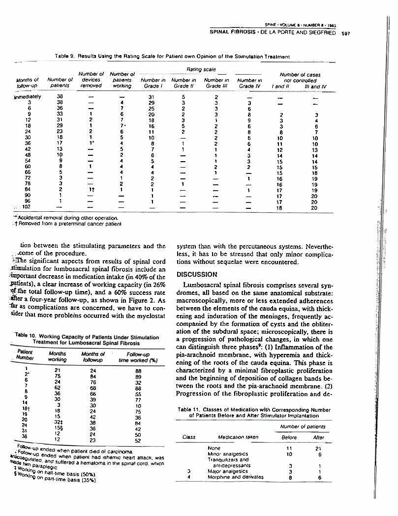
Pain relief is as~essed bv .1 f(~ur-grade rating scale.(Table 8). Table 9 renccI!:. the patient opinion at thedifferent intervals or [("llow-up. Columns ~ and 3 in-dicate. respectively. the number of pali~nts availableat each follow-up and the number of devIces that were
'"
...;.,~ EPidural sIte o;:lectrooe placemenl after connecllon with:.; taneous receiver,

sPINE . vOI.u~E ~ . N\J~BER 8 . 1903--~- 5gB SPINAL FIBROSIS. DE LA PORTE AND SIEGFRIED
Table 6. Occurrence of Clinical and Electrical Device Complications Referred to the Device Used "-DevIce
ComplicationEpiduralunipolar;
E(JlduraibifJOiart TotalLamInectomy. II
!-,;.: 1mNumber
112
95411
11
I
7321
81
21
21322221
7
1
6212
11 -
531611-
26
ClinicalSkin ero!.K>nPain at site of incisionPain at receptor siteInfectionCSF listule
Electrical deviceElectrode displacement
Of not optimal siteElectrodeSystem not workingStimulatorReceptorCable and connectionsAnteMa
Total
. Medtronic system. 12 cases.t Medtronic system. 4 cases; Avery system. 6 cases.~ Avery system. 19 cases.
was classified in the highest class possible;a patient taking a tablet of pentazocine twice awas placed in class 4. The table also indicatesmany patients were in each category, . ..
the department, and after implantation of thelatory at the latest follow-up. Three patients
15,23, and 35) were excluded because ~ not available.
Changes in medication are shown in Thble 12;tients are divided into seven - ~. ,
change in medication intake. Also shown is thenumber in each category after stimulator -
Thirteen patients decreased medication togory II and III). Five increased medicinetion. One patient passed from class 4 to class 2.teen patients (40%) decreased theirmarkedly, 16 (46%) were unchanged, and five
increased.Table 13 ---~..- -~ ---~ ~~ ~ --
eters used. This information was available fromthe cases. The data listed are the parameters .
the patient at the latest follow-up.Most of the patients are using' voltages.
and 7 volts and frequencies from 75 to 100stimula~e three to six times a day. There was
completely and definitively removed. The fourthcolumn shows the number of patients working com-
pared to the number presenting for follow-up (column2). In the last two columns. grades I and II and 111 andIV were added together. Grades I and II were consid-ered successes. and grades 111 and IV. failures. Notethat during the entire follow-up period. the success:failure ratio is always around 50%. These two columnsreflect that at their last follow-up as many patientswere in grades 1 and 11 as in III and IV. It also meansthat not only failure patients were lost or discardedearlier from follow-up, but also success patients.
Working CapacityOf 38 patients. 13 returned to work. The total patient
follow-up time is 1356 months. Of this total. patientsunder stimulation treatment worked 355 months.which represents 26% of total follow-up time. The per-centages mentioned above seem economically impor-tant to us. and give support to the use of spinal cordstimulation in this context (Table 10).
Changes In Medication
We divided the patients into five classes. accordingto the medication they used ("!'able 11). Each patier.:.
Table 7. Personal Appreciation of the Result of thePreimplantatlon Test by the Patient Table 8. Rating Scale According to Patient Appreciation ~:.
Number of patientsPain control (%) Pain rel;el (%)Grade22
3841
I
1\
1\1
IV
. Not completed
SPINE. VOlUME 8. NWBER 8 . 1983
SPINAL FIBROSIS. DE LA PORTE AND SIEGFRIED 597
Table 9. Results Using the Rating Scale lor Patient own Opinion 01 the Stimulation Treatment
Rating scaleNlJmber of
devicesremoved
Number 01 casesnOI conlrolled
I and II 1/1 and IVMonthS 01tollow-uP
Number o(pat,ents
Number In Number inGrade I Grade II
Number InGrade III
Number InGrade IV
ImrTIediately369
121824303642485460667278849096
., 102
383836333129231817131p98533211
312925201816111087654422111
5322352
11
1
23331222211121
-
121211"
1
1t
-47677'65452444121
-368968664332
1
1
- -2338
10111214151515161617171718
3467
10101314141518191919202020
'.Accidental removal during other operation.t Removed from a preterminal cancer patient.
system than with the percutaneous systems. Neverthe-less, it has to be stressed that only minor complica-tions without ~equelae were encountered.
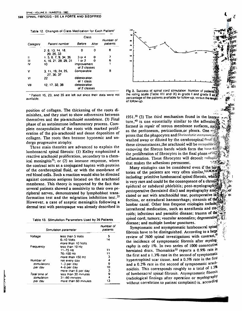
tion between the stimulating parameters and the- ' .come of the procedure.);:7The significant aspects from results of spinal cord
,~u1ation for lumbosacral spinal fibrosis include an,:important decrease in medication intake (in 40% of the~tients), a clear increase of working capacity (in 26%:C!f;the total follow-up time), and a 60% success rate":aTter a four-year follow-up, as shown in Figure 2. Asiar as complications are concerned, we have to con-sider that more problems occurred with the myelostat
DISCUSSION
Lumbosacral spinal fibrosis comprises several syn-dromes, all based on the same anatomical substrate:macroscopically, more or less extended adherencesbetween the elements of the cauda equina, with thick-ening and induration of the meninges, frequently ac-companied by the formation of cysts and the obliter-ation of the subdural space; microscopically, there isa progression of pathological changes, in which onecan distinguish three phases9: (I) Inflammation of thepia-arachnoid membrane, with hyperemia and thick-ening of the roots of the cauda equina. This phase ischaracterized by a minimal fibroplastic proliferationand the beginning of deposition of collagen bands be-tween the roots and the pia-arachnoid membrane. (2)Progression of the fibroplastic proliferation and de-
12.6789
1418t1920243138
2175246236303
181532*15§1212
24847668663930244238362423
88893288557710753684425052
Table 11. Classes of Medication with Corresponding Numberof Patients Before and After Stimulator Implantation
Number of patients
Before AlterClass Medication taken
1110
216
F --," ~kJw-up ended when patient died 01 carcinoma
~ ow-up ended When patient had IS hemiC heart attack. wasrnaae hgUlated. and sullered a hematoma in the spinal cord. WhiCh
1W 1m paraplegic
orklnn 0 h "§ Wor\(j w n ail-time basIs (50%)ng on Part-time basis (35%)
NoneMinor analgesIcsTranquilizers and
antidepressantsMajOr analgesicsMorphine and derivales
338
116
34
Nvmber ofpat,entsworking
SPINE' VOllJl.4E 8' tllJl.4BER 8 . 18&3
l598 SPINAL FIBROSIS' DE LA PORTE AND SIEGFRIED A~:if
~ «~i' ~$.I ~~...;!:,~ ~ yt
'.~ t,:I':'; \t.\'.re
~~~; ~~'~~; ~~.i~1 ",;It ~ ~ ~ ~Cgl
.::; . 1&%
:,': ~' }. .',,~" i 1. dl
. f~lr~ I'tfiilin, I ~~.
Ij.;:na
Table 12. Changes of Class Medication for Each Patient. '0'
'" ..'",
ClassNumber ot
patients
'-'r '
..
.. r'" .
~ ,.v"Before AllerCategory PatIent number
,~..0 80
so
..761
2,8,13,14,18,20. 26. 33
1,5.6.7.9.34.364.16.21.28.29.3110
3 or 4 0lor 2 0
improvement01 2 classes
comparable
IIIIIIV
3.11.19.24.25.27.30.37
Bv
22 deterioration01 1 class
deterioration01 2 classes
VI
12. 17.32.38VII
IFig 2. Success of spinal cord stimulation Number of patietQ.the rating scale (Table VIII and IX) in grade I and grade II as.-percentage of the patients available for lollow-up, versus tt)61e1:igitJ~:
of follow-uo.
. Patient 15. 23. and 35 are leh out since their data were not
available
position of collagen. The thickening of the roots di-minishes, and they start to show adherences betweenthemselves and the pia-arachnoid membrane. (3) Finalphase of an autoimmune inflammatory process. Com-plete encapsulation of the roots with marked prolif-eration of the .pia-arachnoid and dense deposition ofcollagen. The roots then become hypoemic and un-dergo progressive atrophy.
Three main theories are advanced to explain thelumbosacral spinal fibrosis: (1) Kelley emphasized areactive arachnoid proliferation, secondary to a chem-ical meningitis2s; or (2) an immune response, wherethe contrast acts as a conjugated hapten with proteinsof the cerebrospinal fluid. or with the membrane ofred blood cells. Such a reaction would also be directedagainst common antigens located on the arachnoidmembrane. This theory is supported by the fact thatseveral patients showed a sensitivity to their own pe-ripheral nerves, demonstrated by lymphoblast trans-formation test and the migration inhibition test.2SHowever, a case of aseptic meningitis folIowing adermal test with pantopaque was already described in
~~)~-~~;..;
"~ ~:.~;,.~ .~ iJ,. 'j,
--- -r :"'~~C
, 1"B .' ~ :;~.;,~ - ., ~'O[l
1951.30 ~3) The third ~echa?is~ found in the Itte~::'1;; !""",~"i;
ture,40 I.S one ~ssentlally similar to the adhesi~~I.~,~
formed In !epalr of ser,:>us ~embrane surfaces, .s~~~ ,~~~.
as the pentoneum, pencardlum,or pleura. One i, . ~.
poses that the phagocytes and.
washed away or diluted by the cerebrospinal
these circumstances,the arachnoid will be
removing the fibrous bands which form the
the proliferation of fibrocytes in the final phase
inflammation. These fibrocytes will deposit
that makes the adhesions permanent. "~~~il;f~i'-!:
Many etiologies can be considered even if the :biS
l::~ --,c,. "i~tories of the patients' are very often similar,).5.31~~; !:f':~i..
~ncluding: primitive lumbosacral spinal fibrosis, whf: . .:~f'- J
IS very rare and could be the consequence of a ChrO~
I ~";{l\:~r( epidural or subdural phlebitis; post-myelograp~ ,~Jn
postoperative (herniated disc) and myelography as59"
tj ' ; :¥?:.of ciated or not" with arachnoidal tear, postoperative iD-~i ~~ .01 i
fection, or extradural haemorrhage; stenosis of 1;b~~ ~:;
lumbar canal.. Other less fr~quent etiologies includ~,~ ifl
intrathecal medication, such as anesthesia and stc-~~~~:.
roids; infectious and parasitic disease; trauma of the.'~~~
. "'...,,n
s~inal cord; tumo~s; vascular anomalies; degeneraU'~~!:
disease; and multiple lumbar punctures. .".. l ~.
Symptomatic and asymptomatic lumbosacral spit\af:~: i.,'
fibrosis have to be distinguished. According to a lafIC' ir:.:
review of 7600 spinal investigations with contrast, d Z
the incidence of symptomatic fibrosis after myelot, ;',
raphy is only 1%. In two series of 1000 consecutive
herniated discs. Thomalskess reports a 0.9% rate in
the first and a 1.3% rate in the second of symptomatiC
hypertrophied scar tissue. and a 0.5% rate in the firs!
and a 0.2% rate in the second of symptomatic arach'
noiditis. This corresponds roughly to a total of 1.5~
of lumbosacrBI spinal fibrosis. Asymptomatic fibrosIs
(radiological findings after operation or myelographY
without correlation to patient complaint) is. according
Table 13. Stimulation Parameters Used by 26 Patients
Number 01patIentsStimulatIon parameter
5!4
Voltage
Frequency11
11
34
9
10
3
94
13
Number ofstimulatIonsper day
less than 5 Volts6-10 Voltsmore than 10 Voltsless than 10 Hz11-75 Hz76-100 Hzmore than 150 Hznot every cay1-3 per day4-0 per Caymore than 6 per dayless than 30 minutes31-60 minutesmore than 60 minutes
Total time 01stImulatIonper day
SPINE. VOLUME a . NUMBER a . ,~- -- -SPINAL FIBROSIS. DE LA PORTE AND SIEGFRIED 599
.alpe,49.SO present in 16.J to 35% of the casl:s after'myelography with methiodal sodium (Abrodil, Con-lurex. Kontrast U),and in 42 tf) 75% of the cases aftermyelography followed by surgery. When using meg-lumine iothallamate (Conray. Meglumine). the respec-tive figures are 8 to 47% and 23 to 53%, and aftermeglumine iocarmate (Bis-Conray. Dimer-X). 0 to36% and 42 to 73.5%,,The radiologic diagnosis is based on the following
findings on myelography2J,4),4.5: incomplete filling of aroot, as compared with the original myelography; non-filling of sacral or lumbar "oots, usually symmetrical;enlargement of roots; disappearance of the normal.cauda, equina striation; narrowing of the caudal dural:Sac; irregular 'distribution with trapping of contrast;..'jisidual contrast from previous myelography; pseu-.:.docyst formation; incomplete or complete blockage.:: :Benner proposes the following 6 stage classifica-
.:lion.3
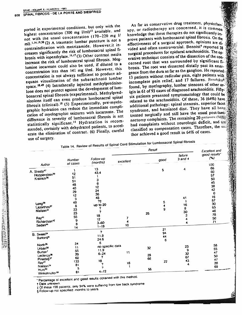
Ag 4. Epidural fibrosis visualized by contrast medium (Telebrix) byCT scan at the level of L4 -5. Note hemilaminectomy at left. Notealso ventrolateral shape of fibrosis.
f- -".-':.; - 0: extradural compression (spondylosis,,v .'-'~,;:- stenosIs)';T°:--
{'~ -;0'-; I : postoperative local alterations1;;:,:- - II : lumbosacral spinal fibrosis at onei, ,';, ~ segment
III : lumbosacral spinal fibrosis at several- -' segments
- ~!" ; IV: block secondary to lumbosacral fi-~:' - 0j,::~:, brosls
-.' -:'- V : progressive lumbosacral spinal fi-~;:...;, brosis ascending more than two seg-- ;':-0 ments above the operated level,. ,. -: -
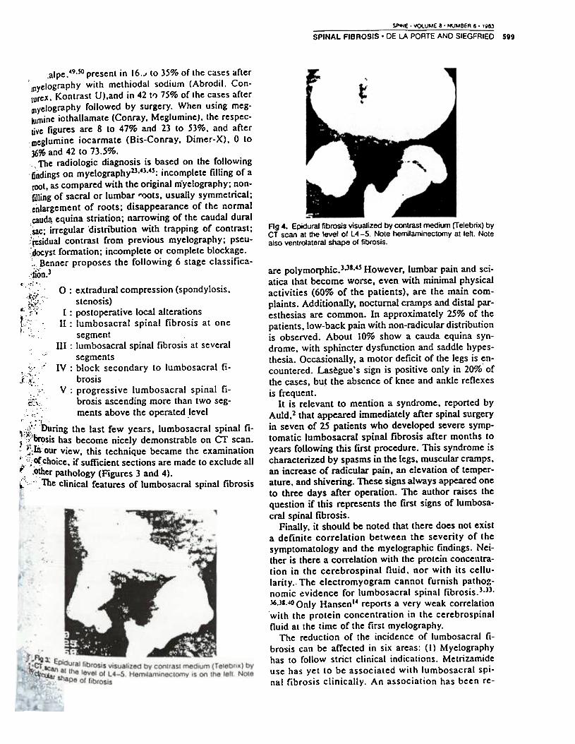
..:;;:': During the last few years, lumbosacral spinal fi-;'~i'b,~sis has become nicely demonstrable on CT scan.': .~:moUr view, this technique became the examination~-'~_ofchoice, if sufficient sections are made to exclude all-' .other pathology (Figures 3 and 4).
~'_o: - The clinical features of lumbosacral spinal fibrosis
are polymorphic.3.3s,4s However, lumbar pain and sci-atica that become worse, even with minimal physicalactivities (60% of the patients), are the main com-plaints. Additionally, nocturnal cramps and distal par-esthesias are common. In approximately 25% of thepatients, low-back pain with non-radicular distributionis observed. About 10% show a cauda equina syn-drome, with sphincter dysfunction and saddle hypes-thesia. Occasionally, a motor deficit of the legs is en-countered. Lasegue's sign is positive only in 20% ofthe cases, but the absence of knee and ankle reflexesis frequent.
It is relevant to mention a syndrome, reported byAuld,2 that appeared immediately after spinal surgeryin seven of 25 patients who developed severe symp-tomatic lumbosacral spinal fibrosis after months toyears following this first procedure. This syndrome ischaracterized by spasms in the legs. muscular cramps.an increase of radicular pain. an elevation of temper-ature. and shivering. These signs always appeared oneto three days after operation. The author raises thequestion if this represents the first signs of lumbosa-cral spinal fibrosis.
Finally. it should be noted that there does not exista definite correlation between the severity of thesymptomatology and the myelographic findings. Nei-ther is there a correlation with the protein concentra-tion in the cerebrospinal fluid. nor with its cellu-larity.. The electromyogram cannot furnish pat hog-nomic evidence for lumbosacral spinal fibrosis.3.}3.)6.}S.oIO Only Hansen 14 reports a very weak correlationwith the protein concentration in the cerebrospinalfluid at the time of the first myelography.
The reduction of the incidence of lumbosacral fi-brosis can be affected in six areas: (I) Myelographyhas to follow strict clinical indications. Metrizamideuse has yet to be associated with lumbosacral spi-nal fibrosis clinically. An association has been re-
~INE . '/OI.UME ~ . NUMBER e . ,~)- -600 SPINAL FIBROSIS' DE LA PORTE AND SIEGFRIED
ported in experimental conditions. but only with thehighest concentration (300 mg [/ml)l~ available. andnot with the usual concentration (170-220 mg IIml).1.14.19.so (2) A traumatic lumbar puncture is not acontraindication with metrizamide. However .it in-creases significantly the risk or lumbosacral spinal fi-brosis with iopenthylate.16.17 (3) Other contrast mediaincrease the risk or lumbosacral spinal fibrosis. Meg-lumine iocarmate could also be used. if diIuted to aconcentration less than t4l mg Uml. However. thisconcentration is not alway~ sufficient to produce ad-equate visualization of the subarachnoid lumbarspace.IS.49 (4) Intrathecatly injected methylpredniso-Ione does not protect against the development of Ium-bosacraI spinal fibrosis (experimental). Methylpred-nisolone itself can even produce lumbosacral spinalfibrosis (clinical).'o (5) Experimentatty, pre.-myelo-graphic hydration can reduce the immediate compli-cation of myelographic seizures with iocarmate. Thedifference in severity of lumbosacraI fibrosis is notstatisticatty significant. II Hydratation is recom-mended, certainly with dehydrated patients, to accel-erate the elimination of contrast. (6) Finally, carefuI
use of surgery.
As far as conscrvative drug treatment. physioth -.
apy. or radiotherapy are concerned, it is commer- "
knowlcdg~ thatt~esc therapies do ~ot significantly i~~prove patients with lumbosacral spinal fibrosis. On theffectiveness of a surgical approach, opinions are d~vided and often controversial. Benoist4 reported 3~surgical procedures for epidural arachnoiditis. The op- .
erative techniquc consists of the dissection of the con- :cerned root that was surrounded by significant fl.:brosis. The root was dissected distally past its emer- .
gence from the dura as far as the ganglion. He reported',13 patients without radicular pain. eight patients with "
incomplete pain relief, and 17 failures. .found. by myelography. lumbar stenoses of other or."igin in 61 of 93 cases of diagnosed arachnoiditis. Fifty."six patients presented symptomatology that could be"related to the arachnoiditis. Of these. 36 (64%) haveadditional pathology: spinal stenosis. superior facetsyndrome. and herniated disc. They have all.treated surgically and still have the usual post-lami-nectomy complaints. The remaining 20 ~had complaints without neurologic deficit. andclassified as compensation cases. Therefore. thethor achieved a good result in 64% of cases.
Result Exce/lentandgoOd results'
(%)failure
3and4Follow-uP(months)
goodNumberof cases
excellentAuthor
1343.4
136
12243648
?'tup to 20
??
18123-601-19
45-A. SheaJy47
Hoppenstein20Krainick26
549
2574
10
Long2iLazorthes27Nielson~
Ray4'RichardsonozSedan"
100666057504538420
1687306546785071
411
16830
244
219444
6
1125151484029123
498
232355
98
14
2411
5520
60
133
81
5
81
?11.924.6
6no specific data11.96-24
?18
??
4-72
239
326743
4
- -
32
2866
11
22161
56
B. SweetS-Burton:j:4
North35Urban~Burton7Leclerca28Pineda§J7Rat'NielsonJ4HIJnt22Winkelmuller~- -. Percentage of excellent and good results obtained with this method
t Data unknown1= or these 198 patients. only 94% were suffering from low back syndrome
§ Follow-uP not specilied: months 10 years
58554750472069- --
J4 published his results from microscopic.. of adhesions of 27 cases. A minimumof seven years showed no difference wilh a
,' .;. He consideredoperation as good exercise for the surgeon. but
--~ benefit for the patient., JI3 found in five patients with a lotal block of
,-- --- -. space (three spinal ste~oses. one fusion.herniated disc L2-3). a severe limited arachno-; that could not be detected in the preoperative
- . . Three of these patients were clearly im-. although the arachnoiditis was not directly
by surgery.
cases.The evidence presented supports very stronglytreatment with ~pidural stimulation when indicationsare present. Lumbosacral spinal fibrosis occurs fre-quently. and can be radiologically very impressive.Fortunately. most of the patients are asymptomatic.For symptomatic patients. different treatments areusually insufficient. It has to be stressed that epiduralstimulation should be considered the treatment of
. . 17 of681 choice in view of its 35 to 60% success rate and lowof herniated disc. (Five several segments. 12: I complication rate. It certainly should be tried before
segment). with 76% improved up to one year any destructive method of therapy is considered.
and 50% over one year. However. neuro-. occurred in 18% of the cases.Thomalskess reported 36.9% of complete re-
after reoperation for hypertrophied scar
or arachnoiditis.of spinal cord stimulation are listed in Tableinto two parts. Part A includes
.::-:..- .. - where the diagnosis of arachnoiditis isientioned specifically and separately. Part B lists the\"Sults from the publications where arachnoiditis was
an integral part of the Anglo-Saxon "Iow-syndrome after multiple interventions for disc
,." This table demonstrates clearly that thes~ries with a follow-up of at least two yearsa success rate from 35 to 55%. These results
. in our series for an even longer follow-(Figure 2). Since Shealy.46 spinal cord stimulation
-:{; h:S been used largely in the treatment of low-back pain~.::.di1e to lumbosacral spinal fibrosis. Table 14 gives an
.L~..o:~erview of the literature.!:
.~ :CONCLUSION",~::~ :. Correct diagnosis of lumbosacral spinal fibrosis is;:i~ of utmost importance. A large percentage of other pa-~ ~logy that can be ~uccessfullY treated by surgery can:- . ~ncealed by this syndrome. There should be no.; hesitation to reinvestigate fully a patient with a history,~~...of multiple procedures and multiple myelograms. if the:.:. tres~lt of the late~t myelography or CT scan is not. aVailable. Once the diagnosis is established. an im-. :o;ement fr~m classical surgical approaches can only
,of hxpected In 0 to 30% of the cases. However. mosl
t ese .. pr successes are not long-lasting. The lreatmenl. "oo~osed is spinal cord stimulation. in view of the. 'a~ re!;~IlS oblained in a series of paliems. who hadl . l.erypre~louSlY all possible trealmems. including sur-
. tion' Without success. Additionally. a 10'" complica-
':. ra porate withoul further sequelae for lhe Palients is, .~ rted I . - . n comparison. Shealy4~ reports that yearly.
: performed on this patient group.

SP'NE' 'iOlUME , . NUMBER" 1')8J
602 SPINAL FIBROSIS. DE LA PORTE AND SIEGFRIED
:1 ,:'-:>[ '" "
~
scrosa spinalis. Bcrl Klin Wochcnschr 45: 15%_::1602. 1908
33. Mooij J: Spinal arachnoiditis: Discasc or coin .5 5 CI, dcncc? Acta Ncurochir 3: I 1-160. 1980
34. Niclson K. Adams J. Hosobuchi Y: ExPCricncwith dorsal column stimulation ror rclicr o~chronic intractable pain: 1968-1973. Surg Ncuror.4:148-152.1975
35. North R. Fischel! T. Long 0: Chronic stimulatioq.:::via percutancously inscrtcd cpidural clcctrodcs.:Ncurosurgcry 1:(2) 215-218.1977 "
36. Parkcr K. Kane J. Wicchcrs D. Johnson E: ,tromyographic changcs .arachnoiditis. Arch Phys Med Rehabil 60:320":'.322.1979 "
37. Pincda A: Complications or dorsal column stirn. ~ulation, J Ncurosurg 48:64-68, 1978 :--
38. Pourcl J. David R. Picard L. ct al: Les arachno.::;cpidurites lombo-sacrecs. Sem HOp Paris 53:(38}.~,2109-2115.1977 .
39. Qucnccr R. Tcnncr M. Rothman L: The c
erativc myclogram. Radiology 123:667-679.40. Quilcs M. MarchiscUo p, Tsairis P: -
hcsivc arachnoiditis. Spinc 3:(1) 45-50, 197841 - .-
A clinical follow-up review. Adv in ~ (Springer) 3:216-224, 1976
42. Richardson R. Siqueira E, Cerullo L: Spinal
chronic intractable pain: Initial and longsuIts. Neurosurgery 5:(3) 344-348, 1979
43. Richter I. Danziger A. . -
myelogram. S.A. Med. J. 55:4Q()-412- 197944. Sedan R. Lazorthes Y. et aI: -
electrique therapeutique. Neurochirurgie(Suppl) I, 1978
45. Shaw MOM. Russell JA, Grossart KW:changing pattern of spinal arachnoiditis. JNeurosurg Psych 41:97-107, 1978
46. Shealy C. Mortimer J. Reswick J: Electrical in-:~hibition of pain by stimulation of the dorsal col-.;"umns: A preliminary report. Anesth --_o-.~46:489-491. 1967 '::
47. Sh.:aIy C. Mortimer J. Hagfors N: Dorsal column'electroanalgesia. J Neurosurg 32:560-564, - .
48.
~~~I.. ,
~.L..
J -. --'-o--r ~sciatic pain. Headache 7: 101-104. 1974
49. Skalpe 10: Adhesive arachnoiditis -lumbar radiculography with water-soluble con-'trast agents. Radiology 121:647-651. 1976
50. Skalpe 10: Adhesive arachnoiditis followinl 'i:
lumbar myelography. Spine 3:(1) 61-64. 1978 ,,-~':~.a;51. Smolik E. Nash F: Lumbar spinal arachnoiditis:. ::,
A complication of the intervertebral disc opera-.tion. Ann Surg 133:490-495. 1951
52. Spiller W. Musser J. Martin E: A case of intraduralspinal cyst with operation and recovery. Univer-sity Penn Med Bull 16:27-31. 1903
53. Stookey B: Adhesive spinal arachnoiditis simu-lating spinal cord tumor. Arch Neurol psychiatr(Chicago) 17:151-178. 1927 r;
water.~oluble myelographic media. Radiology123:681-685.1977
16. Haughton V. Khang-Cheng HO. Larson S. UngerG. Correa-Paz F: Com pari so" of arachnoiditisproduced by meglumine iocarmate and metriza.mide myelography in an animal model. Am JRoentgenol. 131:129-132. 1978
17. Haughton V. Eldevik O. Khang-Cheng HO.Larson S. Unger G: Arachnoiditis from expl:ri-mental myelography with aqueous contrast media.Spine 3:(1) 65-69_. 1978
18. Haughton V. Khang-Cheng HO. Unger G. et al:Severity of postmyelographic arachnoiditis andconcentration of meglumine iocarmate in pri-mates. Am J RoentgenoI130:313-316. 1978
19. Haughton V. Khang-Cheng HO: The risk of ar-achnoiditis from experimental nonionic contrastmedia. Radiology 136:395-397. 1980
20. Hoppenstein R: Electrical stimulation of the ven-tral and dorsal columns of the spinal cord for reliefof chronic intractable pain. Surg Neurol 4:187-
194.197521. Horsley Y: Chronic spinal meningitis: Its differ-
ential diagnosis and surgical treatment. Br Med J
1:513-517. 190922. Hunt W, Goodman J. Bingham W: Stimulation of
the dorsal spinal cord for treatment of intractablepain: A preliminary report. Surg Neurol. 4: 153-
156.197523. Johnson A. Burrows E: Thecal deformity after
lumbar myelography with iophendylate (MyodiOand megiumine iothalamate (Conray 280). Br J Ra-dioI51:196-292. 1978
24. Johnston J. Matheny J: Microscopic lysis oflumbar adhesive arachnoiditis. Spine 3:(1) 36-39.1978
25. Kelley RE. Daroff RB. Sheremata WA, Mc-Cormick JR: Unusual effects of metrizamidelumbar myelography. Arch Neurol 37:588:589,1980
26. Krainick J. Lazorthes Y. Probst CH, et al: Long-term follow-up of dorsal column stimulation forpain of some European clini~s. II Dolore 1:(2) 91-
95.197927. Lazorthes Y. Yerdie J-CL. Arbus L: Stimulation
analgesique medullaire anterieure et posterieurepar technique d'implantation percutanee. ActaNeurochir 41):253-276. 1978
28. Leclercq T. Russo E: La stimulation epiduraledans Ie traitement des douleurs chroniques. Neu-rochirurgie 27:125-128.1981
29. Long D. Hag(ors N: Electrical stimulation in thenervous system: The current status of electricalstimulation of the nervous system for relief ofpain. Pain I: 109-123. 1975
30. Luce J. L~ith W. Burrage W: Pantopaque menin-gitis due to hypersensitivity. Radiology 57:878-881. 1951
31. Mayfield F: Spondylosis and the lame back. ClinNeurosurg 27:335-347. 1980
32. Mendel K. Adler S: Zur kentnis der meningitis

low back pain by the dorsal column stimulation(DCS) system and by the peridural electrodesystem (PISCES) in indications for spinal cordstimulation. proceedings of the 7th InternationalCongress of Neurological Surgery. Munchen. July1981. Excerpta Medica ISBN 90 21905086. pp.
34-41
Addr~ss r~pr;nt r~qu~sts toProf. Dr. J. Siegfried
N~u'ochi,u'gischl' KlinikUniv~rsiI4Isspilal.
CH-809/ Z",ichSchw~iz
Accepted for publication January 21. 1983.
54. Sweet W. Wepsic J: Stimulation of thc postcriorcolumns of the spinal canal for pain control. ClinNeurosurg 21:278-310. 1974
55. Thomalske G. Galow W. Ploke G: Critical com-ments on a comparison of two series (1000 pa-tients each) of lumbar disc surgery. Adv of Neu-rosurg 4 :22-27. 1977
56. Urban B. Nashold B: Percutaneous stimulation ofthe spinal cord for relief of pain. J Neurosurg
48:323-328. 197857. Wadia N. Dastur D: Spinal meningitides with rad-
iculomyelopathy. J Neurol Sci Part one 8:239-260.1969. J Neurol Sci Part two 8:261-297. 1969
58. Wilkinson H. Schuman N: Results of surgical lysisof lumbar adhesive arachnoiditis. Neurosurgery4:(5) 401-409. 1979
59. WinkelmOller W: Experience with the control of
I,,:.