snakes of the lower/middle miocene transition at vieux...
TRANSCRIPT

559GEODIVERSITAS • 2000 • 22 (4) © Publications Scientifiques du Muséum national d’Histoire naturelle, Paris. www.mnhn.fr/publication/
Snakes of the lower/middle Miocene transitionat Vieux Collonges (Rhône, France), with comments on the colonisation of westernEurope by colubroids
Martin IVANOVDepartment of Geology and Palaeontology, Moravian Museum,
Zelnytrh 6, 659 37 Brno (Czech Republic)[email protected]
Ivanov M. 2000. — Snakes of the lower/middle Miocene transition at Vieux Collonges(Rhône, France), with comments on the colonisation of western Europe by colubroids.Geodiversitas 22 (4) : 559-588.
ABSTRACTThe diverse snake assemblage of the lower/middle Miocene (MN 4/5) siteVieux Collonges (France) includes Boidae (Python sp., Eryx sp., Boidae B &C), Colubridae (Texasophis sp., Neonatrix cf. europaea, Natrix aff. sansaniensis,Natricinae A, B, C & D), Elapidae (Micrurus aff. gallicus, Naja cf. romani,Naja sp. 1, Elapidae A & B) and Viperidae (Viperinae A, Vipera [“Orientalvipers”] or Daboia). The Boidae represent an “ancient” component of thesnake fauna which was displaced from Europe by “modern” Asiatic andNorth American immigrants during the lower and the middle Miocene.Although the representatives of the family Boidae were still common in WestEurope at the lower/middle Miocene transition (Vieux Collonges), the repre-sentatives of the Colubridae were predominant in Central Europe (Merkur,Dolnice, Petersbuch 2) already during the lower Miocene. It may be possiblethat the first Colubridae have immigrated into the Central European areasacross the Mazury-Mazowsze continental bridge (Poland) in the lowerOligocene and then penetrated into West Europe across the Rhine Graben inseveral waves of dispersal. The small representatives of family Elapidae appea-red in Europe most probably slightly earlier (MN 3a) than the large represen-tatives of the genus Naja commonly present at Vieux Collonges. Similarly, forthe oldest European viperids are typical small vipers, the large “Oriental”vipers appeared later (MN 3) in Europe. Concerning Vieux Collonges, therepresentatives of the Viperidae are relatively rare.
KEY WORDSSnakes, Boidae,
Colubridae, Elapidae,
Viperidae, lower/middle Miocene transition,
dispersals, France.

INTRODUCTION
Vieux Collonges, former “Mont Ceindre” (part ofthe village of Collonges at the Mont-d’Or) is locat-ed in southeastern France, about 2 km North ofthe northern border of Lyon and 2 km from theright bank of the Saône river. The fossiliferous siteis a fissure within the Jurassic limestone which isfilled with Miocene sediments. These sedimentscontain a rich fauna of vertebrates. The mam-malian fauna has been investigated by Mein (1958)and Guérin & Mein (1971). Vieux Collonges is asignificant locality (Fig. 1) of the lower/middleMiocene transition (MN 4/5) (Bruijn et al. 1992)(Ottnangian/Karpatian [Orleanian] sensuSteininger et al. [1996]). Miocene fauna of snakesindicating relations between Europe and NorthAmerica have been investigated by Rage &Holman (1984). The snake assemblage fromVieux Collonges is considerably diverse, with rep-resentatives of the “ancient” (Boidae) and the
“modern” (Colubridae, Elapidae, Viperidae) snakefauna. The latter mentioned group is representedmostly by Asiatic immigrants.In the systematic part, the “synonymes” includethe references to the material studied.The fossil material of snakes from VieuxCollonges is stored in the Centre des Sciences dela Terre, Université Claude-Bernard, Lyon (col-lected by P. Mein) and in the Laboratoire dePaléontologie, Muséum national d’Histoirenaturelle, Paris.
ABBREVIATIONSVCO Vieux Collonges, France;MNHN Laboratoire de Paléontologie, Muséum
national d’Histoire naturelle, Paris;FSL Faculté des Sciences de Lyon, Lyon;ZZSiD Instytut systematyki i ewolucji zwierzat
PAN, Kraków;n number of specimens;cl centrum length;naw centrum width;or observed range.
Ivanov M.
560 GEODIVERSITAS • 2000 • 22 (4)
RÉSUMÉSerpents du Miocène inférieur/moyen à Vieux Collonges (Rhône, France), avec lescommentaires sur la colonisation de l’Europe de l’Ouest par des colubroides. L’ensemble des serpents du Miocène moyen/inférieur à Vieux Collonges esttrès diversifié : il inclut des Boidae (Python sp., Eryx sp., Boidae B & C),Colubridae (Texasophis sp., Neonatrix cf. europaea, Natrix aff. sansaniensis,Natricinae A, B, C & D), Elapidae (Micrurus aff. gallicus, Naja cf. romani, Najasp. 1, Elapidae A & B) et Viperidae (Viperinae A, Vipera [« Oriental vipers »]ou Daboia). Les Boidae représentent une faune ancienne de serpents qui fut re-foulée de l’Europe par les immigrants modernes asiatiques et nord-américainspendant le Miocène inférieur et moyen. Bien que les représentants des Boidaesoient toujours communs, dans l’Europe de l’Ouest, à la frontière du Miocèneinférieur et moyen, les représentants des Colubridae prédominent en Europecentrale (Merkur, Dolnice, Petersbuch 2) dès le Miocène inférieur. Les pre-miers Colubridae immigrèrent en Europe centrale grâce au « pont continental» Mazury-Mazowsze (Pologne) dès l’Oligocène inférieur et pénétrèrent ensuiteen Europe de l’Ouest à travers le fossé rhénan en plusieurs vagues de dispersion.En ce qui concerne les Elapidae, les petits représentants de cette famille apparu-rent en Europe (MN 3a), le plus probablement un peu avant les grands repré-sentants du genre Naja généralement présents à Vieux Collonges. De façonanalogue, les Viperidae européens les plus anciens sont représentés par de pe-tites vipères, les grandes vipères « orientales » apparaissent plus tard (MN 3) enEurope. En ce qui concerne Vieux Collonges, les représentants de la famille desViperidae sont relativement rares.
MOTS CLÉSSerpents,
Boidae, Colubridae,
Elapidae, Viperidae,
Miocène inférieur/moyen, migration,
France.

SYSTEMATIC PALEONTOLOGY
Suborder ALETHINOPHIDIA Nopcsa, 1923Superfamily BOOIDEA Gray, 1825
Family BOIDAE Gray, 1825Subfamily PYTHONINAE Fitzinger, 1826
Genus Python Daudin, 1803
Python sp.
“unnamed species of Python” – Rage in Thomas et al.1982: 117.“assez gros Boïné” – Rage in Demarcq et al. 1983:256.Python sp. – Ivanov 1997a: 35-36, fig. 18.
MATERIAL EXAMINED. — 1 palatine (FSL 368001), 37 cervical vertebrae (MNHN; VCO 5-VCO 9; FSL368002-FSL 368032, FSL 368033), 321 trunk verte-brae (MNHN; VCO 10-VCO 29; FSL 368034-368334), 2 caudal vertebrae (FSL 368335, FSL368336).
DESCRIPTION
Palatine (Fig. 2A)The complete maxillary process and the base ofthe vomerine process are preserved. Both the ros-tral and the caudal parts of the bone are broken
off. The maxillary process is relatively short, adistinct foramen for maxillary nerve penetrates itsbase. The vomerine process was directed anterior-ly, however its distal end is broken off. The teethare not preserved, with the exception of the baseof one tooth.
Cervical vertebrae (Fig. 2B-D)The vertebrae are massively built. In lateral view,the neural spine is as long as high or somewhathigher. The anterior cervical vertebrae possesscaudally shifted neural spine; both the cranialand caudal margins of the neural spine areinclined posteriorly. In posterior cervical verte-brae, the cranial margin of the neural spine isalmost vertical. The interzygapophyseal ridges areconsiderably developed and they are antero-ventrally directed. Generally, the lateral foraminaare very small and indistinct. The hypapophysesare broken off nearby their base. The subcentralridges are arched slightly dorsally, in some casesthey are straight. The indistinctly divided para-diapophyses are mostly heavy damaged. In dorsalview, the zygosphenal lip is almost straight with
Snakes of the lower/middle Miocene of Vieux Collonges (France)
561GEODIVERSITAS • 2000 • 22 (4)
middle Miocene
lower/middle Miocene
lower MioceneGENÈVE
LIMOGES
GRENOBLE
NÎMES
MONTPELLIER
MARSEILLEDAX
ST-ÉTIENNE
BORDEAUX
TOULOUSE
CLERMONT-FERRAND LYON
FIG. 1. — Distribution of some French lower and middle Miocene ophidian localities; 1, Paulhiac (MN 1); 2, “Saint-Gérand-le-Puy”(MN 2); 3, Marcoin (MN 2); 4, Vieux Collonges (MN 4/5); 5, Sansan (MN 6); 6, La Grive M, L7, L5, L3 (MN 7+8); 7, Isle d´Abeau (MN 7/8).
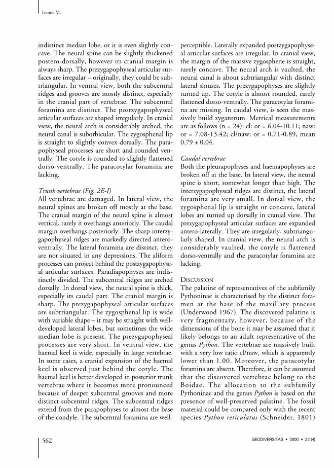
indistinct median lobe, or it is even slightly con-cave. The neural spine can be slightly thickenedpostero-dorsally, however its cranial margin isalways sharp. The prezygapophyseal articular sur-faces are irregular – originally, they could be sub-triangular. In ventral view, both the subcentralridges and grooves are mostly distinct, especiallyin the cranial part of vertebrae. The subcentralforamina are distinct. The postzygapophysealarticular surfaces are shaped irregularly. In cranialview, the neural arch is considerably arched, theneural canal is suborbicular. The zygosphenal lipis straight to slightly convex dorsally. The para-pophyseal processes are short and rounded ven-trally. The cotyle is rounded to slightly flatteneddorso-ventrally. The paracotylar foramina arelacking.
Trunk vertebrae (Fig. 2E-I)All vertebrae are damaged. In lateral view, theneural spines are broken off mostly at the base.The cranial margin of the neural spine is almostvertical, rarely it overhangs anteriorly. The caudalmargin overhangs posteriorly. The sharp interzy-gapophyseal ridges are markedly directed antero-ventrally. The lateral foramina are distinct, theyare not situated in any depressions. The aliformprocesses can project behind the postzygapophyse-al articular surfaces. Paradiapophyses are indis-tinctly divided. The subcentral ridges are archeddorsally. In dorsal view, the neural spine is thick,especially its caudal part. The cranial margin issharp. The prezygapophyseal articular surfacesare subtriangular. The zygosphenal lip is widewith variable shape – it may be straight with well-developed lateral lobes, but sometimes the widemedian lobe is present. The prezygapophysealprocesses are very short. In ventral view, thehaemal keel is wide, especially in large vertebrae.In some cases, a cranial expansion of the haemalkeel is observed just behind the cotyle. Thehaemal keel is better developed in posterior trunkvertebrae where it becomes more pronouncedbecause of deeper subcentral grooves and moredistinct subcentral ridges. The subcentral ridgesextend from the parapophyses to almost the baseof the condyle. The subcentral foramina are well-
perceptible. Laterally expanded postzygapophyse-al articular surfaces are irregular. In cranial view,the margin of the massive zygosphene is straight,rarely concave. The neural arch is vaulted, theneural canal is about subtriangular with distinctlateral sinuses. The prezygapophyses are slightlyturned up. The cotyle is almost rounded, rarelyflattened dorso-ventrally. The paracotylar forami-na are missing. In caudal view, is seen the mas-sively build zygantrum. Metrical measurementsare as follows (n = 24): cl: or = 6.04-10.11; naw:or = 7.08-13.42; cl/naw: or = 0.71-0.89, mean0.79 + 0.04.
Caudal vertebraeBoth the pleurapophyses and haemapophyses arebroken off at the base. In lateral view, the neuralspine is short, somewhat longer than high. Theinterzygapophyseal ridges are distinct, the lateralforamina are very small. In dorsal view, thezygosphenal lip is straight or concave, laterallobes are turned up dorsally in cranial view. Theprezygapophyseal articular surfaces are expandedantero-laterally. They are irregularly, subtriangu-larly shaped. In cranial view, the neural arch isconsiderably vaulted, the cotyle is flatteneddorso-ventrally and the paracotylar foramina arelacking.
DISCUSSION
The palatine of representatives of the subfamilyPythoninae is characterised by the distinct fora-men at the base of the maxillary process(Underwood 1967). The discovered palatine isvery fragmentary, however, because of thedimensions of the bone it may be assumed that itlikely belongs to an adult representative of thegenus Python. The vertebrae are massively builtwith a very low ratio cl/naw, which is apparentlylower than 1.00. Moreover, the paracotylarforamina are absent. Therefore, it can be assumedthat the discovered vertebrae belong to theBoidae. The allocation to the subfamilyPythoninae and the genus Python is based on thepresence of well-preserved palatine. The fossilmaterial could be compared only with the recentspecies Python reticulatus (Schneider, 1801)
Ivanov M.
562 GEODIVERSITAS • 2000 • 22 (4)
(ZZSiD 436 juv., ZZSiD 437 juv.) and Pythonmolurus (Linnaeus, 1758) (ZZSiD 460) – how-ever, because of the scantiness of cranial bones amore precise determination was not possible. Thevertebrae of Python sp. differ from the morpho-types Boidae B & C (see below) in: 1) the muchmore vaulted neural arch; 2) the shape of themassive zygosphenal lip.At present, the available material represents theonly known representative of the subfamilyPythoninae from the European Neogene. Anotherunquestionable Pythoninae comes from theGerman locality Messel (middle Eocene) where agreat number of cranial bones, still articulated(including maxilla and palatine with taxonomi-cally important features), have been discovered(Szyndlar & Böhme 1993). However, the axial
skeleton was not precisely studied (Szyndlar& Böhme 1993). The extinct species Palaeopy-thon sardus Portis, 1901 (= ?Python sardus[Rage, 1984]) belongs most likely also to the sub-family Pythoninae. Both last mentioned cons-trictors were characterised by considerablediameters.
Subfamily ERYCINAE Bonaparte, 1831Genus Eryx Daudin, 1803
Eryx sp.
Bransateryx sp. – Hoffstetter & Rage 1972: 102, pl. II,fig. 11. — Ivanov 1997a: 36-37, fig. 19.
MATERIAL EXAMINED. — 4 trunk vertebrae (FSL368337-FSL 368340), 2 caudal vertebrae (FSL368341, FSL 368342).
Snakes of the lower/middle Miocene of Vieux Collonges (France)
563GEODIVERSITAS • 2000 • 22 (4)
A B C
DE F
G H I
FIG. 2. — Python sp. from the lower/middle (MN 4/5) Miocene of Vieux Collonges; A, left palatine (FSL 368001), labial view; B-D, cer-vical vertebra (FSL 368002); B, dorsal view; C, ventral view; D, lateral view; E-I, trunk vertebra (FSL 368039); E, dorsal view; F, ven-tral view; G, lateral view; H, cranial view; I, caudal view. Scale bars: 2 mm.

DESCRIPTION
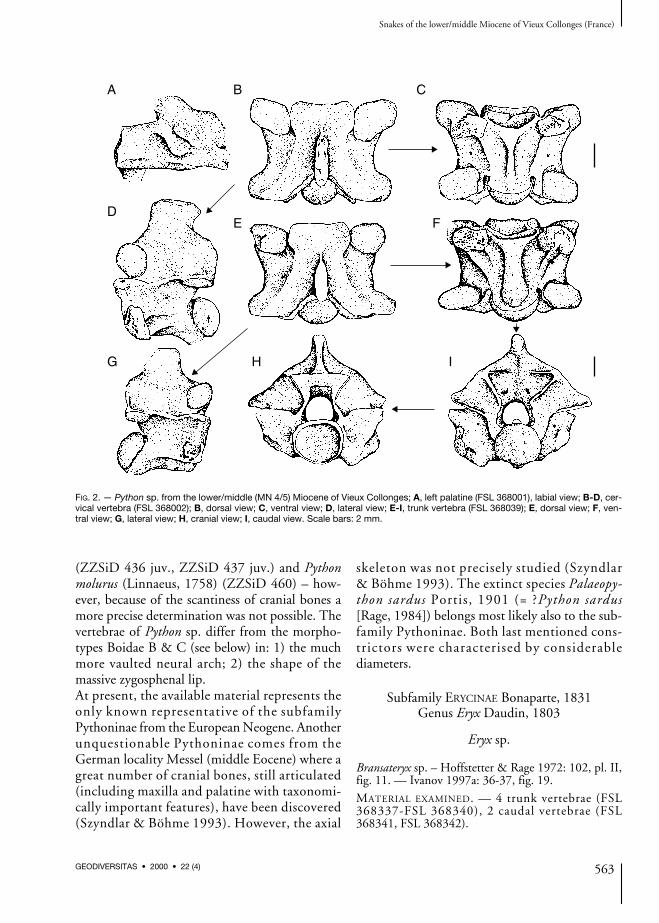
Trunk vertebrae (Fig. 3A-D)In lateral view, the bases of broken off neuralspines are shifted caudally. Short interzygapophyse-al ridges are sharp. The caudal part of the neuralarch is turned up dorsally. The slightly dorsallyarched subcentral ridges extend from the distinct-ly cranially situated synapophyses to the basis ofthe condyle. The paradiapophyses are not dis-tinctly divided, the diapophyseal area is largerthan the parapophyseal one. The lateral foraminaare relatively large and well-visible. In dorsalview, the cranial margin of the zygosphene hasprominent lateral lobes and the median lobe iswide. The neural spine is narrow. The prezy-gapophyseal articular surfaces are subtriangular,well-visible prezygapophyseal processes are veryshort and pointed. In ventral view, the haemalkeel is very wide. It extends from the base of thecotyle to the base of the condyle. In the cranialpart of vertebrae the haemal keel can be strikinglyexpanded. The haemal keel is very distinct inposterior trunk vertebrae. In the case of anteriortrunk vertebrae, the haemal keel is indistinct aswell as the short subcentral ridges and the shallowsubcentral grooves. In all vertebrae, both the sub-central grooves and ridges are more distinct intheir cranial part. The subcentral foramina are
very small and hardly perceptible. In cranial view,the neural arch is conspicuously flattened, theneural canal is approximately circular with dis-tinct lateral sinuses. The cranial margin of thegracile zygosphene is straight. The prezygapophy-ses are distinctly tilted upward. The cotyle is flat-tened dorso-ventrally, the paracotylar foraminaare lacking. Metrical measurements are as follows(n = 4): cl: or = 1.65-3.41; naw: or = 1.88-3.52;cl/naw: or = 0.74-0.97, mean 0.84 + 0.11.
Caudal vertebrae (Fig. 3E-G)Both the zygosphene and the zygantrum areabsent, which proves that these specimens areposterior caudal vertebrae. In lateral view, thevertebrae are high with short vertebral centrum.On the more complete vertebra the right pleura-pophysis is broken off near its base (the left pleu-rapophysis is completely missing like the leftprezygapophysis). The neural spine, broken offat the base, is accompanied laterally with promi-nent pterapophyses which project cranially intoa distinct process. The caudal extension is lessperceptible. The pleurapophysis projects distallyinto a process where the prezygapophysis of thesubsequent vertebra is wedged between thisprocess and the postzygapophysis. In dorsalview, the damaged right prezygapophyseal artic-
Ivanov M.
564 GEODIVERSITAS • 2000 • 22 (4)
A B
CD
E F
G
FIG. 3. — Eryx sp. from the lower/middle (MN 4/5) Miocene of Vieux Collonges; A-D, trunk vertebra (FSL 368339); A, lateral view; B,dorsal view; C, ventral view; D, cranial view; E-G, posterior caudal vertebra (FSL 368341); E, lateral view; F, cranial view; G, dorsalview. Scale bars: 2 mm.

ular surface is irregular, the pterapophyses arerelatively wide. In cranial view, the neural arch isapproximately rounded with lateral sinuses, theprezygapophyses are tilted upward and theprezygapophyseal processes are very short andobtuse. The cotyle is approximately rounded, itsventral margin is thickened. The haemapophysesare directed latero-ventrally, their distal ends arebroken off.
DISCUSSION
The assignment to the subfamily Erycinae and tothe genus Eryx is based on the presence of caudalvertebrae with complicated structure. The dis-covered vertebrae belong most probably to thegenus Eryx – the remaining material of 25 caudaland 70 trunk vertebrae of this genus, discoveredat Vieux Collonges, have recently been investi-gated by Szyndlar & Rage (in prep.). The mor-phology of the caudal vertebrae is verycomplicated in the subfamily Erycinae and theproblems connected with the determination ofsuch vertebrae will be widely discussed (Szyndlar& Rage in prep.).
Subfamily indeterminate
Boidae B
Boidae B – Ivanov 1997a: 41, fig. 21.
MATERIAL EXAMINED. — 2 trunk vertebrae (FSL368343, FSL 368344).
DESCRIPTION
Trunk vertebrae (Fig. 4)In lateral view, the basis of the neural spine isshifted anteriorly. Although the neural spine isincompletely preserved, it may be assumed thatits cranial margin is vertical or slightly inclinedposteriorly like the caudal margin. The neuralarch is turned up caudally, the interzygapophyse-al ridges are distinct and sharp. The lateral fora-mina are large and distinct, situated in shallowdepressions. The subcentral ridges are indistinctand short. The synapophyses are very damaged.In dorsal view, the zygosphenal lip is clearlyconcave. The obovate (?subtriangular) prezy-gapophyseal articular surfaces are damaged, theprezygapophyseal processes are very short,
Snakes of the lower/middle Miocene of Vieux Collonges (France)
565GEODIVERSITAS • 2000 • 22 (4)
A B C
D E
FIG. 4. — Boidae B from the lower/middle (MN 4/5) Miocene of Vieux Collonges, mid-trunk vertebra (FSL 368343); A, lateral view; B, dorsal view; C, ventral view; D, cranial view; E, caudal view. Scale bar: 2 mm.

however, they are well-perceptible from the dor-sal view. Epizygapophyseal spines are lacking. Inventral view, both the subcentral ridges and widesubcentral grooves are relatively indistinct, thesubcentral foramina are large and situated closeto the distinct haemal keel. The haemal keel maybe wide or relatively narrow. The postzygapophyse-al articular surfaces are approximately oval. Incranial view, the neural arch is slightly vaulted,the neural canal is subcircular with distinct lateralsinuses. The massive zygosphenal lip is straight toslightly concave. The prezygapophyses are strong-ly tilted upward. Wide depressions are situatedon either side of the rounded cotyle, the para-cotylar foramina are lacking. Metrical measure-ments: larger vertebra: cl = 3.72; naw = 4.51;cl/naw = 0.82; smaller vertebra: cl = 3.52; naw= 3.99; cl/naw = 0.88.
DISCUSSION
Both the low ratio cl/naw (lower than 1.00) andthe massive structure of vertebrae point to theaffiliation to the family Boidae. The morphotypeBoidae B does most probably not belong to thesubfamily Erycinae because the neural arch is notclearly flattened dorso-ventrally, which is a char-
acteristic of erycine snakes. The vertebrae of themorphotype Boidae B belong most likely to asmall representative of the Boinae or Pythoninae.The Boidae B differs from Python sp. in: 1) theless vaulted neural arch; 2) the prezygapophyseswhich are tilted upward. Based on the limitedmaterial, a more precise determination was notpossible.
Subfamily indeterminate
Boidae C
Boidae C – Ivanov 1997a: 41-42, fig. 22.
MATERIAL EXAMINED. — 4 trunk vertebrae (FSL368345-FSL 368348).
DESCRIPTION
Trunk vertebrae (Fig. 5)In lateral view, the cranial margin of the neuralspine is located at the anterior third of the verte-bral length. The cranial margin of the only pre-served, but very damaged, neural spine isvertical, the caudal margin is broken off. Theinterzygapophyseal ridges are distinct and sharp.
Ivanov M.
566 GEODIVERSITAS • 2000 • 22 (4)
A B C
D E
FIG. 5. — Boidae C from the lower/middle (MN 4/5) Miocene of Vieux Collonges, mid-trunk vertebra (FSL 368346); A, lateral view; B, dorsal view; C, ventral view; D, cranial view; E, caudal view. Scale bar: 2 mm.
The large lateral foramina are not situated indepressions. The subcentral ridges are distinctand vaulted dorsally, extending from stronglydamaged synapophyses to the base of thecondyle. In dorsal view, the zygosphenal lip ischaracterised by distinct lateral lobes, the medianlobe is wide and blunt. The prezygapophysealarticular surfaces are subtriangular, the prezy-gapophyseal processes are very short and invisi-ble from the dorsal view. Epizygapophysealspines are lacking. In ventral view, the subcentralridges and grooves, as well as the deep haemalkeel are distinct; the keel expands near thecotyle. The ventral margin of the haemal keel isrounded in the cranial part of vertebrae but, inthe caudal part, it is relatively sharp. The subcen-tral foramina are distinct and large, they openclose to the haemal keel. The postzygapophysealarticular surfaces are expanded laterally. In cra-nial view, the neural arch is arched, the neuralcanal is subcircular with small lateral sinuses,and the gracile zygosphenal lip is straight. Theprezygapophyses are tilted upward. Deep depres-sions are situated on the both sides of the dorso-ventrally flattened cotyle, paracotylar foraminaare absent. The metrical measurements are asfollows (n = 3): cl: or = 3.49-4.16; naw: or= 3.99-4.93; cl/naw: or = 0.84-0.87, mean 0.86+ 0.02.
DISCUSSION
The vertebrae belong undoubtedly to the familyBoidae based on the massive structure and thelow ratio cl/naw (lower than 1.00). A distinctlyvaulted neural arch excludes the affiliation to thesubfamily Erycinae. The absence of the paracotyl-ar foramina is insufficient for distinction betweensubfamilies Boinae and Pythoninae (Szyndlar1994). The morphotype Boidae C differs fromthe Boidae B on the basis of the following fea-tures: 1) the basis of the neural spine is located onthe anterior third of the vertebral length; 2) thezygosphenal lip is not so massively built; 3) thelateral sinuses of the neural canal are small. TheBoidae C differs from Python sp. mainly in: 1)the less vaulted neural arch; 2) the much moregracile zygosphenal lip.
Superfamily COLUBROIDEA Oppel, 1811Family COLUBRIDAE Oppel, 1811
Subfamily COLUBRINAE Oppel, 1811Genus Texasophis Holman, 1977
Texasophis sp.
Colubrinae C – Ivanov 1997a: 96-97, fig. 43.
MATERIAL EXAMINED. — 7 trunk vertebrae (FSL368349-FSL 368355).
DESCRIPTION
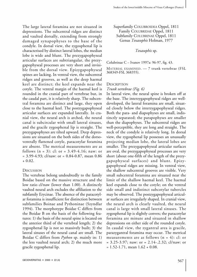
Trunk vertebrae (Fig. 6)In lateral view, the neural spine is broken off atthe base. The interzygapophyseal ridges are well-developed, the lateral foramina are small, situat-ed closely below the interzygapophyseal ridges.Both the para- and diapophyses are small, indis-tinctly separated; the parapophyses are smallerthan the diapophyses. The subcentral ridges arewell-perceptible, they are long and straight. Theneck of the condyle is relatively long. In dorsalview, the zygosphenal lip possesses an unusuallyprojecting median lobe, the lateral lobes aresmaller. The prezygapophyseal articular surfacesare oval, the prezygapophyseal processes are veryshort (about one-fifth of the length of the prezy-gapophyseal surfaces) and blunt. Epizy-gapophyseal ridges are missing. In ventral view,the shallow subcentral grooves are visible. Verysmall subcentral foramina are situated near thelimit of the shallow haemal keel. The haemalkeel expands close to the cotyle; on the ventralside small and indistinct subcotylar tuberclesmay be observed. The postzygapophyseal articul-ar surfaces are irregularly shaped. In cranial view,the neural arch is clearly vaulted, the neuralcanal is large with small lateral sinuses. Thezygosphenal lip is slightly convex; the paracotylarforamina are minute and situated in shallowdepressions on either side of the rounded cotyle.In caudal view, the zygantral area is gracile,parazygantral foramina may occur. The metricalmeasurements are as follows (n = 4): cl: or= 3.25-3.97; naw: or = 2.14-.2.32; cl/naw: or= 1.52-1.71, mean 1.62 + 0.08.
Snakes of the lower/middle Miocene of Vieux Collonges (France)
567GEODIVERSITAS • 2000 • 22 (4)

DISCUSSION
The fossil material is closely related to the extinctgenus Texasophis because of the following fea-tures: 1) the vaulted neural arch; 2) the shape ofthe zygosphenal lip with two distinct lateral lobesand the prominent median lobe; 3) the very shortprezygapophyseal processes; 4) the flat haemalkeel. The only difference from the extinct genusTexasophis is the presence of the subcotylar tuber-cles in some cases. Two species of the genusTexasophis have been reported from the EuropeanMiocene: Texasophis meini Rage & Holman,1984 from the middle Miocene (MN 7+8) of LaGrive M (Rage & Holman 1984) and Texasophisbohemiacus Szyndlar, 1987 from the lowerMiocene (MN 4) of Dolnice (Szyndlar 1987).Texasophis sp. from Vieux Collonges resemblesTexasophis meini in the shape of the zygosphenallip. On the other hand, the relatively narrowhaemal keel and straight (in lateral view) subcen-tral ridges are characteristics of the speciesTexasophis bohemiacus.
Subfamily NATRICINAE Bonaparte, 1838Genus Neonatrix Holman, 1977
Neonatrix cf. europaea Rage & Holman, 1984
Neonatrix cf. europaea – Rage & Holman 1984: 99. —Ivanov 1997a: 99, fig. 45.
MATERIAL EXAMINED. — 7 trunk vertebrae (FSL368823-FSL 368829).
DESCRIPTION
Trunk vertebrae (Fig. 7)In lateral view, the neural spine is about twotimes longer than high; its cranial margin is ver-tical or slightly overhangs anteriorly, the caudalmargin overhangs posteriorly. The interzy-gapophyseal ridges are well-developed, howeverthey are not very sharp. Epizygapophyseal spinesare lacking. The subcentral ridges are approxi-mately straight and reach the vicinity of theproximal margin of the condyle. The lateralforamina are well-perceptible, they are situatedin wide and shallow (rarely deeper) depressions.The para- and diapophyses are clearly separated,the parapophyses are as long as the diapophyses,the antero-ventrally directed parapophysealprocesses are relatively short. Mostly broken offhypapophyses were probably very short. In dor-
Ivanov M.
568 GEODIVERSITAS • 2000 • 22 (4)
A B C
D E
FIG. 6. — Texasophis sp. from the lower/middle (MN 4/5) Miocene of Vieux Collonges, mid-trunk vertebra (FSL 368353); A, lateralview; B, dorsal view; C, ventral view; D, cranial view; E, caudal view. Scale bar: 2 mm.
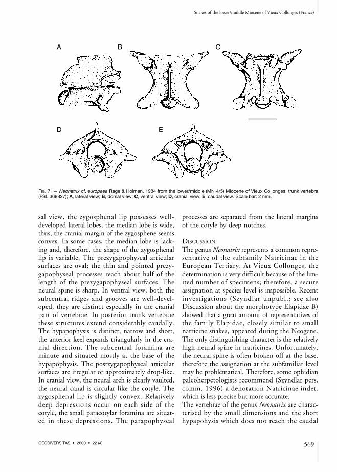
sal view, the zygosphenal lip possesses well-developed lateral lobes, the median lobe is wide,thus, the cranial margin of the zygosphene seemsconvex. In some cases, the median lobe is lack-ing and, therefore, the shape of the zygosphenallip is variable. The prezygapophyseal articularsurfaces are oval; the thin and pointed prezy-gapophyseal processes reach about half of thelength of the prezygapophyseal surfaces. Theneural spine is sharp. In ventral view, both thesubcentral ridges and grooves are well-devel-oped, they are distinct especially in the cranialpart of vertebrae. In posterior trunk vertebraethese structures extend considerably caudally.The hypapophysis is distinct, narrow and short,the anterior keel expands triangularly in the cra-nial direction. The subcentral foramina areminute and situated mostly at the base of thehypapophysis. The postzygapophyseal articularsurfaces are irregular or approximately drop-like.In cranial view, the neural arch is clearly vaulted,the neural canal is circular like the cotyle. Thezygosphenal lip is slightly convex. Relativelydeep depressions occur on each side of thecotyle, the small paracotylar foramina are situat-ed in these depressions. The parapophyseal
processes are separated from the lateral marginsof the cotyle by deep notches.
DISCUSSION
The genus Neonatrix represents a common repre-sentative of the subfamily Natricinae in theEuropean Tertiary. At Vieux Collonges, thedetermination is very difficult because of the lim-ited number of specimens; therefore, a secureassignation at species level is impossible. Recentinvestigations (Szyndlar unpubl.; see alsoDiscussion about the morphotype Elapidae B)showed that a great amount of representatives ofthe family Elapidae, closely similar to smallnatricine snakes, appeared during the Neogene.The only distinguishing character is the relativelyhigh neural spine in natricines. Unfortunately,the neural spine is often broken off at the base,therefore the assignation at the subfamiliar levelmay be problematical. Therefore, some ophidianpaleoherpetologists recommend (Szyndlar pers.comm. 1996) a denotation Natricinae indet.which is less precise but more accurate.The vertebrae of the genus Neonatrix are charac-terised by the small dimensions and the shorthypapohysis which does not reach the caudal
Snakes of the lower/middle Miocene of Vieux Collonges (France)
569GEODIVERSITAS • 2000 • 22 (4)
A B C
D E
FIG. 7. — Neonatrix cf. europaea Rage & Holman, 1984 from the lower/middle (MN 4/5) Miocene of Vieux Collonges, trunk vertebra(FSL 368827); A, lateral view; B, dorsal view; C, ventral view; D, cranial view; E, caudal view. Scale bar: 2 mm.

margin of the condyle. Three species of thisgenus have been discovered in the EuropeanTertiary: Neonatrix europaea Rage & Holman,1984, N. crassa Rage & Holman, 1984 (Rage &Holman 1984) and N. nova Szyndlar, 1987(Szyndlar 1987). Remaining three species,Neonatrix elongata Holman, 1973, N. magnaHolman, 1982 and N. inferna Holman, 1996were discovered in the middle and the upperMiocene of Nebraska, Texas and South Dakota(Holman 1979, 1982, 1996). N. cf. europaea dif-fers from N. nova in its smaller dimensions andthe marked median lobe at the zygosphenal lip.Szyndlar (1987) also considers as distinguishingfeatures the less vaulted neural arch and therounded cotyle in N. nova. However, it is sug-gested that such features may also be seen inN. europaea. N. cf. europaea differs from N. crassain the cranial margin of the zygosphene which isconvex in the first mentioned species. Contraryto the North American species N. elongata andN. magna, N. cf. europaea has a more developedhypapophysis and longer prezygapophysealprocesses (Rage & Holman 1984). N. inferna dif-fers from N. cf. europaea in the elongate vertebraeand the much more lower neural spine.
Genus Natrix Laurenti, 1768
Natrix aff. sansaniensis (Lartet, 1851)
Natrix aff. sansaniensis. — Ivanov 1997a: 102, fig. 47.
MATERIAL EXAMINED. — 2 trunk vertebrae (FSL368830, FSL 368831).
DESCRIPTION
Trunk vertebraeIn lateral view, the neural spine is unusually high,almost as high as long. Its cranial margin overhangsanteriorly and the caudal margin overhangs caudal-ly. The relatively sharp interzygapophyseal ridgesare strikingly developed. The conspicuous lateralforamina are situated in shallow depressions. Thesubcentral ridges are considerably marked and theyare arched dorsally. The para- and diapophyses arewell-separated, the posterolaterally directed
diapophyses are about as large as the parapophyses.The parapophyseal processes are directed anterior-ly. The broken off hypapophyses were distinct anddeep. In dorsal view, the zygosphenal lip has dis-tinct lateral lobes and a prominent median lobe, inanterior (rarely in posterior) trunk vertebrae themedian lobe may be laterally expanded, thus thezygosphenal lip is almost convex. The prezy-gapophyseal articular surfaces are circular to oval,the antero-laterally directed prezygapophysealprocesses are very long – at least as long as theprezygapophyseal surfaces. The dorsal border ofthe neural spine is usually expanded. The epizy-gapophyseal spines are prominent. In ventral view,the subcentral ridges are distinct, however the sub-central grooves are shallow. The subcentral forami-na are minute and hardly visible. The anterior keelof the hypapophysis is triangular; very distinct sub-cotylar tubercles are developed at the ventral mar-gin of the cotyle. The postzygapophyseal articularsurfaces are irregularly shaped. In cranial view, theneural arch is weakly vaulted, the neural canal iscircular and the zygosphenal lip is straight to slight-ly convex. The small paracotylar foramina are situ-ated in prominent depressions on both sides of therounded cotyle.
DISCUSSION
The described shape of the neural spine is charac-teristic for the genus Natrix. N. sansaniensis differsfrom all other Natrix species, living and extinct, inits very high neural spine (Szyndlar & Schleich1993). The following extinct species of the genusNatrix have been reported: Natrix mlynarskiiRage, 1988 (MP 22) (Rage 1988), Natrixsansaniensis (Lartet, 1851) (?MN 4-MN 6) (Rage1981; Szyndlar & Schleich 1993), Natrix longiver-tebrata Szyndlar, 1984 (?MN 7+8-MN16)(Szyndlar 1984, 1991a, b), Natrix parva Szyndlar,1984 (Szyndlar 1984). The described species isclosely related to “Natrix aff. N. sansaniensis(Lartet, 1851)” reported from the German lowerMiocene (MN 4) locality Petersbuch 2 (Szyndlar& Schleich 1993). A denotation “aff.” has beenused because of some morphological resemblancewith the extinct species N. mlynarskii Rage, 1988(Szyndlar & Schleich 1993).
Ivanov M.
570 GEODIVERSITAS • 2000 • 22 (4)
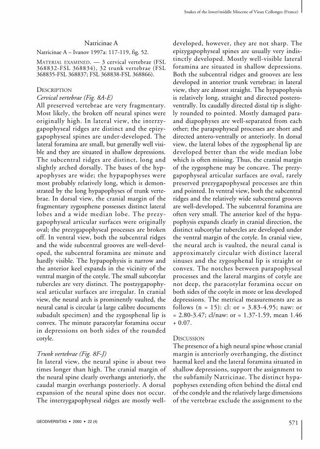
Natricinae ANatricinae A – Ivanov 1997a: 117-119, fig. 52.
MATERIAL EXAMINED. — 3 cervical vertebrae (FSL368832-FSL 368834), 32 trunk vertebrae (FSL368835-FSL 368837; FSL 368838-FSL 368866).
DESCRIPTION
Cervical vertebrae (Fig. 8A-E)All preserved vertebrae are very fragmentary.Most likely, the broken off neural spines wereoriginally high. In lateral view, the interzy-gapophyseal ridges are distinct and the epizy-gapophyseal spines are under-developed. Thelateral foramina are small, but generally well visi-ble and they are situated in shallow depressions.The subcentral ridges are distinct, long andslightly arched dorsally. The bases of the hyp-apophyses are wide; the hypapophyses weremost probably relatively long, which is demon-strated by the long hypapophyses of trunk verte-brae. In dorsal view, the cranial margin of thefragmentary zygosphene possesses distinct laterallobes and a wide median lobe. The prezy-gapophyseal articular surfaces were originallyoval; the prezygapophyseal processes are brokenoff. In ventral view, both the subcentral ridgesand the wide subcentral grooves are well-devel-oped, the subcentral foramina are minute andhardly visible. The hypapophysis is narrow andthe anterior keel expands in the vicinity of theventral margin of the cotyle. The small subcotylartubercles are very distinct. The postzygapophy-seal articular surfaces are irregular. In cranialview, the neural arch is prominently vaulted, theneural canal is circular (a large calibre documentssubadult specimen) and the zygosphenal lip isconvex. The minute paracotylar foramina occurin depressions on both sides of the roundedcotyle.
Trunk vertebrae (Fig. 8F-J)In lateral view, the neural spine is about twotimes longer than high. The cranial margin ofthe neural spine clearly overhangs anteriorly, thecaudal margin overhangs posteriorly. A dorsalexpansion of the neural spine does not occur.The interzygapophyseal ridges are mostly well-
developed, however, they are not sharp. Theepizygapophyseal spines are usually very indis-tinctly developed. Mostly well-visible lateralforamina are situated in shallow depressions.Both the subcentral ridges and grooves are lessdeveloped in anterior trunk vertebrae; in lateralview, they are almost straight. The hypapophysisis relatively long, straight and directed postero-ventrally. Its caudally directed distal tip is slight-ly rounded to pointed. Mostly damaged para-and diapophyses are well-separated from eachother; the parapophyseal processes are short anddirected antero-ventrally or anteriorly. In dorsalview, the lateral lobes of the zygosphenal lip aredeveloped better than the wide median lobewhich is often missing. Thus, the cranial marginof the zygosphene may be concave. The prezy-gapophyseal articular surfaces are oval, rarelypreserved prezygapophyseal processes are thinand pointed. In ventral view, both the subcentralridges and the relatively wide subcentral groovesare well-developed. The subcentral foramina areoften very small. The anterior keel of the hypa-pophysis expands clearly in cranial direction, thedistinct subcotylar tubercles are developed underthe ventral margin of the cotyle. In cranial view,the neural arch is vaulted, the neural canal isapproximately circular with distinct lateralsinuses and the zygosphenal lip is straight orconvex. The notches between parapophysealprocesses and the lateral margins of cotyle arenot deep, the paracotylar foramina occur onboth sides of the cotyle in more or less developeddepressions. The metrical measurements are asfollows (n = 15): cl: or = 3.83-4.95; naw: or= 2.80-3.47; cl/naw: or = 1.37-1.59, mean 1.46+ 0.07.
DISCUSSION
The presence of a high neural spine whose cranialmargin is anteriorly overhanging, the distincthaemal keel and the lateral foramina situated inshallow depressions, support the assignment tothe subfamily Natricinae. The distinct hypa-pophyses extending often behind the distal endof the condyle and the relatively large dimensionsof the vertebrae exclude the assignment to the
Snakes of the lower/middle Miocene of Vieux Collonges (France)
571GEODIVERSITAS • 2000 • 22 (4)
genus Neonatrix. The shape of the relatively highneural spine and the shallow triangular anteriorkeel exclude the referral to the genus Palaeonatrix(Szyndlar in M~lynarski et al. 1982; Szyndlar1987). The morphotype Natricinae A mostprobably belongs to the genus Natrix, which issupported by the shape of the neural spine andthe hypapophysis and by the dimensions of thevertebrae. The morphotype Natricinae A differsfrom the oldest representative – Natrix mly-narskii Rage, 1988 (MP 22) – especially in theshape of the subcentral ridges which are straightin lateral view and slightly medially bent in ven-tral view (Rage 1988). It differs from Natrixsansaniensis (Lartet, 1851) in the lower neuralspine and absence of blunt ridges extending from
the base of the neural spine to each prezygapoph-ysis (Rage 1981). Rage (1984) assumes that theabove mentioned ridges occur also in some livingtaxa. Szyndlar & Schleich (1993) consider theheight of the neural spine as the crucial characterfor the affiliation to the extinct Natrix sansanien-sis (Lartet, 1851). The morphotype Natricinae Adiffers from Natrix longivertebrata Szyndlar,1984 in smaller dimensions, the lower ratiocl/naw, and pointed distal tip of the hypapophysis.
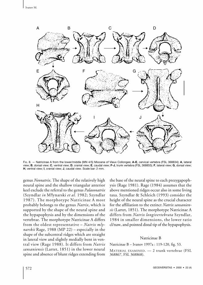
Natricinae B
Natricinae B – Ivanov 1997a : 119-120, fig. 53.
MATERIAL EXAMINED. — 2 trunk vertebrae (FSL368867, FSL 368868).
Ivanov M.
572 GEODIVERSITAS • 2000 • 22 (4)
A B C D
E F G
H
I J
FIG. 8. — Natricinae A from the lower/middle (MN 4/5) Miocene of Vieux Collonges; A-E, cervical vertebra (FSL 368834); A, lateralview; B, dorsal view; C, ventral view; D, cranial view; E, caudal view; F-J, trunk vertebra (FSL 368855); F, lateral view; G, dorsal view;H, ventral view; I, cranial view; J, caudal view. Scale bar: 2 mm.
DESCRIPTION
Trunk vertebrae (Fig. 9)In lateral view, the neural spine is relatively low(the distinct subcentral ridges and grooves showthat this specimen is most probably a posteriortrunk vertebrae), its cranial margin overhangs ante-riorly and the caudal margin overhangs caudally.The blunt interzygapophyseal ridges are well-de-veloped. The subcentral ridges are long, onlyslightly arched dorsally. The hypapophysis is bro-ken off in the vicinity of its base which is unusuallywide in lateral view. The lateral foramina are dis-tinct and large, in one vertebra the foramina occurin wide depressions. The para- and diapophyses areindistinctly separated, the damaged diapophyseswere probably larger than the parapophyses. Thedamaged parapophyses were shifted under the ver-tebral centrum, the parapophyseal processes weremost likely short. The aliform processes extend be-hind the caudal margin of the postzygapophysealsurfaces. In dorsal view, the vertebrae are charac-terised by the long and relatively narrow vertebralconstriction. The zygosphenal lip possesses distinctlateral lobes, the median lobe does not protrudestrikingly and is very wide. The prezygapophysealarticul-ar surfaces are oval to subtriangular, the
prezygapophyseal processes reach about two-thirdsof the length of the prezygapophyseal surfaces, theyare pointed but relatively massively built.Epizygapophyseal spines are lacking. Thediapophyses are directed clearly postero-laterally.In ventral view, the subcentral ridges are sharp, thesubcentral grooves are prominently wide. Thesmall subcentral foramina occur at the base of thehypapophysis. The hypapophysis is expandedproximally into a triangular anterior keel which isinclined antero-ventrally. The anterior keel is sepa-rated from the remaining hypapophysis by aprominent furrow. In cranial view, the neural archis slightly vaulted, the neural canal is approximate-ly circular and the zygosphenal lip is convex. Thedistinct paracotylar foramina are situated in deepdepressions on either side of the rounded cotyle.The deep grooves between damaged parapophysealprocesses and the margin of cotyle are separatedfrom the depressions with paracotylar foramina bypeculiar sharp bony bar. The subcotylar tuberclesare lacking or very slightly developed. Metricalmeasurements: larger vertebra: cl = 4.72; naw= 3.04; cl/naw = 1.55; smaller vertebra: cl = 4.37;naw = 2.92; cl/naw = 1.50.
Snakes of the lower/middle Miocene of Vieux Collonges (France)
573GEODIVERSITAS • 2000 • 22 (4)
A B C
D E
FIG. 9. — Natricinae B from the lower/middle (MN 4/5) Miocene of Vieux Collonges; A-D, posterior trunk vertebra (FSL 368867); A,lateral view; B, dorsal view; C, ventral view; D, cranial view; E, posterior trunk vertebra (FSL 368868), lateral view. Scale bar: 2 mm.

DISCUSSION
The morphotype Natricinae B is defined by theshape of the hypapophysis with prominent anteri-or keel, which is separated form the remaininghypapophysis by a considerable furrow. Thehypapophysis was well-developed and probablyrelatively deep. This feature helps to distinguishthe morphotype Natricinae B from the genusNeonatrix. However, it is impossible to recognizeunambiguously whether Natricinae B belongs toPalaeonatrix or Natrix. The morphotype Natri-cinae B possesses the following features which arecharacteristic of the genus Palaeonatrix: 1) the hy-papophysis markedly extend anteriorly as a keel; 2)the subcentral ridges are prominent (especially inone vertebra). Szyndlar (in M~lynarski et al. 1982;Szyndlar 1987) mentioned that the neural spine ofthe genus Palaeonatrix is low, however, the study atthe locality Petersbuch 2 (Szyndlar & Schleich1993) showed that also high neural spines mayoccur in this genus. The morphotype Natricinae Bis most similar to the species Palaeonatrix silesiacaSzyndlar, 1982 which is only known (Szyndlar inM~lynarski et al. 1982) from the type localityOpole 2, Poland (MN 7). The morphotypeNatricinae B differs from the extinct species ofNatrix in the prominent anterior keel; thus, theNatricinae B differs from Natrix longivertebrataSzyndlar, 1984. From the species Natrix mlynarskiiRage, 1988 the morphotype Natricinae B differsalso in the considerably narrow constriction be-tween the prezygapophyses and the postzy-gapophyses. The Natricinae B differs from Natrixsansaniensis (Lartet, 1851) in the much more lowerneural spine – it is about two to three times longerthan high. The morphotype Natricinae B differsfrom the Natricinae A in indistinct subcotylar tu-bercles. The anterior keel of the hypapophysis isseparated from the remaining hypapophysis by aconsiderable furrow – this feature makes the Natri-cinae B distinguishable from all remaining mor-photypes (Natricinae A, C & D).
Natricinae C
Natricinae C – Ivanov 1997a: 120, fig. 54.
MATERIAL EXAMINED. — 24 trunk vertebrae (FSL368869-FSL 368891).
DESCRIPTION
Trunk vertebraeIn lateral view, the neural spine is about two timeslonger than high, its cranial margin overhangsslightly anteriorly, the posterior overhanging isconsiderable. The largest vertebrae have the top ofneural spine very slightly expanded laterally. Theinterzygapophyseal ridges are distinct but blunt.Well-perceptible lateral foramina are situatedwithin wide and deep depressions. The subcentralridges are straight to arched slightly dorsally. Theventral margin of the peculiar hypapophysis is notstraight (Ivanov 1997a: fig. 54A), the distal end ofthe hypapophysis is rounded and directed caudal-ly. The para- and diapophyses are well-separatedfrom each other, the parapophyses are nearly aslarge as the diapophyses. The parapophysealprocesses are short and directed anteriorly ratherthan antero-ventrally. In dorsal view, the cranialmargin of the zygosphene is mostly concave tostraight, rarely a wide median lobe may occur.The prezygapophyseal articular surfaces are obo-vate, the prezygapophyseal processes are not pre-served. The epizygapophyseal spines are slightlydeveloped. In ventral view, both the subcentralridges and grooves are well-developed only in theposterior trunk vertebrae, in the middle trunkvertebrae these structures are not so distinct. Thesubcentral foramina, situated at the base of thegracile hypapophysis, are minute and hardly per-ceptible. Subcotylar tubercles often occur on theventral margin of the cotyle. The postzygapophy-seal articular surfaces are irregular. In cranial view,the neural arch is more or less vaulted and theneural canal is rounded with small lateral sinuses.The zygosphenal lip is convex. The parapophysealprocesses are separated from the cotyle by shallowfurrows. The paracotylar foramina are situated indeep or shallow depressions. The metrical meas-urements are as follows (n = 8): cl: or = 3.54-4.90;naw: or = 2.25-3.40; cl/naw: or = 1.38-1.63,mean 1.50 + 0.10.
DISCUSSION
Although low (rarely high) neural spine of mostvertebrae is broken off at the base, it may beobserved that its cranial margin overhangs ante-
Ivanov M.
574 GEODIVERSITAS • 2000 • 22 (4)

riorly, the caudal margin overhangs posteriorly.This is the most important criterion for the affil-iation to the subfamily Natricinae. Based on theheight of the neural spine, it is assumed that theNatricinae C may belong to the genus Natrix.However, a peculiar shape of the hypapophysisdistinguishes the morphotype Natricinae C fromall known representatives of the genus Natrix.Regarding the extinct genus Neonatrix, the mor-photype Natricinae C somewhat resembles thenew species of this genus from the middleMiocene (MN 6) of the French locality Sansan(Rage pers. comm. 1996; Augé & Rage 2000).However, the shape of the hypapophysis is sim-ilar to only one vertebra (Augé & Rage 2000:fig. 25A), while the other vertebrae differ con-siderably in the shape of the hypapophysis.Moreover, the vertebrae of the new species ofNeonatrix (Augé & Rage 2000) are smaller thanthose of Natricinae C. The Natricinae C differsfrom the Natricinae A in the following features:1) the hypapophysis is not straight; 2) the lateralsinuses of the neural canal are small andindistinct. Due to the quite different shape ofthe hypapophysis, without furrow separating
the anterior keel from the remaining hypapo-physis, the Natricinae C differs from the Natri-cinae B.
Natricinae D
Natricinae D – Ivanov 1997a: 122, fig. 55.
MATERIAL EXAMINED. — 4 trunk vertebrae (FSL368892-FSL 368895).
DESCRIPTION
Trunk vertebrae (Fig. 10)In lateral view, the neural spine is about four timeslonger than high; its cranial margin overhangs an-teriorly, the caudal margin is broken off in all ver-tebrae. The interzygapophyseal ridges are slightlydeveloped; the small lateral foramina do not openin depressions. The subcentral ridges are straightto weakly arched dorsally. The relatively straightand short hypapophysis is directed caudally; itsdistal termination is pointed or slightly rounded.The synapophyses are very damaged, the para-and diapophyses were most likely well-separatedfrom each other. The anteriorly directed para-pophyseal processes are short. The condyle is situ-
Snakes of the lower/middle Miocene of Vieux Collonges (France)
575GEODIVERSITAS • 2000 • 22 (4)
A B C
D
FIG. 10. — Natricinae D from the lower/middle (MN 4/5) Miocene of Vieux Collonges, trunk vertebra (FSL 368894); A, lateral view;B, dorsal view; C, ventral view; D, cranial view. Scale bar: 2 mm.

ated on a short neck. In dorsal view, the zy-gosphenal lip generally possesses distinct laterallobes and the wide median lobe is slightly devel-oped. The prezygapophyseal articular surfaces areobovate to subtriangular, the prezygapophysealprocesses are relatively short, heavily built andpointed. The epizygapophyseal spines are under-developed. In ventral view, the hypapophysis issharp, its anterior keel expands anteriorly. Theblunt subcentral ridges are well developed; thesubcentral grooves are wide and more distinct inthe cranial part of vertebrae. The subcotylar tuber-cles are absent or very slightly developed. The sub-central foramina are very small. In cranial view,the neural arch is slightly vaulted, the neural canalis subcircular and the zygosphenal lip is convex.The parapophyseal processes are separated fromthe cotyle by relatively deep furrows. The conspic-uous paracotylar foramina are situated on bothsides of the rounded to dorso-ventrally slightlyflattened cotyle. The measurements of vertebraeare as follows (n = 4): cl: or = 3.33-4.37; naw: or= 2.32-2.80; cl/naw: or = 1.42-1.56, mean 1.48+ 0.06.
DISCUSSION
The vertebrae of the morphotype Natricinae Dare characterised by both the relatively high neuralspine and straight as well as short hypapophysis.The Natricinae D resembles especially somerepresentatives of the genus Neonatrix. TheNatricinae D differs from the morphotypesNatricinae A, B & C in the straight and shorthypapophysis. A more precise determination wasnot possible because of damaged neural spines,synapophyses and broken off prezygapophysealprocesses.
Family ELAPIDAE Boié, 1827Genus Micrurus Wagler, 1824
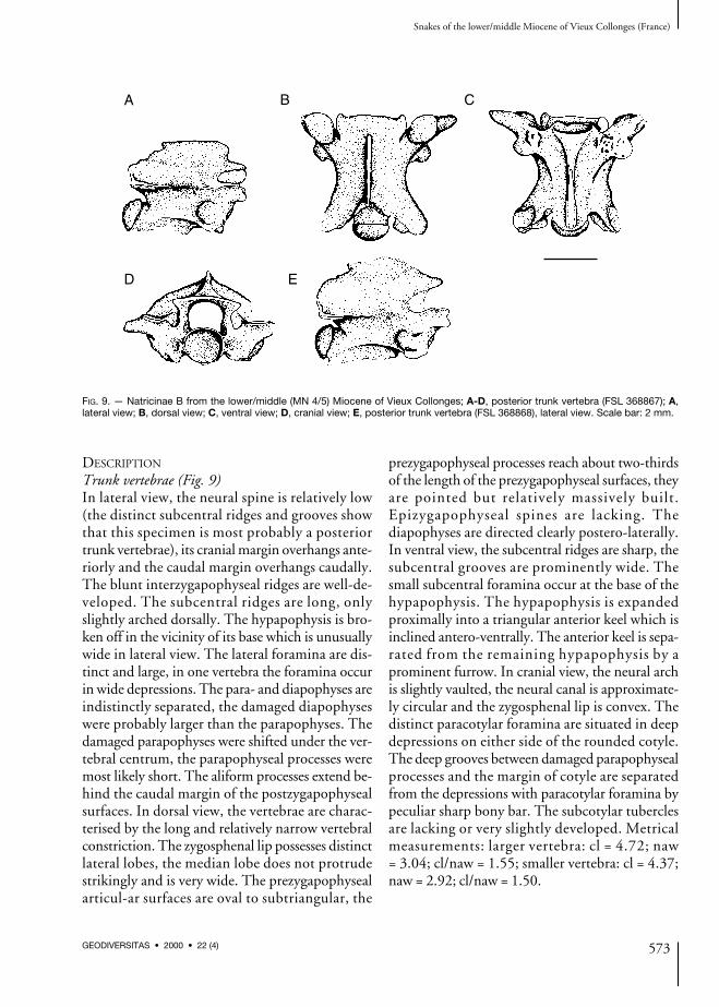
Micrurus aff. gallicus Rage & Holman, 1984
Micrurus cf. gallicus – Rage & Holman 1984: 99.Micrurus aff. gallicus – Ivanov 1997a: 129, fig. 59.
MATERIAL EXAMINED. — 1 trunk vertebra (FSL369450).
DESCRIPTION
Trunk vertebra (Fig. 11)In lateral view, the neural spine is low, both thecranial and caudal margins overhang caudally.The interzygapophyseal ridges are slightly devel-oped. The lateral foramina are small and not sit-uated in depressions. The subcentral ridges areprominent, extending from the synapophyses tothe proximal margin of the condyle. Both thepara- and diapophyses are broken off at thebase. The hypapophysis is damaged, without itsdistal tip. In dorsal view, the zygosphene hasclearly developed lateral lobes. The median lobeis very wide, thus, the zygosphenal lip is moreconvex than crenate. The prezygapophysealarticular surfaces are oval, the prezygapophysealprocesses are broken off at the base.Epizygapophyseal spines are missing. In ventralview, the hypapophysis extends cranially to thebasis of the cotyle where it forms a small andwide tubercle. Both the subcentral ridges andwide subcentral grooves are distinct. The sub-central foramina are very small, located at thebase of the hypapophysis. The postzygapophyse-al articular surfaces are roughly rounded toirregularly shaped. In cranial view, the neuralarch is moderately and regularly vaulted; theneural canal is circular with wide lateral sinuses.The zygosphenal lip is convex. On both sides ofthe rounded cotyle are situated paracotylarforamina in wide depressions. In caudal view,the postzygapophyseal articular surfaces are tilt-ed upward, the zygantral area is relatively wide.The relatively small condyle is rounded. Smallparazygantral foramina are situated above thepostzygapophyseal surfaces. The measurementsof vertebra are as follows: cl = 3.02; naw = 2.05;width between prezygapophyses (pr-pr) = 3.57;width between postzygapophyses (po-po)= 3.40; distance between pre- a postzygapophy-ses (pr-po) = 3.47; zygosphenal width (zw)= 1.73; height of cotyle (cth) = 1.09; width ofcotyle (ctw) = 1.23.
DISCUSSION
The vertebra was originally referred to asMicrurus cf. gallicus by Rage & Holman (1984),
Ivanov M.
576 GEODIVERSITAS • 2000 • 22 (4)
however, without precise description and depic-tion. The vertebra belongs most probably to asmall representative of the family Elapidae on thebasis of the low neural spine overhanging posteri-orly and the presence of a hypapophysis. Mostlikely, it represents an adult individual havingsmall dimensions – small diameter of the neuralcanal, which is as wide as the cotyle (without lat-eral sinuses). Therefore, the vertebra is assignedto the genus Micrurus. The vertebra is very simi-lar to the extinct species Micrurus gallicus report-ed originally from the French locality La Grive M(MN 7) (Rage & Holman 1984). M. aff. gallicusfrom Vieux Collonges differs from M. gallicus inthe following features: 1) the subcentral ridges arebetter developed in M. aff. gallicus but this char-acter may be explained by the fact that this verte-bra is a posterior trunk one; 2) in cranial view,the zygosphenal lip of M. aff. gallicus is convexwhile in known representatives of M. gallicus it isstraight; 3) M. aff. gallicus has a small distincttubercle developed on the ventral margin of thecotyle. In case of M. gallicus this tubercle isabsent. A denotation “aff.” reflected small mor-
phological differences between M. aff. gallicusand M. gallicus.
Genus Naja Laurenti, 1768
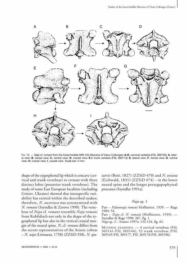
Naja cf. romani (Hoffstetter, 1939)
Part – Palaeonaja romani. — Rage 1984: 54.Part – Naja cf. N. romani (Hoffstetter, 1939). —Szyndlar & Rage 1990: 387, fig. 1.Naja sp. 1 – Ivanov 1997a: 130-131, fig. 60.
MATERIAL EXAMINED. — 2 cervical vertebrae (FSL369104, FSL 369105), 35 trunk vertebrae (FSL369106-FSL 369115, FSL 369116-FSL 369140).
DESCRIPTION
Cervical vertebrae (Fig. 12A-D)The vertebrae have short and wide vertebral cen-tra. In lateral view, the neural spine is lower thanlong, its cranial margin is almost vertical or over-hangs slightly posteriorly, the caudal marginoverhangs posteriorly. The interzygapophysealridges are prominent, the distinct lateral foraminaare situated in deep depressions below the
Snakes of the lower/middle Miocene of Vieux Collonges (France)
577GEODIVERSITAS • 2000 • 22 (4)
A B C
D E
FIG. 11. — Micrurus aff. gallicus Rage & Holman, 1984 from the lower/middle (MN 4/5) Miocene of Vieux Collonges, trunk vertebra(FSL 369450); A, lateral view; B, dorsal view; C, ventral view; D, cranial view; E, caudal view. Scale bar: 2 mm.

interzygapophyseal ridges. The subcentral ridgesare considerably developed, extending from thesynapophyses to the proximal margin of thecondyle. The heavily built hypapophysis is longand directed postero-ventrally. Unfortunately, itsdistal tip is broken off. The para- and diapophysesare well-separated from each other, the postero-laterally directed diapophyses are approximatelyas large as the parapophyses. The parapophysealprocesses are short, directed anteriorly rather thanantero-ventrally. In dorsal view, the zygosphenallip is concave or straight (however, it may be pos-sible that the cranial margin of the zygosphenewas most likely damaged). The damaged prezy-gapophyseal articular surfaces were probably sub-orbicular, the prezygapophyseal processes arebroken off at the base. Epizygapophyseal spinesare indistinct. In ventral view, subcotylar tuber-cles occur on the ventral margin of the cotyle.Both the subcentral ridges and wide subcentralgrooves are well developed, the subcentral forami-na are small. The postzygapophyseal articular sur-faces are roughly rounded. In cranial view, theneural arch is vaulted and the subcircular neuralcanal has distinct lateral sinuses. The zygosphenallip is slightly convex. The paracotylar foraminaare situated in deep and wide depressions on bothsides of the rounded cotyle.
Trunk vertebrae (Fig. 12E-I)The vertebrae are characterised by the relativelyshort and wide centra, which is a typical feature ofthe large colubrids. In lateral view, the neuralspine is somewhat lower than long. The cranialmargin of the neural spine is almost vertical, thecaudal margin overhangs posteriorly. The interzy-gapophyseal ridges are prominent. The lateralforamina are distinct and situated in deep widedepressions. The subcentral ridges are straight orweakly arched dorsally. Epizygapophyseal spinesare under-developed. The para- and diapophysesare not distinct from each other, the parapophysesare as large as the diapophyses or somewhat larg-er. The parapophyseal processes are relatively longand directed anteriorly rather than antero-ventrally.The hypapophyses are broken off at the base. Indorsal view, the zygosphenal lip is concave to
straight (in many vertebrae a mechanical damageis possible), the prezygapophyseal articular sur-faces are wide and oval to suborbicular, the frag-mentary prezygapophyseal processes are heavilybuilt and relatively long. Dorsal expansion of theneural spine has not been noticed. In ventralview, the prominent subcentral ridges and thedeep subcentral grooves are visible. Both struc-tures are best developed in posterior trunk verte-brae. The subcentral foramina are small anddifficult to recognise. The parapophyseal process-es are obtuse. The hypapophysis is gracile, theanterior keel expands near the ventral margin ofthe cotyle. The subcotylar tubercles are oftendeveloped. The postzygapophyseal articular sur-faces are irregularly shaped. In cranial view, theneural arch is moderately arched, the neural canalis circular with short lateral sinuses, thezygosphenal lip is straight to concave. The shal-low furrows separating the parapophyseal process-es from the rounded cotyle are narrowed by thesubcotylar tubercles. The paracotylar foraminaare situated at the inner margins of deep depres-sions on both sides of the cotyle. The metricalmeasurements are as follows (n = 11): cl: or= 6.21-8.75; naw: or = 5.09-8.25; cl/naw: or= 1.01-1.28, mean 1.13 + 0.09.
DISCUSSION
The discovered vertebrae belong to a typical repre-sentative of the family Elapidae. The vertebrae arerelatively large with a low ratio cl/naw and have aflattened and wide vertebral centra. Therefore, thevertebrae are assigned to the genus Naja. Threespecies of large cobras have been discovered in theEuropean Cenozoic: Naja romani (Hoffstetter,1939), Naja iberica Szyndlar, 1985 and “Naja (?)depereti (Hoffstetter, 1939)”. The last knownspecies – Naja antiqua Rage, 1976 has been re-ported from the middle Miocene (MN 7) of BeniMellal in Morocco (Rage 1976). The vertebrae ofNaja cf. romani are most similar to the speciesN. romani and especially to the material from theAustrian locality Kohfidisch (MN 11) originally(Bachmayer & Szyndlar 1985) assigned to the ex-tinct species Naja austriaca. The species Naja aus-triaca was established especially because of the
Ivanov M.
578 GEODIVERSITAS • 2000 • 22 (4)
shape of the zygosphenal lip which is concave (cer-vical and trunk vertebrae) to crenate with threedistinct lobes (posterior trunk vertebrae). Thestudy of some East European localities (includingGritsev, Ukraine) showed that intraspecific vari-ability has existed within the described snakes;therefore, N. austriaca was synonymised withN. romani (Szyndlar & Zerova 1990). The verte-brae of Naja cf. romani resemble Naja romanifrom Kohfidisch not only in the shape of the zy-gosphenal lip but also in the vertical cranial mar-gin of the neural spine. N. cf. romani differs fromthe recent representatives of the Asiatic cobras– N. naja (Linnaeus, 1758) (ZZSiD 358), N. spu-
tatrix (Boié, 1827) (ZZSiD 470) and N. oxiana(Eichwald, 1831) (ZZSiD 474) – in the lowerneural spine and the longer prezygapophysealprocesses (Szyndlar 1991a).
Naja sp. 1
Part – Palaeonaja romani Hoffstetter, 1939. — Rage1984: 54.Part – Naja cf. N. romani (Hoffstetter, 1939). —Szyndlar & Rage 1990: 387, fig. 1.Naja sp. 2 – Ivanov 1997a: 132-134, fig. 61.
MATERIAL EXAMINED. — 4 cervical vertebrae (FSL369141-FSL 369144), 52 trunk vertebrae (FSL369145-FSL 369177, FSL 369178-FSL 369196).
Snakes of the lower/middle Miocene of Vieux Collonges (France)
579GEODIVERSITAS • 2000 • 22 (4)
A B C D
E F G
H I
FIG. 12. — Naja cf. romani from the lower/middle (MN 4/5) Miocene of Vieux Collonges; A-D, cervical vertebra (FSL 369105); A, later-al view; B, dorsal view; C, ventral view; D, cranial view; E-I, trunk vertebra (FSL 369113); E, lateral view; F, dorsal view; G, ventralview; H, cranial view; I, caudal view. Scale bar: 2 mm.
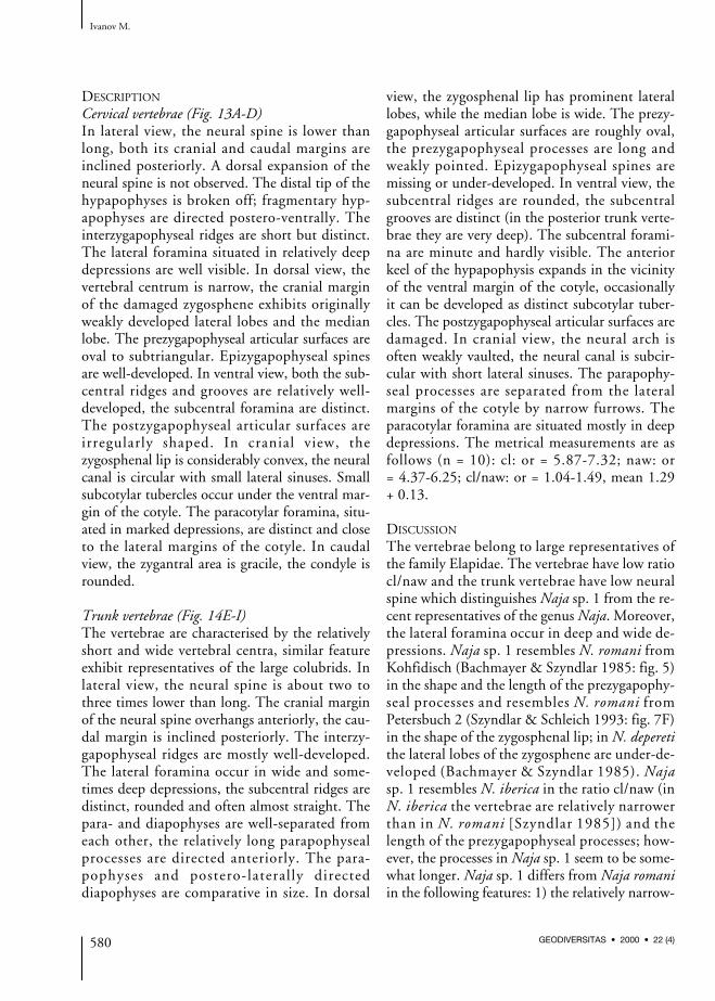
DESCRIPTION
Cervical vertebrae (Fig. 13A-D)In lateral view, the neural spine is lower thanlong, both its cranial and caudal margins areinclined posteriorly. A dorsal expansion of theneural spine is not observed. The distal tip of thehypapophyses is broken off; fragmentary hyp-apophyses are directed postero-ventrally. Theinterzygapophyseal ridges are short but distinct.The lateral foramina situated in relatively deepdepressions are well visible. In dorsal view, thevertebral centrum is narrow, the cranial marginof the damaged zygosphene exhibits originallyweakly developed lateral lobes and the medianlobe. The prezygapophyseal articular surfaces areoval to subtriangular. Epizygapophyseal spinesare well-developed. In ventral view, both the sub-central ridges and grooves are relatively well-developed, the subcentral foramina are distinct.The postzygapophyseal articular surfaces areirregularly shaped. In cranial view, thezygosphenal lip is considerably convex, the neuralcanal is circular with small lateral sinuses. Smallsubcotylar tubercles occur under the ventral mar-gin of the cotyle. The paracotylar foramina, situ-ated in marked depressions, are distinct and closeto the lateral margins of the cotyle. In caudalview, the zygantral area is gracile, the condyle isrounded.
Trunk vertebrae (Fig. 14E-I)The vertebrae are characterised by the relativelyshort and wide vertebral centra, similar featureexhibit representatives of the large colubrids. Inlateral view, the neural spine is about two tothree times lower than long. The cranial marginof the neural spine overhangs anteriorly, the cau-dal margin is inclined posteriorly. The interzy-gapophyseal ridges are mostly well-developed.The lateral foramina occur in wide and some-times deep depressions, the subcentral ridges aredistinct, rounded and often almost straight. Thepara- and diapophyses are well-separated fromeach other, the relatively long parapophysealprocesses are directed anteriorly. The para-pophyses and postero-laterally directeddiapophyses are comparative in size. In dorsal
view, the zygosphenal lip has prominent laterallobes, while the median lobe is wide. The prezy-gapophyseal articular surfaces are roughly oval,the prezygapophyseal processes are long andweakly pointed. Epizygapophyseal spines aremissing or under-developed. In ventral view, thesubcentral ridges are rounded, the subcentralgrooves are distinct (in the posterior trunk verte-brae they are very deep). The subcentral forami-na are minute and hardly visible. The anteriorkeel of the hypapophysis expands in the vicinityof the ventral margin of the cotyle, occasionallyit can be developed as distinct subcotylar tuber-cles. The postzygapophyseal articular surfaces aredamaged. In cranial view, the neural arch isoften weakly vaulted, the neural canal is subcir-cular with short lateral sinuses. The parapophy-seal processes are separated from the lateralmargins of the cotyle by narrow furrows. Theparacotylar foramina are situated mostly in deepdepressions. The metrical measurements are asfollows (n = 10): cl: or = 5.87-7.32; naw: or= 4.37-6.25; cl/naw: or = 1.04-1.49, mean 1.29+ 0.13.
DISCUSSION
The vertebrae belong to large representatives ofthe family Elapidae. The vertebrae have low ratiocl/naw and the trunk vertebrae have low neuralspine which distinguishes Naja sp. 1 from the re-cent representatives of the genus Naja. Moreover,the lateral foramina occur in deep and wide de-pressions. Naja sp. 1 resembles N. romani fromKohfidisch (Bachmayer & Szyndlar 1985: fig. 5)in the shape and the length of the prezygapophy-seal processes and resembles N. romani fromPetersbuch 2 (Szyndlar & Schleich 1993: fig. 7F)in the shape of the zygosphenal lip; in N. deperetithe lateral lobes of the zygosphene are under-de-veloped (Bachmayer & Szyndlar 1985). Najasp. 1 resembles N. iberica in the ratio cl/naw (inN. iberica the vertebrae are relatively narrowerthan in N. romani [Szyndlar 1985]) and thelength of the prezygapophyseal processes; how-ever, the processes in Naja sp. 1 seem to be some-what longer. Naja sp. 1 differs from Naja romaniin the following features: 1) the relatively narrow-
Ivanov M.
580 GEODIVERSITAS • 2000 • 22 (4)
er vertebrae in Naja sp. 1; 2) the anterior marginof the neural spine is clearly inclined anteriorly inNaja sp. 1 while in Naja romani it is almost verti-cal. Concerning the recent representatives of thegenus Naja, the Asiatic members of this genus arecharacterised by the relatively narrower vertebralcentres, especially N. sputatrix (ZZSiD 470), incontrast to the African members – N. haje
(Linnaeus, 1758) (ZZSiD 491) and N. nigricollisReinhardt, 1843 (ZZSiD 492).
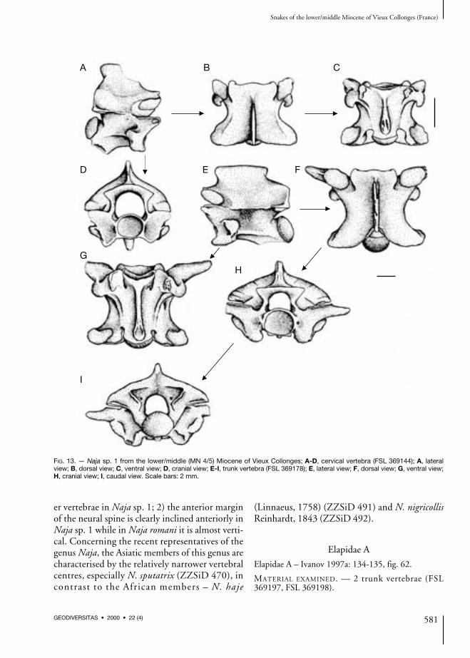
Elapidae A
Elapidae A – Ivanov 1997a: 134-135, fig. 62.
MATERIAL EXAMINED. — 2 trunk vertebrae (FSL369197, FSL 369198).
Snakes of the lower/middle Miocene of Vieux Collonges (France)
581GEODIVERSITAS • 2000 • 22 (4)
A B C
D
G
I
E
H
F
FIG. 13. — Naja sp. 1 from the lower/middle (MN 4/5) Miocene of Vieux Collonges; A-D, cervical vertebra (FSL 369144); A, lateralview; B, dorsal view; C, ventral view; D, cranial view; E-I, trunk vertebra (FSL 369178); E, lateral view; F, dorsal view; G, ventral view;H, cranial view; I, caudal view. Scale bars: 2 mm.
� �
�
�
�
�
�
DESCRIPTION
Trunk vertebraeIn lateral view, the neural spine is very low withvertical margins. The interzygapophyseal ridgesare prominently developed, the lateral foraminaare situated in deep depressions. The para- andthe diapophyses are clearly separated, thediapophysis is strikingly smaller than the para-pophysis. The parapophyseal processes are short.The subcentral ridges are strongly developed andarched dorsally. The hypapophysis is very short,its distal tip is situated closely near the proximalmargin of the condyle; in the posteriormost ver-tebrae the hypapophysis can be bifurcated(FSL 369198). The condyle is borne by a shortneck. In dorsal view, the cranial margin of thezygosphene has distinct lateral lobes, the stronglydamaged median lobe was originally distinct andwide. The prezygapophyseal articular surfaces areobovate, the short prezygapophyseal processes arepointed. Epizygapophyseal spines are missing. Inventral view, both the subcentral ridges andgrooves are noticeable developed. The subcentralforamina are shifted anteriorly at the basis ofwide hypapophysis. The minute subcotylartubercles are developed in one vertebra(FSL 369197). The postzygapophyseal articularsurfaces are obovate. In cranial view, the neuralarch is vaulted, the neural canal is circular withlateral sinuses at the ventral margin; the zygo-sphenal lip is convex. Distinct nerve foraminamay be seen at the bases of the prezygapophysealprocesses. The large paracotylar foramina are sit-uated in depressions on either side of the round-ed cotyle.
DISCUSSION
The vertebrae of the morphotype Elapidae A arecharacterised by the following features: 1) thevery small dimensions; 2) the very low neuralspine; 3) the vaulted neural arch; 4) short para-pophyseal processes directed anteriorly. The ver-tebrae belong most likely to small representativesof cobras. The morphotype Elapidae A resemblesextinct species Micrurus gallicus Rage & Holman,1984 because of the elongated centra of verte-brae, the shape of the zygosphene and the rela-
tively shallow caudal notch. The hypapophysis isdirected caudally, however, in M. gallicus thehypapophysis is shorter. The most visible differ-ence is the very low and indistinct neural spine inthe morphotype Elapidae A; in the case of M. gal-licus the low neural spine is distinctly developed(Szyndlar & Schleich 1993). Moreover, Elapi-dae A is characterised by shorter prezygapophy-seal processes.
Elapidae B
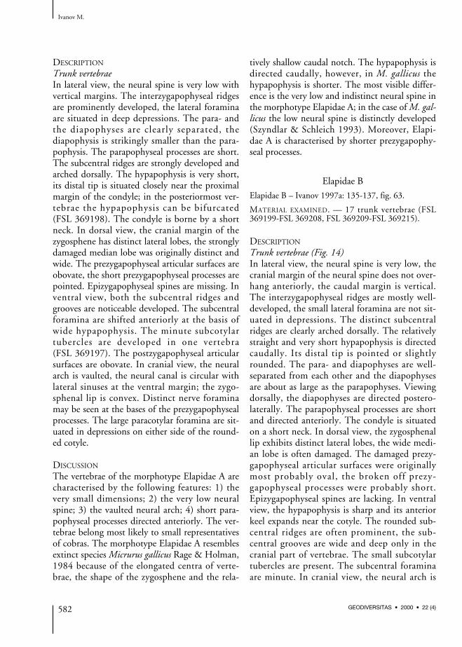
Elapidae B – Ivanov 1997a: 135-137, fig. 63.
MATERIAL EXAMINED. — 17 trunk vertebrae (FSL369199-FSL 369208, FSL 369209-FSL 369215).
DESCRIPTION
Trunk vertebrae (Fig. 14)In lateral view, the neural spine is very low, thecranial margin of the neural spine does not over-hang anteriorly, the caudal margin is vertical.The interzygapophyseal ridges are mostly well-developed, the small lateral foramina are not sit-uated in depressions. The distinct subcentralridges are clearly arched dorsally. The relativelystraight and very short hypapophysis is directedcaudally. Its distal tip is pointed or slightlyrounded. The para- and diapophyses are well-separated from each other and the diapophysesare about as large as the parapophyses. Viewingdorsally, the diapophyses are directed postero-laterally. The parapophyseal processes are shortand directed anteriorly. The condyle is situatedon a short neck. In dorsal view, the zygosphenallip exhibits distinct lateral lobes, the wide medi-an lobe is often damaged. The damaged prezy-gapophyseal articular surfaces were originallymost probably oval, the broken off prezy-gapophyseal processes were probably short.Epizygapophyseal spines are lacking. In ventralview, the hypapophysis is sharp and its anteriorkeel expands near the cotyle. The rounded sub-central ridges are often prominent, the sub-central grooves are wide and deep only in thecranial part of vertebrae. The small subcotylartubercles are present. The subcentral foraminaare minute. In cranial view, the neural arch is
Ivanov M.
582 GEODIVERSITAS • 2000 • 22 (4)

more or less vaulted with the circular neuralcanal and the convex zygosphenal lip. The para-pophyseal processes are separated from thecotyle by relatively shallow furrows. The promi-nent paracotylar foramina occur within deepdepressions at both sides of the cotyle which isrounded to weakly flattened dorso-ventrally.The measurements of the largest vertebrae are asfollows (n = 8): cl: or = 4.35-6.71; naw: or= 2.98-4.53; cl/naw: or = 1.42-1.50, mean 1.47+ 0.03.
DISCUSSION
The vertebrae have considerably elongated verte-bral centra, thus at the first sight, they may sug-gest representatives of the subfamily Natricinae,especially Natrix or Neonatrix, rather thanelapids. However, these two genera are charac-terised by the markedly higher neural spine andsmaller dimensions (Neonatrix). The vertebraeof the morphotype Elapidae B are relativelylarge. They are assigned to the family Elapidaeon the basis of the occurrence of hypapophysesin trunk vertebrae and as well as on the basis ofthe low neural spine. Based on the shape of the
neural spine, the vertebrae belong most probablyto the genus Naja. Because of the high ratiocl/naw, it is assumed that the vertebrae probablybelong to the representatives of the Asiaticrather than African cobras. The morphotypeElapidae B differs from the Elapidae A in: 1) thelarger dimensions of vertebrae; 2) the higherneural spine.
Family VIPERIDAE Oppel, 1811Subfamily VIPERINAE Oppel, 1811
“European vipers” group“aspis complex”
Viperinae A
Viperinae A – Ivanov 1997a: 147, fig. 68.
MATERIAL EXAMINED. — 12 trunk vertebrae (FSL369451-FSL 369462).
DESCRIPTION
Trunk vertebrae (Fig. 15)All vertebrae are fragmentary. In lateral view, theneural spine is relatively low (about three timeslonger than high). Both its cranial and caudal
Snakes of the lower/middle Miocene of Vieux Collonges (France)
583GEODIVERSITAS • 2000 • 22 (4)
A
D
B
E
C
FIG. 14. — Elapidae B from the lower/middle (MN 4/5) Miocene of Vieux Collonges, trunk vertebra (FSL 369214); A, lateral view; B, dorsal view; C, ventral view; D, cranial view; E, caudal view. Scale bar: 2 mm.
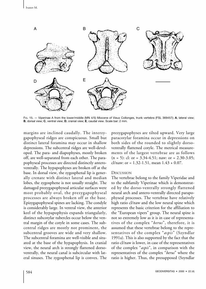
margins are inclined caudally. The interzy-gapophyseal ridges are conspicuous. Small butdistinct lateral foramina may occur in shallowdepressions. The subcentral ridges are well-devel-oped. The para- and diapophyses, mostly brokenoff, are well-separated from each other. The para-pophyseal processes are directed distinctly antero-ventrally. The hypapophyses are broken off at thebase. In dorsal view, the zygosphenal lip is gener-ally crenate with distinct lateral and medianlobes, the zygosphene is not usually straight. Thedamaged prezygapophyseal articular surfaces weremost probably oval, the prezygapophysealprocesses are always broken off at the base.Epizygapophyseal spines are lacking. The condyleis considerably large. In ventral view, the anteriorkeel of the hypapophysis expands triangularly,distinct subcotylar tubercles occur below the ven-tral margin of the cotyle in some cases. The sub-central ridges are mostly not prominent, thesubcentral grooves are wide and very shallow.The subcentral foramina are well-visible and situ-ated at the base of the hypapophysis. In cranialview, the neural arch is strongly flattened dorso-ventrally, the neural canal is subcircular with lat-eral sinuses. The zygosphenal lip is convex. The
prezygapophyses are tilted upward. Very largeparacotylar foramina occur in depressions onboth sides of the rounded to slightly dorso-ventrally flattened cotyle. The metrical measure-ments of the largest vertebrae are as follows (n = 5): cl: or = 3.34-4.51; naw: or = 2.30-3.05;cl/naw: or = 1.32-1.51, mean 1.43 + 0.07.
DISCUSSION
The vertebrae belong to the family Viperidae andto the subfamily Viperinae which is demonstrat-ed by the dorso-ventrally strongly flattenedneural arch and antero-ventrally directed parapo-physeal processes. The vertebrae have relativelyhigh ratio cl/naw and the low neural spine whichrepresents the basic criterion for the affiliation tothe “European vipers” group. The neural spine isnot so extremely low as it is in case of representa-tives of the complex “berus”, therefore, it isassumed that these vertebrae belong to the repre-sentatives of the complex “aspis” (Szyndlar1991a). This is also supported by the fact that theratio cl/naw is lower, in case of the representativesof the complex “aspis”, in comparison with therepresentatives of the complex “berus” where theratio is higher. Thus, the presupposed (Szyndlar
Ivanov M.
584 GEODIVERSITAS • 2000 • 22 (4)
A B C
D E
FIG. 15. — Viperinae A from the lower/middle (MN 4/5) Miocene of Vieux Collonges, trunk vertebra (FSL 369457); A, lateral view; B, dorsal view; C, ventral view; D, cranial view; E, caudal view. Scale bar: 2 mm.

& Rage 1999) occurrence of the “aspis” group atVieux Collonges is supported.
“Oriental vipers” group
Vipera (“Oriental vipers”) or Daboia
Viperinae B – Ivanov 1997a: 153, fig. 69.
MATERIAL EXAMINED. — 2 trunk vertebrae (FSL369463, FSL 369464).
DESCRIPTION
A great number of viperine vertebrae from VieuxCollonges (233 trunk vertebrae), belonging actu-ally to “Vipera (‘Oriental vipers’) or Daboia”, has
recently been reported by Szyndlar & Rage(1999: fig. 9).
CONCLUSION
Vieux Collonges is a significant locality of thelower/middle Miocene (MN 4/5) transitionwhich has exhibited a very diverse snake assem-blage. This fauna includes the families Boidae(Python sp., Eryx sp., Boidae B & C), Colubridae(Texasophis sp., Neonatrix cf. europaea, Natrix aff.sansaniensis, Natricinae A, B, C & D), Elapidae(Micrurus aff. gallicus, Naja cf. romani, Naja sp. 1,Elapidae A & B), and Viperidae (Viperinae A,
Snakes of the lower/middle Miocene of Vieux Collonges (France)
585GEODIVERSITAS • 2000 • 22 (4)
France CzechRepublic
SlovakRepublic
VIEUX COLLONGES
IBDNV
MERKUR
low
er M
ioce
nem
iddl
e M
ioce
neup
per
Mio
cene
MN
1M
N 2
MN
3M
N 4
MN
5M
N 6
MN
7M
N 8
MN
9M
N 1
0
Pyt
hon
sp.
Ery
x sp
.B
oida
e B
Boi
dae
C
Boi
dae
A
Boi
dae
D
Nat
ricin
ae A
Nat
ricin
ae B
Nat
ricin
ae C
Naj
a cf
. rom
ani
Naj
a sp
. 1
Ela
pida
e A
Neo
natr
ix c
f. eu
ropa
ea
Nat
rix a
ff. s
ansa
nien
sis
Mic
ruru
s af
f. ga
llicu
s
Vip
erin
ae A
Vip
era
(“O
rient
al v
iper
s”)
or D
aboi
a
Nat
rix a
ff. s
ansa
nien
sis
Ela
pida
e A
Col
uber
dol
nice
nsis
Col
uber
sue
vicu
sC
olub
rinae
A
Col
ubrin
ae B
Col
ubrin
ae D
Neo
natr
ix s
p.
Ela
pida
e C
FIG. 16. — Stratigraphic range of the Miocene snake taxa recently reported from some West- and Central European localities.Abbreviations: IB, Ivancice near Brno; DNV, Devínska Nová Ves (“Bonanza”).

Vipera [“Oriental vipers”] or Daboia). The Boidaerepresent members of an “ancient” snake faunawhich were displaced from Europe by “modern”Asiatic and North American immigrants duringthe lower and the middle Miocene. Although rep-resentatives of Boidae are still common in WestEurope by the lower/middle Miocene transition(Vieux Collonges), representatives of Colubridaeare predominant in Central Europe (Merkur,Dolnice, Petersbuch 2) already during the lowerMiocene (Figs 16; 18). The last Central and EastEuropean representative of large Boidae (Boinaeor Pythoninae) – i.e. the Boidae D – has been re-ported (Ivanov 1996) from the lower/middleMiocene transition (MN ?4-6) of Ivancice nearBrno (Czech Republic, South Moravia). The firstColubridae might have immigrated into CentralEuropean areas probably across the Mazury-Mazowsze continental bridge in the lowerOligocene (MP 22) and subsequently penetrated
into West Europe across the Rhine Graben in sev-eral waves of dispersal (Ivanov 1997b). During themarine incursion 3 (MN 1-MN 2a) (sensu Martini1990) the connection of the Rhine Graben withthe northern Sea Basin was interrupted and therepresentatives of the oldest Miocene colubridsmight have dispersed into West Europe for abouttwo to three million years (Fig. 17A). The disper-sal wave of the eastern colubrid immigrants, close-ly preceding the incursion 4 (MN 3-MN 4) (sensuMartini 1990), participated in the last displace-ment of the Boidae in Europe (Fig. 17B). As forthe Elapidae, the small representatives of this fam-ily appeared in Europe most probably somewhatearlier than the large representatives of the genusNaja commonly present at Vieux Collonges.Similarly, the oldest European viperids are repre-sented by small vipers, the large “Oriental vipers”appeared later (MN 3) in Europe. At VieuxCollonges, the representatives of the Viperidae are
Ivanov M.
586 GEODIVERSITAS • 2000 • 22 (4)
FIG. 17. — Approximate coastlines in Central Europe during the earliest Miocene (MN 1-MN 2a); A, till the lower-lower/middleMiocene (MN 3-MN 4/5); B, numbered arrows indicate source of incursions 3 to 5 (sensu Martini 1990). Other possible connectionsbetween the Upper Rhine Graben system and the North Sea Basin and/or Paratethys are marked with arrows and a question mark.Abbreviations: H, Hamburg; M, München (Martini 1990). Grey arrows indicate single dispersal waves of terrestrial vertebrates withsnakes of the family Colubridae.
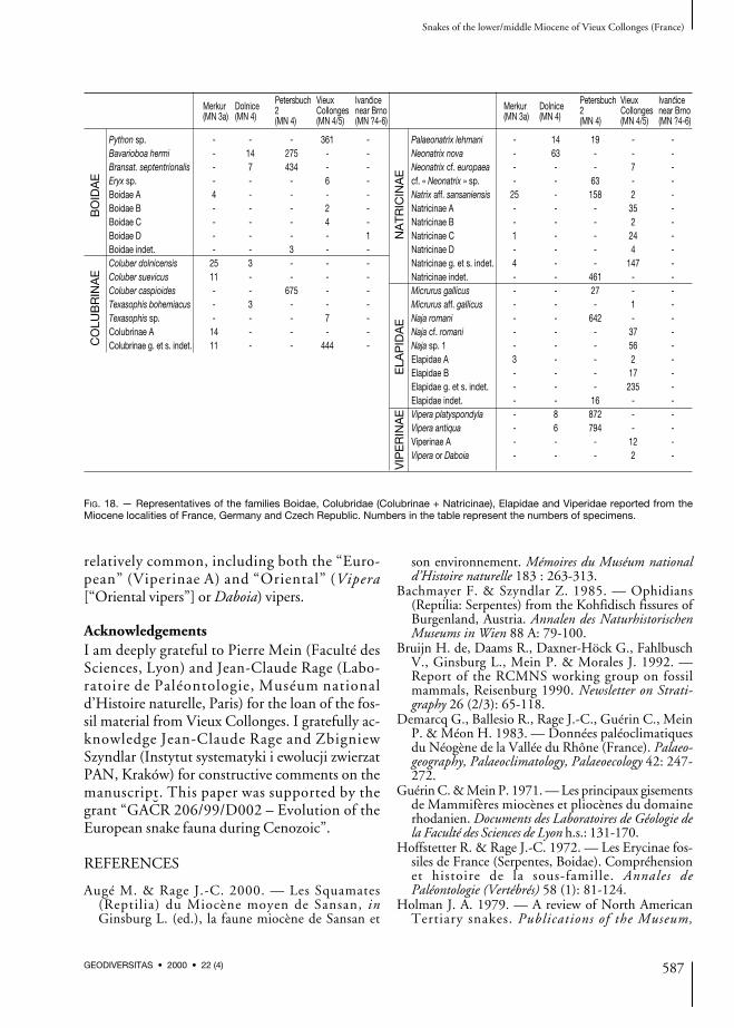
relatively common, including both the “Euro-pean” (Viperinae A) and “Oriental” (Vipera[“Oriental vipers”] or Daboia) vipers.
AcknowledgementsI am deeply grateful to Pierre Mein (Faculté desSciences, Lyon) and Jean-Claude Rage (Labo-ratoire de Paléontologie, Muséum nationald’Histoire naturelle, Paris) for the loan of the fos-sil material from Vieux Collonges. I gratefully ac-knowledge Jean-Claude Rage and ZbigniewSzyndlar (Instytut systematyki i ewolucji zwierzatPAN, Kraków) for constructive comments on themanuscript. This paper was supported by thegrant “GACR 206/99/D002 – Evolution of theEuropean snake fauna during Cenozoic”.
REFERENCES
Augé M. & Rage J.-C. 2000. — Les Squamates(Reptilia) du Miocène moyen de Sansan, inGinsburg L. (ed.), la faune miocène de Sansan et
son environnement. Mémoires du Muséum nationald’Histoire naturelle 183 : 263-313.
Bachmayer F. & Szyndlar Z. 1985. — Ophidians(Reptilia: Serpentes) from the Kohfidisch fissures ofBurgenland, Austria. Annalen des NaturhistorischenMuseums in Wien 88 A: 79-100.
Bruijn H. de, Daams R., Daxner-Höck G., FahlbuschV., Ginsburg L., Mein P. & Morales J. 1992. —Report of the RCMNS working group on fossilmammals, Reisenburg 1990. Newsletter on Strati-graphy 26 (2/3): 65-118.
Demarcq G., Ballesio R., Rage J.-C., Guérin C., MeinP. & Méon H. 1983. — Données paléoclimatiquesdu Néogène de la Vallée du Rhône (France). Palaeo-geography, Palaeoclimatology, Palaeoecology 42: 247-272.
Guérin C. & Mein P. 1971. — Les principaux gisementsde Mammifères miocènes et pliocènes du domainerhodanien. Documents des Laboratoires de Géologie dela Faculté des Sciences de Lyon h.s.: 131-170.
Hoffstetter R. & Rage J.-C. 1972. — Les Erycinae fos-siles de France (Serpentes, Boidae). Compréhensionet histoire de la sous-famille. Annales dePaléontologie (Vertébrés) 58 (1): 81-124.
Holman J. A. 1979. — A review of North AmericanTertiary snakes. Publications of the Museum,
Snakes of the lower/middle Miocene of Vieux Collonges (France)
587GEODIVERSITAS • 2000 • 22 (4)
FIG. 18. — Representatives of the families Boidae, Colubridae (Colubrinae + Natricinae), Elapidae and Viperidae reported from theMiocene localities of France, Germany and Czech Republic. Numbers in the table represent the numbers of specimens.
Dolnice Petersbuch Vieux Ivancice Dolnice Petersbuch Vieux IvanciceMerkur(MN 4) 2 Collonges near Brno Merkur
(MN 4) 2 Collonges near Brno(MN 3a) (MN 4) (MN 4/5) (MN ?4-6) (MN 3a) (MN 4) (MN 4/5) (MN ?4-6)
Python sp. - - - 361 - Palaeonatrix lehmani - 14 19 - -Bavarioboa hermi - 14 275 - - Neonatrix nova - 63 - - -Bransat. septentrionalis - 7 434 - - Neonatrix cf. europaea - - - 7 -Eryx sp. - - - 6 - cf. « Neonatrix » sp. - - 63 - -Boidae A 4 - - - - Natrix aff. sansaniensis 25 - 158 2 -Boidae B - - - 2 - Natricinae A - - - 35 -Boidae C - - - 4 - Natricinae B - - - 2 -Boidae D - - - - 1 Natricinae C 1 - - 24 -Boidae indet. - - 3 - - Natricinae D - - - 4 -Coluber dolnicensis 25 3 - - - Natricinae g. et s. indet. 4 - - 147 -Coluber suevicus 11 - - - - Natricinae indet. - - 461 - -Coluber caspioides - - 675 - - Micrurus gallicus - - 27 - -Texasophis bohemiacus - 3 - - - Micrurus aff. gallicus - - - 1 -Texasophis sp. - - - 7 - Naja romani - - 642 - -Colubrinae A 14 - - - - Naja cf. romani - - - 37 -Colubrinae g. et s. indet. 11 - - 444 - Naja sp. 1 - - - 56 -
Elapidae A 3 - - 2 -Elapidae B - - - 17 -Elapidae g. et s. indet. - - - 235 -Elapidae indet. - - 16 - -Vipera platyspondyla - 8 872 - -Vipera antiqua - 6 794 - -Viperinae A - - - 12 -Vipera or Daboia - - - 2 -
CO
LUB
RIN
AE
BO
IDA
E
VIP
ER
INA
EE
LAP
IDA
EN
ATR
ICIN
AE
Michigan State University, Paleontological series1 (6): 323-335.
Holman J. A. 1982. — New herpetological species andrecords from the Norden Bridge fauna (Miocene;Late Barstovian) of Nebraska. Transactions,Nebraska Academy of Sciences 10: 31-36.
Holman J. A. 1996. — Herpetofauna of the TrinityRiver Local Fauna (Miocene: Early Barstovian), SanJacinto County, Texas, USA. Tertiary Research17 (1-2): 5-10.
Ivanov M. 1996. — Nález Boidního hada (Serpentes;Boidae) z miocénu u Ivancic (jizní Morava).[= Discovery of a boid snake (Serpentes; Boidae)from the Miocene near Ivancice (South Moravia)].Geologické v yzkumy na Morave a ve Slezsku v r.1995: 62-64.
Ivanov M. 1997a. — Hadi evropského kenozoika (Snakesof the European Cenozoic). Ph.D. dissertation,Masaryk University, Brno, Czech Republic, 217 p.
Ivanov M. 1997b. — Vyvoj kenozoické hadí fauny
Evropy. [= The evolution of European Cenozoicsnake fauna]: 59-91, in Hladilová S. (ed.),Dynamika vztah°u marinního a kontinentálníhoprostredí. [= Dynamics of interaction between marineand continental environments]. Masaryk University,Brno.
Martini E. 1990. — The Rhinegraben system, a con-nection between northern and southern seas in theEuropean Tertiary. Veröffentlichungen des Übersee-Museums A (10): 83-98.
Mein P. 1958. — Les mammifères de la faunesiderolithique de Vieux-Collonges. NouvellesArchives du Muséum d’Histoire naturelle 5: 1-122.
M~lynarski M., Szyndlar Z., Estes R. & Sanchíz B.1982. — Lower vertebrate fauna from the Mio-cene of Opole (Poland). Estudios Geológicos 38: 103-119.
Rage J.-C. 1976. — Les Squamates du Miocène de BeniMellal, Maroc. Géologie méditerranéenne 3 (2): 57-69.
Rage J.-C. 1981. — Les Colubridae (Reptilia, Serpentes)du Néogène français. Le problème des espèces duMiocène de Sansan. Geobios 14 (4): 537-542.
Rage J.-C. 1984. — Serpentes: 1-80, in Wellnhofer P.(ed.), Handbuch der Paläoherpetologie (Encyclopediaof Paleoherpetology). Part 11. Gustav Fischer, NewYork; Stuttgart.
Rage J.-C. 1988. — The oldest known colubridsnakes. The state of the art, in Szyndlar Z. (ed.), AFestschrift in honour of Professor Marian M~lynars-ki on the occasion of his retirement. Acta ZoologicaCracoviensia 31 (13): 457-474.
Rage J.-C. & Holman J. A. 1984. — Des Serpents(Reptilia, Squamata) de type Nord-Américain dans
le Miocène francais. Évolution parallèle ou disper-sion ? Geobios 17: 89-104.
Steininger F. F., Berggren W. A., Kent D. V., BernorR. L., Sen S. & Agusti J. 1996. — Circum-Mediterranean Neogene (Miocene and Pliocene)marine-continental chronologic correlations ofEuropean mammal units: 7-46, in Bernor R. L.,Fahlbusch V. & Mittmann H.-W. (eds), TheEvolution of Western Eurasian Neogene MammalFaunas. Columbia University Press, New York.
Szyndlar Z. 1984. — Fossil snakes from Poland. ActaZoologica Cracoviensia 28 (1): 3-156.
Szyndlar Z. 1985. — Ophidian fauna (Reptilia,Serpentes) from the Upper Miocene of Algora(Spain). Estudios Geológicos 41: 447-465.
Szyndlar Z. 1987. — Snakes from the Lower Miocenelocality of Dolnice (Czechoslovakia). Journal ofVertebrate Paleontology 7: 55-71.
Szyndlar Z. 1991a. — A review of Neogene andQuaternary snakes of Central and Eastern Europe.Part II: Natricinae, Elapidae, Viperidae. EstudiosGeológicos 47: 237-266.
Szyndlar Z. 1991b. — Ancestry of the Grass Snake(Natrix natrix): Paleontological evidence. Journal ofHerpetology 25 (4): 412-418.
Szyndlar Z. 1994. — Oligocene snakes of the South-ern Germany. Journal of Vertebrate Paleontology14 (1): 24-37.
Szyndlar Z. & Böhme W. 1993. — Die fossilenSchlangen Deutschlands: Geschichte der Faunenund ihrer Erforschung. Mertensiella 3: 381-431.
Szyndlar Z. & Rage J.-C. 1990. — West Palearcticcobras of the genus Naja (Serpentes: Elapidae):interrelationships among extinct and extant species.Amphibia - Reptilia 11: 385-400.
Szyndlar Z. & Rage J.-C. 1999. — Oldest fossil vipers(Serpentes: Viperidae) from the Old World. Kaupia8: 9-20.
Szyndlar Z. & Schleich H. H. 1993. — Description ofMiocene Snakes from Petersbuch 2 with commentson the Lower and Middle Miocene Ophidian fau-nas of Southern Germany. Stuttgarter Beiträge zurNaturkunde 192 (B): 1-47.
Szyndlar Z. & Zerova G. A. 1990. — Neogene cobrasof the genus Naja (Serpentes: Elapidae) of EastEurope. Annalen des Naturhistorischen Museums inWien 91(A): 53-61.
Thomas H., Sen S., Khan M., Battail B. & Ligabue G.1982. — The Lower Miocene fauna of Al-Sarrar(Eastern province, Saudi Arabia). Atlal 5: 109-136.
Underwood G. 1967. — A contribution to the classi-fication of snakes. Trustees of the British Museum(Natural History) 663: 1-179.
Submitted on 12 October 1999;accepted on 31 January 2000.
Ivanov M.
588 GEODIVERSITAS • 2000 • 22 (4)