miocene arthropods from the mojave desert california · 2011-05-23 · miocene arthropods from the...
TRANSCRIPT
Miocene Arthropods from the Mojave Desert CaliforniaBy ALLISON R. PALMER
With sections by
J. C. M. CARVALHO, D. R. COOK, KELLIE O'NEILL,ALEXANDER PETRUNKEVITCH, and R. I. SAILER
CONTRIBUTIONS TO GENERAL GEOLOGY
GEOLOGICAL SURVEY PROFESSIONAL PAPER 294-G
Seventeen species of aquatic and terrestrial
arthropods from lacustrine beds of the
Ear stow formation are described and
illustrated
UNITED STATES GOVERNMENT PRINTING OFFICE, WASHINGTON : 1957
UNITED STATES DEPARTMENT OF THE INTERIOR
FRED A. SEATON, Secretary
GEOLOGICAL SURVEY
Thomas B. Nolan, Director
For sale by the Superintendent of Documents, U. S. Government Printing Office Washington 25, D. C. - Price $1 (paper cover)
CONTENTS
Page Abstract—_______________________________________ 237Introduction _______________________________________ 237
Methods and procedures-------.------.---------- 238Acknowledgements. __--___-_-___-_____--________ 239
Geology-__________________________________ 239Characteristics of the nodules_______________________ 240Preservation of the fossils-_-__-_-____________________ 241Age of the fauna_--__-__-__--____-_______-________ 242Composition and ecology of the fauna _________________ 2 42Zoogeographic relations_________________-___-________ 243Observations of evolutionary significance.____________ 244Systematic paleontology__-___-_--_________--___----_ 245
Class Crustacea.-_____________________________ 245Order Anostraca.___________________________ 245
Class Arachnida________-_______________________ 246Order Aranei, by Alexander Petrunkevitch_____ 246Order Acarina, by D. R. Cook______________ 248
Systematic paleontology — ContinuedClass Insecta__ _ ________________________________
Order Odonata. _______ ____ _________ _ __
Order Thysanoptera, by Kellie O'Neill___ _ ___
Order Hemiptera _ _________________________
Heteroptera, by R. I. Sailer and J. C. M.
Carvalho _ _ _________________________
Homoptera__ _ ________________________
Order Coleoptera___-______--_-_----___--__-
Order Diptera_-__ _ _____________ _ _______
Collecting localities _ _______________________________
Literature cited-__--__---_-------__-------___-------
Page
249
249
251
255
255
258
259
265
275
276
279
ILLUSTRATIONS[Plates 30-34 follow index]
PLATE 30. External views and cross sections of fossil-bearing nodules.
31. Anatomy of Orthemis? sp.32. Anostraca, Arachnida, Odonata.33. Hemiptera, Coleoptera.34. Diptera.
Page FIGUEE 83. Index map_-_--_--_---_---_---_--------- 237
84. Key to collecting areas.___-_--__---__--__ 24085. Detail of principal collecting area___-_---_- 24186. Anostraca_-_-_-----------__---__---_--_- 24587. Argenna fossilis Petrunkevitch, n. sp_ ______ 24688. Hydracarina---.-_____-____--__-----__ 24989. Orthemis? sp__.___________________ 24990. Thysanoptera_--__--_--__-_-____-------- 25191. Miogonates subimpunctatus Sailer and Car
valho, n. gen., n. sp____--_____---__--_- 257
Page FIGURE 92. Deltocephalini_________________________ 258
93. Schistomerus californense Palmer, n. gen.,n. sp., external morphology. ____________ 260
94. Schistomerus californense Palmer, n. gen.,n. sp., tracheal system__-_______________ 263
95. Coleoptera______________________ 26696. Dasyhelea australis antiqua Palmer, n. subsp.,
adult characters_-____-_____---_---____ 26797. Dasyhelea australis antiqua Palmer, n. subsp.,
tracheae- ______________________________ 26798. Dasyhelea australis antiqua Palmer, n. subsp.,
larva and pupa characters______________ 26999. Dasyhelea stenoceras Palmer, n. sp__________ 272
100. Culicoides megacanthus Palmer, n. sp _______ 273101. Calopsectra sp-_-----------__----------- 275
in

SHORTER CONTRIBUTIONS TO GENERAL GEOLOGY
MIOCENE ARTHROPODS FROM THE MOJAVE DESERT, CALIFORNIA
By ALLISON R. PALMER
ABSTRACT
Seventeen species of aquatic and terrestrial arthropods representing Anostraca, Aranei, Acarina, and five orders of insects are described and illustrated from lacustrine beds of the lower part of the Barstow formation of middle (?) Miocene age in the southern Calico Mountains, Mojave Desert, southeastern California. External details of many specimens are perfectly preserved. Internal anatomical features are preserved in larval water beetles and dragonflies, and adult midges. The arthropods are replaced by at least six minerals, most commonly a crypto- crystalline variety of quartz. Most specimens have been ob tained from formic acid residues of calcareous nodules that are abundant at three or more levels within the upper part of the lower member of the Barstow formation.
New species and genera include Argenna fossilis Petrunkevitch, n. sp. (Aranei); Protoarrenurus convergens Cook, n. gen., n. sp. (Hydracarina); Anaphothrips (Proscirtothrips) vitreus O'Neill, n. sp. (Thysanoptera); Procymophes lithax Sailer and Carvalho, n. gen., n. sp., Miogonates subimpunctatus Sailer and Carvalho, n. gen., n. sp., and Miomonalonion conoidifrons Sailer and Carvalho, n. gen., n. sp. (Hemiptera: Heteroptera); Schistomerus californense Palmer, n. gen., n. sp. (Coleoptera); and Dasyhelea ste;rioceras Palmer, n. sp., and Culicoides megacanthus Palmer, n. sp. (Diptera).
All larvae of the dytiscid beetle described as Schistomerus californense Palmer have characteristics found only in first stage larvae of the most closely related modern species. They are designated as representatives of a new tribe, Schistomerini Palmer.
A modern species, Dasyhelea australis Wirth (Diptera), from the Islas Juan Fernandez west of central Chile, is represented in the fossil fauna by a subspecies, D. australis antiqua Palmer, n. subsp.
The aquatic fauna seems to be most like that of an alkaline pond. It is composed almost entirely of immature individuals and dominated by Schistomerus californense Palmer, Dasyhelea australis antiqua Palmer, and a fairy shrimp. The presence of specimens with excellently preserved internal anatomical features suggests a nearly aseptic chemical environment in the bottom sediments.
INTRODUCTION
Well-preserved fossil nonmarine arthropods are known from only a few deposits in the world. The best known of these are deposits of amber which have also yielded by far the greatest numbers of fossils. This paper is concerned with fossil nonmarine arthropods
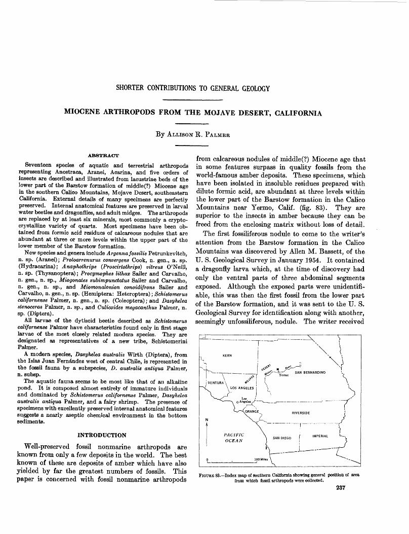
from calcareous nodules of middle(?) Miocene age that in some features surpass in quality fossils from the world-famous amber deposits. These specimens, which have been isolated in insoluble residues prepared with dilute formic acid, are abundant at three levels within the lower part of the Barstow formation in the Calico Mountains near Yermo, Calif, (fig. 83). They are superior to the insects in amber because they can be freed from the enclosing matrix without loss of detail.
The first fossiliferous nodule to come to the writer's attention from the Barstow formation in the Calico Mountains was discovered by Alien M. Bassett, of the U. S. Geological Survey in January 1954. It contained a dragonfly larva which, at the time of discovery had only the ventral parts of three abdominal segments exposed. Although the exposed parts were unidentifi able, this was then the first fossil from the lower part of the Barstow formation, and it was sent to the U. S. Geological Survey for identification along with another, seemingly unfossiliferous, nodule. The writer received
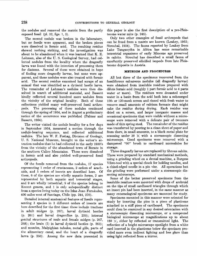
-—Index map of southern California showing general position, of area from which fossil arthropods were collected.
237
238 CONTRIBUTIONS TO GENERAL GEOLOGY
the nodules and removed the matrix from the partly exposed fossil (pi. 32, figs. 1, 5).
The second nodule was broken in the laboratory, but no fossils were apparent, and the broken pieces were dissolved in formic acid. The resulting residue showed nothing striking, and the investigation was about to be abandoned when it was learned that K. E. Lohman, also of the U. S. Geological Survey, had col lected nodules from the locality where the dragonfly larva was found with the intention of processing them for diatoms. Several of these were obtained in hope of finding more dragonfly larvae, but none were ap parent, and these nodules were also treated with formic acid. The second residue examined had scraps of an animal that was identified as a dytiscid beetle larva. The remainder of Lohman's nodules were then dis solved in search of additional material, and Bassett kindly collected several hundred more nodules from the vicinity of the original locality. Both of these collections yielded many well-preserved fossil arthro pods. The processing of these nodules continued through the spring of 1954, and in August a preliminary notice of the occurrence was published (Palmer and Bassett, 1954).
The writer visited the nodule locality for a few days in September 1954, measured a section through the nodule-bearing sequence, and collected additional nodules. The late W. F. Foshag, Division of Geology, U. S. National Museum, brought to the writer's at tention nodules that he had collected in the early 1930's from the vicinity of the abandoned town of Borate in the southern Calico Mountains. These were dissolved in formic acid and also yielded well-preserved fossil arthropods.
Of the fossils removed from the nodules, 17 species representing 1 order of crustaceans, 2 orders of arach nids, and 5 orders of insects are described here. Of these, 6 of the species are wholly aquatic forms, 2 are represented by both aquatic and terrestrial stages, and 9 are wholly terrestrial; 9 of the species belong to Recent genera, and 1 is only subspecifically distinct from a species living today on the Islas Juan Fernandez, 400 miles west of Santiago, Chile (pp. 244, 270).
Detailed internal anatomical features of fossils repre senting 3 species in 3 different orders of insects are here described for the first time: these include tracheae in adult midges (p. 267), larval dytiscid beetles (p. 261) and larval dragonflies (p. 250); internal genital structures of male and female midges (p. 267, 268); the brain (I) in larval dytiscid beetles (p. 263); and muscles, Malpighian tubules, rectal gills, parts of the alimentary canal, and the heart of a dragonfly larva (p. 250). Among the new data presented in
this paper is also the first description of a pre-Pleis- tocene water mite (p. 248).
Only two other examples of fossil arthropods that can be freed from a matrix are known (Leakey, 1952; Strouhal, 1954). The fauna reported by Leakey from Lake Tanganyika in Africa has some remarkable terrestrial organisms of early Miocene age preserved in calcite. Strouhal has described a small fauna of excellently preserved silicified isopods from late Pleis tocene deposits in Austria.
METHODS AND PROCEDURES
All but three of the specimens recovered from the fossiliferous calcareous nodules (all dragonfly larvae) were obtained from insoluble residues prepared with dilute formic acid (roughly 1 part formic acid to 4 parts water or more). The residues were decanted under water in a basin from the acid bath into a submerged 100- or 120-mesh screen and rinsed with fresh water to remove small amounts of calcium formate that might cake the residue during drying. Each residue was dried on a screen, and larger inorganic masses and occasional specimens that were visible without a micro scope were removed with a delicate pair of tweezers made of thin spring steel. The remainder of the residue was transferred by pouring to a clean sheet of paper and from there, in small amounts, to a black metal plate for scanning under 30 X with a stereoscopic dissecting microscope. Good specimens were removed with a dampened "00" brush to cardboard microslides for storage.
Three dragonfly larvae are replaced by fibrous calcite. These were prepared by standard mechanical methods, using a grinding wheel on a dental machine, a Burgess Vibro-tool with a special chuck for holding needles, and a chisel-edged needle in a pin vise. All operations but the grinding were performed under a stereoscopic dis secting microscope.
Some of the better preserved specimens from the insoluble residues were mounted with drops of ambroid on the tips of small cardboard triangles through which an insect pin had been inserted, in the same manner as many entomological specimens are mounted for study.
Specimens mounted in this manner were oriented for study by inserting the pins in a piece of plasticene attached to a stiff piece of cardboard. The specimens could then be examined in any desired orientation with a stereoscopic dissecting microscope, or a compound biological microscope at magnifications up to about 180 X, either by reflected or transmitted light. The reflection of a bright microscope spotlight from a white card inserted in the plasticene below the specimen pro vided more even indirect lighting and less glare than using light reflected from a mirror.
MIOCENE ARTHROPODS FROM THE MOJAVE DESERT, CALIFORNIA 239
Specimens with possible internal structures were immersed in xylene, which has an index of refraction reasonably close to silica, the commonest replacement material. Where internal structures were visible, the specimens were transferred to liquid Canada balsam in hollowed glass slides and protected with cover glasses for permanent mounts. Desired orientation was ob tained by first placing such a specimen in a small drop of clear acetate cement which is not soluble in xylene, the solvent for Canada balsam. This held the specimen in place while the balsam hardened.
Internal features and some surface details of specimens mounted in Canada balsam on glass slides were best observed by placing a white card beneath each slide, directing a spot light on the specimen from above and viewing the specimen in a combination of direct and indirect lighting.
Minute details observed on thrips were seen only with a phase microscope.
Internal structures of a dragonfly larva replaced by fibrous calcite were observed by making a transverse cut through the abdomen and a longitudinal cut, slightly off the median line, both in vertical planes, using a thin-bladed 2-incli-diameter diamond saw. After the transverse cut, the specimen was mounted in a clear resin (Kosto casting compound). To avoid sur face air bubbles and unfilled cavities within the speci men, it was placed in a bell jar after being surmerged in liquid resin, and most of the air was evacuated. After the resin hardened, the longitudinal cut was made, and the resulting surfaces were protected by cover glasses applied with drops of liquid Canada balsam. Polished surfaces of the resin and the cover glass were found to be unsatisfactory for viewing the specimen at high mag nifications because of minute scratches that reflected the light. This problem was eliminated by spraying the desired surfaces with Krylon plastic spray which pro duced a clear, scratch-free surface.
Photographs of the specimens were prepared by N. W. Shupe, of the U. S. Geological Survey. Specimens mounted on insect pins were lightly coated with ammo nium chloride to reduce glare and were photographed with normal lighting, as were the polished surfaces. Specimens in balsam on glass slides were illuminated by placing them against a white card and directing a light on them from above.
Drawings were prepared with a camera lucida or by using an ocular grid and graph paper. The authors of the various systematic sections prepared their own drawings unless otherwise indicated.
ACKNOWLEDGEMENTS
This study could not have been completed without the generous cooperation of many individuals. The
following contributed descriptions in their field of specialization: Alexander Petrunkevitch, Yale Univer sity, New Haven, Conn. (Aranei); D. R. Cook, Wayne University, Detroit, Mich. (Hydracarina); R. I. Sailer (Hemiptera: Heteroptera) and Kellie O'Neill (Thysa- noptera), of the Entomology Research Branch of the United States Department of Agriculture, Washington, D. C., and J. C. M. Carvalho, Museo Nacional, Rio de Janeiro, Brazil (Hemiptera: Heteroptera). The fol lowing people, through correspondence or personal consultation, provided identifications, references, and guidance on many taxonomic and morphologic prob lems: W. W. Wirth (Diptera); D. A. Young, Jr. (Hemiptera: Homoptera); W. H. Anderson and L. M. Walkley (Coleoptera), A. B. Gurney (Odonata), and E. W. Baker (Arachnida), all of the Entomology Research Branch of the United States Department of Agriculture, Washington, D. C.; R. E. Snodgrass, Research Associate, United States National Museum, Washington, D. C. (arthropod anatomy); P. P. Calvert, Cheney, Pa. (Odonata); R. W. Dexter, Kent State University, Kent, Ohio, and F. A. Chace, Division of Zoology, United States National Museum, Washington, D. C. (Anostraca); R. A. Crowson, the University of Glasgow, Glasgow, Scotland, and R. E. Blackwelder, Society of Systematic Zoology, Washington, D. C. (Coleoptera).
Particular thanks are due the writer's colleagues on the United States Geological Survey: T. H. McCulloh, who provided unpublished information on the geology of the Calico Mountains, and A. M. Bassett, who found the first fossiliferous nodule and who collected many additional nodules during the early phases of this study.
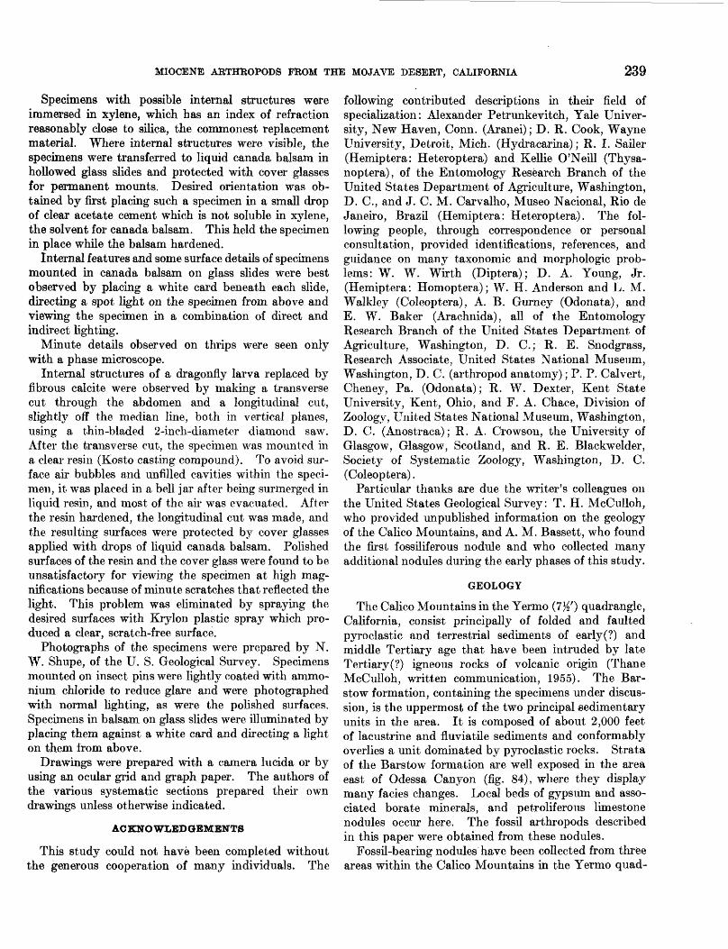
GEOLOGY
The Calico Mountains in the Yermo (7K') quadrangle, California, consist principally of folded and faulted pyroclastic and terrestrial sediments of early(?) and middle Tertiary age that have been intruded by late Tertiary(?) igneous rocks of volcanic origin (Thane McCulloh, written communication, 1955). The Bar- stow formation, containing the specimens under discus sion, is the uppermost of the two principal sedimentary units in the area. It is composed of about 2,000 feet of lacustrine and fluviatile sediments and conformably overlies a unit dominated by pyroclastic rocks. Strata of the Barstow formation are well exposed in the area east of Odessa Canyon (fig. 84), where they display many facies changes. Local beds of gypsum and asso ciated borate minerals, and petroliferous limestone nodules occur here. The fossil arthropods described in this paper were obtained from these nodules.
Fossil-bearing nodules have been collected from three areas within the Calico Mountains in the Yermo quad-
240 CONTRIBUTIONS TO GENERAL GEOLOGY
14 13
T.10N
o
X'Area 2
Figure 85^S%5%%2^§^^# -iS
Area 1O
25
36
18
Area 3
19
CM 30
31
Yermo
IMile
FIGURE 84.—Part of the Yermo OW) quadrangle. California, showing collecting areas for arthropod-bearing nodules
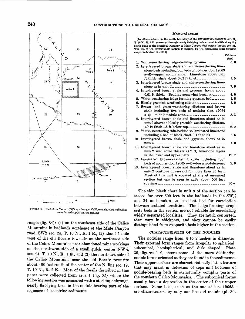
rangle (fig. 84): (1) on the southeast side of the Calico Mountains in badlands northeast of the Mule Canyon road, SW% sec. 24, T. 10 N., K. 1 E., (2) about 1 mile west of the old Borate townsite on the northeast side of the Calico Mountains near abandoned mine workings on the northwest side of a small gulch, center NW#, sec. 24, T. 10 N., R. 1 E., and (3) the northeast side of the Calico Mountains near the old Borate townsite about 400 feet south of the center of the N. line sec. 19, T. 10 N., R. 2 E. Most of the fossils described in this paper were collected from area 1 (fig. 85) where the following section was measured with a steel tape through nearly flat-lying beds in the nodule-bearing part of the sequence of lacustrine sediments.
Measured section[Location,—About on the south boundary of the SWMSWHNEMSWM see. 24,
T. 10 N., B. 1 E.; measured through nearly flat-lying beds exposed in cliffs along the south bank of the principal tributary to Mule Canyon that passes through sec. 24. The top of the stratigraphic section is marked by the prominent ledge-forming evaporite horizon of unit 1]
Thieknest (feet)
1. White-weathering ledge-forming gypsum—— ———— - 5, 02. Interlayered brown shale and white-weathering lime
stone beds including four beds of nodules (loc. 19063 a-d)—upper nodule zone. Limestone about 0.01 ft thick; shale about 0.02 ft thick...__. —— __—— 1. 5
3. Interlayered brown shale and white-weathering lime stone as in unit 2___ — __ — —— __ ——— — ____ 7. 0
4. Interlayered brown shale and gypsum; layers about0.01 ft thick. Bedding somewhat irregular...—— 4. 0
5. White-weathering ledge-forming gypsum bed._____ 1. 56. Blocky greenish-weathering siltstone.-—___________ 1.07. Brown- and green-weathering siltstone and brown
shale including five beds of nodules (loc. 19064 a-e)—middle nodule zone____ — _____ — _____ 3. 3
8. Interlayered brown shale and limestone about as in unit 2 above; a blocky greenish-weathering siltstone 1.7 ft thick 1.5 ft below top..____________ 6. 9
9. White-weathering thin-bedded to laminated limestoneincluding a bed of black chert 0.1 ft thick____ 1. 6
10. Interlayered brown shale and gypsum about as inunit 4.___________________________ 1. 0
11. Interlayered brown shale and limestone about as in unit 2 with some thicker (1.2 ft) limestone layers in the lower and upper parts..------------------ 12. 7
12. Laminated brown-weathering shale including fourbeds of nodules (loc. 19065 a-d)—lower nodule zone. 2. 6
13. Interlayered brown shale and limestone about as in unit 2 continue downward for more than 30 feet. Most of this unit is covered at site of measured section but can be seen in gully about 500 feet southeast___________________________________ 30+
The thin black chert in unit 9 of the section can be traced for over 500 feet in the badlands in the SW^ sec. 24 and makes an excellent bed for correlation between isolated localities. The ledge-forming evap orite beds in the section are not reliable for correlating widely separated localities. They are much contorted, they vary in thickness, and they cannot be easily distinguished from evaporite beds higher in the section.
CHARACTERISTICS OF THE NODULES
The nodules range from % to 2 inches in diameter. Their external form ranges from irregular to spherical, subconical, hemispherical, and disk shaped. Plate 30, figures 1-9, shows some of the more distinctive nodule forms oriented as they are found in the sediments. Their upper surfaces are characteristically flat, a feature that may assist in detection of tops and bottoms of nodule-bearing beds in structurally complex parts of the southern Calico Mountains. , The subconical forms usually have a depression in the center of their upper surface. Some beds, such as the one at loc. 19063d are characterized by only one form of nodule (pi. 30,
MIOCENE ARTHROPODS FROM THE MOJAVE DESERT, CALIFORNIA 241
Measured section ^19063-65
500i_. 1500 Feet
Contour interval 20 feetDatum is mean sea level
FIGURE 85.—Enlargement of collecting area 1, the principal collecting area, showing exact locations from which fossil-bearing nodules were obtained.
figs. 3, 6). More commonly, a variety of nodule-forms characterize each bed.
Polished sections in a vertical plane through the nodules (pi. 30, figs. 4-6, 8) show them to have alternat ing layers of light and dark crystalline calcium carbonate and occasional silicified layers about parallel to the bedding of the enclosing sediments. These bedding- layers are generally bowed downward within the nodules. Some of the layers are composed almost entirely of short rods that may represent fecal material of fairy shrimp (light-colored layers in the lower half of figs. 8, 9, pi. 30).
The nodules are petroliferous, and an oily foam accumulates on the surface of the acid solution in which the nodules are dissolved.
The origin of the nodules is still in doubt. It is being investigated as a part of geochemical studies of the fossil-bearing sequence.
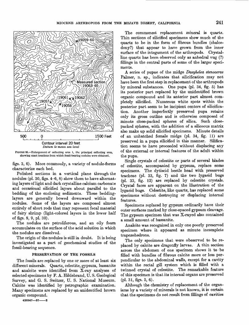
PRESERVATION OF THE FOSSILS
The fossils are replaced by one or more of at least six different minerals. Quartz, celestite, gypsum, bassanite and analcite were identified from X-ray analyses of selected specimens by F. A. Hildebrand, U. S. Geological Survey, and G. S. Switzer, U. S. National Museum. Calcite was identified by petrographic examination. Many specimens are replaced by an unidentified brown organic compound.
426947—57———2
The commonest replacement mineral is quartz. Thin sections of silicified specimens show much of the quartz to be in the form of fibrous bundles (chalce dony?) that appear to have grown from the inner surface of the integument of the arthropods. Crystal line quartz has been observed only as anhedral vug (?) fillings in the central parts of some of the larger speci- ments.
A series of pupae of the midge Dasyhelea stenoceras Palmer, n. sp., indicates that silicification may not have been the first step in replacement of the arthropods by mineral substances. One pupa (pi. 34, fig. 5) has its posterior part replaced by the unidentified brown organic compound and its anterior part almost com pletely silicified. Numerous white spots within the posterior part seem to be incipient centers of silicifica tion. Another imperfectly preserved pupa retains only its gross outline and is otherwise composed of minute close-packed spheres of silica. Such close- packed spheres, with the addition of a siliceous matrix also make up solid silicified specimens. Minute details of an unhatched female midge (pi. 34, fig. 11) are preserved in a pupa silicified in this manner. Silifica- tion seems to have proceeded without displacing any of the external or internal features of the adult within the pupa.
Single crystals of celestite or parts of several Wades of celestite, accompanied by gypsum, replace some specimens. The dytiscid beetle head with preserved tracheae (pi. 33, fig. 7) and the two lygaeid bugs (pi. 33, fig. 12) are replaced by celestite crystals. Crystal faces are apparent on the illustration of the lygaeid bugs. Celestite, like quartz, has replaced some specimens without destroying or displacing internal features.
Specimens replaced by gypsum ordinarily have their outer surfaces marked by close-spaced gypsum cleavage. The gypsum specimen that was X-rayed also contained a small amount of bassanite.
Analcite was recognized in only one poorly preserved specimen where it appeared as minute incomplete trapezohedrons.
The only specimens that were obeserved to be re placed by calcite are dragonfly larvae. A thin section across the abdomen of one specimen shows it to be filled with bundles of fibrous calcite more or less per pendicular to the abdominal walls, except for a cavity within the rectal gill system which is filled with a twinned crystal of celestite. The remarkable feature of this specimen is that its internal organs are preserved (pi. 31, figs. 5, 6).
Although the chemistry of replacement of the organ isms by a variety of minerals is not known, it is certain that the specimens do not result from fillings of cavities
242 CONTRIBUTIONS TO GENERAL GEOLOGY
left by leaching the specimens away. The replacement took place soon enough after death, or in such a chem ical environment, that internal organs in many instances had not disintegrated.
AGE OF THE FAUNA
The age of the fauna cannot be determined directly from the fossils because there are no comparable fossil faunas. Vertebrate remains from beds in the Barstow formation that are conformable with but slightly younger than those producing the arthropods have been dated by G. E. Lewis (1957) as early late Miocene and perhaps late middle Miocene. The fossil arthropod fauna is thus no younger than late Miocene. The comformable stratigraphic relations of the insect and bone-bearing beds in an area that had a complex structural history during the Tertiary indicate that the arthropod fauna comes from beds probably no older than middle Miocene.
COMPOSITION AND ECOLOGY OF THE FAUNA
The fauna contains both aquatic and nonaquatic assemblages that are treated separately below.
The aquatic assemblage in the nodule beds examined is dominated by three species: dytiscid beetle larvae belonging to Schistomerus calij'ornense Palmer, n. sp., larvae and pupae of Dasyhelea australis antigua Pal mer, n. subsp., and an indeterminate fairy shrimp.
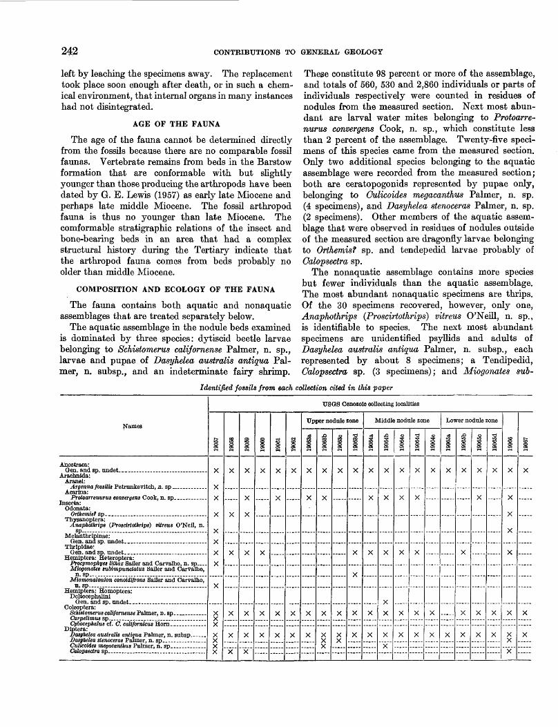
These constitute 98 percent or more of the assemblage, and totals of 560, 530 and 2,860 individuals or parts of individuals respectively were counted in residues of nodules from the measured section. Next most abun dant are larval water mites belonging to Protoarre- nurus convergens Cook, n. sp., which constitute less than 2 percent of the assemblage. Twenty-five speci mens of this species came from the measured section. Only two additional species belonging to the aquatic assemblage were recorded from the measured section; both are ceratopogonids represented by pupae only, belonging to Culicoides megacanthus Palmer, n. sp. (4 specimens), and Dasyhelea stenoceras Palmer, n. sp. (2 specimens). Other members of the aquatic assem blage that were observed in residues of nodules outside of the measured section are dragonfly larvae belonging to Orthemisf sp. and tendepedid larvae probably of Calopsectra sp.
The nonaquatic assemblage contains more species but fewer individuals than the aquatic assemblage. The most abundant nonaquatic specimens are thrips. Of the 30 specimens recovered, however, only one, Anaphothrips (Proscirtothrips) vitreus O'Neill, n. sp., is identifiable to species. The next most abundant specimens are unidentified psyllids and adults of Dasyhelea australis antiqua Palmer, n. subsp., each represented by about 8 specimens; a Tendipedid, Calopsectra sp. (3 specimens); and Miogonates sub-
Identified fossils from each collection cited in this paper
Names
Ahostraca: Gen. and sp. undet _ _______________
Arachnida: Aranei:
Argenna fossilis Petnmkevitch, n. sp ______Acarina:
Protoanenurus convergens Cook, n. sp ............ Insects:
Odonata:
Thysanoptera: Anaphothrips (Proscirtothrips) vitreus O'Neil, n.
Melanthripinae:
Thripidae:
Hemiptera: Heteroptera: Procymophyes Itthax Sailer and Carvalho, n. sp ....Miogonates subimpunctatus Sailer and Carvalho,
n. sp .Miomonalonion conoidifrons Sailer and Carvalho,
Hemiptera: Homoptera: Deltocephalini
Coleoptera: Schistomerus calif ornense Palmer, u.sp.. _____
Cpbocephalus cf. C. calif ornicus Horn __.....—.„Diptera:
Dasyhelea australis antigua Palmer, n. subsp ___
Calopsectra sp ___________________
USQS Oenozoic collecting localities
s
X
X
X
X
X
X
X
X
X
fS 3
X
X
X
X
X
X
§3
X
X
X
X
X
X
X
IX
.....
X
X
X
s
X
X
X
X
,X
-----
X
X
Upper nodule zone
|
X
X
X
X
£1
X
X
X
X X
iX
X
X X
iX
X
X
X
X
Middle nodule zone
1
X
X
X
X
X
,0
X
X
X
X
X
X
X
o
X
X
X
X
X
T3
X
X
X
X
X
1X
-----
-----
X
X
Lower nodule zone
iX
.....
.....
— ...
X
1
X
-----
X
X
X
1
X
X
-----
X
X
•a
X
.....
.....
X
X
.X
X
X
X
X
X
X X
X
r~
X
.....
.....
X
X
MIOCENE ARTHROPODS FROM THE MOJAVE DESERT, CALIFORNIA 243
impunctatus Sailer and Carvalho, n. sp. (2 specimens). Species represented by single specimens are a spider Argenna fossttis Petrunkevitch, n. sp., the beetles Carpelimus sp. and Cybocephalus of C. calijornicus Horn, a leafhopper belonging to the Deltocephalini, and the bugs Procymophyes lithax Sailer and Carvalho, n. sp., and Miomonalonion conoidifrons Sailer and Car valho, n. sp.
Columns for USGS Cenozoic localities 19063a- 19065d in the table show the species identified from each nodule bed in the measured section. Only 2 species, Dasyhelea australis antiqua and the fairy shrimp, are present in all. Schistomerus californense is present in all nodule beds except 19065a. Protoarre- nurus convergens, thrips and unidentified psyllids are present in each of the 3 nodule zones although the first 2 forms are recorded from only 7 beds and the last from only 4 beds. Culicoides megacanthus has been found in only 2 beds, 1 in each of the upper 2 zones. Dasy helea stenoceras is recorded from 2 beds in the upper zone only. The remaining elements of the aquatic assemblage are recorded either from single beds within the measured section of from nodules collected outside of the measured section. Thus the 6 most abundant species are found in all 3 nodule zones. The remaining species form such a small percent of the total fauna that their absence from a particular bed or zone cannot be taken to mean their absence from the fauna at that time. The stratigraphic persistence of the more com mon species indicates stability of conditions favorable for their development. These same conditions were probably also favorable for the existence of the less common species, and it is possible that most or all of the species recorded from the nodules existed in the area throughout the period of nodule formation.
A striking feature of the aquatic assemblages at all of the nodule horizons in and outside of the measured section is the absence of any adult arthropods except perhaps fairy shrimp. Each bed contains only im mature stages of species other than fairy shrimp that spend their entire existence in the water. Midges, which have aquatic immature stages but are non- aquatic adults, are the only species for which aquatic and adult representatives have been recognized.
Immature aquatic arthropods are dominant elements of the fauna of a lake or pond principally in the spring or late winter, although many aquatic species breed and develop at later times of the year. The presence of fairy shrimp, many of which normally hatch between late January and May in temperate latitudes (Pennak, 1953, p. 334), is further evidence for considering the fossil fauna at each horizon as a late winter or spring- tune fauna. The complete absence of adults is also most likely only in the springtime or earlier.
The formation of calcareous nodules that incorporate only an early season fauna at every level of occurrence indicates that the conditions favorable for the preserva tion of the fauna and the growth of nodules, can most probably be correlated with seasonal physicochemical conditions of the water and the bottom muds.
The restricted nature of the fauna, and the fact that some specimens have internal anatomical features pre served, suggest the possibility of unusual chemical conditions in the aquatic environment. In July 1956 the writer and W. T. Edmundson, of the Department of Zoology, University of Washington, visited Soap Lake, a highly alkaline lake in the southern Grand Coulee, central Washington. At the time of the visit, larval dytiscid bettles similar to the Miocene fossils, and floating pupal skins of aquatic diptera, were abun dant. No other evidence of aquatic arthropqds was seen. According to Edmundson (oral communica tion, July 1956), fairy shrimp have also recently been observed in Soap Lake. The striking similarity of the aquatic fauna of this lake to the fossil aquatic fauna described in this paper suggests that the principal ecologic factor controlling the composition of the fossil fauna of the Miocene lake may have been high alkalinity.
ZOOGEOGRAPHIC RELATIONS
Only 8 of the 17 species described in this paper show affinities with living groups that have restricted geographic ranges. The remaining 9 species belong to genera or suprageneric groups without well-defined geographic ranges.
At the species level Dasyhelea australis antiqua Palmer is only subspecifically distinct from Dasyhelea australis australis Wirth, a species now living on the Mas Juan Fernandez in the Pacific Ocean about 400 miles west of Santiago, Chile; in many characteristics Orthemis? sp. resembles 0. jerruginea, a Caribbean species; Cybocephalus cf. C. californicus Horn is iden tical in all observable features with the living species from California; and Schistomerus californense Palmer is most similar to Deronectes striatellus LeConte from Southwestern United States. At the subgeneric level Proscirtothrips, a subgenus presently known only from the drier regions of Western North America, is repre sented by Anaphothrips (Proscirtothrips) vitreus O'Neill. At the genus level Argenna fossttis Petrunkevitch represents a genus whose only certainly identified species are from Europe; and Procymophyes lithax Sailer and Carvalho is most similar to the living genus Cymophyes, which is represented by 1 species in North Africa, 2 species in Asia Minor, and 1 species that ranges from Greece to Turkestan. At the suprageneric level Miomonalonion conoidifrons Sailer and Carvalho is
244 CONTRIBUTIONS TO GENERAL GEOLOGY
assigned to the tribe Monaloniini, which has repre sentatives living only in the tropical regions of south eastern Asia and the Western Hemisphere.
Most of the fossil species have relationships to forms from the warm temperate and tropical regions. There is no clear-cut resemblance, however, of this fossil fauna to faunas of Central or South America such as Matthew (1915) and Darlington (1948) postulated for the Tertiary faunas of North America.
OBSERVATIONS OF EVOLUTIONARY SIGNIFICANCE
Possible examples indicating evolutionary reduction and evolutionary retardation are provided by the dytiscid beetle Schistomerus californense Palmer and the dragonfly Orthemis? sp., respectively.
S. californense, like all dytiscids, has three larval stages. The distinguishing feature of the species is the presence of a well-defined sternal plate on the seventh abdominal segment of the mature stage. A subsidiary characteristic is the presence of spinules on the surfaces of the cerci and legs of all first- and some second-stage larvae. Closely related living dytiscids, that may be descendants of S. californense, have these characteristics only on the first-stage larvae. The spinules are absent, and the sternal plate is fused with the tergite of the seventh segment to form an unbroken ring in second- and third-stage larvae.
It appears here that characters present throughout the larval developmental stages of an ancestral form are present only in the earliest developmental stages of a descendant. This conforms with the idea of evolution ary reduction as stated by De Beer (1951, p. 31).
Tillyard (1917, p. 180-187) describes rectal gill sys tems of dragonfly larvae, discusses their ontogeny, and speculates on their phylogenetic development. The rectal gill system in the Libellulidae, to which Orthemis? sp. belongs, is a sacklike organ at the rear of the abdo men in the larva. This organ bears six longitudinal rows of lobate gill lamellae on its inner surface. The number of gill lamellae in each row ranges from 12 to 30 within the family. A hierarchy has been established by Tillyard in which forms with the greatest number of gill lamellae in each row are considered to be the most specialized forms. Those with the least number of gill lamellae are considered to be archaic. He supports these phylogenetic speculations with evidence from ontogenetic studies in accordance with the theory of recapitulation which was still popular when his book was written.
The maximum number of gill lamellae for an individ ual row in the rectal gill system of living libellulid larvae is 30. Orthemis? sp. has between 50 and 60 gill lamellae in each row. This evidence is incompatible with the theoretical conclusions of Tillyard.
Tillyard shows that the libellulids with 30 gill lamellae in each row pass through stages in their ontogeny with fewer gill lamellae. It thus seems reason able to expect that a form with 50 or 60 lamellae proba bly also passed through earlier ontogenetic stages with fewer lamellae. The fact that no living libellulid in cludes more than 30 lamellae in any row may now be interpreted as evidence for evolutionary retardation in which the ontogenetic development 6^ these structures in living libellulids never reaches as far as it did in the ancestral forms. Tillyard's whole phylogenetic scheme may be reversed. The libellulids with the fewest gill lamellae in each row of the rectal gill system may represent the extremes of evolutionary retardation in this group, and may be the most recent rather than the most ancient members of the group.
This example illustrates the inherent unreliability of phylogenetic reconstructions based only on contempo rary information. The importance of paleontologic sequence in phylogenetic study cannot be over emphasized.
The morphologic similarity of the midge Dasyheka austmlis antiqua Palmer to D. australis australis Wirth from the Islas Juan Fernandez and its dissimilarity to possibly related forms in California raises the question of the factors that determine rates of evolution.
Midges are minute complex organisms whose accurate identification to species requires knowledge of male and female sexual characters, details of color pattern, proportions of appendages, form of wing venation, and distribution of body hairs and setae. They are generally restricted in their habitat. Dasyhelea australis antiqua belongs to the D. dncta species group. This group includes eight species now living in either North or South America, or Africa. D. australis antiqua differs from D. australis australis Wirth in only three char acters, none of which are considered to be critical specific characters. The next most similar species in the D. dncta group lives on the Chilean mainland. The North American and African species in this group are distinguishable from the fossil on many characters. Thus, D. australis has maintained its critical taxonomic features for about 20 million years, although its living representatives are now 6,000 miles from the area where the fossil representatives have been found.
Several species in the D. dncta group now live in California. These have dark distal spines on the tarsi of their middle legs. D. australis antiqua has such spines on the tarsi of all of its legs, and this is one of the char acters that distinguishes it from D. australis australis which lacks such spines. The common presence of dark distal spines on the tarsi of both the living and fossil species in California may indicate that the living species are descendants of D. austratis antiqua. If this
MIOCENE AETHROPODS FROM THE MOJAVE DESERT, CALIFORNIA 245
is true D. australis has evolved sufficiently in California to produce a new species.
If D. australis has evolved significantly in California but hardly at all in the Islas Juan Ferndndez, as it appears, its rate of evolution is probably controlled by environmental factors and is greatest in the area where these factors have changed the most.
SYSTEMATIC PALEONTOLOGY
The quality of preservation of the fossil arthropods is comparable to that of Recent forms. For this reason the fossils have been treated as Recent forms for taxonomic purposes. Identifications are refined only as far as the quality of the material permits. Species are named only if critical morphologic features of species rank are preserved on the specimens.
Collection numbers are those recorded in the Cenozoic catalog of the U. S. Geological Survey. Descriptions of the collecting localities are presented on pages 275,276. The table on page 242 indicates the species identified in each collection.
All types and figured specimens are in the collections of the U. S, National Museum.
Class CRUSTACEA Order ANOSTRACA
Genus and species undet.
Figure 86; plate 32, figure 7
A species of fairy shrimp is one of the three most abundant species in the fossil fauna. Fairy shrimp are soft bodied, and it is remarkable that they are preserved at all. Commonly, only the II antennae and the mandibles remain as fossils in the collections studied here. Only 6 of the 3,000 or more specimens observed have enough of the body remaining to show critical structural features.
Overall length of the body appears to have been 4-5 millimeters. It is possible, however, that the preserved individuals might not have been full grown.
Mandibles, II antennae, I maxillae, and II maxillae are the only features of the head region that have been preserved. Of these, the mandibles (fig. 866) and the I maxillae are of the general form characteristic of all fairy shrimp. The I maxillae each have a single short, relatively broad seta at the ventral end of the row of long setae that reaches to the region of the mouth. The II antennae (fig. 86&) have broad basal joints that appear to be fused. Near the distal end of each basal joint, a serriform outgrowth is directed inward. On the posterior surface, behind each outgrowth, an elongate narrow slit or notch reaches to the dorsal
FIGURE 86.—Anostraca, genus and species undetermined, (a) II antennae, posterior aspect, X 60; (b) left mandible, ventral aspect, X 60; (c) n maxillary, lateral aspect, X120; (d) anterior setae of third and fourth endites of fifth leg,xioo.
margin. The apical joint is fused with the basal joint. It is simple and has a flattened cross section with a slight median constriction. The II maxillae (fig. 86c) each bear two distal spines and single disk-shaped protuberances along their posterior margins.
There are 11 pairs of legs having typical fairy shrimp form. The principal critical structures are the anterior setae on endites 3-5. On the first leg of 1 specimen, endite 3 bears 2 anterior setae, and a group of 7 setae lies distal to it. These may all be the anterior setae of endite 4, or, since endite 5 is not apparent, they may represent both endites. In this instance, a slight divi sion within the group could be interpreted to indicate 3 anterior setae for endite 4 and 4 for endite 5. On the other legs endites 3 and 4 consistently have 2 anterior setae (fig. 86d) that arise from a common base. The proximal seta is always considerably longer than the distal one. Endite 5 appears also to have 2 anterior setae on most specimens. The abdomen and genital structures have not been observed.
Because even the best of the fossil fairy shrimp are imperfect, the precise taxonomic affinities of the fossils cannot be determined. The combination of observed characteristics does not fit any of the described fairy shrimp families. Simple apical joints and serriform outgrowths from the II antennae, 2 distal setae on the maxillae, and seemingly 2 anterior setae on endite 5 of
246 CONTRIBUTIONS TO GENERAL GEOLOGY
the 2d to llth pairs of legs indicate a possible relation ship with the Chirocephalidae. The first leg, however, clearly has only 2 anterior setae on endite 3. According to Linder (1941, p. 159), the Chirocephalidae have 3-5 anterior setae on this endite. Also, the basal joints and the basal and apical joints of the II antennae appear fused. These characteristics seem to eliminate the Chirocephalidae from consideration. The remaining families can be ruled out on one or more of the following characters: Number of pairs of legs, form of the II anteamae and II maxillae, or number of anterior setae on endite 5 of the 2d to llth pairs of legs.
Despite the taxonomic uncertainty, the quality of preservation of some features is remarkable. The imperfect condition of most specimens is due not so much to the nature of preservation as it is to the fact that the organism to begin with had practically no hard parts. An example of unusual structural detail can be seen on the marginal setae of the exopodite of one specimen. These are covered with setules and, at the bases of several of the setae, pectinate scales such as those illustrated by Linder (1941, fig. 2, p. 118) are preserved.
Th« only other record of abundant fossil remains of fairy shrimp is by Woodward (1879) from fresh-water deposits on the Isle of Wight.
Occurrence: USGS Cenozoic localities 19057-19067 Figured specimens: USNM 561976 a-d from USGS Cenozoic
locality 19057
Class ARACHNIDA Order ARANEI (Spiders)
By ALEXANDER PETBUNKEVITCH, Yale University
Family DICTYNIDAE Genus ARGENNA Thorell
Argenna fossilis Petrunkevitch, n. sp.
Figure 87; plate 32, figure 3
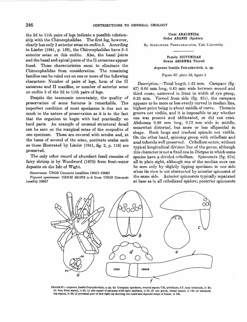
Description.—Total length 1.42 mm. Carapace (fig. 87) 0.60 mm long, 0.45 mm wide between second and third coxae, narrowed in front to width of eye group, 0.25 mm. Viewed from side (fig. 87c), the carapace appears to be more or less evenly curved in median line, highest point being in about middle of curve. Thoracic groove not visible, and it is impossible to say whether one was present and obliterated, or did not exist. Abdomen 0.80 mm long, 0.72 mm wide in middle, somewhat distorted, but more or less ellipsoidal in shape. Book lungs and tracheal spiracle not visible. On the other hand, spinning group with cribellum and anal tubercle well preserved. Cribellum entire, without typical longitudinal division line of the genus, although this character is not a fixed one in Dictyna in which some species have a divided cribellum. Spinnerets (fig. 87a) all in plain sight, although one of the median ones can be seen only by slightly tipping specimen to one side when its view is not obstructed by anterior spinneret of the same side. Anterior spinnerets typically separated at base as in all cribellated spiders; posterior spinnerets
AT-
a
FIGURE 87.—Argenna fossilis Petrunkevltch, n. sp. (o) Complete specimen, ventral aspect: CR, eribellum; AT, anal tubereule, X 37; (6) face, front aspect, X 85; (c) side aspect of carapace with right chellcera, X 85; (<f) eye group, dorsal aspect, X 140; (e) carapace, top aspect, X 60; (/) proximal part of first right leg showing two hairs and sigmoid shape of femur, X 140.
MIOCENE ARTHROPODS FROM THE MOJAVE DESERT, CALIFORNIA 247
still wider apart. Second joint of anterior and posterior spinnerets considerably more slender, short and rounded at end.
Four anterior eyes with pair of posterior lateral eyes form continuous curve which appears strongly recurved when viewed from above (fig. 87d) and slightly upcurved when viewed from front (fig. 876). Posterior eye row straight, with four posterior eyes about equidistant, perhaps median ones slightly more separated from each other than from lateral eyes.
Sternum (fig. S7a) shield shaped, with convex sides and pointed posterior end which separates fourth coxae by more than their width. Sternum distinctly convex, anteriorly about three times as wide as lip. Maximum width of sternum 0.35 mm, length in median line also 0.35 mm. Surface seems to be glabrous. Chelicerae straight, vertical, short (fig. 876) with short, transverse fangs. Figure 876 shows also that clypeus—distance between anterior edge of carapace and lower edge of anterior median eyes—is a little higher than diameter of latter. First coxae wide apart.
Although flexed, legs can be easily and exactly measured by rotating specimen in fluid medium such as xylene. Dorsal view of first femur (fig. 87/) shows that it is distinctly sigmoidal. No spines on any legs, and two structures shown on the first femur are simple hairs.
Leg formula 12.3 2.0 2.0 1.7
The following table gives the leg dimensions of Argenna fossilis Petrunkevitch.
Leg dimensions of Argenna fossilis Petrunkevitch in millimeters
Leg
I——— ——.———-- ———II—— ——_.————————III...—— ....... .—....—..IV..........— ...............
Femur
0.60.42.35.45
Patella and Tibia
0.45.42.30.37
Meta tarsus
0.25.22.20.25
Tarsus
70.17.17.15.17
Total
1.371.231.001.24
Discussion.—This species is represented by one prob ably very immature young specimen. It is complete except for the loss of the tarsi of the first pair of legs and of the metatarsus and tarsus of the right leg of the second pair. For this reason the length of the first tarsus is not known but is here assumed to be the same as that of the second tarsus, an assumption which is probably very nearly correct. The measurements in the table were made under microscope with the aid of a measuring ocular. The individual segments of the appendages are clearly visible; consequently, the measurements are correct to the second decimal. Spines are wanting, but a few hairs are preserved on various segments of the legs. Apparently they are of
the simple, not serrated variety. Many hairs are bro ken off at or close to their base. No hair can be seen either on the carapace or on the abdomen.
Since the spider clearly shows the presence of a cribellum in its normal place in front of the spinnerets, the so-called calamistrum should be present on the metatarsi of the fourth pair of legs, but not a trace of it can be seen. My own observations on first instar spiderlings of the genera Uloborus and HypochUus show that their calamistrum can be clearly seen at that age. Since Uloborus and HypochUus belong to two families of cribellated spiders, the assumption is warranted that representatives of the family Dictynidae also possess a calamistrum at that early stage of their life. Its absence in the present fossil specimen may be therefore due to its loss in the process of the removal of the specimen from the surrounding matrix. The calamis trum is present only in cribellated spiders and is cor related with the cribellum. The latter, unlike the spinnerets which possess a musculature of their own and are used in spinning without the help of any other organ, is nothing but a perforated plate from the open ings of which the silk threads have to be combed out by the calamistrum. A calamistrum is always wanting in all noncribellated spiders, and it never occurs on any other pair of legs than the fourth, because that is the only pair that can reach the cribellum.
I mention all this because a person not specializing in the study of spiders may not realize the strict connec tion between the two organs, the cribellum and the calamistrum, and make the mistake of finding the latter on a wrong pair of legs, or even in spiders without a cribellum. Thus, Protescu (1937) described a Theridium from the Rumanian amber with some lateral denticulations resembling a "calamistrum" on one of the anterior legs. The genus Theridium belongs to the noncribellated family Theridiidae, which has neither a cribellum nor a calamistrum. Whether Protescu's identification of the genus is correct or wrong, the row of denticles figured by him cannot be a calamistrum. They may be the curved spines present on the tibia and metatarsus of the anterior two pairs of legs in the family Mimetidae, but that could be determined only by an examination of the type under high power.
In Argenna fossUis the claws are missing, but presuma bly there were three claws, because the spiders belong ing to the only two families with a cribellum and a calamistrum but with only two claws (Acanthoctenidae and Zoropsidae) are so different from the 3-clawed cribellated spiders to which the genus Argenna, belongs. Trichobothria are also missing, but they are so fragile that their loss is less remarkable than would be their presence. The lip looks very thin and transparent. Its outline is not very clear.
248 CONTRIBUTIONS TO GENERAL GEOLOGY
The eyes, except for the posterior median pair, are very well visible. The latter are recognizable as such, because of the reflection of light by them, but their outline, as shown in the figure is approximate. The genera of the family Dictynidae, as it is now usually delimited, are distinguished primarily by the configura tion of their eye group. Unfortunately, the characters as given originally for the genotypes, show in many genera considerable variations. When Thorell, in 1870, established the genus Argenna he stated that it is distinguished "by the eyes of the anterior row being situated very close together, not more distant than are the lateral eyes from each other * * *", and Simon in 1892 in his key to the genera of Dictynidae gave as distinction of the genus Argenna from three other, closely related genera, the same character, stating that "four anterior and lateral and posterior eyes close together."
The genotype and three other species are European. Five species were referred to the genus Argenna by various authors. No fossil species have been pre viously described either in Europe or America. That I have not yet found any representative of the genus Argenna in the Baltic amber is, of course, no proof that it did not exist in the Oligocene of Europe. Not withstanding their beautiful preservation, spiders in amber are difficult to study; the material is scattered; and all Dictynidae are small in size, in no way con spicuous, and require attention to characters which would easily escape the notice of any but an experienced arachnologist. Emerton's (1911) description and fig ures of the Recent Argenna obesa from Long Island make me doubt that his species belongs to this genus because he states that the front eyes are "at equal distances from each other." Neither of the other Recent American species conforms fully with the characters of the genotype mentioned by either Thorell or Simon. For that matter the fossil species described here also differs in some ways from the genotype. Consequently, it is very difficult to decide whether any of the American species really belong to the genus Argenna, but it seems certain that the fossil species has the greatest right to be regarded as a slightly modified representative of Argenna.
Occurrence: USGS Cenozoic locality 19057. Figured specimen: Holotype USNM 561977.
Order ACARINA
Suborder HYDRACARINA
By D. R. Cook, Wayne University
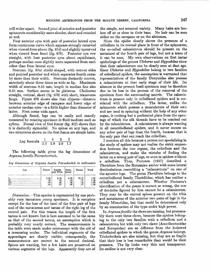
Genus PROTOARRENURUS Cook, n. gen.
Diagnosis.—(Based on larval stage.) Large dorsal shield present; coxae large and platelike, occupying
most of ventral surface; suture lines between all coxae complete; median border of second coxae reduced to a median angle.
Type species.—Protoarrenurus convergens Cook, n. sp.The affinities of Protoarrenurus are somewhat obscure.
The presence of a large dorsal shield, structure of the legs, and the large size of the coxae place this genus among the more highly evolved of the Hydracarina. In the higher water-mites only members of the super- families Arrenurae and Mideopsae have the suture line between the second and third coxae complete. Proto arrenurus can be placed in one of these two super- families but cannot be assigned to a family on the basis of larval characters. In all living genera of Arrenurae and Mideopsae, in which the larval stage is known, the median borders of the second coxae are not reduced to a median angle but are of approximately the same width throughout. This difference in the new genus would strongly suggest that it does not belong to any of the described families of Hydracarina. Because, however, the systematics of the water mites are set up primarily on adult characters, the establishment of a new family for Protoarrenurus based on larval characters alone would serve no useful function. It is hoped that con tinued collecting will uncover nymphs or adults of this mite, thereby permitting a more refined placement of the genus within the system.
Description of unengorged larva.—Length of body 220/4-260/4, width of body 170/4-190ju; dorsal surface covered with a large dorsal shield; coxae large, occupy ing most of ventral surface; first coxae forming a moderately deep, V-shaped notch at anterior end; first coxae widest at median border; second coxae much narrowed at median border; urstigma present between
Protoarrenurus convergens Cook, n. sp.
Figure 886; plate 32, figure 4
first and second coxae; third coxae very large, occupying more area than first and second coxae combined; posterior borders of third coxae rounded, median borders touching along anterior half; suture lines between all coxae complete; anal plate slightly longer than wide; plate widest at posterior end; anal opening located somewhat anterior to middle of plate; pseudocapitulum relatively narrow, palpi projecting only slightly laterally; palpal hooks not seen on any of the specimens, probably broken off. Setae—extremely delicate in larval hy- drachnids—not observed. However, chaetotaxy of pseudocapitulum, coxae, and anal region remarkably constant in higher water-mites and Protoarrenurus should have setal arrangement similar to that of living genus Arrenurus (fig. 88a); legs of swimming type; claw sockets present on first and second legs; claws not
MIOCENE ARTHROPODS FROM THE MOJAVE DESERT, CALIFORNIA 249
FIGURE 88.—Hydracarina. (a) Anenurus sp., larva, ventral aspect (chaetotaxy of legs not shown), X 160; (&) Protoarrenurus concergens Cook, n. sp.; larva, ventral aspect, X 160.
preserved; following are lengths of leg segments which were not too foreshortened to permit accurate measure ments: I-Leg-5, 36/i; II-Leg-5, 44/i; III-Leg-1, 58/x; III-Leg-2, 60M ; III-Leg-3, 44M ; III-Leg-4, 64M .
Nymphs and adults.—Nymphs and adults from these larvae are unknown. These larvae are unengorged, which means that they emerged from eggs deposited by adults which would also have to be in the same body of water. Therefore, there is every reason to believe that nymphs and adults may be found in future collections.
Hosts.—One of the fossils taken in the collection is a female midge of the genus Dasyhelea with a larval water mite attached to the underside of the abdomen. Un fortunately, the ventral side of the hydrachnid was not visible, and it was felt that attempted removal might completely destroy the specimen. However, the total length of the mite and the lengths of the leg segments agree closely with those given in the description of P. convergent. Although positive identification is im possible without an examination of the ventral side, there is good reason to believe that the parasite is P. convergens and that at least one of its hosts was Dasyhe lea.
Occurrence: USGS Cenozoic localities 19057, 19059, 19061, 19063a, b, 19064a-d, 19065c, 19066.
Figured specimen: Holotype, USNM 561978, from USGS Cenozoic locality 19065c.
Class INSECTAOrder ODONATA
Family HBELLULIDAESubfamily LIBELLULINAE
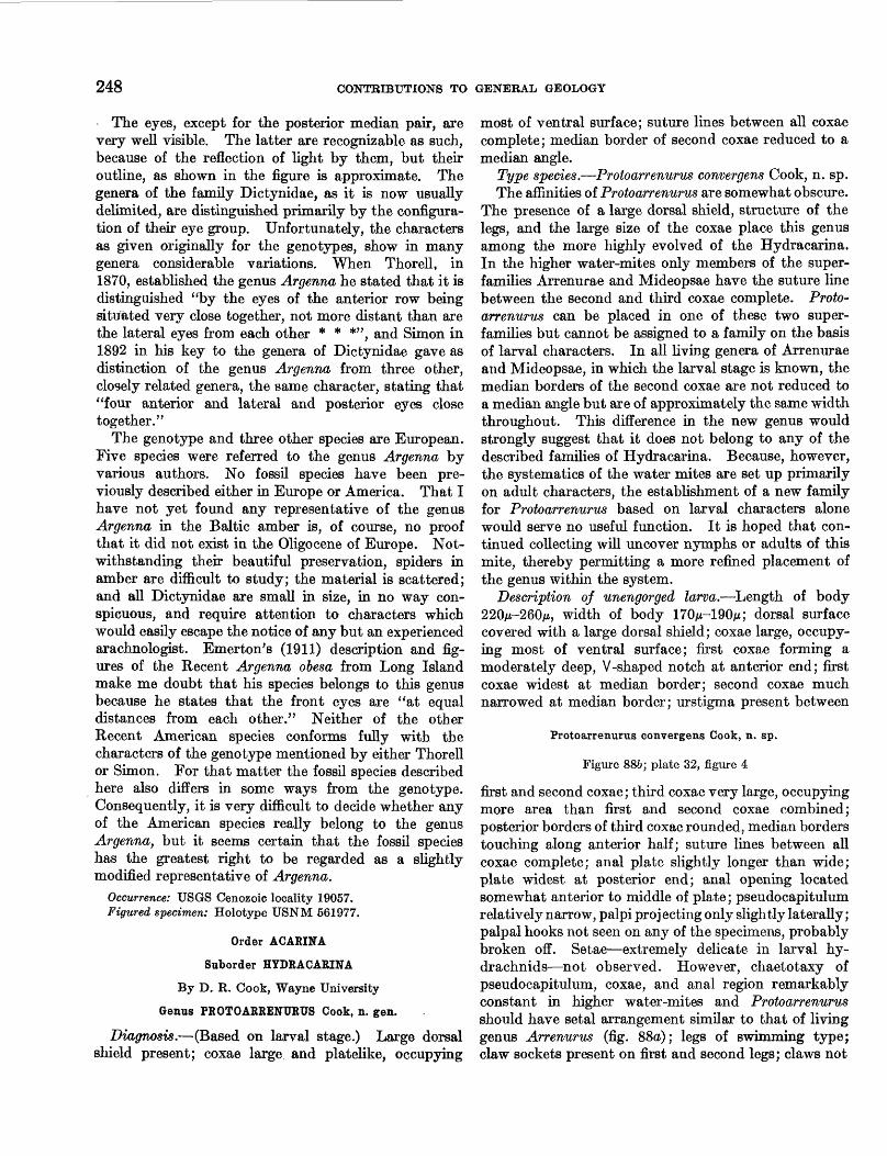
Or the mis? sp.
Figure 89; plate 31, figures 1-6; plate 32, figures 1, 2, 5, 6, 8-10
Description.—Anterior margin of head convex. Eyes projecting forward and slightly laterad from antero- lateral margins of head in dorsal view. On tilting head
426947—57———3
FIGURE 89.— Orthemis? sp., right labial palp, anterior aspect, X 13.
of specimen upward towards the spectator in dorsal view, the upper surface of frons remains convex and eyes project dorsad. A transverse frontal ridge present. Antennae lacking, but their circular sockets are visible on frons above bases of mandibles. Hind edges of sockets about on a line joining anterior margins of eyes. Labium covers face up to eyes. Mentum distinct, length about three-fourths width at distal margin. Details of distal margin not preserved. Labial palp (fig. 89; pi. 32, fig. 10) with seven distinct scallops along its distal margin. Submentum with median notch at its distal end. Sutures between frons and clypeus and clypeus and labrum distinct. Mandibles with bases not quite as far dorsad as frontoclypeal suture; apices acute; each apex forms an angle of about 40°.
Pronotum distinctly divided into an anterior and a posterior division; anterior division nearly the same width as hind margin of head; posterior division about one-thiid wider than anterior division. Hind margins of both divisions convex, posterior division more strongly so. Anterior margins of both divisions concave. Wing pads with slight notch on dorsal margin near tip, and indistinct ridges indicating some of the more prominent veins. Veins appear to be costa, subcosta, radius, media 1, 3, and 4, cubitus, aid anal of Needham (1903, p. 238).
Only coxa, trochanter, and parts of femur of legs observed. Coxa covered with short spines or hairs on one specimen. Femur triangular in cross section with single rows of short hairs along angular ridges.
Abdomen with neither middorsal hooks nor lateral spines. Superior appendage wide at its base; its sides slightly concave; its apex very acute. Lateral append ages slender, about one-fourth as long as superior appendage. Only one inferior appendage observed. It is broken and its length relative to that of superior appendage cannot be determined.
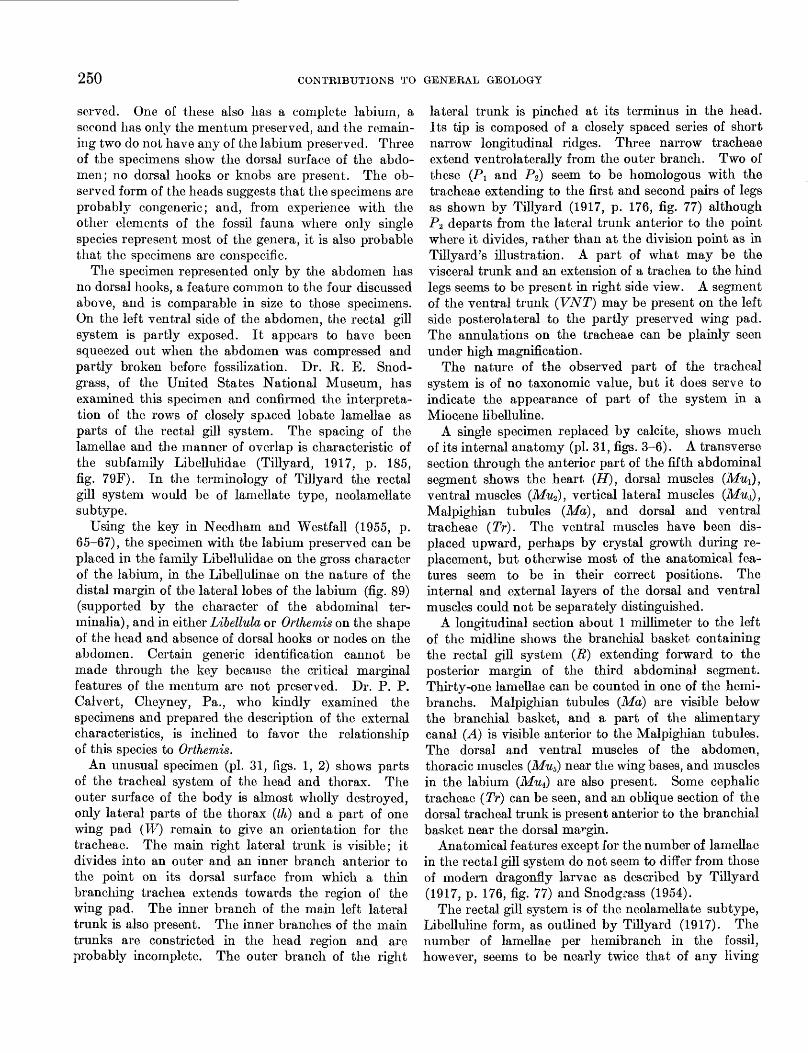
Rectal gill system (pi. 31, fig. 5) of neolamellate subtype, libelluline form (Tillyard, 1917, p. 185). More than 30, perhaps 50-60 lamellae in each hemibranch.
Discussion.—Five imperfectly preserved larvae are present in the collection—4 are represented only by the anterior parts of the body, and a fifth is represented only by an abdomen. The shape of the head is shown on the four specimens with their anterior parts pre-
250 CONTRIBUTIONS TO GENERAL GEOLOGY
served. One of these also has a complete labium, a second has only the mentum preserved, and the remain ing two do not have any of the labium preserved. Three of the specimens show the dorsal surface of the abdo men; no dorsal hooks or knobs are present. The ob served form of the heads suggests that the specimens are probably congeneric; and, from experience with the other elements of the fossil fauna where only single species represent most of the genera, it is also probable that the specimens are conspecific.
The specimen represented only by the abdomen has no dorsal hooks, a feature common to the four discussed above, and is comparable in size to those specimens. On the left ventral side of the abdomen, the rectal gill system is partly exposed. It appears to have been squeezed out when the abdomen was compressed and partly broken before fossilization. Dr. R. E. Snod- grass, of the United States National Museum, has examined this specimen and confirmed the interpreta tion of the rows of closely spaced lobate lamellae as parts of the rectal gill system. The spacing of the lamellae and the manner of overlap is characteristic of the subfamily Libellulidae (Tillyard, 1917, p. 185, fig. 79F). In the terminology of Tillyard the rectal gill system would be of lamellate type, neolamellate subtype.
Using the key in Needham and Westfall (1955, p. 65-67), the specimen with the labium preserved can be placed in the family Libellulidae on the gross character of the labium, in the Libellulinae on the nature of the distal margin of the lateral lobes of the labium (fig. 89) (supported by the character of the abdominal ter- minalia), and in either Libellula or Orthemis on the shape of the head and absence of dorsal hooks or nodes on the abdomen. Certain generic identification cannot be made through the key because the critical marginal features of the mentum are not preserved. Dr. P. P. Calvert, Cheyney, Pa., who kindly examined the specimens and prepared the description of the external characteristics, is inclined to favor the relationship of this species to Orthemis.
An unusual specimen (pi. 31, figs. 1, 2) shows parts of the tracheal system of the head and thorax. The outer surface of the body is almost wholly destroyed, only lateral parts of the thorax (th) and a part of one wing pad (W) remain to give an orientation for the tracheae. The main right lateral trunk is visible; it divides into an outer and an inner branch anterior to the point on its dorsal surface from which a thin branching trachea extends towards the region of the wing pad. The inner branch of the main left lateral trunk is also present. The inner branches of the main trunks are constricted in the head region and are probably incomplete. The outer branch of the right
lateral trunk is pinched at its terminus in the head. Its tip is composed of a closely spaced series of short narrow longitudinal ridges. Three narrow tracheae extend ventrolaterally from the outer branch. Two of these (Pi and P2) seem to be homologous with the tracheae extending to the first and second pairs of legs as shown by Tillyard (1917, p. 176, fig. 77) although PZ departs from the lateral trunk anterior to the point where it divides, rather than at the division point as in Tillyard's illustration. A part of what may be the visceral trunk and an extension of a trachea to the hind legs seems to be present in right side view. A segment of the ventral trunk (VNT) may be present on the left side posterolateral to the partly preserved wing pad. The annulations on the tracheae can be plainly seen under high magnification.
The nature of the observed part of the tracheal system is of no taxonomic value, but it does serve to indicate the appearance of part of the system in a Miocene libelluline.
A single specimen replaced by calcite, shows much of its internal anatomy (pi. 31, figs. 3-6). A transverse section through the anterior part of the fifth abdominal segment shows the heart (H), dorsal muscles (Mu^), ventral muscles (Mu2), vertical lateral muscles (Mut), Malpighian tubules (Ma), and dorsal and ventral tracheae (Tr). The ventral muscles have been dis placed upward, perhaps by crystal growth during re placement, but otherwise most of the anatomical fea tures seem to be in their correct positions. The internal and external layers of the dorsal and ventral muscles could not be separately distinguished.
A longitudinal section about 1 millimeter to the left of the midline shows the branchial basket containing the rectal gill system (R} extending forward to the posterior margin of the third abdominal segment. Thirty-one lamellae can be counted in one of the hemi- branchs. Malpighian tubules (Ma) are visible below the branchial basket, and a part of the alimentary canal (A) is visible anterior to the Malpighian tubules. The dorsal and ventral muscles of the abdomen, thoracic muscles (Mu5) near the wing bases, and muscles in the labium (Mu^) are also present. Some cephalic tracheae (Tr) can be seen, and an oblique section of the dorsal tracheal trunk is present anterior to the branchial basket near the dorsal margin.
Anatomical features except for the number of lamellae in the rectal gill system do not seem to differ from those of modern dragonfly larvae as described by Tillyard (1917, p. 176, fig. 77) and Snodgrass (1954).
The rectal gill system is of the neolamellate subtype, Libelluline form, as outlined by Tillyard (1917). The number of lamellae per hemibranch in the fossil, however, seems to be nearly twice that of any living
MIOCENE ARTHROPODS PROM THE MOJAVE DESERT, CALIFORNIA 251
libelluline. Tillyard cites 30 as the maximum number of lamellae in a hemibranch of the libelluline form. Thirty-one lamellae can be counted in the longitudinal section of the fossil from the front of the brachial basket to the anterior part of the fifth abdominal segment. A transverse section through the posterior part of the sixth segment, about 2 millimeters to the rear, still shows lamellae and their bases. There are about 13 evenly spaced lamellae per millimeter in the forward part of the system. Projecting this backward across the 2-millimeter gap into the transverse section men tioned above would add another 26 lamellae, making a total of at least 57. For larvae of Orthemis there is no published record of the number of lamellae in the rectal gill system, but if they have no more than 30 lamellae, then this character may be an important means of distinguishing the fossil from living libellulines.
Tillyard (1917, p. 186) discusses the phylogenetic relations of the various types of rectal gill systems in the dragonfry larvae. He indicates that the most primitive libellulid has only 12 lamellae in each hemi branch. The evidence given above invalidates this concept and this conclusion. (See p. 244.)
Occurrence: USGS Cenozoic localities 19057-19059, 19066.Figured specimens: USNM 561979a-d from USGS Cenozoic
locality 19057, 561980 from USGS Cenozoic locality 19059, 561981 from USGS Cenozoic locality 19066.
Order THYSANOPTERA: Suborder TEREBRANTIA
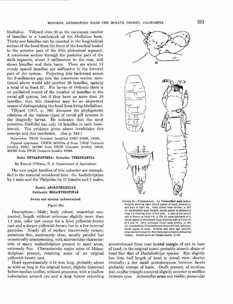
By KELLIE O'NEILL, U. S. Department of Agriculture
The two major families of this suborder are exempli fied in the material considered here: the Aeolothripidae by 1 male and the Thripidae by 17 females and 5 males.
Family AEOLOTHRIPIDAE Subfamily MEIANTHRIPINAE
Genus and species undetermined
Figure 90a
Descriptions.—Male; body robust, somewhat con tracted; length without antennae slightly more than 1.1 mm, color lost except for a faint yellowish-brown cast and a deeper yellowish-brown hue in a few internal particles. Nearly all of surface transversely striate; striations fine, moderately close, mostly parallel but occasionally anastomosing, with microsetulae character istic of many melanthripines present in most areas, extremely fine. Characteristic major setae of Melan- thripinae present, retaining some of an original yellowish-brown cast.
Head approximately 0.16 mm long, probably about as wide as long in the original insect, slightly depressed before median ocellus, without processes, with a shallow indentation around eye and a deep furrow extending
FIGUKE 90.—Thysanoptera. (a) Unidentified male melan- ttiripine showing right lateral aspect of head, pronotum, and part of right leg. Only dorsal seta* shown, X 125; (6) unidentified male thripid, dorsal aspect of abdominal terga 1-8 showing setae of first pair. A seta of the second pair is shown on terga 5-8, X 125; (c) same specimen as 6, dorsal aspect of abdominal terga showing part of 8 and all of 9 and 10. Only principal dorsal setae shown, X 250; (d) Anaphothrips (Proscirtothrips) vitreus O'Neill, holotype, dorsal aspect of male. Midlegs and hind legs omitted; setae omitted except for first three pairs of ninth abdominal tergum. Shaded areas are foreign matter, X 125.
posterodorsad from near lateral margin of eye to base of head; in the original insect probably absent; shape of head like that of Dactuliothrips species. Eye slightly less than half length of head in dorsal view, shorter ventrally; a few small protuberances between facets probably stumps of hairs. Ocelli present, of medium size; ocellar triangle centered slightly anterior to midline between eyes. Anteocellar setae not visible; postocular
252 CONTRIBUTIONS TO GENERAL GEOLOGY
row of setae characteristic of subfamily well developed. Antenna represented by stump of first segment; mouth cone obscured.
Pronotum about 0.15 mm long and 0.16 mm wide; greatest width subposterior; sides converge slightly anteriorly. Major setae as in robust Dactuliothrips species, not confined to posterior marginal row, a few on anterior angle characteristically directed anteriorly. Pterothorax robust, not depressed, about 0.20 mm wide through point of attachment of forewing. Legs normal for males of the subfamily, with robust setae; forefemur slightly enlarged. Approximate length of forefemur, 0.21 mm; tibia, 0.16 mm; tarsus, 0.07 mm; midfemur, 0.14 mm; tibia, 0.16 mm; tarsus, 0.08 mm; hind femur, 0.22 mm; tibia, 0.25 mm; tarsus, 0.08 mm. Forefemur with two, possibly more, weak spurs near apex anteriorly; tibia and tarsus without evidence of presence or absence of spurs or teeth; midlegs and hind legs unarmed except for spinelike setae at apex of tibia. Tegula with sockets for robust setae as in Dactuliothrips. Wings broken, their length probably normal for Aelothripidae; width less than normal for this family but greater than for most other Tere- brantia; margins probably nearly parallel or slightly diverging apically. Forewing with anterior marginal and two longitudinal veins visible, beset with robust setae; the longitudinal veins seemingly closer to mar gins than to each other (possibly because of crushing of wing); no cross vein visible; attachment of scale not definable. Longest measurable wing seta on scale 0.06 mm. Fringe setae not visible on foremargin, on hind margin with roughened appearance at magnification of 440 times, probably feathered or ridged as in coarser modern melanthripine species. Hind wing typical of family, without longitudinal vein beyond base and with fringe indicated by stumps of hairs on either margin; length of residue of wing (nearly complete), 0.62 mm; maximum measurable width, near tip, 0.08 mm.
Abdomen entirely typical of Dactuliothrips, slightly constricted at juncture with pterothorax. Tergum of first segment roughly triangular, with posterior margin convex at middle but surface without longitudinal hump and apodemes not visible. Terga of intermediate seg ments strongly transverse; those of terminal segments less so; ninth tergum with setae normal, not thornlike; tenth with enlarged posterolateral seta-bearing areola. Venter with striations and microsetulae pronounced, accessory setae not discernible, certainly lacking on intermediate segments. Tips of median hypophallic process and lateral hypophallic arms extruded beyond tenth segment, those of hypophallic arms possibly (by no means certainly) with interlocking thumblike proc esses in addition to normal sensory areas.
Discussion.—The habitus, the absence of a longi tudinal vein beyond the base in the hind wing, and the elongate first abdominal segment of this fossil place it in the family Aeolothripidae; and the presence and distribution of robust setae in the postocular region and on the anterior part of the pronotum, the legs, and the wings place it in the subfamily Melanthripinae. Beyond this the loss of the antennae and palpi prevent this specimen from being named further, as the antennae and palpi bear the principal characteristics normally used to identify genera of the Melanthripinae; however, it can be seen that the genus Dactuliothrips Moulton is probably the most closely related modern genus in this group. This relationship is shown by the femoral teeth that are known only in Dactuliothrips, though not in all its species, and by the presence of microsetulae over a large part of the body, particularly the venter of the abdomen. Among modern North American melanthripine genera, the presence of microsetulae over so much of the surface is peculiar to Dactuliothrips although the Chilean genus Dorythrips Hood has micro setulae similarly distributed and the Australian and South African genus Cranothrips Bagnall may have them distributed so, but it is not available for observa tion. The absence of cephalic processes is common to Dactuliothrips and Melanthrips Haliday and distin guishes them from the three remaining modern genera of Melanthripinae—Cranothrips, Dorythrips, and Anko- thrips D. L. Crawford.
All genera representing the Aeolothripidae in North America are found in the drier western regions corres ponding to that in which the fossil was found, and most of them, including all melanthripine genera in North America, are confined to that part of the continent. Most of the aeolothripine genera not limited to the western part of North America, notably Aeolothrips Haliday, have an extensive distribution. Outside North America no pattern of distribution is evident in either subfamily. The genus Dactuliothrips is repre sented in North America by four described and one or more undescribed species, and outside the continent, by a single species in Argentina. The humid forested region in which the Argentine species was found is not comparable to the habitat of North American species.
Other fossil Aeolothripidae have been found in North America (Scudder 1875 and 1890), but the details preserved in the present specimen were seemingly not preserved in Scudder's, which from their descriptions cannot be identified beyond the family according to present knowledge of the group.
Occurrence: USGS Cenozoic locality 19057. Figured specimen: USNM 561987.
MIOCENE ARTHROPODS FROM THE MOJAVE DESERT, CALIFORNIA 253
Family THRIPIDAE
Anaphothrips (Proscirtothrips) vitreus O'Neill, n. sp.
Figure 90d
Description.—Body and appendages, where not broken, with configuration typical of Thripinae, slender, delicate; length approximately 0.8 mm excluding antenna; surface not hispid but with usual sculpture.
Head transverse, approximately rectangular but with eyes and cheeks gently rounded; length 0.09 mm; width 0.11 mm. Interantennal notch narrow. Eye bordering base of antenna longer than wide, about half dorsal length of head; the pair of eyes separated by more than width of one; facets of medium size and number. Ocelli large; ocellar triangle centered between eyes. An anteocellar seta possibly present; no other seta discernible. Vertex with transverse striations extend ing ventrally on cheeks, lines occasionally anastomosing. Left antenna with five segments remaining, right with three; all segments with typical sculpture and propor tions of subgenus; the first two segments with normal fine setae visible; second segment with subapicil dorsal areola, third with simple lateral sense cone, fourth with forked sense cone. Length of measurable segments, 1, 0.01 mm; 2, 0.03 mm; 3, 0.03 mm; 4, slightly less than 0.03 mm. Mouth cone short; maxillary palpus of normal length, three segmented; labial palpus with one segment visible, probably two present.
Pronotum scarcely longer and wider than head (length 0.09 mm; width 0.12 mm); surface transversely striate, lines occasionally anastomosing, setae lost or absent. Prosternum with ver/ucose texture of mem branous area distinguishable.
Mesonotum with faint transverse striations and faint reticulations; median interstices roughly circular; lateral ones elongate, longitudinal. Legs short, slender, sculptured as usual and still bearing a few fragments of fine clothing hairs; unarmed but with one, possibly two, toothlike dorsal setae at apex of hind tibia; tarsi subdivided. Approximate lengths, forfemur 0.09 mm; tibia more than 0.07 mm; tarsus 0.03 mm; midfemur 0.08 mm; tibia 0.08 mm; tarsus 0.06 nun; hind femur more than 0.09 mm; tibia 0.11 mm; tarsus 0.06 mm. Wings broken off near base; costa and one longitudinal vein visible in forewing; a third vein unidentifiable, probably belonging to hind wing.
Abdominal terga with typical faint sculpture on seg ments 1-7, segment 8 obscured; a few very faint lines on segment nine. Several terga with a few teeth on extreme lateroposterior margin; no comb present; terga of segments 2-8 with first dorsal seta (or stump of seta) characteristic of subgenus, approximate and more conspicuous as usual on posterior segments. Ninth tergum with a central group of two pairs of thornlike
setae, second pair directly posterior to first. Principal segments of venter showing fold of intersegmental membrane, antecosta, and posterior margin, on which the three regular setae are extremely fine. Accessory setae absent, no glandular area visible.
Discussion.—Several other specimens that agree with it in some characteristics but lack enough of the signifi cant ones to be identifiable belong to this species. One of the males from USGS Cenozoic locality 19057 has similar thornlike setae on the ninth tergum and in the absence of differences is considered as probably the same. A female from USGS Cenozoic locality 19066 agrees with P. vitreus in all observable characters except secondary sexual ones. This specimen has addi tional characters that are not preserved on P. vitreus but may have been present. These characteristics are the presence or substantial evidence of at least one major seta at the posterior angle of the pronotum; presence of a marginal and two longitudinal veins in the forewing, each with one to several setae; unreduced setae as in many Anaphothrips species but longer, as in the subgenus Proscirtothrips Karny, those opposite beginning of fringe on posterior margin are about 0.03 mm long; and straight wing fringe.
The species P. vitreus, like others of its subgenus, differs from the subgenus Anaphothrips by the presence of the combination of simple and forked sense cones and the distinctive first dorsal abdominal seta. It probably also resembles the other Proscirtothrips species in having a straight wing fringe and lacking the glandu lar areas that appear on the male venter in the subgenus Anaphothrips, although the wing fringe is lost and the absence of the glandular areas cannot be certainly determined. However, the fossil species shows a link between Proscirtothrips and the rest of the genus in the presence of the two pairs of thornlike setae on the ninth male tergum, typical of many species of Anaphothrips and also of several related genera. The setae on the ninth male tergum of other Proscirtothrips species are longer and more delicate, resembling those of many thripine species. As was noted in the description, the habitus of A. vitreus is also thripine rather than ana- phothripine, in contrast to that of strict sense An aphothrips species and A. zeae Moulton, which have the head and pronotum distinctly longer than wide.
In addition to the thornlike ninth tergal setae, a specific difference between the fossil and modern Proscirtothrips species is the absence of a comb on the posterior margin of the eighth tergum.
The subgenus Proscirtothrips is presently known only from western North America.
Occurrence: USGS Cenozoic collections 19057, 19066. Figured specimen: Holotype USNM 561986 from USGS
Cenozjoic collection 19057.
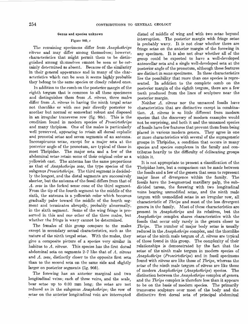
254 CONTRIBUTIONS TO GENERAL GEOLOGY
Genus and species unknown
Figure 906, c
The remaining specimens differ from Anaphothrips vitreus and may differ among themselves; however, characteristics that might permit them to be distin guished among thamselves cannot be seen or be cer tainly determined as absent. Because of the similarity in their general appearance and in many of the char acteristics which can be seen it seems highly probable they belong to the same species or closely related ones.
In addition to the comb on the posterior margin of the eighth tergum that is common to all these specimens and distinguishes them from A, vitreus, three males differ from A. vitreus in having the ninth tergal setae not thornlike or with one pair directly posterior to another but normal or somewhat robust and disposed in an irregular transverse row (fig. 90c). This is the condition found in modern species of Proscirtothrips and many thripines. One of the males is particularly well preserved, appearing to retain all dorsal cephalic and proriotal setae and seven segments of an antenna. Inconspicuous setae, except for a major seta at the posterior angle of the pronotum, are typical of those in most Thripidae. The major seta and a few of the abdominal setae retain some of their original color as a 3Tellowish cast. The antenna has the same proportions as that of Anaphothrips zeae, the type species of the subgenus Proscirtothrips. The third segment is decided ly the longest, and the distal segments are successively shorter, but the antenna of the fossil differs from that of A. zeae in the forked sense cone of the third segment. From the tip of the fourth segment to the middle of the sixth, the antenna is a yellowish color which becomes gradualty paler toward the middle of the fourth seg ment and terminates abruptly, probably abnormally, in the sixth segment. Some of the wing fringe is pre served in this and one other of the three males, but whether the fringe is wavy cannot be determined.
The females of this group compare to the males except in secondary sexual characteristics, such as the nature of the ninth tergal setae. With the males, they give a composite picture of a species very similar in habitus to A. vitreus. This species has the first dorsal abdominal seta on segments 2-7 like that of A. vitreus and A. zeae, distinctly closer to the opposite first seta than to the second seta on the same side and slightly larger on posterior segments (fig. 906).
The forewing has an anterior marginal and two longitudinal veins; each of these veins, and the scale, bear setae up to 0.03 mm long; the setae are not reduced as in the subgenus Anaphothrips; the row of setae on the anterior longitudinal vein are interrupted
distad of middle of wing and with two setae beyond interruption. The posterior margin with fringe setae is probably wavy. It is not clear whether there are fringe setae on the anterior margin of the forewing in any specimen. It is also not clear whether all of the group could be expected to have a well-developed anteocellar seta and a single well-developed seta at the posterior angle of the pronotum, although these features are distinct in some specimens. In these characteristics lies the possibility that more than one species is repre sented. In addition to the complete comb on the posterior margin of the eighth tergum, there are a few teeth produced from the lines of sculpture near the posterior margin.
Neither A. vitreus nor the unnamed fossils have characteristics that are distinctive except in combina tion. A. vitreus is so little different from modern species that the discovery of modern examples would not be surprising, and both it and the unnamed species of fossils have few features that prevent them from being placed in various modern genera. They agree in one or more characteristics with several of the suprageneric groups in Thripidae, a condition that occurs in many species and species complexes in the family and con tributes heavily to the difficulty of delineating groups within it.
It is not appropriate to present a classification of the Thripidae here, but a comparison can be made between the fossils and a few of the genera that seem to represent major lines of divergence within the family. The fossils have the 3-segmented maxillary palp, the sub divided tarsus, the forewing with two longitudinal veins bearing unmodified setae, and the ninth male tergum with unmodified setae in an irregular row, all characteristic of Thrips and most of the less divergent genera in the family. Most of these characteristics are present in Anaphothrips and its relatives, but the Anaphothrips complex shares characteristics with the fossils that occur only rarely in the genera closer to Thrips. The number of major body setae is usually reduced in the Anaphothrips complex, and the thornlike setae of the ninth male tergum of A. vitreus are typical of those found in this group. The complexity of their relationships is demonstrated by the fact that the setae of the ninth male tergum in modern species of Anaphothrips (Proscirtothrips} and in fossil specimens found with vitreus are like those of Thrips, whereas the setae of the ninth male tergum of vitreus are like those of modern Anaphothrips (Anaphothrips} species. The distinction between the Anaphothrips complex of genera and the Thrips complex is therefore less than it appears to be on the basis of modern species. The primarily transverse sculpture over most of the body and the distinctive first dorsal seta of principal abdominal
MIOCENE ARTHROPODS FROM THE MOJAVE DESERT, CALIFORNIA 255
segments in the fossils are found also in most species of the Sericothrips complex, which differs conspicuously from Thrips and Anaphothrips by the presence of microsetae over most of the surface of the body. The development of the distinctive first dorsal seta reaches an extreme in Heliothrips. The complex including this genus differs from the first three in features of major importance such as the lack of subdivision in the tarsus but may show a relationship to the Anaphothrips complex in the thornlike ninth male tergal setae, here consisting of three pairs in a longitudinal line.
As yet no other fossil specimens of Thripidae have been found in North America, although many, includ ing four species of Anaphothrips, have been described from Baltic amber and other sources. It is not possible to compare these species to A. vitreus on the basis of the descriptions, but a few details of the four fossil Anaphothrips species described by Priesner (1924, 1929) from Baltic amber may be mentioned. The male syntype of A. perspicuus and a male that Priesner considered to be probably A. mcinus show only one pair of thornlike setae; however, it is much easier to determine the presence than the absence of structures in fossils, and the second pair of setae in related modern species o f genera such as Oxythrips and Pseudothrips are distinctly smaller, sometimes hairlike. The setae on the whig veins of A. perspicuus, A. vicinus, and A. arcanus Priesner are weak or delicate; A. magniceps Priesner is brachypterous. Only A. magniceps has the head wider than long.
Occurrence: USGS Cenozoic localities 19057-19060; 19063d; 19064a-d; 19065b; 19066.
Figured specimens: USNM 561988 from USGS Cenozoic locality 19064b.
ORDER HEMIPTERA
Suborder HETEROPTERABy R. I. SAILER, U. S. Department of Agriculture
and J. C. M. CARVALHO, Museum Nacional, Brazil
Family LYGAEIDAE
Three imperfectly preserved specimens, including two species, have been recovered from two nodules, one nodule having contained two specimens of the same species. The species have distinct characters, which should make it possible to recognize either of them if additional specimens are ever found.
Scudder (1890) listed 26 genera and 51 species of Lygaeidae as having been found as fossils in sedimen tary rocks in North America. Few if any of them can be placed confidently in a subfamily, to say nothing of tribe. The characters on which the classification of the family is based are not preserved in most fossils, and only rarely can one be placed by general facies.
Usinger (1940) described a fossil from Florissant, Colo. ; which he was able to place in the Cyminae, with the note that when compared with living lygaeids it appeared closest to an Australian genus.
Of the 2 new genera described here, Procymophyes appears to be far removed from the living genera of Pachygronthinae now known from the New World; but is close to Cymophyes, which is known from 1 North African species, 2 species from Asia Minor, and 1 that ranges from Greece to Turkestan. Thus, this onetime resident of North America and first known fossil representative of the Pachygronthinae seems to have its nearest living relatives distributed in the southwestern Palaearctic region. Miogonates, on the other hand, is at least as closely related to the New World genera Gonates, Paragonates, and Cryphula as to the Old World Lethaeus. It can be placed in the subfamily Rhyparochrominae and the tribe Lethaeini on the basis of characters now used in classifying living genera, and it is the first representative of the Lethaeini to be recognized from the Miocene.
Genus PROCYMOPHYES Sailer and Carvalho, n. gen.
Type species.—Procymophyes lithax Sailer and Car- valho, n. sp.
Description.—Head porrect, coarsely punctured on frons and vertex, produced before the antennal tuber- cules a distance equal to one-half width of vertex; each ocellus removed from inner posterior angle of eye by distance equal to its diameter; eyes removed from anterior margin of pronotum by a distance greater than diameter of an ocellus; bucculae almost oval, three-fifths as long as first rostral segment; genae swollen beneath antennal tubercules; apex of second segment of rostrum reaches base of head; third segment reaches base of forecoxae.
Pronotum trapezoidal, wider than long, slightly transversely constricted near middle; humeral angles rounded, somewhat swollen and smooth; rest of pro notum except hind margin coarsely and deeply punc tate. Front coxae open behind; proxyphus bifid at apex, approaching but not seating against mesosternum. Forefemur enlarged, armed beneath with a row of spines along apical two-thirds. Mesothorax a little swollen, evenly punctured; sternum with a broad shallow groove along apical half; mesoxyphus small, acute, apex barely reaching half the length of middle coxae. All coxae smooth, without evidence of lateral spines. Scutellum broader than long, surface punc tured, apex acute. Metathorax with auricular process of each ostiole small, ovate, little elevated, barely longer than apical width of middle coxa.
Discussion.—The characteristic appearance of the head, pronotum, and femur of the single specimen repre-
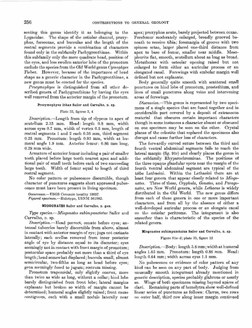
256 CONTRIBUTIONS TO GENERAL GEOLOGY
senting this genus identify it as belonging to the Lygaeidae. The shape of the ostiolar channel, proxy- phus, forecoxae, and bucculae and the length of the rostral segments provide a combination of characters found only in the subfamily Pachygronthinae. Within this subfamily only the more quadrate head, position of the eyes, and less swollen anterior lobe of the pronotum exclude the species from the Old World genus Cymophyes Fieber. However, because of the importance of head shape as a generic character in the Pachygronthinae, a new genus must be erected for the species.
Procymophyes is distinguished from all other de scribed genera of Pachygronthinae by having the eyes well removed from the anterior margin of the pronotum.
Procymophyes lithax Sailer and Carvalho, n. sp.
Plate 33, figures 3, 4
Description.—Length from tip of clypeus to apex of scutellum 2.35 mm. Head: length 0.8 mm, width across eyes 0.7 mm, width of vertex 0.5 mm, length of rostral segments 1 and 2 each 0.36 mm, third segment 0.25 mm. Pronotum: length 0.93 mm, width at hu meral angle 1.9 mm. Anterior femur: 0.86 mm long, 0.28 mm wide.
Armature of anterior femur including a pair of smaller teeth placed before large tooth nearest apex and addi tional pair of small teeth before each of two succeeding large teeth. Width of femur equal to length of third rostral segment.
No color pattern or pubescence discernible, though character of punctures suggests short appressed pubes cence must have been present in living specimen.
Occurrence.—TJSGS Cenozoic locality 19057. Figured specimen.—Holotype, USNM 561982.
MIOGONATES Sailer and Carvalho, n. gen.
Type species.—Miogonates subimpunctatus Sailer and Carvalho, n. sp.
Description.—Head porrect, conate before eyes; an- tennal tubercles barely discernible from above, almost in contact with anterior margin of eye; juga not carinate laterally; each ocellus removed from inner posterior angle of eye by distance equal to its diameter; eyes seemingly not in contact with front margin of pronotum; postocular space probably not more than a third of eye length; head somewhat displaced; buccula small, almost semicircular, two-fifths as long as head before eyes; gena seemingly fused to jugum; rostrum missing.
Pronotum trapezoidal, only slightly convex, more than twice as wide as long, without a collar; hind lobe barely distinguished from front lobe; lateral margins explanate but broken so width of margin cannot be determined; humeral angles slightly raised; front coxae contiguous, each with a small nodule laterally near
apex; proxyphus acute, barely projected between coxae. Forefemur moderately enlarged, broadly grooved be neath to receive tibia; foremargin of groove with two spinose setae, larger placed one-third distance from apex to base of femur, smaller near middle. Meso- pleurite flat, smooth, scutellum about as long as broad. Metathorax with osteolar opening raised but not enlarged to form either an auricular process or an elongated canal. Forewings with embolar margin well defined but not explanate.
Body generally quite smooth with scattered small punctures on hind lobe of pronotum, prostethium, and lines of small punctures along veins and intervening areas of forewings.
Discussion.—This genus is represented by two speci mens of a single species that are fused together and in considerable part covered by a deposit of extraneous material that obscures certain important characters though in some instances a character absent or obscured on one specimen may be seen on the other. Crystal planes of the celestite that replaced the specimens also disrupt and cause further loss of characters.
The forwardly curved suture between the third and fourth ventral abdominal segments fails to reach the lateral margin (fig. 91c) and clearly places the genus in the subfamily Rhyparochrominae. The positions of the three opaque glandular spots near the margin of the fourth ventral abdominal segment are typical of the tribe Lethaeini. Within the Lethaeini there are at least four genera that appear closely related to Miogo nates. Three of these, Cryphula, Gonates, and Parago- nates, are New World genera, while Lethaeus is widely distributed in the Old World. The new genus differs from each of these genera in one or more important characters, and from all by the absence of either a well-developed auricular process or an elongate canal on the ostiolar peritreme. The integument is also smoother than is characteristic of the species of the related genera.
Miogonates subimpunctatus Sailer and Carvalho, n. sp.
Figure 91a-d; plate 33, figure 12
Description.—Body: length 5.8 mm; width at humeral angles 1.65 mm. Pronotum: length 0.86 mm. Head: length 0.64 mm; width across eyes 1.1 mm.
No pubescence or evidence of color pattern of any kind can be seen on any part of body. Judging from unusually smooth integument already mentioned in generic description, species probably glabrous or nearly so. Wings of both specimens missing beyond apices of clavi. Remaining parts of hemelytra show well-defined linear series of punctures as follows: Clavus, two rows on outer half, third row along inner margin continue^
MIOCENE ARTHROPODS FROM THE MOJAVE DESEET, CALIFORNIA 257
FIGUKE 91.—Miogonaf.es sMmpunctatus, Sailer and Carvalho, n. sp. (a) Head of holo type, dorsal aspect, X 30; (6) head of holotype, lateral aspect, X 30; (c) abdomen of paratype, lateral aspect, X 30; (d) metapleurite showing ostiolar peritreme, X 30.
along commissure, scattered punctures in intervening area; corium with two well-defined rows adjacent to claval suture and one row along inner margin of em- bolium; intervening area with about six less well defined rows. The only punctures evident on venter are on the prostethium and posterior flange of the metapleurite.
Discussion.—This species appears to have its closest living relatives in the neotropical genus Para- gonates. The characters of color and pubescence have been used to a great extent for distinguishing species in Paragonates and related genera. Because these characters are lost in the fossil specimens at hand, the specific description must be less than adequate until additional specimens in a better state of preservation are found at the same locality and horizon.
Occurrence: USGS Cenozoic locality 19063d. Figured specimens: Holotype, specimen with head, USNM
561984. Paratype, without head, fused to type. 426947—57———i
Family MIRIDAE
The single specimen representing this family is a fragment, but it exhibits a number of distinctive char acters that justify its placement in the subfamily Byrocorinae. The specimen is excluded from the described genera of that subfamily by the characters of the frons and antennal segments and, accordingly, is described below as a representative of a new genus and new species.
Of the mirids preserved as a fossil in sedimentary rocks Miomonalonion conoidifrons Sailer and Carvalho, n. sp., is one of the few that can be placed in subfamily and tribe with some confidence. Scudder (1868, 1875, 1890) reported 13 species from Florissant and, except for one which he placed in a new genus, all were assigned to well-known genera now present in North America. The present authors are indebted to Dr. F. M. Carpenter for the privilege of examining the types of Scudder's Florissant species now in the Museum of Comparative Zoology at Harvard University. In our opinion, not one of these fossil specimens can be referred to a tribe, and any subfamily assignments would be hazardous.
The tribe Monaloniini, in which we have placed M. conoidifrons, is a tropical group well represented in the Australasian region, but in the New World it is repre sented only by the neotropical genus Monalonion.
Genus MIOMONALONION Sailer and Carvalho, n. gen.
Type species.—Miomonalonion conoidifrons Sailer and Carvalho, n. sp.
Description.—Head, with frons conately produced between antennae, but projected little beyond anterior margin of eyes; vertex immarginate, smooth; neck distinct; eyes pedunculate, projected laterally; anten nae with first segment incrassate; clypeus vertical, scarcely prominent; rostrum with second segment thickened toward apex and with bilateral papilliform projections near middle of inner surface. Pronotum smooth; calli not visible; disk of posterior lobe con- vexly declivous. Scutellum smooth (shape distorted). (Hemelytra badly damaged.) Embolium very narrow; ci'neus apparently much longer than wide. (Abdomen and legs fragmentary), one visible tibia incrassate to ward apex and densely pilose.
Discussion.—This genus is tentatively placed in the Byrocorinae near Felisacus and Monalonion. The absence of a pronotal collar excludes the genus from the subfamilies Mirinae, Deraeocorinae and Cylapinae. The flattened necklike anterior lobe of the pronotum excludes the genus from the Phylinae and Orthotylinae, but associates it with the above-mentioned genera of Byrocorinae. The conate frons and incrassate first antennal segments are distinctive of the genus.
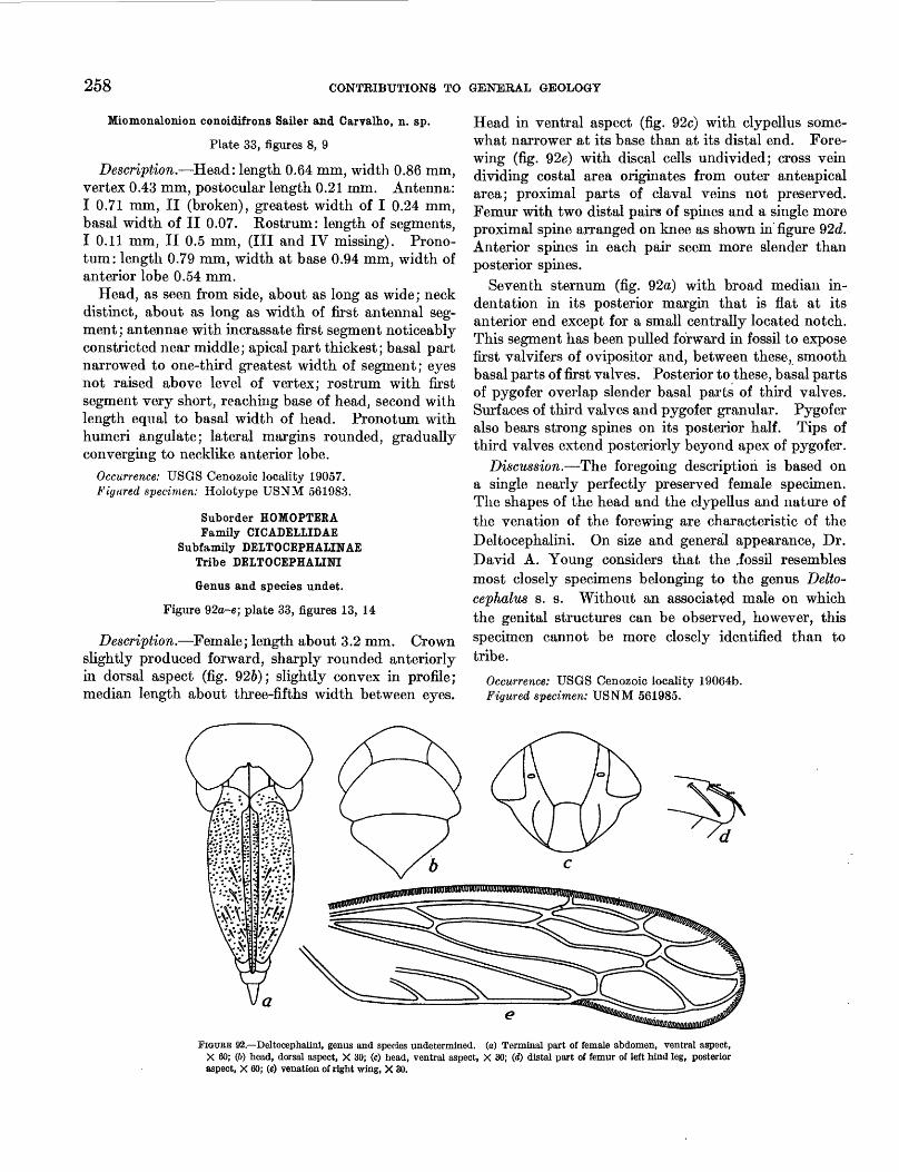
258 CONTRIBUTIONS TO GENERAL GEOLOGY
Miomonalonion conoidifrons Sailer and Carvalho, n. sp.
Plate 33, figures 8, 9
Description.—Head: length 0.64 mm, width 0.86 mm, vertex 0.43 mm, postocular length 0.21 mm. Antenna: I 0.71 mm, II (broken), greatest width of I 0.24 mm, basal width of II 0.07. Rostrum: length of segments, I 0.11 mm, II 0.5 mm, (III and IV missing). Prono- tum: length 0.79 mm, width at base 0.94 mm, width of anterior lobe 0.54 mm.
Head, as seen from side, about as long as wide; neck distinct, about as long as width of first antennal seg ment ; antennae with inerassate first segment noticeably constricted near middle; apical part thickest; basal part narrowed to one-third greatest width of segment; eyes not raised above level of vertex; rostrum with first segment very short, reaching base of head, second with length equal to basal width of head. Pronotum with humeri angulate; lateral margins rounded, gradually converging to necklike anterior lobe.
Occurrence: USGS Cenozoic locality 19057. Figured specimen: Holotype USNM 561983.
Suborder HOMOPTERAFamily CICADELLIDAE
Subfamily DELTOCEPHALINAETribe DELTOCEPHAIINI
Genus and species undet.
Figure 92a-e; plate 33, figures 13, 14
Description.—Female; length about 3.2 mm. Crown slightly produced forward, sharply rounded anteriorly in dorsal aspect (fig. 926); slightly convex in profile; median length about three-fifths width between eyes.
Head in ventral aspect (fig. 92c) with clypellus some what narrower at its base than at its distal end. Fore- wing (fig. 92e) with discal cells undivided; cross vein dividing costal area originates from outer anteapical area; proximal parts of claval veins not preserved. Femur with two distal pairs of spines and a single more proximal spine arranged on knee as shown in figure 92d. Anterior spines in each pair seem more slender than posterior spines.
Seventh sternum (fig. 92a) with broad median in dentation in its posterior margin that is flat at its anterior end except for a small centrally located notch. This segment has been pulled forward in fossil to expose first valvifers of ovipositor and, between these, smooth basal parts of first valves. Posterior to these, basal parts of pygofer overlap slender basal parts of third valves. Surf aces of third valves and pygofer granular. Pygofer also bears strong spines on its posterior half. Tips of third valves extend posteriorly beyond apex of pygofer.
Discussion.—The foregoing description is based on a single nearly perfectly preserved female specimen. The shapes of the head and the clypellus and nature of the venation of the forewing are characteristic of the Deltocephalini. On size and general appearance, Dr. David A. Young considers that the .fossil resembles most closely specimens belonging to the genus Delto- cephalus s. s. Without an associated male on which the genital structures can be observed, however, this specimen cannot be more closely identified than to tribe.
Occurrence: USGS Cenozoic locality 19064b. Figured specimen: USNM 561985.
FIGURE 92.—Deltoeephalini, genus and species undetermined, (a) Terminal part of female abdomen, ventral aspect, X 60; (&) head, dorsal aspect, X 30; (c) head, ventral aspect, X 30; (d) distal part of femur of left hind leg, posterior aspect, X 60; («) venation of right wing, X 30.
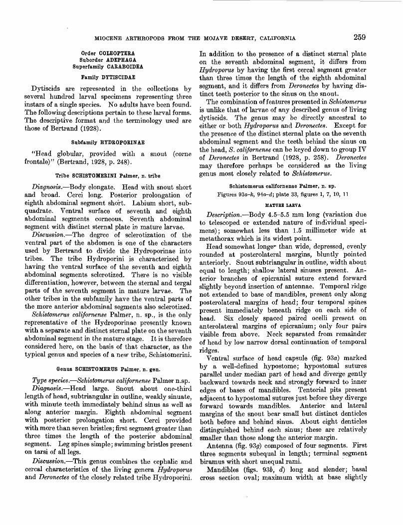
MIOCENE AETHROPODS FROM THE MOJAVE DESERT, CALIFORNIA 259
Order COIEOPTERASuborder ADEPHAGA
Superfamily CARABOIDEA
Family DYTISCIDAE
Dytiscids are represented in the collections by several hundred larval specimens representing three instars of a single species. No adults have been found. The following descriptions pertain to these larval forms. The descriptive format and the terminology used are those of Bertrand (1928).
Subfamily HYDROPORINAE
"Head globular, provided with a snout (corne frontale)" (Bertrand, 1928, p. 248).
Tribe SCHISTOMERINI Palmer, n. tribe
Diagnosis.—Body elongate. Head with snout short and broad. Cerci long. Posterior prolongation of eighth abdominal segment short. Labium short, sub- quadrate. Ventral surface of seventh and eighth abdominal segments corneous. Seventh abdominal segment with distinct sternal plate in mature larvae.
Discussion,—The degree of sclerotization of the ventral part of the abdomen is one of the characters used by Bertrand to divide the Hydroporinae into tribes. The tribe Hydroporini is characterized by having the ventral surface of the seventh and eighth abdominal segments sclerotized. There is no visible differentiation, however, between the sternal and tergal parts of the seventh segment in mature larvae. The other tribes in the subfamily have the ventral parts of the more anterior abdominal segments also sclerotized.
Schistomerus californense Palmer, n. sp., is the only representative of the Hydroporinae presently known with a separate and distinct sternal plate on the seventh abdominal segment in the mature stage. It is therefore considered here, on the basis of that character, as the typical genus and species of a new tribe, Schistomerini.
Genus SCHISTOMERUS Palmer, n. gen.
Type species.—Schistomerus californense Palmer n.sp.Diagnosis.—Head large. Snout about one-third
length of head, subtriangular in outline, weakly sinuate, with minute teeth immediately behind sinus as well as along anterior margin. Eighth abdominal segment with posterior prolongation short. Cerci provided with more than seven bristles; first segment greater than three tunes the length of the posterior abdominal segment. Leg spines simple; swimming bristles present on tarsi of all legs.
Discussion.—This genus combines the cephalic and cereal characteristics of the living genera Hydroporus and Deronectes of the closely related tribe Hydroporini.
In addition to the presence of a distinct sternal plate on the seventh abdominal segment, it differs from Hydroporus by having the first cereal segment greater than three times the length of the eighth abdominal segment, and it duffers from Deronectes by having dis tinct teeth posterior to the sinus on the snout.
The combination of features presented in Schistomerus is unlike that of larvae of any described genus of living dytiscids. The genus may be directly ancestral to either or both Hydroporus and Deronectes. Except for the presence of the distinct sternal plate on the seventh abdominal segment and the teeth behind the sinus on the head, S. californense can be keyed down to group IV of Deronectes in Bertrand (1928, p. 258). Deronectes may therefore perhaps be considered as the living genus most closely related to Schistomerus.
Schistomerus californense Palmer, n. sp. Figures 93o-fe, 94a-d; plate 33, figures 1, 7, 10, 11
MATURE IAEVA
Description.—Body 4.5-5.5 mm long (variation due to telescoped or extended nature of individual speci mens); somewhat less than 1.5 millimeter wide at metathorax which is its widest point.
Head somewhat longe*1 than wide, depressed, evenly rounded at posterolateral margins, bluntly pointed anteriorly. Snout subtriangular in outline, width about equal to length; shallow lateral sinuses present. An terior branches of epicranial suture extend forward slightly beyond insertion of antennae. Temporal ridge not extended to base of mandibles, present only along posterolateral margins of head; four temporal spines present immediately beneath ridge on each side of head. Six closely spaced paired ocelli present on anterolateral margins of epicranium; only four pairs visible from above. Neck separated from remainder of head by low narrow dorsal continuation of temporal ridges.
Ventral surface of head capsule (fig. 93a) marked by a well-defined hypostome; hypostomal sutures parallel under median part of head and diverge gently backward towards neck and strongly forward to inner edges of bases of mandibles. Tentorial pits present adjacent to hypostomal sutures just before they diverge forward towards mandibles. Anterior and lateral margins of the snout bear small but distinct denticles both before and behind sinus. About eight denticles distinguished behind each sinus; these are relatively smaller than those along the anterior margin.
Antenna (fig. 930) composed of four segments. First three segments subequal in length; terminal segment biramus with short unequal rami.
Mandibles (figs. 936, d) long and slender; basal cross section oval; maximum width at base slightly
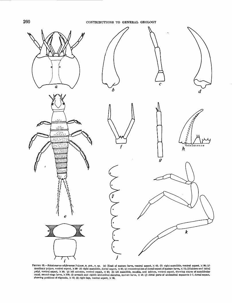
260 CONTRIBUTIONS TO GENERAL GEOLOGY
FIQUEE 93.—Schistomerua colifornense Palmer, n. gen., n. sp. (a) Head of mature larva, ventral aspect, X 45; (6) right mandible, ventral aspect, X 80; (c) maxillary palpus, ventral aspect, X 90 1 (d) right mandible, dorsal aspect, X 90; (e) reconstruction of dorsal aspect of mature larva, X12; (f) labium and labial palpi, ventral aspect, X 90; (a) left antenna, ventral aspect, X 90; (ft) left mandible, maxilla, and labium, ventral aspect, showing course of mandibular canal, second-stage larva, X120; (z) seventh and eighth abdominal sternites, mature larva, X 45; (j) distal parts of abdominal segments 1-7, dorsal aspect, showing positions of stigmata, X 45; (fc) right legs, ventral aspect, X 30.
MIOCENE ARTHROPODS FROM THE MOJAVE DESERT, CALIFORNIA
more than one-fourth mandibular length. Ventral condyle prominent. Mandible not provided with dis tinct groove; rather, there is a sub terminal opening in ventral surface of tip and a canal extending gradually dorsad from this opening along inner edge of mandible and leaving mandible at its inner dorsal margin (fig. 93A). From this point it turns sharply inward in horizontal plane perpendicular to axial line of head and extends to region of mouth.
Maxilla (fig. 93c) with coxal part subconical in out line, length slightly greater than width. Maxillary palpus composed of three segments; second segment shorter than first; terminal segment very short. Single secondary segment developed at base of primary seg ment.
Labium (fig. 93/) short, broad; width about 1% times length. Labial palpus composed of two segments; terminal segment shorter than proximal segment. Slight median anterior indentation on ligula. Sides of indentation, adjacent to inner dorsal margins of each labial palpus, marked by a cluster of three fine hairs.
Thorax (fig. 93e) with length of prothorax about 1% length of mesothorax and of metathorax. Ventral features not known. Spines apparent on specimens studied only along the posterior margin and near mid- line on posterior part of each segment. Posterior margin of each segment bears about 20 evenly spaced spines.
Legs (fig. 93&) lengthen from front to back. Tibia and tarsus of each leg subequal in length and each only about yz-% length of femur. Trochanter small and essentially like that of living dytiscids. Coxa of anterior leg slightly longer than femur and about 1% times greater in diameter. Coxae of middle and posterior pairs of legs slightly shorter than respective femora but have diameters nearly twice those of femora.
Femur, tibia, and tarsus of each leg are provided with single rows of relatively strong spines along anterodorsal and anteroventral margins, in addition to a few strong spines scattered on anterior and posterior surfaces. Number of spines in each row ranges from 3 each on tibia and tarsus of anterior pair of legs to about 7 each on tibia and tarsus of middle and posterior pair of legs. In addition to these spines, tibia and tarsus of each leg are provided with posterodorsal row of slender closely spaced swimming bristles. A second stage larva shows swimming bristles also on femur. Distribution of spines on femur, tibia, and tarsus shown diagrammati- cally in reconstruction (fig. 93e). Scattered spines have been observed on coxae but details of distribution could not be accurately determined.
Abdomen relatively long and slender, tapered pos teriorly Anterior segments short and broad; width and
length of posterior segments subequal. Length of seventh segment slightly less than that of eighth. Distribution of stigmata on segments 1-7 shown on figure 93J, Positions of stigmata of eighth segment shown in figure 93e.
Only seventh and eighth segments have sclerotized ventral parts. Seventh segment has a well-defined transversely subovate sternal plate in mature larval stage (fig. 93; pi. 33, fig. 1). This plate is separated by membranes from ends of tergal part of segment. Eighth segment completely sclerotized in all stages. Posterior projection of eighth segment short and broad.
Dorsal and ventral surfaces of abdomen bear scattered spines. Because of membranous nature of most of ventral surface, distribution of spines on ventral surface of first six segments cannot be accurately determined. Sternal plate bears many evenly dis tributed moderately strong spines. On dorsal surface there is a row of strong spines along posterior margin of each of first seven segments. Other dorsal surface spines seem to be more abundant on posterior segments than on anterior segments.
Cerci of second larval stage, long and slender probably not greatly different from those of mature larva. Length of first cereal segment about seven times that of length of eighth abdominal segment. Second cereal segment short and slender, about one-tenth length of first segment. Relatively strong spines scattered throughout length of first segment without any order. Entire surface of first segment of some specimens covered with minute closely spaced spinules. These spinules do not seem to be present on preserved parts of cerci of mature larvae. Unbroken cerci have not been found for mature larvae.
TRACHEAL SYSTEM
Parts of the tracheal system of the larval head have been preserved in several specimens of mature larvae. The best of these is illustrated on plate 33, figure 7, and has been used as the basis for the following discussion. The specimens studied all have been replaced by celestite. Only the tracheal system and the brain (?) can be distinguished. The dorsal and ventral tracheal systems maintain the same relative positions to each other and to the brain (?) that might be expected in a live specimen. It is probable that there has not been a great deal of collapse of the system by the disintegra tion of the body tissues before fossilization. Annula- tions could not be observed on the tracheae of any specimens.
In the illustrated specimen the tracheae may be hollow, as some of the branches are incomplete and their courses are indicated by lines of small bubbles. The tracheae in another specimen appear as transparent
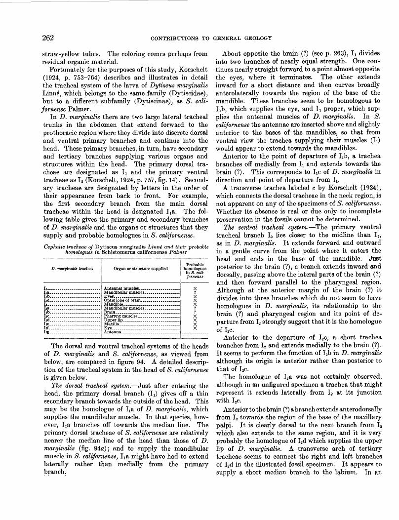
262 CONTRIBUTIONS TO GENERAL GEOLOGY
straw-yellow tubes. The coloring comes perhaps from residual organic material.
Fortunately for the purposes of this study, Korschelt (1924, p. 753-764) describes and illustrates in detail the tracheal system of the larva of Dytiscus marginalis Linne", which belongs to the same family (Dytiscidae), but to a different subfamily (Dytiscinae), as S. cali fornense Palmer.
In D. marginalis there are two large lateral tracheal trunks in the abdomen that extend forward to the prothoracic region where they divide into discrete dorsal and ventral primary branches and continue into the head. These primary branches, in turn, have secondary and tertiary branches supplying various organs and structures within the head. The primary dorsal tra cheae are designated as Ix and the primary ventral tracheae as I2 (Korschelt, 1924, p. 757, fig. 14). Second ary tracheae are designated by letters in the order of their appearance from back to front. For example, the first secondary branch from the main dorsal tracheae within the head is designated Jia. The fol lowing table gives the primary and secondary branches of D. marginalis and the organs or structures that they supply and probable homologues in S. ealijornense.
Cephalic tracheae of Dytiscus marginalis Linn& and their probable homologues in Schistomerus californense Palmer
D. marginalis trachea
Ii....— .............. ......Iia.. ........................lib..........................IlC--_ —— . —— —— —— —— -. —— -J2.. .........................l».. ........................I2b.. ........................T,nI2d.......— ................l«.. ........................I2f... .......................I2g.. ........................
Organ or structure supplied
Brain.-.......-.-...-..-...—......
Probable homologues In S. cali fornense
X1
X??
The dorsal and ventral tracheal systems of the heads of D. marginalis and S. californense, as viewed from below, are compared in figure 94. A detailed descrip tion of the tracheal system in the head of S. californense is given below.
The dorsal tracheal system.—Just after entering the head, the primary dorsal branch (10 gives off a thin secondary branch towards the outside of the head. This may be the homologue of Ija of D. marginalis, which supplies the mandibular muscle. In that species, how ever, Ija branches off towards the median line. The primary dorsal tracheae of S. californense are relatively nearer the median line of the head than those of D. marginalis (fig. 94a); and to supply the mandibular muscle in S. californense, Iia might have had to extend laterally rather than medially from the primary branch.
About opposite the brain (?) (see p. 263), Ii divides into two branches of nearly equal strength. One con tinues nearly straight forward to a point almost opposite the eyes, where it terminates. The other extends inward for a short distance and then curves broadly anterolaterally towards the region of the base of the mandible. These branches seem to be homologous to lib, which supplies the eye, and Ii proper, which sup plies the antennal muscles of D. marginalis. In S. californense the antennae are inserted above and slightly anterior to the bases of the mandibles, so that from ventral view the trachea supplying their muscles (Ii) would appear to extend towards the mandibles.
Anterior to the point of departure of lib, a trachea branches off medially from Ii and extends towards the brain (?). This corresponds to Ixc of D. marginalis in direction and point of departure from It .
A transverse trachea labeled c by Korschelt (1924), which connects the dorsal tracheae in the neck region, is not apparent on any of the specimens of S. californense. Whether its absence is real or due only to incomplete preservation in the fossils cannot be determined.
The ventral tracheal system.—The primary ventral tracheal branch I2 lies closer to the midline than I1; as in D. marginalis. It extends forward and outward in a gentle curve from the point where it enters the head and ends in the base of the mandible. Just posterior to the brain (?), a branch extends inward and dorsally, passing above the lateral parts of the brain (?) and then forward parallel to the pharyngeal region. Although at the anterior margin of the brain (?) it divides into three branches which do not seem to have homologues in D. marginalis, its relationship to the brain (?) and pharyngeal region and its point of de parture from I2 strongly suggest that it is the homologue of I2c.
Anterior to the departure of I2c, a short trachea branches from I2 and extends medially to the brain (?). It seems to perform the function of I2b in D. marginalis although its origin is anterior rather than posterior to that of I2c.
The homologue of I2a was not certainly observed, although in an unfigured specimen a trachea that might represent it extends laterally from I2 at its junction with I2c.
Anterior to the brain (?) a branch extends anterodorsally from I2 towards the region of the base of the maxillary palpi. It is clearly dorsal to the next branch from I2 which also extends to the same region, and it is very probably the homologue of I2d which supplies the upper lip of D. marginalis. A transverse arch of tertiary tracheae seems to connect the right and left branches of I2d in the illustrated fossil specimen. It appears to supply a short median branch to the labium. In an
MIOCENE ARTHROPODS PROM THE MOJAVE DESERT, CALIFORNIA 26S
a
FIGURE 84.—Comparison of cephalic tracheae of Schistomerm californense Palmer, n. gen., n. sp., and Dytiscus marginalia Linn6 hi ventral aspect, (a) Dorsal tracheae, D. marginalis, X 5; (&) dorsal tracheae, S. cali fornense, X 46; (c) ventral tracheae, D. marginalif, X 5; (d) ventral tracheae, S. californense, X 45. Figures a, c after Korschelt, 1924, fig. 14. br brain; Ii primary dorsal tracheae; Ii primary ventral tracheae; sub script letters refer to secondary tracheae. (See table p. 262.)
unfigured specimen a single branch appears to extend from near the base of I2d on the left side of the head to the region of the labium. This is a generally homologous condition to that observed in D. marginalis where the labium is supplied by tertiary branches from Pd.
Just anterior to the departure of I2d, a branch extends to the region of the base of the maxillary palp. It is the probable homologue of I2e which supplies the same structure in D. marginalis.
Anterior to the departure of I2e, a short trachea extends laterally from I2 towards the termination of lib. If the homologies of lib are correctly interpreted, then the short branch from I2 would also extend to the eye region and be the homologue of I2f.
The homologue of I2g was not observed.The homology of the tracheal systems in the heads of
larvae of S. californense and D. marginalis, at opposite ends of the Dytiscidae, indicates that within the family the system maintains considerable uniformity in the functions and disposition of the primary and secondary tracheal branches.
THE BRAIN (?)
The heads of 8. californense that have the tracheal system preserved also have a poorly defined semi- opaque subquadrate centrally located area that may represent the remains of the brain. Its position is shown by the shaded areas in figure 946, d. Its outline
264 CONTRIBUTIONS TO GENERAL GEOLOGY
cannot be determined accurately, and the prominent optic lobes characteristic of living dytiscids are not apparent. The relative positions of the brain and tracheae of D. marginalis and the brain (?) and tracheae in S. californense, however, seem to support the infer ence that this semiopaque area does indeed represent the remains of the brain. In both species tracheae I2c of the ventral system branch from I2 posterior to the brain and pass over it to the region of the pharynx, thus being partly obscured in ventral view by the brain; tracheae I2b of the ventral system go to the ventral surface of the brain; and tracheae l^e go to the antero- lateral corners of the brain.
SECOND-STAGE LARVA
The larvae of this stage seem to be like the mature larvae except for the fact that some individuals have minute, closely spaced spinules on the cerci. Their presence may be a function of preservation.
LARVUIE
The larvule differs from the mature larva principally in the relative lengths of the segments of the antennae, maxillary palpi, and labial palpi; in the distribution of spines on the legs and cerci; and by lacking any ap parent sternal plate on the seventh abdominal segment. The distal segments of the antennae, maxillary palpi, and labial palpi are relatively longer than the proximal segments in the larvule. In the mature larva the lengths of these segments are equal to or shorter than the more proximal segments. The legs of the larvule do not appear to bear swimming bristles, and the claws seem to be relatively longer and more slender than in the mature larvae. The leg surfaces are covered with closely spaced minute spines. The cerci also bear a few long, slender spines in this stage. Their number is less, and their length is relatively greater than in the second-stage and mature larvae.
In accordance with Dyar's law (Thompson, 1942, p. 169, 165), the width of the head capsule increases by a constant factor from instar to instar. In successive instars of this species, the width of the head capsule increases by a factor between 1.54 and 1.57.
Comparative dimensions, in millimeters, for the successive larval stages of S. californense Palmer
Width of head....— ........... _ ...............
Larvule
1.3.45.38.20.10.10.80
2d-stage larva
3.75.62.32.20.20
1.40
Mature larva
4. 5-5. 51.150.90
0. 40-0. 450.300.35
12.5
• Estimated.
DISCUSSION
Among living dytiscids, this species most nearly resembles Deronectes striatettus LeConte. The dimen sions of the head capsule and the details of the cephalic appendages are nearly identical. The presence of distinct denticles behind the sinus of S, californense Palmer is one of the principal characters that serves to distinguish the head of this species from that of D. striatellus. S. californense also has fewer temporal spines (4 versus 7), and some of the finer hairlike spines that are present on the head of D. striatettus have not been observed on the fossil specimens.
The principal differences in the spine distribution on the postcephalic parts of the body in the two species is the greater number of spines on the dorsal surface of the thorax in D. striatellus. The distribution of spines on the cerci and legs of the two species is similar.
D. striatellus lacks any indication of a sternal plate on the seventh abdominal segment in the mature larva.
S. californense Palmer may be the direct ancestor of D. striatellus LeConte. From the available information Schistomerus could well be the "root stock" from which the living genera Hydroporus and Deronectes were derived. The presence of teeth behind the sinus on the snout is one of the characteristics of Hydroporus; the general outline of the head is most similar to species of both Hydroporus and Deronectes; and the relatively long cerci are one of the characteristics of Deronectes.
In S. californense perhaps the most striking features of evolutionary significance are the presence in second- stage and mature larvae of characteristics found only in larvules of living hydroporines. These are the distinct sternal plate on the seventh abdominal segment in the second-stage and the mature larvae and the presence of minute closely spaced spinules on the cerci of some second-stage larvae. The small number of temporal spines may also have some evolutionary significance.
Occurrence: USGS Cenozoic localities 19057-19064e; 19065b- 19067.
Figured specimens: Holotype USNM 561989 from USGS Cenozoic locality 19057, USNM 561990a-c from USGS Cenozoic locality 19057, USNM 561991 from USGS Cenozoic locality 19066, USNM 561992 from USGS Cenozoic locality 19058.
Family STAPHYUNIDAE Genus CARPELLMUS Leech, 1819 ( = Trogophloeus Mannerheim,
1831) Carpelimus sp.
Figure 95c, d; plate 33, figure 2
A single imperfectly preserved male staphylinid beetle approximately 2 millimeters long is present in the collections. The observable features are the head and its appendages, except for the distal antennal segments and the right mandible; the pronotum and left elytron
MIOCENE ARTHROPODS FROM THE MOJAVE DESERT, CALIFORNIA 265
of the thorax in dorsal view and the metasternum and some of the proximal leg segments in ventral view; six abdominal segments; and the tips of the genitalia. Adventitious material obscures the posterior part of the head and most of the sternum in ventral view. The margins of the elytron are somewhat ragged, apparently owing to imperfect silicification, so that the outline shown on figure 95c might be modified slightly if a better specimen were available.
The distinctive features of the specimen are the size and position of the eyes, position of the antennae, shape of the pronotum, character of the leg segments, and number and character of the abdominal segments.
The eyes are prominent on the sides of the head with a distinct area slightly more than one-half their length separating them from the posterior margin of the head. The antennae are inserted at the front corners of the head.
The pronotum has a modified hexagonal outline. The sides of the anterior two-thirds are strongly bowed outward, and the sides of the posterior third are sub- parallel. The anterior margin is straight, and the pos terior margin is bluntly pointed towards the rear.
The coxa of the first pair of legs is elongate. That of the second pair of legs is subhemispherical and bears many closely spaced spines on its convex posterior face. That of the third pair of legs is subtriangular in outline and bears an elevated protuberance to which the trochanter is attached. The trochanters of all three pairs of legs appear to be small but prominent, especially on the second and third legs. Exact details of their character could not be determined. The femur of the left foreleg has a slender base and gradually expands to a moderately bulbous tip. It bears a row of spines along its anterior margin.
Six abdominal segments are visible in ventral view (fig. 94d). The first segment is relatively long compared with those that follow, but this may be due only to the fact that the abdomen is somewhat telescoped. The sixth visible segment has a distinct median slit in its posterior margin. Posterior to this segment a smaller, broadly bilobed structure probably represents part of the genitalia.
The shape of the pronotum and position of the antennae and eyes are characteristic of Carpelimus, and it is on these characters combined with the overall body shape that the generic identification of the speci men is based. The morphologic position of the ab dominal segment bearing the slit might have a bearing on the identification of the specimen. According to R. E. Blackwelder (oral communication, April 1955), the slit could be on either the seventh or eighth mor phologic segment of the abdomen. It is not possible to determine from the specimen which morphologic seg
ment bears the slit. The slit should be in the eighth morphologic segment if the specimen belongs in Carpel imus.
Occurrence: USGS Cenozoic locality 19057. Figured specimen: USNM 561993.
Family CYBOCEPHALIDAE Genus CYBOCEPHALUS Erichson
Cybocephalus cf. C. californicus Horn
Figure 95a, 6; plate 33, figures 5, 6Cybocephalus californicus Horn, 1879, Entomol. Soc. Am. Trans.,
v. 7, p. 320.
A single specimen of a small smooth beetle with 4- jointed tarsi and 11 segments to the antennae is assign able on these characters to the Cybocephalidae. The shape and size of the body and the head; evenly rounded posterolateral margins of the prothorax (pi. 33, fig. 5); form of the antenna (fig. 95<z), which consists of 2 conspicuous basal segments, 3 slender poorly defined segments, 3 slender well-defined segments, and a broad, 3-segmented club; and slender segments of the tarsi (fig. 956) are features common to the fossil specimen and the living species Cybocephalus californicus Horn. The sternum of the fossil is covered with debris that cannot be removed, color is gone, the wings and genitalia cannot be observed, and some surface details are obscure. For these reasons, determination of the exact identity of the fossil with C. californicus is not possible. Its affinities according to L. M. Walkley (oral communication June 1955) are closer to C. californicus, however, than to any other species of Cybocephalus in the collections of the United States National Museum.
R. A. Crowson, who provided the generic identifica tion, kindly compared the fossil with species of Cybo cephalus at the British Museum. He indicated need for a modern revision of the genus before accurate comparisons could be made at the species level. He also noted that the specimen is preserved in fully extended condition, a common appearance in drowned insects.
Occurrence: USGS Cenozoic locality 19057. Figured specimen: USNM 561994.
Order DIPTERA Family CERATOPOGONIDAE
Genus DASYHEIEA Kieffer
This genus is represented by larvae, pupae, and male and female adults. The quality of preservation of the pupae and adults makes comparison with modern forms at the species level possible. One new species (Dasyhelea stenoceras) based on pupae and a new subspecies of a
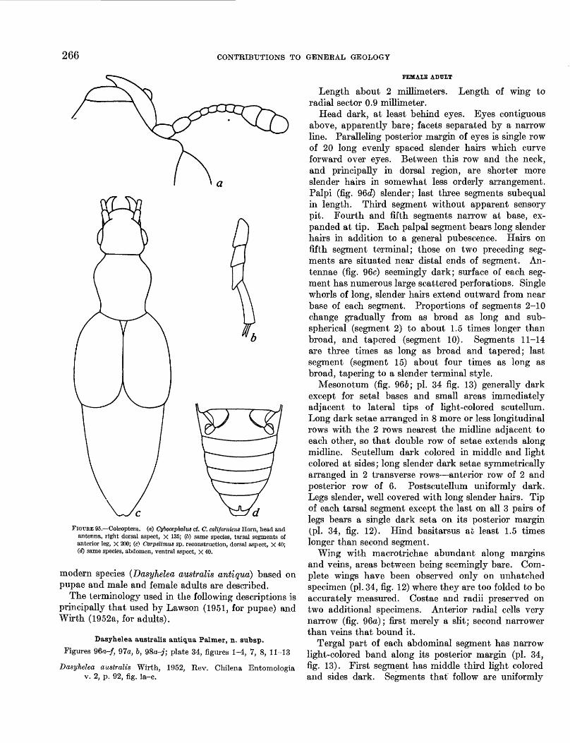
266 CONTRIBUTIONS TO GENERAL GEOLOGY
FIGURE 95.—Coleoptera. (a) Cybocepkalw cf. C. californiais Horn, head and antenna, right dorsal aspect, X 135; (6) same species, tarsal segments of anterior leg, X 200; (c) Carpelimiis sp. reconstruction, dorsal aspect, X 40; (d) same species, abdomen, ventral aspect, X 40.
modern species (Dasyhelea australis antiqua) based on pupae and male and female adults are described.
The terminology used in the following descriptions is principally that used by Lawson (1951, for pupae) and Wirth (1952a, for adults).
Dasyhelea australis antiqua Palmer, n. subsp.
Figures 96o-/, 97a, b, QSa-j; plate 34, figures 1-4, 7, 8, 11-13
Dasyhelea australis Wirth, 1952, Rev. Chilena Entomologia v. 2, p. 92, fig. la-c.
FEMALE ADULT
Length about 2 millimeters. Length of wing to radial sector 0.9 millimeter.
Head dark, at least behind eyes. Eyes contiguous above, apparently bare; facets separated by a narrow line. Paralleling posterior margin of eyes is single row of 20 long evenly spaced slender hairs which curve forward over eyes. Between this row and the neck, and principally in dorsal region, are shorter more slender hairs in somewhat less orderly arrangement. Palpi (fig. 96c?) slender; last three segments subequal in length. Third segment without apparent sensory pit. Fourth and fifth segments narrow at base, ex panded at tip. Each palpal segment bears long slender hairs in addition to a general pubescence. Hairs on fifth segment terminal; those on two preceding seg ments are situated near distal ends of segment. An tennae (fig. 96c) seemingly dark; surface of each seg ment has numerous large scattered perforations. Single whorls of long, slender hairs extend outward from near base of each segment. Proportions of segments 2-10 change gradually from as broad as long and sub- spherical (segment 2) to about 1.5 times longer than broad, and tapered (segment 10). Segments 11-14 are three times as long as broad and tapered; last segment (segment 15) about four times as long as broad, tapering to a slender terminal style.
Mesonotum (fig. 966; pi. 34 fig. 13) generally dark except for setal bases and small areas immediately adjacent to lateral tips of light-colored scutellum. Long dark setae arranged in 8 more or less longitudinal rows with the 2 rows nearest the midline adjacent to each other, so that double row of setae extends along midline. Scutellum dark colored in middle and light colored at sides; long slender dark setae symmetrically arranged in 2 transverse rows—anterior row of 2 and posterior row of 6. Postscutellum uniformly dark. Legs slender, well covered with long slender hairs. Tip of each tarsal segment except the last on all 3 pairs of legs bears a single dark seta on its posterior margin (pi. 34, fig. 12). Hind basitarsus at least 1.5 times longer than second segment.
Wing with macrotrichae abundant along margins and veins, areas between being seemingly bare. Com plete wings have been observed only on unhatched specimen (pi. 34, fig. 12) where they are too folded to be accurately measured. Costae and radii preserved on two additional specimens. Anterior radial cells very narrow (fig. 96a); first merely a slit; second narrower than veins that bound it.
Tergal part of each abdominal segment has narrow light-colored band along its posterior margin (pi. 34, fig. 13). First segment has middle third light colored and sides dark. Segments that follow are uniformly
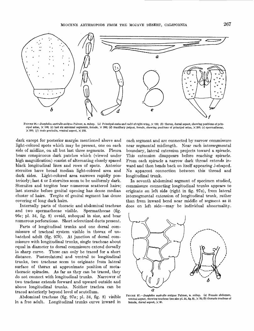
MIOCENE ARTHROPODS FROM THE MOJAVE DESERT, CALIFORNIA 267
FIGURE $b.—Dasyhelea australis antiqua Palmer, n. subsp. (a) Principal costs and radii of right-wing, X 125; (6) thorax, dorsal aspect, showing positions of prin cipal setae, X 100; (c) last six antennal segments, female, X 200; (rf) maxillary palpus, female, showing positions of principal setae, X 250; (e) spermathecae, X 500; (/) male genitalia, ventral aspect, X 210.
dark except for posterior margin mentioned above and light-colored spots which may be present, one on each side of midline, on all but last three segments. Pleura bears conspicuous dark patches which (viewed under high magnification) consist of alternating closely spaced black longitudinal lines and rows of spots. Anterior sternites have broad median light-colored area and dark sides. Light-colored area narrows rapidly pos teriorly; last 4 or 5 sternites seem to be uniformly dark. Sternites and tergites bear numerous scattered hairs; last sternite before genital opening has dense median cluster of hairs. Tergite of genital segment has dense covering of long dark hairs.
Internally parts of thoracic and abdominal tracheae and two spermathecae visible. Spermathecae (fig. 96e; pi. 34, fig. 8) ovoid, subequal in size, and bear numerous perforations. Short sclerotized ducts present.
Parts of longitudinal trunks and one dorsal com missure of tracheal system visible in thorax of un- hatched adult (fig. 976). At junction of dorsal com missure with longitudinal trunks, single tracheae about equal in diameter to dorsal commissure extend dorsally in sharp curve. These can only be traced for a short distance. Posterolateral and ventral to longitudinal trunks, two tracheae seem to originate from lateral surface of thorax at approximate position of meta- thoracic spiracles. As far as they can be traced, they do not connect with longitudinal trunks. Narrower of two tracheae extends forward and upward outside and above longitudinal trunks. Neither trachea can be traced anteriorly beyond level of scutellum.
Abdominal tracheae (fig. 97a; pi. 34, fig. 8) visible in a free adult. Longitudinal trunks curve inward in
each segment and are connected by narrow commissure near segmental midlength. Near each intersegmental boundary, lateral extension projects toward a spiracle. This extension disappears before reaching spiracle. From each spiracle a narrow dark thread extends in ward and then bends back on itself appearing J-shaped. No apparent connection between this thread and longitudinal trunk.
In seventh abdominal segment of specimen studied, commissure connecting longitudinal trunks appears to originate on left side (right in fig. 97a), from lateral intersegmental extension of longitudinal trunk, rather than from inward bend near middle of segment as it does on left side—may be individual abnormality.
FIGURE W.—Dasyhelea australis antiqua Palmer, n. subsp. (a) Female abdomen, ventral aspect, showing tracheae (see also pi. 34, flg.8), X 70; (6) thoracic tracheae of female, dorsal aspect, X 80.
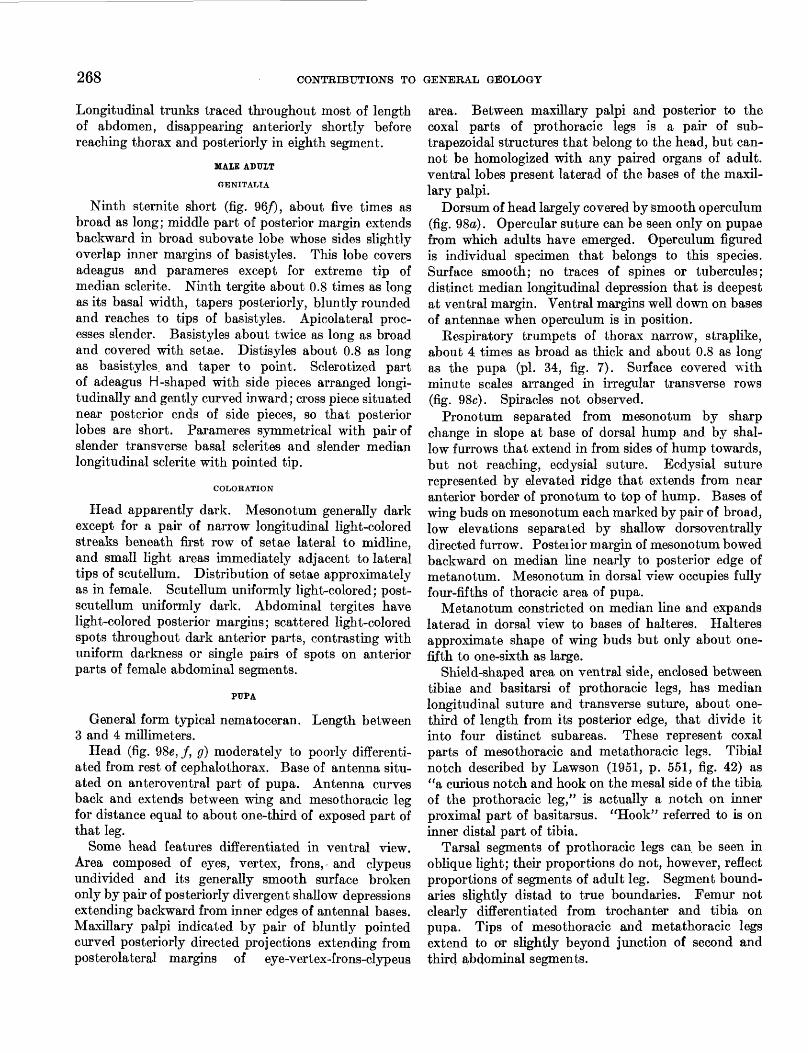
268 CONTRIBUTIONS TO GENERAL GEOLOGY
Longitudinal trunks traced throughout most of length of abdomen, disappearing anteriorly shortly before reaching thorax and posteriorly in eighth segment.
MAIE ADUIT
GENITALIA
Ninth sternite short (fig. 96/), about five times as broad as long; middle part of posterior margin extends backward in broad subovate lobe whose sides slightly overlap inner margins of basistyles. This lobe covers adeagus and parameres except for extreme tip of median sclerite. Ninth tergite about 0.8 times as long as its basal width, tapers posteriorly, bluntly rounded and reaches to tips of basistyles. Apicolateral proc esses slender. Basistyles about twice as long as broad and covered with setae. Distisyles about 0.8 as long as basistyles and taper to point. Sclerotized part of adeagus H-shaped with side pieces arranged longi tudinally and gently curved inward; cross piece situated near posterior ends of side pieces, so that posterior lobes are short. Parameres symmetrical with pair of slender transverse basal sclerites and slender median longitudinal sclerite with pointed tip.
COLORATION
Head apparently dark. Mesonotum generally dark except for a pair of narrow longitudinal light-colored streaks beneath first row of setae lateral to midline, and small light areas immediately adjacent to lateral tips of scutellum. Distribution of setae approximately as in female. Scutellum uniformly light-colored; post- scutellum uniformly dark. Abdominal tergites have light-colored posterior margins; scattered light-colored spots throughout dark anterior parts, contrasting with uniform darkness or single pairs of spots on anterior parts of female abdominal segments.
PUPA
General form typical nematoceran. Length between 3 and 4 millimeters.
Head (fig. Q8e, f, g) moderately to poorly differenti ated from rest of cephalothorax. Base of antenna situ ated on anteroventral part of pupa. Antenna curves back and extends between wing and mesothoracic leg for distance equal to about one-third of exposed part of that leg.
Some head features differentiated in ventral view. Area composed of eyes, vertex, frons,- and clypeus undivided and its generally smooth surface broken only by pair of posteriorly divergent shallow depressions extending backward from inner edges of antennal bases. Maxillary palpi indicated by pair of bluntly pointed curved posteriorly directed projections extending from posterolateral margins of eye-vertex-frons-clypeus
area. Between maxillary palpi and posterior to the coxal parts of prothoracic legs is a pair of sub- trapezoidal structures that belong to the head, but can not be homologized with any paired organs of adult, ventral lobes present laterad of the bases of the maxil lary palpi.
Dorsum of head largely covered by smooth operculum (fig. 98a). Opercular suture can be seen only on pupae from which adults have emerged. Operculum figured is individual specimen that belongs to this species. Surface smooth; no traces of spines or tubercules; distinct median longitudinal depression that is deepest at ventral margin. Ventral margins well down on bases of antennae when operculum is in position.
Respiratory trumpets of thorax narrow, straplike, about 4 times as broad as thick and about 0.8 as long as the pupa (pi. 34, fig. 7). Surface covered with minute scales arranged in irregular transverse rows (fig. 98c). Spiracles not observed.
Pronotum separated from mesonotum by sharp change in slope at base of dorsal hump and by shal low furrows that extend in from sides of hump towards, but not reaching, ecdysial suture. Ecdysial suture represented by elevated ridge that extends from near anterior border of pronotum to top of hump. Bases of whig buds on mesonotum each marked by pair of broad, low elevations separated by shallow dorsoventrally directed furrow. Posteiior margin of mesonotum bowed backward on median line nearly to posterior edge of metanotum. Mesonotum in dorsal view occupies fully four-fifths of thoracic area of pupa.
Metanotum constricted on median line and expands laterad in dorsal view to bases of halteres. Halteres approximate shape of wing buds but only about one- fifth to one-sixth as large.
Shield-shaped area on ventral side, enclosed between tibiae and basitarsi of prothoracic legs, has median longitudinal suture and transverse suture, about one- third of length from its posterior edge, that divide it into four distinct subareas. These represent coxal parts of mesothoracic and metathoracic legs. Tibial notch described by Lawson (1951, p. 551, fig. 42) as "a curious notch and hook on the mesal side of the tibia of the prothoracic leg," is actually a notch on inner proximal part of basitarsus. "Hook" referred to is on inner distal part of tibia.
Tarsal segments of prothoracic legs can be seen in oblique light; their proportions do not, however, reflect proportions of segments of adult leg. Segment bound aries slightly distad to true boundaries. Femur not clearly differentiated from trochanter and tibia on pupa. Tips of mesothoracic and metathoracic legs extend to or slightly beyond junction of second and third abdominal segments.
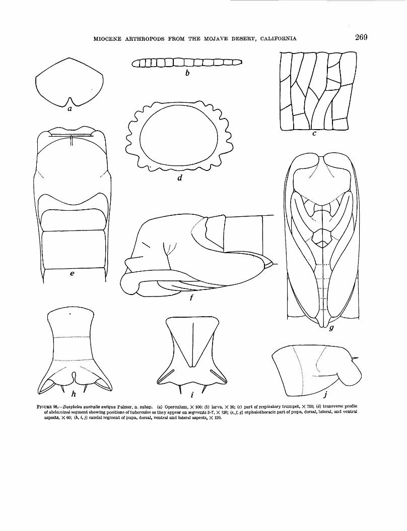
MIOCENE ARTHROPODS FROM THE MOJAVE DESERT, CALIFORNIA 269
FIGURE VS.—Dasyhelea australis antiqua Palmer, n. subsp. (a) Operculum, X 100; (6) larva, X 20; (c) part of respiratory trumpet, X 750; (d) transverse profile of abdominal segment showing positions of tubercules as they appear on segments 3-7, X 120; (e,f, g) cephalothoracic part of pupa, dorsal, lateral, and ventral aspects, X 60; (A, i, j) caudal segment of pupa, dorsal, ventral and lateral aspects, X 120.
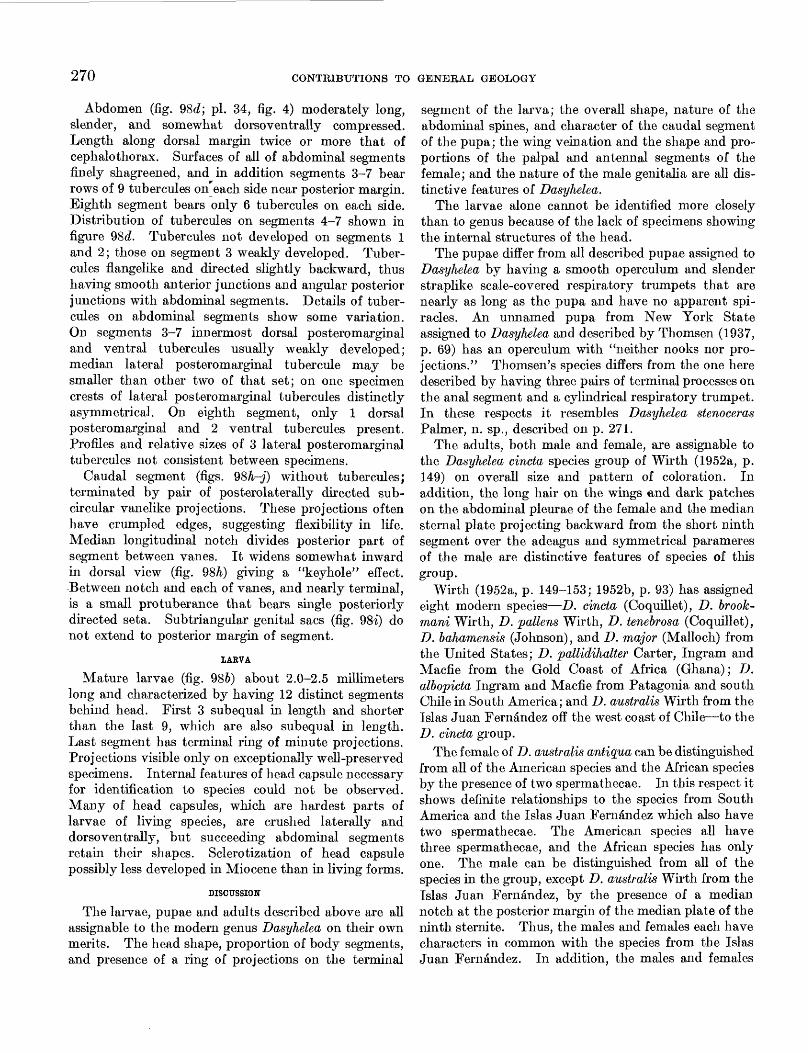
270 CONTRIBUTIONS TO GENERAL GEOLOGY
Abdomen (fig. 98d; pi. 34, fig. 4) moderately long, slender, and somewhat dorsoventrally compressed. Length along dorsal margin twice or more that of cephalothorax. Surfaces of all of abdominal segments finely shagreened, and in addition segments 3-7 bear rows of 9 tubercules on^each side near posterior margin. Eighth segment bears only 6 tubercules on each side. Distribution of tubercules on segments 4-7 shown in figure 98d. Tubercules not developed on segments 1 and 2; those on segment 3 weakly developed. Tuber cules flangelike and directed slightly backward, thus having smooth anterior junctions and angular posterior junctions with abdominal segments. Details of tuber cules on abdominal segments show some variation. On segments 3-7 innermost dorsal posteromarginal and ventral tubercules usually weakly developed; median lateral posteromarginal tubercule may be smaller than other two of that set; on one specimen crests of lateral posteromarginal tubercules distinctly asymmetrical. On eighth segment, only 1 dorsal posteromarginal and 2 ventral tubercules present. Profiles and relative sizes of 3 lateral posteromarginal tubercules not consistent between specimens.
Caudal segment (figs. 9Sh-j) without tubercules; terminated by pair of posterolaterally directed sub- circular vanelike projections. These projections often have crumpled edges, suggesting flexibility in life. Median longitudinal notch divides posterior part of segment between vanes. It widens somewhat inward in dorsal view (fig. 98h) giving a "keyhole" effect. Between notch and each of vanes, and nearly terminal, is a small protuberance that bears single posteriorly directed seta. Subtriangular genital sacs (fig. Q8i) do not extend to posterior margin of segment.
IABVA
Mature larvae (fig. 986) about 2.0-2.5 millimeters long and characterized by having 12 distinct segments behind head. First 3 subequal in length and shorter than the last 9, which are also subequal in length. Last segment has terminal ring of minute projections. Projections visible only on exceptionally well-preserved specimens. Internal features of head capsule necessary for identification to species could not be observed. Many of head capsules, which are hardest parts of larvae of living species, are crushed laterally and dorsoventrally, but succeeding abdominal segments retain their shapes. Sclerotization of head capsule possibly less developed in Miocene than in living forms.
DISCUSSION
The larvae, pupae and adults described above are all assignable to the modern genus Dasyhelea on their own merits. The head shape, proportion of body segments, and presence of a ring of projections on the terminal
segment of the larva; the overall shape, nature of the abdominal spines, and character of the caudal segment of the pupa; the wing veination and the shape and pro portions of the palpal and antennal segments of the female; and the nature of the male genitalia are all dis tinctive features of Dasyhelea.
The larvae alone cannot be identified more closely than to genus because of the lack of specimens showing the internal structures of the head.
The pupae differ from all described pupae assigned to Dasyhelea by having a smooth operculum and slender straplike scale-covered respiratory trumpets that are nearly as long as the pupa and have no apparent spi racles. An unnamed pupa from New York State assigned to Dasyhelea and described by Thomsen (1937, p. 69) has an operculum with "neither nooks nor pro jections." Thomson's species differs from the one here described by having three pairs of terminal processes on the anal segment and a cylindrical respiratory trumpet. In these respects it resembles Dasyhelea stenoceras Palmer, n. sp., described on p. 271.
The adults, both male and female, are assignable to the Dasyhelea cincta species group of Wirth (1952a, p. 149) on overall size and pattern of coloration. In addition, the long hair on the wings and dark patches on the abdominal pleurae of the female and the median sternal plate projecting backward from the short ninth segment over the adeagus and symmetrical parameres of the male are distinctive features of species of this group.
Wirth (1952a, p. 149-153; 1952b, p. 93) has assigned eight modern species—D. cincta (Coquillet), D. brook- mani Wirth, D. pattens Wirth, D. tenebrosa (Coquillet), D. bahamensis (Johnson), and D. major (Malloch) from the United States; D. pallidihalter Carter, Ingram and Macfie from the Gold Coast of Africa (Ghana); D. albopicta Ingrain and Macfie from Patagonia and south Chile in South America; and D. australis Wirth from the Islas Juan Fernandez off the west coast of Chile—to the D. cincta group.
The female of D. australis antigua can be distinguished from all of the American species and the African species by the presence of two spermathecae. In this respect it shows definite relationships to the species from South America and the Islas Juan Fernandez which also have two spermathecae. The American species all have three spermathecae, and the African species has only one. The male can be distinguished from all of the species in the group, except D. australis Wirth from the Islas Juan Fernandez, by the presence of a median notch at the posterior margin of the median plate of the ninth sternite. Thus, the males and females each have characters in common with the species from the Islas Juan Fernandez. In addition, the males and females
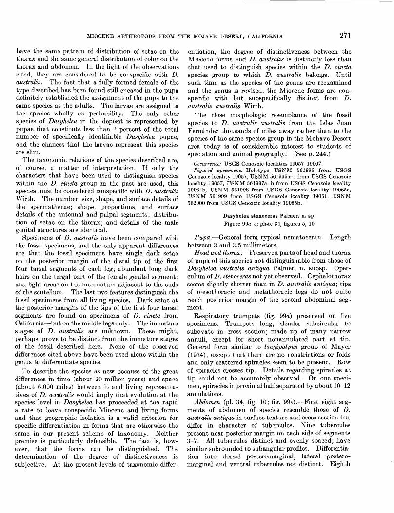
MIOCENE ARTHROPODS FEOM THE MOJAVE DESEET, CALIFORNIA 271
have the same pattern of distribution of setae on the thorax and the same general distribution of color on the thorax and abdomen. In the light of the observations cited, they are considered to be conspecific with D. australis. The fact that a fully formed female of the type described has been found still encased in the pupa definitely established the assignment of the pupa to the same species as the adults. The larvae are assigned to the species wholly on probability. The only other species of Dasyhelea in the deposit is represented by pupae that constitute less than 2 percent of the total number of specifically identifiable Dasyhelea pupae, and the chances that the larvae represent this species are slim.
The taxonomic relations of the species described are, of course, a matter of interpretation. If only the characters that have been used to distinguish species within the D. cincta group in the past are used, this species must be considered conspecific with D. australis Wirth. The number, size, shape, and surface details of the spermathecae; shape, proportions, and surface details of the antennal and palpal segments; distribu tion of setae on the thorax; and details of the male genital structures are identical.
Specimens of D. australis have been compared with the fossil specimens, and the only apparent differences are that the fossil specimens have single dark setae on the posterior margin of the distal tip of the first four tarsal segments of each leg; abundant long dark hairs on the tergal part of the female genital segment; and light areas on the mesonotum adjacent to the ends of the scutellum. The last two features distinguish the fossil specimens from all living species. Dark setae at the posterior margins of the tips of the first four tarsal segments are found on specimens of D. cincta from California—but on the middle legs only. The immature stages of D. australis are unknown. These might, perhaps, prove to be distinct from the immature stages of the fossil described here. None of the observed differences cited above have been used alone within the genus to differentiate species.
To describe the species as new because of the great differences in time (about 20 million years) and space (about 6,000 miles) between it and living representa tives of D. australis would imply that evolution at the species level in Dasyhelea has proceeded at too rapid a rate to leave conspecific Miocene and living forms and that geographic isolation is a valid criterion for specific differentiation in forms that are otherwise the same hi our present scheme of taxonomy. Neither premise is particularly defensible. The fact is, how ever, that the forms can be distinguished. The determination of the degree of distinctiveness is subjective. At the present levels of taxonomic differ
entiation, the degree of distinctiveness between the Miocene forms and D. australis is distinctly less than that used to distinguish species within the D. cincta species group to which D. australis belongs. Until such tune as the species of the genus are reexamined and the genus is revised, the Miocene forms are con- specific with but subspecifically distinct from D. australis australis Wirth.
The close morphologic resemblance of the fossil species to D. australis australis from the Islas Juan Fernandez thousands of miles away rather than to the species of the same species group hi the Mohave Desert area today is of considerable interest to students of speciation and animal geography. (See p. 244.)
Occurrence: USGS Cenozoic localities 19057-19067.Figured specimens: Holotype USNM 561996 from USGS
Cenozoic locality 19057, USNM 561995a-c from USGS Cenozoic locality 19057, USNM 561997a, b from USGS Cenozoic locality 19064b, USNM 561998 from USGS Cenozoic locality 19065c, USNM 561999 from USGS Cenozoic locality 19061, USNM 562000 from USGS Cenozoic locality 19065b.
Dasyhelea stenoceras Palmer, n. sp.
Figure 99a-e; plate 34, figures 5, 10
Pupa.—General form typical nematoceran. Length between 3 and 3.5 millimeters.
Head and thorax.—Preserved parts of head and thorax of pupa of this species not distinguishable from those of Dasyhelea australis antiqua Palmer, n. subsp. Oper- culum of D. stenoceras not yet observed. Cephalothorax seems slightly shorter than hi D. australis antiqua; tips of mesothoracic and metathoracic legs do not quite reach posterior margin of the second abdominal seg ment.
Kespiratory trumpets (fig. 99a) preserved on five specimens.. Trumpets long, slender subcircular to subovate in cross section; made up of many narrow annuli, except for short nonannulated part at tip. General form similar to longipalpus group of Mayer (1934), except that there are no constrictions or folds and only scattered spiracles seem to be present. Row of spiracles crosses tip. Details regarding spiracles at tip could not be accurately observed. On one speci men, spiracles in proximal half separated by about 10-12 annulations.
Abdomen (pi. 34, fig. 10; fig. 99e).—First eight seg ments of abdomen of species resemble those of D. australis antiqua in surface texture and cross section but differ in character of tubercules. Nine tubercules present near posterior margin on each side of segments 3-7. All tubercules distinct and evenly spaced; have similar subrounded to subangular profiles. Differentia tion into dorsal posteromarginal, lateral postero- marginal and ventral tubercules not distinct. Eighth
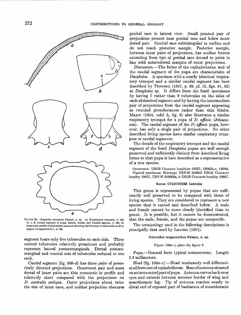
272 CONTKIBUTIONS TO GENERAL GEOLOGY
FIOUBE 99.—Dasyhelea stenoceras Palmer, n. sp. (a) Respiratory trumpet, X 120; (6, c, d) caudal segment of pupa, lateral, dorsal, and ventral aspects, X 120; (e) transverse profile of abdominal segment showing distribution of tubercules as they appear on segments 3-7, X 120.
segment bears only five tubercules on each side. Three central tubercules relatively prominent and probably represent lateral posteromarginals. Dorsal postero- marginal and ventral sets of tubercules reduced to one each.
Caudal segment (fig. 99b-d) has three pairs of poste riorly directed projections. Outermost pair and most dorsal of inner pah's are thin crescentic in profile and relatively short compared with the projections on D. australis antiqua. Outer projections about twice the size of inner ones, and neither projection obscures
genital sacs in lateral view. Small pointed pair of projections present near genital sacs and below inner dorsal pair. Genital sacs subtriangular hi outline and do not reach posterior margin. Posterior margin, between inner pairs of projections, has median furrow extending from tips of genital sacs dorsad to point hi line with anterodorsal margins of outer projections.
Discussion.—The forms of the cephalothorax and of the caudal segment of the pupa are characteristic of Dasyhelea. A specimen with a nearly identical respira tory trumpet and a similar caudal segment has been described by Thomsen (1937, p. 69, pi. 15, figs. 81, 82) as Dasyhelea sp. It differs from the fossil specimens by having 7 rather than 9 tubercules on the sides of each abdominal segment and by having the intermediate pair of projections from the caudal segment appearing as rounded protuberances rather than thin blades. Mayer (1934, tafel A, fig. 6) also illustrates a similar respiratory trumpet for a pupa of D. affinis (Johann- sen). The caudal segment of the D. affinis pupa, how ever, has only a single pair of projections. No other described living species have similar respiratory trum pets or caudal segments.
The details of the respiratory trumpet and the caudal segment of the fossil Dasyhelea pupae are well enough preserved and sufficiently distinct from described living forms so that pupa is here described as a representative of a new species.
Occurrence: USGS Cenozoic localities 19057; 19063b,e; 19066.Figured specimens: Holotype USNM 562001 USGS Cenozoic
locality 19057, USNM 562002a, b USGS Cenozoic locality 19057.
Genus CULICOIDES Latrielle
This genus is represented by pupae that are suffi ciently well preserved to be compared with those of living species. They are considered to represent a new species that is named and described below. A male and female cannot be more closely identified than to genus. It is possible, but it cannot be demonstrated, that the male, female, and the pupae are conspecific.
The terminology used hi the following descriptions is principally that used by Lawson (1951).
Culicoides megacanthus Palmer, n. sp.
Figure lOOa-i; plate 34, figure 6
Pupa.—General form typical nematoceran. Length 2.4 millimeters.
Head (fig. lOOa-c).—Head moderately well differenti ated from rest of cephalothorax. Base of antenna situated on anteroventral part of pupa. Antenna curves back over eyes and extends between anterior border of wing and mesothoracic leg. Tip of antenna reaches nearly to distal end of exposed part of basitarsus of mesothoracic
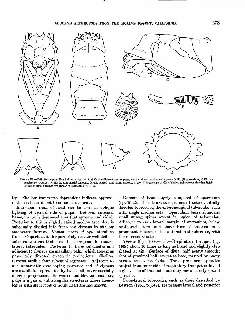
MIOCENE ARTHROPODS FROM THE MOJAVE DESERT, CALIFORNIA 273
FIGTJBE 100.—ddicoides megacanthtts Palmer, n. sp. (a, 6, c) Cephalothoracic part of pupa, ventral, dorsal, and lateral aspects, X 80; (d) opercolum, X 120; («) respiratory trumpet, X 180; (/, g, A) caudal segment, dorsal, ventral, and lateral aspects, X 120; (i) transverse profile of abdominal segment showing distri bution of tubercules as they appear on segments 3-7, X 120.
leg. Shallow transverse depressions indicate approxi mate positions of first 10 antennal segments.
Individual areas of head can be seen in oblique lighting of ventral side of pupa. Between antennal bases, vertex is depressed area that appears undivided. Posterior to this is slightly raised median area that is subequally divided into frons and clypeus by shallow transverse furrow. Ventral parts of eye lateral to frons. Opposite anterior part of clypeus are well-defined subcircular areas that seem to correspond to ventro- lateral tubercules. Posterior to these tubercules and adjacent to clypeus are maxillary palpi, which appear as posteriorly directed crescentic projections. Shallow furrows outline four subequal segments. Adjacent to and apparently overlapping posterior end of clypeus are mandibles represented by two small posteromesially directed projections. Between mandibles and maxillary palpi is a pair of subtriangular structures whose homo- logies with structures of adult head are not known.
Dorsum of head largely composed of operculum (fig. 100</). This bears two prominent anteroventrally directed tubercules, the anteromarginal tubercules, each with single median seta. Operculum bears abundant small strong spines except in region of tubercules. Adjacent to each lateral margin of operculum, below prothoracic horn, and above base of antenna, is a prominent tubercule, the anterodorsal tubercule, with three terminal setae.
Thorax (figs. 100<J-c, e).—Kespiratory trumpet (fig. lOOe) about 10 times as long as broad and slightly club shaped at tip. Surface of distal half nearly smooth; that of proximal half, except at base, marked by many narrow transverse folds. Three prominent spiracles project from inner side of respiratory trumpet in folded region. Tip of trumpet crossed by row of closely spaced spiracles.
Dorsolateral tubercules, such as those described by Lawson (1951, p.J549), are present lateral and posterior
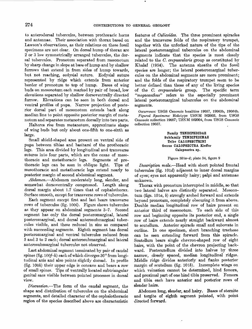
274 CONTEIBUTIONS TO GENERAL GEOLOGY
to anterodorsal tubercules, between prothoracic horns and antennae. Their association with thorax based on Lawson's observations, as their relations on these fossil specimens are not clear. On dorsal hump of thorax are 2 or 3 low symmetrically arranged tubercules, the dor sal tubercules. Pronotum separated from mesonotum by sharp change in slope at base of hump and by shallow furrows that extend in from sides of hump towards, but not reaching, ecdysial suture. Ecdysial suture represented by ridge which extends from anterior border of pronotum to top of hump. Bases of wing buds on mesonotum each marked by pair of broad, low elevations separated by shallow dorsoventrally directed furrow. Elevations can be seen in both dorsal and ventral profiles of pupa. Narrow projection of poste rior dorsal part of mesonotum extends back along median line to point opposite posterior margin of meta- notum and separates metanotum dorsally into two parts.
Halteres rise from metanotum, approximate shape of wing buds but only about one-fifth to one-sixth as large.
Small shield-shaped area present on ventral side of pupa between tibiae and basitarsi of the prothoracic legs. This area divided by longitudinal and transverse sutures into four parts, which are the coxae of meso- thoracic and metathoracic legs. Segments of pro- thoracic legs can be seen in oblique light. Tips of mesothoracic and metathoracic legs extend nearly to posterior margin of second abdominal segment,
Abdomen.—Abdomen moderately long, slender, and somewhat dorsoventrally compressed. Length along dorsal margin about 1.7 times that of cephalothorax. Surface smooth, except for transverse rows of tubercules.
Each segment except first and last bears transverse rows of tubercules (fig. lOOi). Figure shows tubercules as they appear on abdominal segments 3-7. Second segment has only the dorsal posteromarginal, lateral posteromarginal, and dorsal anterosubmarginal tuber cules visible, and these reduced in size as compared with succeeding segments. Eighth segment has dorsal posteromarginal and ventral tubercules reduced from 5 and 3 to 2 each; dorsal anterosubmarginal and lateral anterosubmarginal tubercules not observed.
Last abdominal segment terminated by pair of caudal spines (fig. 10Q/-A.) each of which diverges 30° from longi tudinal axis and also points slightly dorsad. In profile (fig. 100A) their upper edge is concave and bears a row of small spines. Tips of ventrally located subtriangular genital sacs visible between pointed processes in dorsal view.
Discussion.—The form of the caudal segment, the shape and distribution of tubercules on the abdominal segments, and detailed character of the cephalothoracic region of the species described above are characteristic
features of Culicoides. The three prominent spiracles and the transverse folds of the respiratory trumpet, together with the unforked nature of the tips of the lateral posteromarginal tubercules on the abdominal segments indicate that the species is most closely related to the C. crepuscularis group as constituted by Khalaf (1954). The antenna sheaths of the fossil species are longer; the lateral posteromarginal tuber cules on the abdominal segments are more prominent; and the folds of the respiratory trumpet seem to be better defined than those of any of the living species of the C. crepuscularis group. The specific term "megaca/nthus" refers to the especially prominent lateral posteromarginal tubercules on the abdominal segments.
Occurrence: USGS Cenozoic localities 19057, 19063b, 19064b.Figured Specimens: Holotype USNM 562003, from USGS
Cenozoic collection 19057; USNM 562004, from USGS Cenozoic collection 19057.
Family TENDIPEDIDAE Subfamily TENDIPEDINAE
Tribe CALOPSECTRINIGenus CALOPSECTRA Kieffer
Calopsectra sp.
Figure Wla-d; plate 34, figure 9
Description male.—Head with short pointed frontal tubercules (fig. 10Id) adjacent to inner dorsal margins of eyes; eyes not apparently hairy; palpi and antennae unknown.
Thorax with pronotum interrupted in middle, so that two lateral halves are distinctly separated. Mesono tum (figs. 10la, 6) strongly arched forward and extends beyond pronotum, completely obscuring it from above. Double median longitudinal row of hairs present on anterior third of mesonotum. To each side of this row and beginning opposite its posterior end, a single row of hairs extends nearly straight backward almost to scutellum. Anterior spiracle small and subovate in outline. In one specimen, short branching tracheae can be seen extending forward from this spiracle. Scutellum bears single chevron-shaped row of eight hairs, with the point of the chevron projecting back ward. Postscutellum divided into halves by three narrow, closely spaced, median longitudinal ridges. Middle ridge divides anteriorly and flanks posterior margin of scutellum (fig. 1016). Incomplete wings on which vernation cannot be determined, hind femora, and proximal part of one hind tibia preserved. Femora and tibia each have anterior and posterior rows of slender hairs.
Abdomen long, slender, and hairy. Bases of sternite and tergite of eighth segment pointed, with point directed forward,
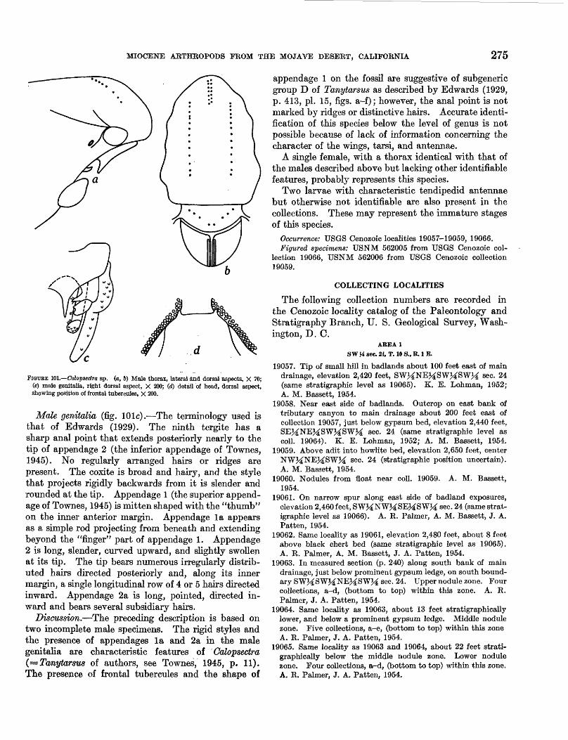
MIOCENE ARTHROPODS FROM THE MOJAVE DESERT, CALIFORNIA 275
FIGURE 101.—Calopsedra sp. (a, 6) Male thorax, lateral and dorsal aspects, X 70; (c) male genitalia, right dorsal aspect, X 200; (d) detail of head, dorsal aspect, showing position of frontal tubercoles, X 200.
Male genitalia (fig. lOlc).—The terminology used is that of Edwards (1929). The ninth tergite has a sharp anal point that extends posteriorly nearly to the tip of appendage 2 (the inferior appendage of Townes, 1945). No regularly arranged hairs or ridges are present. The coxite is broad and hairy, and the style that projects rigidly backwards from it is slender and rounded at the tip. Appendage 1 (the superior append age of Townes, 1945) is mitten shaped with the "thumb" on the inner anterior margin. Appendage la appears as a simple rod projecting from beneath and extending beyond the "finger" part of appendage 1. Appendage 2 is long, slender, curved upward, and slightly swollen at its tip. The tip bears numerous irregularly distrib uted hairs directed posteriorly and, along its inner margin, a single longitudinal row of 4 or 5 hairs directed inward. Appendage 2a is long, pointed, directed in ward and bears several subsidiary hairs.
Discussion.—The preceding description is based on two incomplete male specimens. The rigid styles and the presence of appendages la and 2a in the male genitalia are characteristic features of Calopsectra (=Tanytarsus of authors, see Townes, 1945, p. 11). The presence of frontal tubercules and the shape of
appendage 1 on the fossil are suggestive of subgeneric group D of Tanytarsus as described by Edwards (1929, p. 413, pi. 15, figs, a-f); however, the anal point is not marked by ridges or distinctive hairs. Accurate identi fication of this species below the level of genus is not possible because of lack of information concerning the character of the wings, tarsi, and antennae.
A single female, with a thorax identical with that of the males described above but lacking other identifiable features, probably represents this species.
Two larvae with characteristic tendipedid antennae but otherwise not identifiable are also present in the collections. These may represent the immature stages of this species.
Occurrence: USGS Cenozoic localities 19057-19059, 19066.Figured specimens: USNM 562005 from USGS Cenozoic col
lection 19066, USNM 562006 from USGS Cenozoic collection 19059.
COLLECTING LOCALITIES
The following collection numbers are recorded in the Cenozoic locality catalog of the Paleontology and Stratigraphy Branch, tJ. S. Geological Survey, Wash ington, D. C.
AREA 1 SW H sec. 24, T. 10 S., R. I E.
19057. Tip of small hill in badlands about 100 feet east of main drainage, elevation 2,420 feet, SWMNE^SWMSWM sec. 24 (same stratigraphic level as 19065). K. E. Lohman, 1952; A. M. Bassett, 1954.
19058. Near east side of badlands. Outcrop on east bank of tributary canyon to main drainage about 200 feet east of collection 19057, just below gypsum bed, elevation 2,440 feet, SE^NE^SW^SWM sec. 24 (same stratigraphic level as coll. 19064). K. E. Lohman, 1952; A. M. Bassett, 1954.
19059. Above adit into howlite bed, elevation 2,650 feet, center NWJ^NEJ^SW^ sec. 24 (stratigraphic position uncertain). A. M. Bassett, 1954.
19060. Nodules from float near coll. 19059. A. M. Bassett, 1954.
19061. On narrow spur along east side of badland exposures, elevation 2,460feet, SW^NW^SE^SW^ sec. 24 (same strat igraphic level as 19066). A. R. Palmer, A. M. Bassett, J. A. Patten, 1954.
19062. Same locality as 19061, elevation 2,480 feet, about 8 feet above black chert bed (same stratigraphic level as 19065). A. R. Palmer, A. M. Bassett, J. A. Patten, 1954.
19063. In measured section (p. 240) along south bank of main drainage, just below prominent gypsum ledge, on south bound ary SWMSWMNEMSWM sec. 24. Upper nodule zone. Four collections, a-d, (bottom to top) within this zone. A. R. Palmer, J. A. Patten, 1954.
19064. Same locality as 19063, about 13 feet stratigraphically lower, and below a prominent gypsum ledge. Middle nodule zone. Five collections, a-e, (bottom to top) within this zone A. R. Palmer, J. A. Patten, 1954.
19065. Same locality as 19063 and 19064, about 22 feet strati graphically below the middle nodule zone. Lower nodule zone. Four collections, a^d, (bottom to top) within this zone. A. R. Palmer, J. A. Patten, 1954.
276 CONTRIBUTIONS TO GENERAL GEOLOGY
AREA 2 NEM sec. 24, T. 10 S., R. 1 E,
19066. On west side of north-draining canyon, adjacent to aban doned mine workings, elevation about 2,860 feet, NE24SWM-
NE^NEM sec. 24. A. R. Palmer, A. M. Bassett, J. A. Patten, 1954.
AREA 3 See. 19, T. 10 N., R. 2 E.
19067. Near old Borate townsite, about 400 feet south of the center of the north edge of sec. 19. W. F. Foshag, 1932 or 1933.
LITERATURE CITED
Bailey, S. F., 1937, The genus. Dactuliothrips Moulton: Pan- Pacific Entomol., v. 13, p. 121-126, figs. 1-15.
———- 1939, A new species of thrips from the Mojave Desert: Pan-Pacific Entomology, v. 15, p. 168-172, figs. 1-2.
———— 1954, A review of the Melanthripinae with descriptions of two new species (Thysanoptera, Terebrantia): Entomol. Soc. Wash. Proc., v. 56, p. 78-85, pis. 1-2.
Bertrand, Henri, 1928, Les larves et nymphes des Dytiscides, Hygrobi ides, Haliplides: Encyclopedic Entomologique, v. 10, Paris, 358 p.
Carter, H. F., Ingram, A., and Macfie, J. W. S., 1920, Observa tions on the Ceratopogonine midges of the Gold Coast with descriptions of new species, Part II: Annals Trop. Med. Parasit., v. 14, no. 2, p. 211-274.
Darlington, P. J., Jr., 1948, The geographical distribution of cold-blooded vertebrates: Quart. Rev. Biol., v. 23, no. 2, p. 105-121.
DeBeer, G. R., 1951, Embryos and Ancestors: London, Oxford University Press, 159 p.
Edwards, F. W., 1929, British non-biting midges (Diptera, Chironomidae): London Entomol. Soc. Trans., v. 77, pt. 2, p. 279-430.
Emerton, J. H., 1911, New Spiders from New England: Conn. Acad. Arts and Sci. Trans., v. 16, p. 383-407.
Horn, G. H., 1879, Nitidulidae of the United States: Entomol. Soc. Am. Trans., v. 7, p. 267-336.
Ingram, A., and Macfie, J. W. S., 1931, Ceratopogonidae, Diptera of Patagonia and South Chile, Part II, Fasc. 4: British Museum (Nat. History), p. 135-232.
Kettle, D. S., and Lawson, J. W. H., 1952, The early stages of British biting midges Culicoides Latreille (Diptera: Cerato pogonidae) and allied genera: Bull. Entomol. Research, v. 43, pt. 3, p. 421-467.
Khalaf, Kamel, 1954, Speciation in Culicoides: Entomol. Soc. Am. Annals., v. 47, p. 34-51.
Korschelt, E., 1924, Der gelbrand Dyliscus marginalis L., Band 1: Bearbeitung einheimischer tiere, Mon 1, Leipzig, 863 p.
Lawson, J. W. H., 1951, The anatomy and morphology of the early stages of Culicoides nubeculosus Meigen (Diptera: Ceralopogonidae-Heleidae: Royal Entomol. Soc. London Trans., v. 102, pt. 9, p. 511-574.
Leakey, L. S. B., 1952, Lower Miocene invertebrates from Kenya: Nature, v. 169, no. 4302, p. 624, 625.
Lewis, G. E., 1957, Stratigraphic paleontology of the Barstow formation in the Barstow syncline: Calif. State Survey (in
Mayer, Karl, 1934, Die Metamorphose der Ceratopogonidae (Dipt.) Ein Beitrag zur Morphologic Systematik, Oekologie und Biologic der Jugenstadien dieser Dipterenfamilie: Archiv Naturgesell. (Leipzig), v. 3, p. 205-288.
———— 1935, Ceratopogoniden-Metamorphosen (C. Intermediaeund C. Vermiformes) Der Deutschen LimnologischenSunda-Expedition: Archiv Hydrobiologie supp., v. 13, p.166-202.
Matthew, W. D., 1915, Climate and evolution: N. Y. Acad.Sci. Annals, v. 24, p. 171-318 [reprinted, 1939, as SpecialPub. 1, N. Y. Acad. Sci.].
Needham, J. G., 1903, Aquatic Insects in New York State:N. Y. State Mus. Bull. 68, 501 p.
Needham, J. G., and Westfall, M. J., Jr., 1955, The Dragonfliesof North America: Berkeley, Calif., Calif. Univ. Press, 606 p.
Oman, P. W., 1949, The Nearctic Leafhoppers (Homoptera:Cicadellidae), A Generic classification and check list:Entomol. Soc. Wash., Mem. 3, 253 p.
O'Neill, Kellie, 1955, A note on the nomenclature and taxonomyof the subgenus Proscirtothrips Karny (Thysanoptera,Thripidae): Entomol. News., v. 66, no. 9, p. 239-242.
Palmer, A. R., and Bassett, A. M., 1954, Nonmarine Miocenearthropods from California: Science, v. 120, no. 8, p. 228.
Pennak, R. W., 1953, Fresh-water invertebrates of the UnitedStates: New York, The Ronald Press Company, 769 p.
Priesner, Herman, 1924, Bernstein-Thysanopteren: Entomol.Mitt., Band 13, no. 4/5, p. 130-151, figs. 1-21.
———— 1929, Bernstein-Thysanopteren II: Bernstein-Forsch- ungen, Heft 1, p. 111-138, pi. 8, Berlin, Germany, Walter de Gruyter & Co.
1949, Genera Thysanopterorum: Soc. Fouad ler Entomol.Bull., v. 33, p. 31-157.
Protescu, O., 1937, Etude, ge"ologique et pale"obiologique de1'ambre roumain. Les inclusions organiques de 1'ambre deBuzan: Bulletin de la Soci4te" Roumaine de Geologic, v. 3,p. 65-110.
Readio, P. A., 1922, Ovipositors of Cicadellidae: Kans. Univ.Sci. Bull., v. 14, no. 8 (whole ser. 24, no. 8), Entomology,no. 5, p. 217-298.
Scudder, S. H., 1868, The fossil insects of North America:Geol. Mag., v. 5, no. 47, p. 221.
———— 1875, The Tertiary Physapoda of Colorado; U. S. Geol.and Geog. Survey Terr. Bull., v. 1, 2d ser., no. 4, p. 221-223.
1890, The tertiary insects of North America: U. S. Geol.
Linder, Folke, 1941, Contributions to the morphology and the taxonomy of the Branchipoda Anostraca: Zool. Bidr. Uppsa la, Band 20, p. 103-302.
Survey Terr. Rept., v. 13, p. 371-374, pi. 5, figs. 88-91, 102-105, 115.
Simon, Eugene, 1892, Histoire Naturelle des Araigne"es, v. I.: Paris, 540 p.
Snodgrass, R. E., 1954, The dragonfly larva: Smithsonian Misc. Coll., v. 123, no. 2, p. 1-38.
Speyer, E. R., and Parr, W. J., 1950, Entomological Investiga tions 3. Studies upon Thysanoptera: Cheshunt, Herts [England], Exper. and Research Sta. Ann. Rept. (36th yr), p. 35-40, pis. 1-4.
Strouhal, Hans, 1954, Isopodenreste aus der altiplistozanen Spaltenfullung von Hundsheim be Deutsche-Altenburg: Osterr. Akad. Wiss. Wien Sitzungsber. Math.-naturwiss. Kl., Abt. 1, p. 163, p. 51-61.
Tapia, E. A., 1953, Espeeie nueva de Dactuliothrips (Thysanop tera) : Argentina Soc. Entomol. Rev., v. 16, p. 77-79, figs. a-e.
Thompson, D. W., 1942, On Growth and Form: New York, The Macmillan Co.
MIOCENE ARTHROPODS FROM THE MOJAVE DESERT,, CALIFORNIA 277
Thomsen, L. C., 1937, Aquatic Diptera. Part V. Ceratopo-gonidae: Cornell Univ. Agr. Expt. Sta. Mem. 210, p. 57-80.
Thorell, T. T., 1869, On European Spiders: K. ret. soc. NovaActa Regia Soc. Sci. Upsala, ser. 3, v. 7, 224 p.
Tillyard, R. J., 1917, The biology of Dragonflies: Cambridge,England, Cambridge Univ. Press, 396 p.
Townes, H. K., Jr., 1945, The Neartic species of Tendipedini(Diptera, Tendipedidae=Chironomidae): Am. MidlandNaturalist, v. 34, no. 1, p. 1-206.
Usinger, R. L., 1940, Fossil Lygaeidae (Hemiptera) from Floris-sant: Jour. Paleontology, v. 14, no. 1, p. 79-80.
Wirth, W. W., 1951, The genus Culicoides in Alaska: Entomol.Soc. Am. Annals, v. 44, no. 1, p. 75-85.
———— 1952a, The Heleidae of California: Calif. Univ. Pub.Entomology, v. 9, no. 2, p. 95-266.
1952b, Los insectos de las Islas Juan Fernandez: Rev»Chilena Entomologia v. 2, p. 87-104.
Woodward, Henry, 1879, On the occurrence of Branchipus (or Chirocephalus) in a fossil state, associated with Eosphaeroma and with numerous msect remains in the Eocene freshwater (Bembridge) limestone of Gurnet Bay, Isle of Wight: Geol. Soc. London Quart. Jour., v. 35, p. 342-350.

INDEX
[Italic page numbers indicate descriptions]
PageAbstract________________________-____________ 237 Acanthoctenidae.__-_________________________———.. 247 Acarina_____——————————————————_——————_ 242,248Acknowledgements____________________._____...—-..- 239 Adephaga__—————_.___———....——————__—.———— 259-265Aeolothripidae Melanthripinae gen., sp. undet.____________- 251, fig. 90a Aeolothrfps................................................................ ..- 252affinis, Dasyhelea............................................................. 272Age of fauna..———....______——_______-_____————.- 242albopicta, Dasyhelea........................................................... 270Ammonium chloride, use of in specimen photography...._____———_. 239Anaphothrips arcanus.................................... .____-_——.. 255
———————————————.———————————— 255.._....._............................_....—„........ 255
(Proscirtothrips) vitreus———....- ....——............ 242,243, 253, 254, fig. 90<fsp..———. ......._........ .........————__.___————— 242
vicinus.................................................................... 255zeae.............._...........__...............__....._........ 253,254
Ankothrips-.................................................................. 252Anostraca..____________________________________ 242 Anostraca gen. andsp. undet__._...________....___. 242, 245-246, pi. 32antiqua, Dasyhelea australis..........................................———— 242,
243, 244, ^66, 270, 271, 272, pi. 34; figs. 96o-/, 97o, 6, Q&a-j Arachnida..—————————.-——.._........._._...——... 242,246-249Aranei.....—-——--———_._—————————————————— 242,246-248arcanus, Anaphothrips........................................................ 256Argenna fossilis.......................................... 242, 243, S46, pi. 32; fig. 87
obesa.................................................................. ... 248Arrenurus sp. __________________________________ fig. 88a australis, Dasyhelea..................................................... 245,266,270
Dasyhelea australis................................................. 243,244,271australis antiqua, Dasyhelea................................................ 242,243,
244, S66, 270, 271, 272, pi. 34; figs. 96a-/, 97a, 6, 98a-j australis australis, Dasyhelea............................................ 243,244,271
bahamensis, Dasyhelea........................................................ 270Barstow formation...._______...________________... 237,239Bibliography...........................................................—— 276-277brookmani, Dasyhelea-........................................................ 270Burgess Vibro-tool, use of in specimen preparation_______________ 238 Byrocorinae..___________.___________________..... 257
Calico Mountains......................—————————————— 237,238,239californense, Schistomerus.................. 242, 243, 244, 259, pi. 33; figs. 93o-fc. 94<z-dcalifornicus, Cybocephalus.............................. 242, 243, 265, pi. 33; fig. 95a-6Calopsectra sp...__........................___._._______ 242, 274, pi- 34Calopsectrini_________________________________—.. 274 Canada balsam, use of in specimen preparation_...__________--.-. 239 Caraboidea____.....__................___..........._........— 259-265Carpelimus sp.-...................................... 242, 243, 264, pi. 33; fig. 95c, dCeratopogonidae.. ____________________________ 265-274Cicadellidae._.......................___.___........__......._.... 258dncta, Dasyhelea........................................................ 244,270,271Coleoptera..—....__........__............_..__............ 242,259-265Collecting localities,..._______________________________ 275-276 Composition of fauna___________.________—__..———————— 242 conoidifrons, Miomonalonion. ................................ 242,243,257,258, pi. 33convergens, Protoanenurus................................... 242, 248, pi. 32; fig. 886Cranothrips__________________________.___________.... 252crepuscularis, Culicoides.... ________.._____..___....———.. 274Crustacea..________.__________________________ 245-246 Cryphula................................................................... 255,256Oulicoides____.-..-_____________________________ 272
crepuscularis.............................................................. 274megacanthus..................................... 242, 243, 272, pi. 34; fig. lOOa-i
PageCybocephalidae.._________________________________ 265 Cybocephalus................................................................. 265
californicus........................................ 242, 243, 265, pi. 33; fig. 95a, 6Cymophyes................................................................. 243,255
Dactuliothrips............................................................... 251,252Dasyhelea................................................................... 248,265
.................................................................. 272albopicta....................._______________________ 270australis............................................................ 245,266,270
antiqua............................................................... 242,243, 244, 266, 270, 271, 272, pi. 34; figs. 96a-/, 97a, 6, 98a-j
australis........................................................ 243,244,271bahamensis................................................................ 270brookmani-. ____________________________________ 270 dncta............................................................... 244,270,271longipalpus............................................................... 271major..................................................................... 270pollens.................................................................... 270pallidihalter........................................................ .__. 270stenoceras........................... ... 241,242,243,265,270,271, pi. 34; fig. 99a-etenebrosa.................................................................. 270sp........................................................................ 272
Deltocephalinae................................................................ 258Deltocephalini gen. andsp. undet————.—————————————242,243, 258, pi. 33 Deronectes.................................................................. 259,264
__________________.__..__........._.__ 243,264Descriptions________._________________.___—_. 245-276 Diamond saw, use of in specimen preparation—————————————————— 239 Dictyna....................................................................... 246Dictynidae_____.________________________....__ 246-248Diptera——....,-.„..—.....——....__.__——........——— 242,265-276Dorythrips.................................................................... 252Dytiscidae_________________________________.__— 259 Dytiscus marginatts—.................................................. 262, fig. 94<z, C
Ecology of the fauna.._____________________________ 242-243 Evolution, significant observations on____________——_—————— 244
Felisacus..................................................................... 257ferruginea, Orthemis........................................................... 243Formic acid, use in specimen preparation__.___.__——.——————— 238 fossilis, Argenna.......................................... 242, 243, 246, pi. 32; fig.87
Geology——....———...———.....................__....._———— 239-240donates--................................................................. 255,266
Heliothrips.................................................................... 255Hemiptera—————————————........_..........__....242,255-258Heteroptera...____.__________________________ 242,255-258 Hildebrand, F. A., identification of minerals replacing fossils by_——__— 241 Homoptera____._._______________________.______ 242,258 Hydracarina__—__...___.____.___.___.._..————— 248 Hydroporinae.._.__._.__________________..___——.—— 259 Hydroporus................................................................. 259,264Hypochilus......................................... .________—__. 247
Insecta.-——--.-....—___—______._.___...__———— 249-276Islas Juan Fernandez._._.........._—............. . 238,243,244,245,270,271Internal organs preservation of_ 250, 261, 263, 267, 268, figs. 94, 96, 97; pis. 31,33,34 Introduction_______________________________.___ 237-238
Krylon, use of in specimen preparation_______.___..__—___ 239
Lethaeini________________________________________ 256 Lethaeus.................................................................... 256,256
279
280 INDEX
Page Libellula...................................................................... 250Libellulidae-.—————__._...———————_————————— 249-251 lithax Procymophyes-........................................ 242,243,255,#50, pi. 33longipalpus, Dasyhelea........................................................ 271Lygaeidae____—.....___—___.__________________ 255
magniceps, Anaphothrips...................................................... 255major, Dashyhelea............................................................. 270marginalis, Dytiscus................................................. 262, fig. 94o, cmegacanthus, Culicoides............................ 242, 243, 272, pi. 34; fig. lOOo-zMelanthripinae gen., sp. undet___________________ 242, 251, fig. 90o Melanthrips.................................................................. 252Microscope, use of in specimen preparation.„_________________ 238 Minerals replacing fossils. ____________________________ 241 Miogonates................................................................. 255,256
subimpunctatus........................................ 242, 866, pi. 33; fig. 91o-dMiomonalonion............................................................... 257
conoidifrons.............................................. 242,243,257, 258, pi. 33Miridae..._.________.____.____„_._..„._.__.__ 257 Monalonion................................................................... 257
Nodules, characteristics of—.————......————_——..____ 240-241, pi. 30
obesa, Argenna................................................................ 24sOdonata—-...—.—————....—......————.._.—..._._... 242,249-251Odessa Canyon...——————————————————__———____.___ 239 Orthemisferruginea........................................................... 243
sp-——......——-—...————....————-_..— 242, 243, 249, pis. 31, 32Orthotylinae..__________________________________ 257 Oxythrips..................................................................... 255
Pachygronthinae. —_______________________________ 250 pattens, Dasyhelea.......................................................•...... 270pattidihalter, Dasyhelea........................................................ 270Paragonates............................................................. 255,256,257perspicuus, Anaphothrips______________. _______________ 255 Phylinae—.........—....................___.....—.................... 257Plastic spray, use of in specimen preparation__________________ 239 Plasticene, use of in specimen preparation___________________ 238 Preparation of specimens. ____________________________ 238 Preservation of fossils______________________________ 241-242 Procymophyes.............................. ,__________________ 255
lithax................................................... 242, 243, 255, S58, pi. 33
PageProscirtothrips.. _____________—————_——————————————— 243 (Proscirtothrips) vitreus, Anaphothrips-... ._.___.—_ 242, 243, 253, 254, fig. 90<i(Prosocirtothrips) sp., Anaphothrips........................................... 242Protoarrenurus-............ ————————————————-——-————— 248
convergent................................................ 242, 248, pi. 32; fig. 886Pseudothrips.................................................................. 255
Resin, liquid, use of in specimen preparation—————————————————— 239 Rhyparoehrominae____________._____——......———————— 255
Sehistomerini______________________——_._———————— 259 Schistomerus.................................................................. 259
californense............................ 242, 243, 244, 259, pi. 33; figs. 93o-ft, 94<z-dSericothrips................................................................... 255Soap Lake, Wash___.______________—————.————....~ 243Staphylinidae...—.......—..—..————.—————————- 264stenoceras, Dasyhelea ..................... 241, 242, 243, 265, 270, 871, pi. 34; fig. 99o-«striateUus, Deronectes....................................................... 243,264subwipunctatvs, Miogonates............................... 242, $56, pi. 33; fig. 910-nfSwitzer, Q. S., identification of minerals replacing fossils by.————————— 241
Tanytarsus....——————.————————————————————————————— 275Tendipedidae.-_.___—._.__..-....-„——.———————-——-— 274tenebrosa, Dasyhelea........................................................... 270Theridium.................................................................... 247Thorell, T. T., quoted————————————————————— 248Thripidae..———————————————————————— 242,253-255
gen. and sp. unknown_________——..———.——————————— 254 Thnjx.—...................... ........... ...........................——— 254Thysanoptera._——————————————————————— 242,251-255Tweezers, use of in specimen preparation. _____—————————————— 238
UkBorus...................................................................... 247
meinus, Anaphothrips......................................................... 255vitreus, Anaphothrips (Proscirtothrips)....................... 242,243, 253, 254,fig.90<i
Xylene, use of hi specimen preparation....____————.———————— 239
zeae, Anaphothrips.......................................................... 253,254Zoogeographic relations__._____....._....————————————— 243-244Zoropsidae..______________—__._——__———————— 247
o
PLATES 30-34
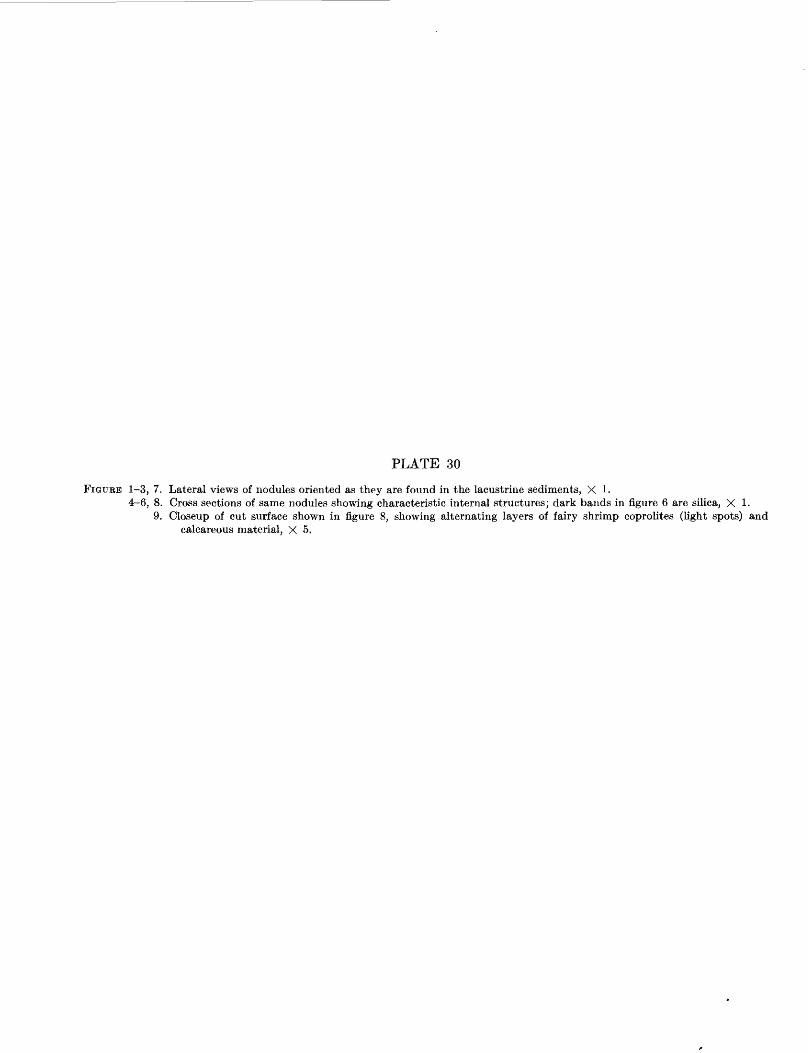
PLATE 30
FIGURE 1-3, 7. Lateral views of nodules oriented as they are found in the lacustrine sediments, X 1.4-6, 8. Cross sections of same nodules showing characteristic internal structures; dark bands in figure 6 are silica, X 1.
9. Closeup of cut surface shown in figure 8, showing alternating layers of fairy shrimp coprolites (light spots) and calcareous material, X 5.
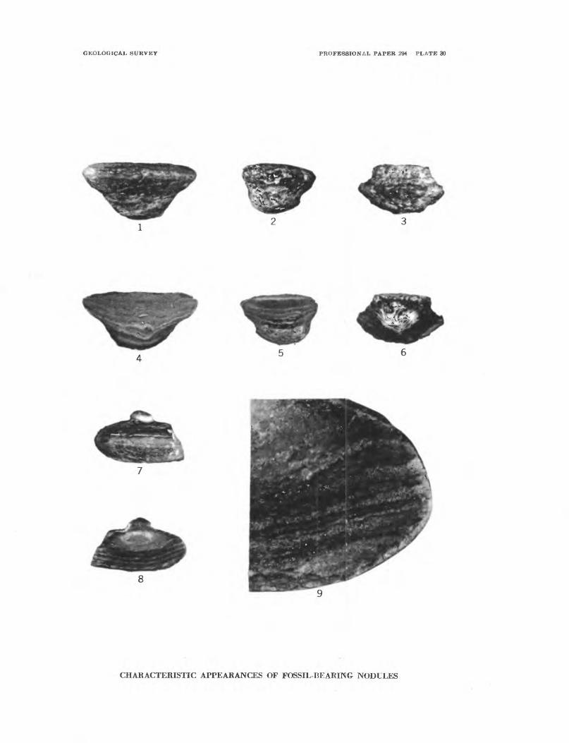
GEOLOGICAL SURVEY PROFESSIONAL PAPER 294 PLATE 30
CHARACTERISTIC APPEARANCES OF FOSSIL-BEARING NODULES
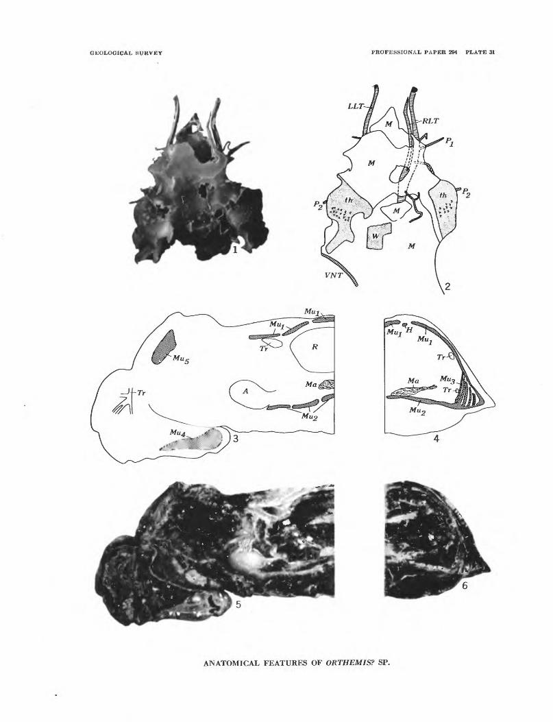
GEOLOGICAL SURVEY PROFESSIONAL PAPER 294 PLATE 31
LLT
MU
ANATOMICAL FEATURES OF ORTHEMIS? SP.
PLATE 31FIGURES 1-6. Orthemis? sp. (p. 249).
1, 2. Thoracic tracheae, X 1. 1, Photo of incomplete specimen with silicified thoracic tracheae, USNM 561980,USGS loc. 19059. 2, Key to structures in figure 1: PI, P2 , tracheae to first and second legs; VNT, ventral trunk;RLT, right lateral trunk; LLT, left lateral trunk; th, surface of thorax; W, part of wing pad; M, matrix.
3-6. Anatomy, X 8. 3, 4, Keys to structures shown in figures 5, 6: A, alimentary canal; H, heart; Ma, Mal-pighian tubules; Mui, dorsal muscles; Mu2, ventral muscles; Muz, vertical lateral muscles; Mm, muscle in labium;Mm, muscle in thorax; R, rectal gill system; Tr, tracheae. 5, Longitudinal section cut slightly to the left of themidline of the specimen figured on plate 32, figures 1, 5, USNM 561979a'. 6, Transverse section through fifthabdominal segment of same specimen, USNM 561979a", USGS locality 19057.
PLATE 32FIGURES 1, 2, 5, 6, 8-10. Orthemisf sp. (p. 249).
1, 5, Anterior and ventral views of original specimen found by Bassett. This specimen also had its internal anatomy preserved (see pi. 31); X 4, USNM 561909a, USGS loc. 19057. 2, Silicified thorax of nearly full-grown larva, X 2, USNM 561981, USGS loc. 19066. 6, Abdominal terminalia, dorsal view, X 10, USNM 561979c, USGS loc. 19057. 8, 9, Dorsal and lateral views of young larva, X 4, USNM 561979d, USGS loc. 19057. 10, Anterior view of specimen with labial palps preserved, X 4, USNM 561979b, USGS loc. 19057.
3. Argenna fossil-is Petrunkevitch, n. sp. (p. 246).Holotype, ventral view, X 20, USNM 561977, USGS loc, 19057.
4. Protoarrenurus convergens Cook, n. gen., n. sp. (p. 248).Holotype, ventral view, X 40, USNM 561978, USGS loc. 19065c.
7. Anostraca, genus and species undertermined (p. 245).Ventral view of most nearly complete specimen, X 20, USNM 561976a, USGS loc. 19057.
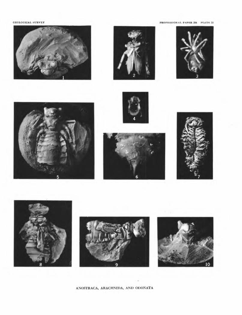
GEOLOGICAL SURVEY PROFESSIONAL PAPER 294 PLATE 32
ANOSTRACA, ARACHNIDA, AND ODONATA
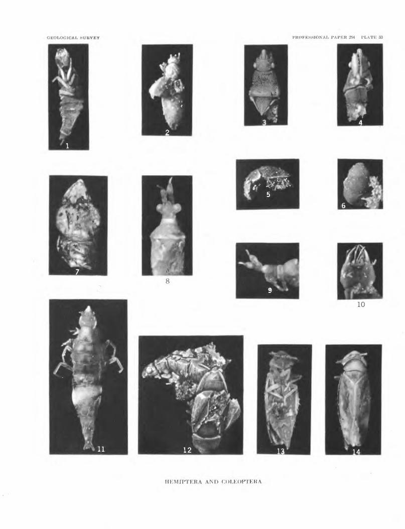
GEOLOGICAL SURVEY PROFESSIONAL PAPER 294 PLATE 33
10
HEMIPTERA AND COLEOPTERA
PLATE 33
FIGURES 1, 7, 10, 11. Schistomerus californense Palmer, n. gen., n. sp. (p. 259).1, Mature larva; ventral view showing sternal plate on seventh abdominal segment, X 10, USNM 561990a,
USGS loc. 19057. 7, Head of mature larva; ventral view showing some cephalic tracheae, X 30, USNM 561991, USGS loc. 19066. 10, Head of mature larva; ventral view showing mouth parts, X 20, USNM 561990b, USGS loc. 19057. 11, Holotype, mature larva; dorsal view, X 10, USNM 561989, USGS loc. 19057.
2. Carpelimus sp., (p. 264).Dorsal view, X 20, USNM 561993, USGS loc. 19057.
3, 4. Procymophyes lithax Sailer and Carvalho, n. gen., n. sp., (p. 256).Dorsal and ventral views of holotype, X 15, USNM 561982, USGS loc. 19057.
5, 6. Cybocephalus cf. C. californicus Horn, (p. 265).Lateral and dorsal views, X 20, USNM 561994, USGS loc. 19057.
8, 9. Miomonalonion conoidifrons Sailer and Carvalho, n. gen., n. sp. (p. 258).Dorsal and lateral views of holotype, X 15, USNM 561983, USGS loc. 19057.
12. Miogonates subimpunctatus Sailer and Carvalho, n. gen., n. sp. (p, 256).Dorsal view of headless paratype, lateral view of holotype, X 10. Note cleavage planes of celestite across
eye and margin of abdomen of holotype, USNM 561984, USGS loc. 19063d. 13, 14. Deltocephalini, genus and species undetermined (p. 258).
Ventral and dorsal views of female, X 15, USNM 561985, USGS loc. 19064b.
PLATE 34
FIGURES 1-4, 7, 8, 11-13. Dasyhelea australis antiqua Palmer, n. subsp. (p. 266).1, Nearly complete male, lateral view, X 15, USNM 562000, USGS loc. 19065b. 2, Female, ventral
view, X 20, USNM 561995b, USGS loe. 19057. 3, Incomplete larva, X 20, USNM 561995d, USGS loc. 19057. 4, Pupa, ventral view, X 10, USNM 561995c, USGS loe. 19057. 7, Pupa showing extent of respiratory trumpets, X 10, USNM 561999, USGS loc. 19061. 8, Terminal part of abdomen of specimen in figure 2, mounted in balsam, showing tracheae and spermathecae (see also text fig. 97a), X 30. 11, 12, Holotype, unhatched female in pupa; 11, closeup of tarsi and wing tips ventral view, X 150 (This photo by L. G. Henbest, U. S. Geological Survey); 12, anterior part of holotype, ventral view, X 50, USNM 561996, USGS loc. 19064b. 13, Female with color markings, dorsal view, X 20, USNM 561998, USGS loc. 19065c.
5, 10. Dasyhelea stenoceras Palmer, n. sp. (p. 271).5, Partly silicified pupa, showing respiratory trumpets, X 10, USNM 562002a, USGS loc. 19057. 10,
Holotype, pupa with partly emerged adult, ventral view, X 15, USNM 562001, USGS loc. 19057. 6. Culicoides megacanthus Palmer, n. sp., (p. 272).
Holotype, pupa, ventral view, X 20, USNM 562003, USGS loc. 19057. 9. Calopsectra sp., (p. 274).
Male, dorsal view of terminal part of abdomen, X 40, USNM 562005, USGS loc. 19066.
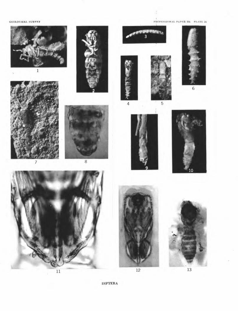
GEOLOGICAL SURVEY PROFESSIONAL PAPER 294 PLATE 34
13
DIPTERA