smith-magenis syndrome: a new contiguous gene syndrome
TRANSCRIPT
J Med Genet 1991; 28: 627-632
Smith-Magenis syndrome: a new contiguous genesyndrome. Report of three new cases
A Moncla, M 0 Livet, M Auger, J F Mattei, M G Mattei, F Giraud
AbstractInterstitial deletion of the short arm of chro-mosome 17 was detected in three patients.They all had a similar phenotype with mentalretardation, behavioural problems, facial dys-morphism, brachycephaly, a broad face with aflat midface, and short and broad hands. Allthree cases were ascertained over a six monthperiod by two neuropaediatricians aware ofthis specific anomaly, which suggests that thismicrodeletion is not particularly rare. Com-parison ofthe clinical and cytogenetic findingsin a total of 24 patients allows a new contigu-ous gene syndrome to be defined that only highresolution analysis can detect. In two cases,molecular analysis confirmed the cytogeneticresults. The Charcot-Marie-Tooth type Iagene has recently been localised to the 17pll.2sub-band.
The introduction in the early 1980s ofprometaphaseanalysis led to the identification of submicroscopicchromosomal anomalies. Small deletions or duplica-tions have been detected in some well delineatedsyndromes, such as Prader-Willi syndrome, Angel-man syndrome, Miller-Diecker syndrome, andBeckwith-Wiedemann syndrome, and have alsobeen found to be at the origin of new clinicalsyndromes.
Smith et al,' in 1982, first described an interstitialdeletion in the proximal short arm of chromosome17: del(l7)(p 11.2). The 17p11.2 band was deleted intwo unrelated patients presenting with cleft palateand cardiac malformation. In 1984, Patil and Bart-ley2 reported a smaller deletion in the 17p1 1.2 sub-band, del(l7)(p 1l.2-p 11.2), in a single patient withmental and growth retardation, hypotonia, hearing
INSERM U242 and Centre de G6n6tique Medicale,H6pital d'Enfants de la Timone, 13385 MarseilleCedex 5, France.A Moncla, M 0 Livet, M Auger, J F Mattei, M G Mattei,F GiraudCorrespondence to Dr Moncla.
loss, and facial dysmorphism. This microdeletionhas now been reported in 21 patients2-7 and a newclinical syndrome has emerged. Thus it can beassumed that deletion 17pll.2 is not such a rareconstitutional structural chromosomal aberration.We present three new patients with partial
17pll.2 deletion and review previously publishedcases. The three new cases confirm that the clinicalspectrum of the syndrome is specific enough to leadto clinical diagnosis at about 3 years of age.
Case reportsCASE 1The patient, a boy, was born at term after anuncomplicated pregnancy. The parents, a 24 yearold mother and a 25 year old father, were bothhealthy. He was the second child of this couple, thefirst was stillborn at 7 months of gestation. Thecouple had two other normal children, a boy and agirl.At birth his weight was 2800 g, length 47 cm, and
head circumference 31-5 cm. The Apgar score wasnormal. The neonatal period was unremarkable, butthe parents described him as being "too calm ababy". Slight motor delay was observed during thisperiod; he sat at 10 months and walked at 16 months.He spoke single words at 2 years. Delayed develop-ment became increasingly obvious with age.At 3 years, he was referred to a neuropaediatric
clinic for evaluation of his speech delay and beha-vioural disturbance. He spoke only about 20 isolatedwords. He was hyperactive with a very short atten-tion span and frequent temper tantrums. He showedpsychotic behaviour with repetitive activity, makingabnormal noises with his nose and tongue, bruxism,and insomnia.On physical examination, his height was 88 cm
(-2 SD), weight 13 kg, and head circumference48 cm (-2 SD). He was brachycephalic with frontalbossing, mild facial hypoplasia, mild epicanthus, ashort philtrum, broad nasal bridge, downturnedmouth, and small, turned under ears. His voice washoarse and low pitched. The hands were short andbroad with bilateral clinodactyly of the fifth fingers.Bilateral testicular ectopia was present. Audiogramwas normal and the EEG subnormal with slow
Received for publication 16 December 1990.Revised version accepted for publication 5 February 1991.
627
on March 24, 2022 by guest. P
rotected by copyright.http://jm
g.bmj.com
/J M
ed Genet: first published as 10.1136/jm
g.28.9.627 on 1 Septem
ber 1991. Dow
nloaded from
Moncla, Livet, Auger, Mattei, Mattei, Giraud
rhythms. Psychomotor testing using the TermanMerill scale at 6-5 years gave a score of 65.
CASE 2The patient, a girl, was the second child of healthyparents; the mother was 35 and the father 31 yearsold. The first child was normal. The patient wasborn at term after an uncomplicated pregnancy. Herbirth weight was 3200 g, height 47 cm, and headcircumference 35 cm. She developed respiratory dis-tress owing to a pneumothorax but recoveredquickly.When she was 8 days old she was referred to the
Medical Genetics Centre because of facial dysmor-phism. She was brachycephalic with a broad, flatface, epicanthus, strabismus, a broad nasal bridge,small nose, small philtrum, carp shaped mouth,micrognathia, and low set ears with hypoplastichelices. Karyotyping with R banding was performedand found to be normal. During her first year shewas very quiet and she walked at 19 months.When she was 3 years old, she was examined in a
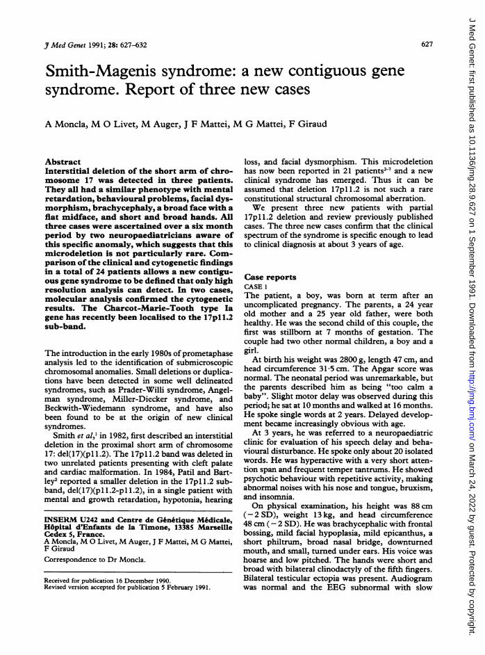
neuropaediatric department for speech delay andbehavioural problems. She was overactive andanxious with poor language development; she usedonly single words. Physical examination showednormal growth: weight 15 kg (+ 1 SD), height 90 cm(-1 SD), and head circumference 49 cm. Facialdysmorphism was increasingly obvious with age (fig1). The hands were short, she had genu valgum, andher feet were flat and short with partial syndactyly oftoes 2/3. Her voice was hoarse. Audiogram showed amoderate conductive hearing loss. Serous otitis me-dia was treated with myringotomy tubes. EEG was
subnormal with slow rhythms. Psychomotor testingwas possible at 4 5 years and the Terman Merillscale showed moderate mental retardation with fullscale IQ of 65.
CASE 3The patient, a girl, was born at term after a normalpregnancy. The mother was 23 years old and thefather 24. Birth weight was 3300 g, height 49 cm,and head circumference 35 cm. The family historywas unremarkable. She sat at 8 months and walkedat 19 months.When she was 30 months old, she was evaluated in
a neuropaediatric department because of markedspeech delay: she was only able to say "mam". Shewas hyperactive, irritable, and autoaggressive, withno concentration span. She made frequent gutturalnoises.
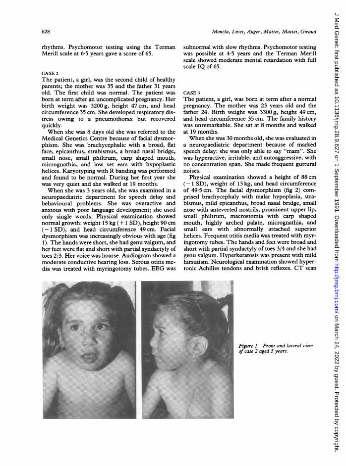
Physical examination showed a height of 88 cm(- 1 SD), weight of 13 kg, and head circumferenceof 49-5 cm. The facial dysmorphism (fig 2) com-prised brachycephaly with malar hypoplasia, stra-bismus, mild epicanthus, broad nasal bridge, smallnose with anteverted nostrils, prominent upper lip,small philtrum, macrostomia with carp shapedmouth, highly arched palate, micrognathia, andsmall ears with abnormally attached superiorhelices. Frequent otitis media was treated with myr-ingotomy tubes. The hands and feet were broad andshort with partial syndactyly of toes 3/4 and she hadgenu valgum. Hyperkeratosis was present with mildhirsutism. Neurological examination showed hyper-tonic Achilles tendons and brisk reflexes. CT scan
Figure 1 Front and lateral viewOf case 2 aged S years.
I,.'4:
628
on March 24, 2022 by guest. P
rotected by copyright.http://jm
g.bmj.com
/J M
ed Genet: first published as 10.1136/jm
g.28.9.627 on 1 Septem
ber 1991. Dow
nloaded from
Smith-Magenis syndrome: a new contiguous gene syndrome
Figure 2 Front and lateral viewof case 3 aged 8 years.
was normal. Routine cytogenetic analysis with Rbanding failed to show any anomaly.At the age of 8 years, she was examined by the
neuropaediatricians. Because of the clinical similari-ties with recently diagnosed cases 1 and 2, wedecided to re-evaluate her karyotype with high reso-lution banding.
CYTOGENETIC STUDIESIn these three cases, chromosomal studies wereperformed on phytohaemagglutinin stimulated lym-phocytes, with both standard and high resolutiontechniques. In the standard technique the lympho-cytes were cultured for 72 hours and harvested by
standard methods. R banding was performed byheat controlled denaturation and Giemsa staining.No precise chromosomal rearrangement could bedetected in any of the three cases but in cases 1 and 3there was a query concerning the short arm of onechromosome 17, which looked slightly shorter thanthe other.High resolution banding was performed accord-
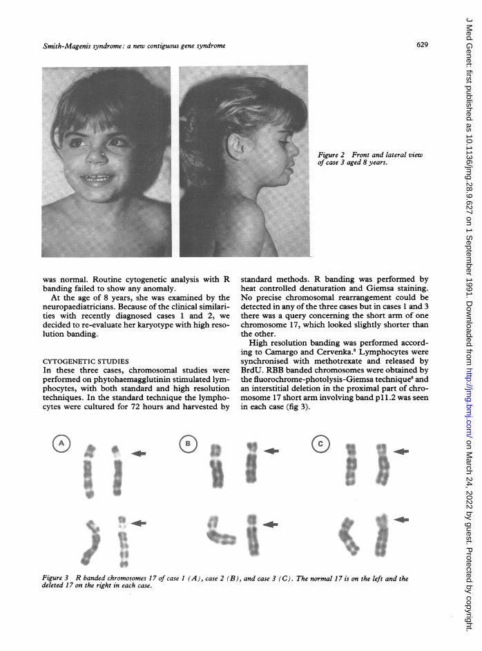
ing to Camargo and Cervenka.8 Lymphocytes weresynchronised with methotrexate and released byBrdU. RBB banded chromosomes were obtained bythe fluorochrome-photolysis-Giemsa technique8 andan interstitial deletion in the proximal part of chro-mosome 17 short arm involving band p1 1.2 was seenin each case (fig 3).
014
M.,0.
I., *Z4
....
A
I.*Jkw--I1
Figure 3 R banded chromosomes 17 of case 1 (A), case 2 (B), and case 3 (C). The normal 17 is on the left and thedeleted 17 on the right in each case.
629
on March 24, 2022 by guest. P
rotected by copyright.http://jm
g.bmj.com
/J M
ed Genet: first published as 10.1136/jm
g.28.9.627 on 1 Septem
ber 1991. Dow
nloaded from
Moncla, Livet, Auger, Mattei, Mattei, Giraud
In case 1, about 50% of the 17p 1l.2 band seemedto be deleted. The size of the deletion in cases 2 and3 was difficult to determine accurately but seemed tobe slightly smaller than in case 1.
DiscussionHigh resolution cytogenetics have led to the defini-tion of a new recognisable syndrome comprising a
distinct clinical phenotype and microdeletion(17pl 1.2). We have described three children evalu-ated in the Department of Neuropaediatrics at about3 years of age because of speech delay associatedwith behavioural disturbances. Case 1 was alsoreferred to the medical genetics department becauseof his facial dysmorphism. Standard karyotypingshowed an anomaly of 17p which was confirmed andrefined by high resolution banding techniques.Because of their clinical similarities with case 1, anddespite a normal karyotype using standard tech-niques, both cases 2 and 3 were reassessed cytogene-tically using high resolution banding. A 17pll.2microdeletion was detected in the two cases, con-
firming the clinical diagnosis.
The considerable clinical similarity between thethree patients was remarkable. Comparison of our
cases with 21 previously published cases'-7 makes itpossible to delineate the clinical features whichsuggest this diagnosis (table). All patients havemoderate to severe mental retardation with markedspeech delay. Speech is more affected than motor
ability as walking usually starts between 18 and 24months. Behavioural disturbances are always foundincluding hyperactivity with short attention span,
irritability, and aggressiveness. Some autisticfeatures are frequent with self-mutilation and self-stimulation activity.
All patients have a strikingly similar facial appear-
ance. A combination of brachycephaly, mild facialhypoplasia, and broad nasal bridge is alwaysobserved. Frequent features are apparent telecan-thus, short philtrum with carp shaped mouth, lowset and malformed ears, and mandibular prognath-ism. The degree of prognathism, however, is age
dependent and is present in the older patients. Theyoungest patients have micrognathia rather thanprognathism.3 The voice is hoarse with a deep,rasping quality, as pointed out by Smith et al,3 and
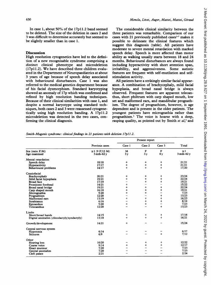
Smith-Magenis syndrome: clinicalfindings in 21 patients with deletion 17pll.2.
Present report
Previous cases Case 1 Case 2 Case 3 Total
Sex (ratio F/M) 1 (9 F/12 M) M F F 1Age examined 3 mth-62 y 3 y 3 y 8 y 3 mth-62 y
Mental retardationSpeech delay 18/18 + + + 21/21Hyperactivity 17/17 + + + 21/21Behavioural problems 14/17 + + + 17/20
CraniofacialBrachycephaly 20/21 + + + 23/24Mild facial hypoplasia 19/21 + + + 22/24Broad face 17/19 - + + 19/21Prominent forehead 14/21 + - - 15/22Broad nasal bridge 19/21 + + + 22/24Carp shaped mouth 16/18 + + + 19/21Micrognathia 5/20 + + - 7/23Prognathism 12/20 - - + 13/23Malformed ears 13/19 + + + 16/23Strabismus 6/16 - + + 8/19Epicanthus 5/20 + + - 7/23Telecanthus 12/20 - - + 13/23
LimbsShort/broad hands 14/15 + + + 17/18Digital anomalies (clinodactyly/syndactyly) 13/18 + + + 16/21
Growth/development 14/21 + - - 15/24
Central nervous systemHypotonia 6/14 - - - 6/17Seizures 6/9 - - + 7/12
OtherHearing loss 10/20 - + + 12/22Coarse voice 9/14 + + + 12/17Heart murmur 7/20 - - - 7/23Genital anomalies 7/20 - - - 7/23Cleft palate 2/21 - - - 2/24
630
on March 24, 2022 by guest. P
rotected by copyright.http://jm
g.bmj.com
/J M
ed Genet: first published as 10.1136/jm
g.28.9.627 on 1 Septem
ber 1991. Dow
nloaded from
Smith-Magenis syndrome: a new contiguous gene syndrome
may be a good diagnostic clue. Otitis media isfrequent with conductive hearing loss. The handsare short and broad with brachydactyly, clinodactylyof the fifth fingers, and syndactyly of the toes.
Other clinical signs have been reported. Growthretardation is present in about 50% of patients.Ophthalmological anomalies have also been de-scribed,7 as have dermatological signs like hyperker-atosis and hirsutism.6 Visceral malformations, facialclefts, congenital heart defects, cardiac murmur, andrenal and genital anomalies are rare.The sex ratio is equal: among the 24 reported
cases, 13 were females and 14 were males, asreported by Lockwood et al.6 The ages at which thepatients have been described range from the neo-natal period to 62 years. This explains the phenoty-pic variability, particularly for facial dysmorphism.In our case 2, the typical facial features were presentat birth but the diagnosis was difficult without theother specific signs. A review of published reportsindicates that diagnosis is clinically possible at 3years when phenotypic expression of the three majorsigns is complete: speech delay, facial dysmorphism,and short hands. This syndrome should be con-sidered when evaluating children with speech delayand behavioural disturbances and knowledge ofthese characteristic signs allows high resolutionanalysis to be focused on a specific chromosome.The frequency of this microdeletion is difficult toestablish but appears to be high. Our three patientswere identified over a 6 month period in a singlecentre and Lockwood et a16 reported a similar fre-quency.High resolution analysis is necessary to reveal this
microdeletion because of its small size. The deletioninvolved at least 50% of band 17pl.1.2 both in ourcases and the other previously published cases. Intwo cases reported by Smith et al' the 17pl1.2 bandwas completely missing but the phenotype includedcardiac defect and cleft palate.These microdeletions always appear de novo and
no parental chromosomal rearrangements have beenfound. In contrast with the deletion in Miller-Dieker syndrome, which has been localised to thedistal short arm of chromosome 17 (17p13) throughan inherited translocation,9'0 the Smith-Magenissyndrome was discovered only through the improve-ment in cytogenetic techniques. Four translocationsinvolving a breakpoint in 17pl 1.2 have beenreported." In all the cases, the unbalanced re-arrangement led to a trisomy 17p by adjacent 1segregation. No monosomy 17p 1.2-pter has beenobserved, probably because this type of rearrange-ment is lethal. The short arm of chromosome 17seems to be a hot spot for rearrangement in twospecific regions, 17p1 1.2 and 17p 13. This non-ran-dom distribution of breakpoints may be explained
by repetitive sequences in these regions of chromo-some 17. The sequences may be of Alu type found inR + bands, which are frequently associated withrearrangements, as described by Lehrman et al.'2Deletion might be the consequence of asymmetricalpairing and unequal exchange arising withinrepeated sequences during meiosis.13
Several genes, such as the ubiquitine gene,'4 thegenes for the cytokeratin family,'5 and the gene forCharcot-Marie-Tooth disease type Ia, have alreadybeen located in the 17pll .2 region. The mapping ofthe Charcot-Marie-Tooth disease gene was obtainedby linkage analysis.'6-8 Using three probes linked tothe Charcot-Marie-Tooth disease gene (pEW 301,pUC 10-41, and pYNM 67, supplied by ATCC), wehave shown that probes pU 10-41 andpYNM 67 aredeleted in our cases 2 and 3, but neither the pre-viously published patients, nor our three patientsshow any neuromuscular anomaly (Moncla et al,submitted). It is likely that the gene for Charcot-Marie-Tooth disease is located outside the observeddeletions. Construction of a somatic cell line is inprogress for our patients.A more sophisticated molecular approach to this
new 'contiguous gene syndrome"9 is now necessaryto characterise the genes involved in the deletionbetter.
1 Smith ACM, McGavran L, Waldstein G. Deletion of the 17short arm in two patients with facial clefts. Am 7 Hum Genet1982;34:410A.
2 Patil SR, Bartley JA. Interstitial deletion of the short arm ofchromosome 17. Hum Genet 1984;67:237-8.
3 Smith ACM, McGavran L, Robinson J, et al. Interstitialdeletion of (17)(pll.2pll.2) in nine patients. Am J MedGenet 1986;24:393-414.
4 Stratton RF, Dobyns WB, Greenberg F, et al. Report of sixadditional patients with a new chromosome deletion syn-drome. Am J Med Genet 1986;24:421-32.
5 Hamill MA, Roberts SH, Maguire MJ, Laurence KM. Intersti-tial deletion of 17pll.2: case report and review. Ann Genet(Paris) 1988;31:36-8.
6 Lockwood D, Hecht F, Dowman C, et al. Chromosome sub-band 17p 1l.2 deletion: a minute deletion syndrome. J MedGenet 1988;25:732-7.
7 Cabral de Almeida JC, Fagundes Reis D, Martins RR. Intersti-tial deletion of (17)(p 1l.2) - a microdeletion syndrome. AnnGenet (Paris) 1989;32:184-6.
8 Camargo M, Cervenka J. Patterns ofDNA replication ofhumanchromosomes. II. Replication map and replication model. AmJ Hum Genet 1982;34:757-80.
9 Stratton RF, Dobyns WB, Susan DA, Ledbetter DH. Newchromosomal syndrome: Miller-Dieker syndrome and mono-somy 17pl3. Hum Genet 1984;67:193-200.
10 Schinzel A. Microdeletion syndromes, balanced translocations,and gene mapping. J Med Genet 1988;25:454-62.
11 Schrander-Stumpel C, Schrander J, Fryns JP, Hamers G.Trisomy 17p due to a t(8;17)(p23;pll.2)pat translocation.Case report and review of the literature. Clin Genet1990;37: 148-52.
12 Lehrman MA, Russel DW, Goldstein GL, Brown MS. Exon-Alu recombination deletes 5 kilobases from the low densitylipoprotein receptor gene producing a null phenotype infamilial hypercholesterolemia. Proc Natl Acad Sci USA1989;83:3679-83.
13 Chandley AC. Asymmetry in chromosome pairing: a majorfactor in de novo mutation and the production of geneticdisease in man. J Med Genet 1989;26:546-52.
631
on March 24, 2022 by guest. P
rotected by copyright.http://jm
g.bmj.com
/J M
ed Genet: first published as 10.1136/jm
g.28.9.627 on 1 Septem
ber 1991. Dow
nloaded from
Moncla, Livet, Auger, Mattei, Mattei, Giraud
14 Webb GC, Baker RT, Fagan K, Board PG. Localization of thehuman UbB polyubiquitin gene to chromosome band17pl 11.1-17p12. Am I Hum Genet 1990;46:308-15.
15 Solomon E, Barker DF. Report of the committee on the geneticconstitution of chromosome 17. Cytogenet Cell Genet1989;51:319-37.
16 Raeymaekers P, Timmerman V, De Jonghe P, et al. Localiza-tion of the mutation in an extended family with Charcot-Marie-Tooth neuropathy (HMSNI). Am Hum Genet1989;45:953-8.
17 Middleton P, Harding AE, Monteiro C, Berciano J, Malcolm S.Linkage of hereditary motor and sensory neuropathy type I inthe pericentromeric region of chromosome 17. Am Jf HumGenet 1990;46:92-4.
18 Patel P, Franco B, Garcia C, et al. Genetic mapping of auto-somal dominant CMT disease in a large French Canadiankindred. Identification of new linked markers of chromosome17. Am Hum Genet 1990;46:801-9.
19 Schmickel RD. Contiguous gene syndromes: a component ofrecognisable syndromes. I Pediatr 1986;109:231-41.
632
on March 24, 2022 by guest. P
rotected by copyright.http://jm
g.bmj.com
/J M
ed Genet: first published as 10.1136/jm
g.28.9.627 on 1 Septem
ber 1991. Dow
nloaded from