nih public access a,b christopher w. carr omar a. … contiguous rudd.pdf · large contiguous gene...
TRANSCRIPT
Large Contiguous Gene Deletions in Sjögren-Larsson Syndrome
Holly Engelstada, Gael Carneya, Dana S'Aulisa, Janae Risea, Warren G. Sangera,b, M.Katharine Ruddc, Gabriele Richardd, Christopher W. Carre, Omar A. Abdul-Rahmane, andWilliam B. Rizzoa,b
a Department of Pediatrics, University of Nebraska Medical Center, Omaha, NE 68198, U.S.A.b Munroe-Meyer Institute for Genetics and Rehabilitation, University of Nebraska Medical Center,Omaha, NE 68198, U.S.A.c Department of Human Genetics, Emory University School of Medicine, Atlanta, GA 30322,U.S.A.d GeneDx Inc, Gaithersburg, MD 20877, U.S.A.e Department of Pediatrics, University of Mississippi Medical Center, Jackson, MS 39216, U.S.A.
AbstractSjögren-Larsson syndrome (SLS) is an autosomal recessive disorder characterized by ichthyosis,mental retardation, spasticity and mutations in the ALDH3A2 gene for fatty aldehydedehydrogenase, an enzyme that catalyzes the oxidation of fatty aldehyde to fatty acid. More than70 mutations have been identified in SLS patients, including small deletions or insertions,missense mutations, splicing defects and complex nucleotide changes. We now describe 2 SLSpatients whose disease is caused by large contiguous gene deletions of the ALDH3A2 locus on17p11.2. The deletions were defined using long distance inverse PCR and microarray-basedcomparative genomic hybridization. A 24-year-old SLS female was homozygous for a 352-kbdeletion involving ALDH3A2 and 4 contiguous genes including ALDH3A1, which codes for themajor soluble protein in cornea. Although lacking corneal disease, she showed severe symptomsof SLS with uncommon deterioration in oral motor function and loss of ambulation. The other 19-month-old female patient was a compound heterozygote for a 1.44-Mb contiguous gene deletionand a missense mutation (c.407C>T, P136L) in ALDH3A2. These studies suggest that large genedeletions may account for up to 5% of the mutant alleles in SLS. Geneticists should consider thepossibility of compound heterozygosity for large deletions in patients with SLS and other inbornerrors of metabolism, which has implications for carrier testing and prenatal diagnosis.
KeywordsALDH3A1; fatty aldehyde; ichthyosis; mutation; SLC47A1; ULK2
© 2010 Elsevier Inc. All rights reserved.Address correspondence to Dr. William Rizzo, Department of Pediatrics, University of Nebraska Medical Center, 985456 NebraskaMedical Center, Omaha, NE 68198-5456, U.S.A. [email protected], Telephone: 402 559-2560, FAX: 402 559-2540.Publisher's Disclaimer: This is a PDF file of an unedited manuscript that has been accepted for publication. As a service to ourcustomers we are providing this early version of the manuscript. The manuscript will undergo copyediting, typesetting, and review ofthe resulting proof before it is published in its final citable form. Please note that during the production process errors may bediscovered which could affect the content, and all legal disclaimers that apply to the journal pertain.
NIH Public AccessAuthor ManuscriptMol Genet Metab. Author manuscript; available in PMC 2012 November 1.
Published in final edited form as:Mol Genet Metab. 2011 November ; 104(3): 356–361. doi:10.1016/j.ymgme.2011.05.015.
NIH
-PA Author Manuscript
NIH
-PA Author Manuscript
NIH
-PA Author Manuscript

INTRODUCTIONSjögren-Larsson syndrome (SLS; OMIM 270200) is an autosomal recessive inborn error ofmetabolism caused by mutations in the ALDH3A2 gene for fatty aldehyde dehydrogenase(FALDH) [1,2]. Clinical features of SLS include ichthyosis, spastic diplegia or tetraplegia,mental retardation, seizures and a distinctive retinal crystalline maculopathy characterizedby perifoveal glistening white dots. The ichthyosis is usually congenital in onset and is oftenpruritic in nature. Neurological symptoms of mental retardation and spasticity typicallydevelop by the 2nd year of life and present with delay in achieving motor and cognitivemilestones. The symptoms of SLS vary from mild to profound, and are generally non-progressive.
FALDH catalyzes the oxidation of fatty aldehyde to fatty acid, and is a necessary componentof the fatty alcohol:NAD+ oxidoreductase enzyme complex that catalyzes the sequentialoxidation of fatty alcohol to fatty acid [3,4,5]. SLS patients consequently have elevated fattyalcohols in plasma, urine and cultured cells [6-9]. The symptoms of SLS are thought to arisedirectly or indirectly from accumulation of fatty aldehyde, fatty alcohol or related lipidproducts in the skin and brain [10].
The ALDH3A2 gene is located on chromosome 17p11.2. More than 70 mutations have beendiscovered in SLS patients, including small deletions or insertions, missense mutations,splicing defects and complex mutations composed of deletion/insertions and nucleotidesubstitutions [11]. Intragenic deletions of one or more exons have also been rarely described[12-14]. Most mutations in SLS are private, and many patients have been found to behomozygous due to consanguinity or founder effects.
We now present 2 unique SLS patients in whom the disease was caused by unusually largedeletions involving ALDH3A2 and surrounding genes on chromosome 17p11.2.
MATERIALS AND METHODSThe Institutional Review Board at the University of Nebraska Medical Center approved thisresearch, and all subjects consented to the study.
Patient DescriptionsPatient 1—This 24-year-old female was born at 32 weeks gestation to consanguineousfirst-cousin Pakistani parents. She was noted to have erythematous dry scaly skin at birth,but had no collodion membrane. She developed several seizures at 6 weeks of age, whichwere associated with hypocalcemia. Delays in achieving motor milestones and speech werenoted in infancy. After 2 febrile illnesses at 6-7 months of age, she lost the ability to rollover and her limbs became very stiff. A brain CT at 9 months of age showed moderatecerebral atrophy. However, she began to sit unsupported and crawl at about 18 months.Physical examination at 3 years of age showed developmental delay, spastic diplegia andgeneralized ichthyosis. Speech consisted of occasionally saying “mama” and “baba”.Laboratory studies were normal, including karyotype, EEG, electroretinogram, thyroidfunction tests, plasma phytanic acid, urine amino acids and metabolic screen. Her ichthyosisresponded well to etretinate therapy, which was subsequently switched to isotretinoin andthen discontinued several years later because of concerns about retinoid toxicity. By 10years of age, she was no longer speaking and began having difficulty swallowing. Physicalexam showed a pruritic, generalized ichthyosis along with spastic diplegia, leg contracturesand ankle clonus (Fig 1A). She had photophobia and avoided bright lights. She was able toambulate only with a walker using a crouched gait. Brain MRI revealed bilateral,symmetrical abnormal T2-weighted signal involving the fronto-parietal and superior
Engelstad et al. Page 2
Mol Genet Metab. Author manuscript; available in PMC 2012 November 1.
NIH
-PA Author Manuscript
NIH
-PA Author Manuscript
NIH
-PA Author Manuscript
periventricular regions. Several months later she developed increased drooling with tongueprotrusion and dysphagia. Oral motor incoordination and loss of swallowing slowlyprogressed and necessitated placement of a gastrostomy tube at 12 years of age. Bilateralhamstring lengthening was also performed at this age. At 14 years, she was noted to havespastic tetraplegia and be non-ambulatory. At 17 years of age, ophthalmologic examinationunder sedation showed chronic blepharitis, but no cataracts, retinal or corneal abnormalities.The diagnosis of SLS was confirmed by demonstrating FALDH deficiency (8% of meannormal activity) in cultured fibroblasts.
Patient 2—This 19-month-old female infant was born at 36 weeks gestation to a 27-year-old mother and non-consanguineous father. Mother was of Irish and Cherokee Indiandescent; father was English and American Indian. At birth, the infant was noted to have acollodion membrane with shiny, “tight” skin that transformed into dry, scaling skin after 2-3weeks. Over time, she began scratching continually, often to the point of bleeding. At 10months of age, a skin biopsy revealed hyperkeratosis, papillomatosis and acanthosis.
At 12 months of age, physical examination revealed height, weight, and head circumferencewithin normal age limits. Significant findings included ichthyotic skin, ears adherent to thescalp bilaterally, and shortened fifth fingers bilaterally. She was noted to have spasticdiplegia. Truncal hypotonia prevented her from sitting up for more than short periods oftime. A Denver II Developmental Assessment found her to have social, language, finemotor, and gross motor abilities consistent with an age of 9, 7, 6, and 4 months old,respectively. Ophthalmologic examination revealed no ocular or visual abnormalities,including no evidence of macular glistening white dots. Her skin was dry withhyperkeratosis or scaling on the extremities, trunk, neck, palms, and soles (Fig 2D). Thehyperkeratosis was most severe on the patient's legs, forearms, neck and trunk, where it wasreminiscent of lamellar ichthyosis. Her hair and fingernails were normal. At 19 months ofage, the patient suffered a tonic-clonic seizure, which subsequently recurred on severaloccasions.
Fibroblast CultureCultured skin fibroblasts were grown from skin biopsies in Dulbecco's minimal essentialmedium containing 10% fetal bovine serum, penicillin, and streptomycin at 37°C in anatmosphere of 5% CO2. The cells were collected by trypsinization and washed twice withphosphate-buffered saline. Cell pellets were stored at -70°C for DNA isolation.
FALDH AssayFibroblasts were homogenized and assayed for FALDH activity using octadecanal assubstrate as described [5], except that all reactions were run at 37° C in a 96-well fluorescentplate reader (Molecular Dynamics) in a total volume of 0.38 ml. Cell protein wasdetermined according to Lowry et al [15]. Enzyme activity was determined as pmol/min/mgcell protein, and expressed as percentage of normal mean enzyme activity.
DNA IsolationGenomic DNA was purified from cultured fibroblasts and blood using the Wizard GenomicDNA Purification kit (Promega). Buccal DNA was collected from controls and familymembers for PCR as described [16].
Deletion Characterization by Long Distance Inverse-PCR (LDI-PCR)LDI-PCR was performed essentially as described by Willis et al [17]. A PstI restrictionenzyme cut site was identified in the DNA sequence flanking one end of the deletion. One
Engelstad et al. Page 3
Mol Genet Metab. Author manuscript; available in PMC 2012 November 1.
NIH
-PA Author Manuscript
NIH
-PA Author Manuscript
NIH
-PA Author Manuscript
microgram of genomic DNA was therefore digested with 0.5 Unit of PstI (Patient 1) in atotal volume of 25 μl according to the manufacturer's instructions (New England Biolabs).The DNA digest was purified using a QIAquick PCR Purification Kit (Qiagen). CircularDNA was created by ligating 660-700 ng digested DNA with 30 Units of T4 ligase(Promega) in a total volume of 660-700 μl overnight at 25° C. The ligated DNA waspurified by a double ethanol precipitation and resuspended in TE buffer.
Using oligonucleotide primers within the known DNA sequence flanking the restrictionenzyme cut site, we inversely amplified the circular DNA using the Expand Long TemplatePCR System (Roche). All PCR reactions were performed in a final volume of 50 μl. Thereactions contained 1 Unit Taq polymerase, 0.05 mM dNTPs, 12-20 ng purified ligatedDNA, 17.5 mM MgCl2, and 15-20 ng of each primer (primer sequence is available byrequest). The amplification occurred in three steps: 1) an initial denaturation at 94° C for 5min; 10 cycles of denaturation at 94° C for 30 sec, annealing at 64° C for 1 min, andelongation at 72° C for 4 min; 2) 10 cycles of denaturation at 94° C for 30 sec, annealing at64° C for 1 min, and elongation at 72° C for 5 min; 3) 20 cycles of denaturation at 94° C for30 sec, annealing at 64° C for 1 min, and elongation at 72° C for 6 min; a final extension at72° C for 1 min. The resulting PCR products were separated on a 1% agarose gel, purifiedwith a MinElute Spin Column and sequenced.
Genomic DNA Amplification across the Deletion in Patient 1After identifying deletion breakpoints by LDI-PCR, primers were designed to amplifyacross the breakpoint in undigested genomic DNA (Table 1). PCR amplification of thepatient-specific DNA fragment was multiplexed with amplification of exon 8 of ALDH3A2as a control. The PCR contained 1 U Taq polymerase, 0.05 mM dNTPs, 50 ng of DNA, 17.5mM MgCl2, 10 ng of the patient-specific primers, and 20 ng of the exon 8 primers (Table 1).The cycling conditions consisted of 1) an initial denaturation at 94° C for 5 min; 2) 30 cyclesof denaturation at 94° C for 30 sec, annealing at 68° C for 1 min, and elongation at 72° C for1 min; 3) a final extension at 72° C for 5 min.
DNA SequencingLDI-PCR products and ALDH3A2 exons amplified from genomic DNA [13] weresequenced using the BigDye Terminator Cycle Sequencing Kit (Applied Biosystems, Inc,Foster City, CA) and an ABI 377A sequencer.
Screening for the c.407C>T (P136L) Mutation in Patient 2The c.407C>T (P136L) mutation in exon 3 creates a new HindIII restriction enzyme cut site.To screen for this mutation, a 102 bp fragment from exon 3 containing the mutation wasamplified from genomic DNA and digested with HindIII. The PCR contained 0.75 U Taqpolymerase, 2.5 mM dNTPs, 17.5 mM MgCl2, 1 μl buccal DNA and 10 ng of each primer(Table 1) in reaction volume of 50 μl. The cycling conditions were 1) initial denaturation at95° C for 7 min, 2) 40 cycles of 95° C for 30 sec, 62° C for 1 min and 72° C for 1 min, 3)final extension at 72° C for 5 min. The PCR product (0.25 μg) was digested in 50 μl reactionvolume containing Buffer 2 and 0.5 Unit HindIII overnight at 37° C according to themanufacturer's instructions (New England Biolabs). The digestion products were separatedon a 3% agarose gel. The c.407C>T mutation produces a HindIII cut site with generation ofa 80bp fragment, whereas the wild-type DNA is undigested.
Engelstad et al. Page 4
Mol Genet Metab. Author manuscript; available in PMC 2012 November 1.
NIH
-PA Author Manuscript
NIH
-PA Author Manuscript
NIH
-PA Author Manuscript

Array Comparative Genomic Hybridization (CGH) and Fluorescence In Situ Hybridization(FISH)
CGH was performed using a whole genome 44K custom oligonucleotide array (AgilentTechnologies, Inc) as described [18]. The deletion was confirmed via FISH, using abacterial artificial chromosome corresponding to the deleted region (RP11-147i5) and alphasatellite DNA from chromosome 17. Probes were labeled and hybridized to metaphasechromosomes as described previously [18].
RESULTSPatient 1
Patient 1 was first suspected to have a large gene deletion when ALDH3A2 exons could notbe amplified from genomic DNA. Southern blot analysis subsequently confirmed a completedeletion of the gene. The deletion boundaries were initially narrowed down by generatingPCR products from flanking DNA sequences and sequentially walking closer to the deletionuntil no PCR products were produced; however, attempts to amplify across the deletionusing multiple combinations of primers were unsuccessful. We therefore used LDI-PCR todetermine the precise deletion breakpoints. This method depends on knowing DNAsequence flanking one end of the deletion breakpoint. Restriction enzymes that cut withinthe known sequence (and presumably somewhere on the other sides of the deletionbreakpoints) were used to generate DNA fragments that were subsequently ligated intocircular DNA. Primers situated within the known DNA sequence were then used toinversely amplify across the deleted DNA region in the circular DNA and locate the DNAsequence from the other side of the deletion to determine the precise breakpoints.
Using this approach, a 4.1 kb PCR product was produced by LDI-PCR of the patient'scircularized DNA, and sequencing of the PCR product identified the deletion breakpoints onchromosome 17 at nucleotide 19,446,110 and nucleotide 19,798,450 (in GRCh37/hg19 ofthe human genomic assembly). This 352 kb deletion includes ALDH3A2, ALDH3A1, ULK2,SLC47A1 and SLC47A2 (Fig 3). The homozygous deletion was confirmed by array CGH(data not shown).
Patient 1 was a child of consanguineous parents. Using primers that flanked the deletionbreakpoints, a 496 bp PCR product was amplified from the patient's genomic DNA and herparents, thus confirming the parental carrier status for this deletion, but not from controls(Fig 1B). A control PCR product of exon 8 was produced using DNA from the parents andher unaffected brother, but not from the patient, consistent with her homozygous genotype.
Patient 2Because of the patient's constellation of clinical findings, CGH analysis was initiallyundertaken using a 44K oligonucleotide genomic microarray. Patient 2 was found to carry aheterozygous 1.44 Mb interstitial deletion of 17p11.2 [chr17:18,716,455 - 20,160,197] inNCBI36/hg18 of the human genomic assembly that spans 15 genes, including ALDH3A1and ALDH3A2 (Fig 2A, Fig 3). The deletion lies within the recurrent Smith-Magenissyndrome (SMS) deletion region, but does not include the complete SMS critical region.FISH analysis confirmed the heterozygous deletion (Fig 2B).
Sequence analysis of the patient's only remaining ALDH3A2 gene copy identified a novelhemizygous missense mutation (c.407C>T, P136L) in exon 3 (Fig 2C). This mutationgenerates a new HindIII restriction enzyme cut site. By screening a PCR amplicon of exon 3and digesting with HindIII, we did not detect the mutation in 50 unrelated Caucasian controlsubjects, suggesting that it is not a common polymorphism. The P136 amino acid residue is
Engelstad et al. Page 5
Mol Genet Metab. Author manuscript; available in PMC 2012 November 1.
NIH
-PA Author Manuscript
NIH
-PA Author Manuscript
NIH
-PA Author Manuscript
invariantly conserved among FALDH proteins in vertebrate species ranging from zebra fishto humans and undoubtedly has a critical function in the protein. In this regard, the P136residue in human FALDH corresponds to P138 in a related rat class 3 ALDH protein, forwhich the crystalline structure has been reported [19]. P138 of the rat protein initiates a kinkbetween the β2 β-strand and the αB α-helical domain within the Rossmann fold, which isimportant for NAD binding.
Investigation of the parents by microarray and DNA sequencing revealed that the patient'sfather carried the deletion mutation and her mother was heterozygous for the P136Lmutation.
DISCUSSIONThe SLS patients reported here have unusually large deletions, including the entireALDH3A2 gene on chromosome 17p11.2. Previously reported intragenic deletions in SLShave ranged from 1 or 2 nucleotides to as large as 6 kb [11], but no complete gene deletionshave been reported. Our patients carried contiguous gene deletion alleles that includedALDH3A2 and multiple flanking genes. Indeed, this region of chromosome 17 is associatedwith large de novo contiguous gene mutations that are a well-known marker of SMS [20],which is caused by haploinsufficiency for the RAI1 gene [21].
Patient 1 represents a unique “experiment in nature”. She is homozygous for a contiguousgene mutation that deletes ALDH3A2 and 4 neighboring genes, including ALDH3A1, ULK2,SLC47A1 and SLC47A2. ALDH3A1 encodes an aldehyde dehydrogenase that is expressed athigh levels in stomach and comprises up to 40% of the total soluble protein in cornea [22].The enzyme oxidizes medium-chain aliphatic aldehydes [23], and is thought to eliminatetoxic aldehydes generated during lipid peroxidation [24]. However, its unusual abundance incornea suggest that the protein may also have a non-catalytic role in absorbing UV light andprotecting the underlying lens from damage [25]. A naturally occurring strain of mice(SWR/J) that is deficient in ALDH3A1 enzyme activity [26] and Aldh3a1-/- gene knockoutmice [25] are abnormally susceptible to cataract formation when exposed to UV light. TheULK2 gene codes for a UNC-51-like serine/threonine protein kinase that is widely expressedin tissues [27] and is involved in cell signaling pathways for apoptosis [28], autophagy[29,30], axonal outgrowth and endocytosis [31], and fibroblast growth factor receptorsubstrates [32]. The SLC47A1 and SLC47A2 genes encode multidrug and toxin extrusionproteins (MATE1 and MATEK-2, respectively) that are localized in the kidney and areimportant for actively secreting certain organic cationic molecules and drugs, includingmetformin and cimetidine, from the blood into the urine [33,34,35]. Non-synonymouspolymorphisms in the genes may be responsible for decreasing, or even eliminating, renalclearance of these ionic drugs and thereby affect drug pharmacokinetics and toxicity [35,36].Mice naturally lack the SLC47A2 gene, but genetic knockout of the murine SLC47A1 generesults in a profound reduction in metformin excretion and increased blood concentrations ofthis drug [37].
Although missing ALDH3A2 and 4 additional genes, the clinical phenotype of Patient 1 waswithin the spectrum seen in other SLS patients, albeit at the more severe end. Some of hersymptoms, however, may originate from the deleted neighboring genes. Her oral motordysfunction and excessive drooling is not usually seen in SLS, and most patients do notrequire a gastrostomy tube for feeding. Despite lacking the ALDH3A1 protein, she did notexhibit corneal or lens opacities. Her long-standing photophobia and squinting, which arecommon features of SLS patients, may have limited her ocular exposure to UV light andprevented eye damage. The absence of perimacular glistening white dots, which are oftenseen in SLS, indicates that her photophobia does not arise from light hitting and refracting
Engelstad et al. Page 6
Mol Genet Metab. Author manuscript; available in PMC 2012 November 1.
NIH
-PA Author Manuscript
NIH
-PA Author Manuscript
NIH
-PA Author Manuscript
off these abnormal structures. Without the MATE1 and MATEK-2 transporters, Patient 1should be at higher risk for adverse effects of MATE-dependent drugs. The lack of ULK2would seem to impair several cell signaling pathways and may contribute to her more severeSLS phenotype with progressive loss of speech, oral motor function and worseningspasticity. Most SLS patients have a static disease with little or no symptom progressionover time. Additional subtle phenotypic effects of her deleted genes could be missed on herclinical background of severe SLS. Moreover, the extent to which the clinical phenotype ofPatient 2 is influenced by haploinsufficiency for the many deleted genes is also not known.
The genetic analysis of Patient 2 underscores the importance for considering large genedeletions in patients with ostensibly homozygous ALDH3A2 mutations and the need forparental testing to confirm carrier status. If this patient had only undergone standardmutation analysis by sequencing genomic exons amplified by PCR, her genotype could havebeen initially misidentified as homozygous P136L. In the absence of parental testing, aheterozygous deletion or duplication would have gone undetected, and subsequent genetictesting of family relatives or prenatal diagnosis could have led to erroneous results. Mutationanalysis of her parents, however, would have raised the possibility of a co-existing deletionmutation in the patient and prompted further DNA analyses by array CGH or high-densitysingle nucleotide polymorphism (SNP) chips.
The frequency of large gene deletions in SLS is not precisely known. We are aware,however, of two other SLS patients who also carry unique large gene deletions that involvethe complete ALDH3A2 gene or most of it (unpublished observations). Together with thetwo mutations reported here, these large deletions account for approximately 5% of themutant alleles reported so far.
Large deletion mutations have been detected in several other inborn errors of metabolism,including Canavan disease [38-40], cystinosis [41], steroid sulfatase deficiency [42-44], X-linked adrenoleukodystrophy [45], phenylketonuria [46], pyruvate dehydrogenase deficiency[47] and ornithine transcarbamylase deficiency [44,49]. Although CGH and SNP arrays arerarely done for the evaluation of suspected metabolic disorders, they are becoming astandard diagnostic test for patients with non-specific developmental delay. Our findings inSLS suggest that these tests also have utility for detecting contiguous gene deletions in anincreasing number of inborn errors of metabolism.
AcknowledgmentsThis work was supported by grant AR044552 from the National Institute of Arthritis and Musculoskeletal and SkinDiseases of the National Institutes of Health, and by the Sjögren-Larsson Syndrome Research Fund at theUniversity of Nebraska.
Abbreviations
CGH comparative genomic hybridization
FALDH fatty aldehyde dehydrogenase
FISH fluorescence in situ hybridization
LDI-PCR long distance inverse-PCR
OMIM Online Mendalian Inheritance in Man
SLS Sjögren-Larsson syndrome
SMS Smith-Magenis syndrome
Engelstad et al. Page 7
Mol Genet Metab. Author manuscript; available in PMC 2012 November 1.
NIH
-PA Author Manuscript
NIH
-PA Author Manuscript
NIH
-PA Author Manuscript
SNP single nucleotide polymorphism
References1. Sjögren T, Larsson T. Oligophrenia in combination with congenital ichthyosis and spastic disorders.
Acta Psychiatr. Neurol. Scand. 1957; 32(suppl 113):1–113. [PubMed: 13434968]2. De Laurenzi V, Rogers GR, Hamrock DJ, Marekov LN, Steinert PM, Compton JG, et al. Sjögren-
Larsson syndrome is caused by mutations in the fatty aldehyde dehydrogenase gene. Nat. Genet.1996; 12:52–7. [PubMed: 8528251]
3. Rizzo WB, Craft DA. Sjögren-Larsson syndrome. Deficient activity of the fatty aldehydedehydrogenase component of fatty alcohol:NAD+ oxidoreductase in cultured fibroblasts. J. Clin.Invest. 1991; 88:1643–8. [PubMed: 1939650]
4. Rizzo WB, Dammann AL, Craft DA. Sjögren-Larsson syndrome. Impaired fatty alcohol oxidationin cultured fibroblasts due to deficient fatty alcohol:nicotinamide adenine dinucleotideoxidoreductase activity. J. Clin. Invest. 1988; 81:738–44. [PubMed: 3343337]
5. Kelson TL, Secor McVoy JR, Rizzo WB. Human liver fatty aldehyde dehydrogenase: microsomallocalization, purification, and biochemical characterization. Biochim. Biophys. Acta. 1997;1335:99–110. [PubMed: 9133646]
6. Rizzo WB, Dammann AL, Craft DA, Black SH, Tilton AH, Africk D, et al. Sjögren-Larssonsyndrome: inherited defect in the fatty alcohol cycle. J. Pediatr. 1989; 115:228–34. [PubMed:2666627]
7. Rizzo WB, Craft DA. Sjögren-Larsson syndrome: accumulation of free fatty alcohols in culturedfibroblasts and plasma. J. Lipid Res. 2000; 41:1077–81. [PubMed: 10884288]
8. Willemsen MA, de Jong JG, van Domburg PH, Rotteveel JJ, Wanders RJ, Mayatepek E. Defectiveinactivation of leukotriene B4 in patients with Sjögren-Larsson syndrome. J. Pediatr. 2000;136:258–60. [PubMed: 10657837]
9. Rizzo WB, Craft DA, Somer T, Carney G, Trafrova J, Simon M. Abnormal fatty alcoholmetabolism in cultured keratinocytes from patients with Sjögren-Larsson syndrome. J. Lipid Res.2008; 49:410–9. [PubMed: 17971613]
10. Rizzo WB. Sjögren-Larsson syndrome: molecular genetics and biochemical pathogenesis of fattyaldehyde dehydrogenase deficiency. Mol. Genet. Metab. 2007; 90:1–9. [PubMed: 16996289]
11. Rizzo WB, Carney G. Sjögren-Larsson syndrome: diversity of mutations and polymorphisms in thefatty aldehyde dehydrogenase gene (ALDH3A2). Hum. Mutat. 2005; 26:1–10. [PubMed:15931689]
12. Sillén A, Anton-Lamprecht I, Braun-Quentin C, Kraus CS, Sayli BS, Ayuso C, et al. Spectrum ofmutations and sequence variants in the FALDH gene in patients with Sjögren-Larsson syndrome.Hum. Mutat. 1998; 12:377–84. [PubMed: 9829906]
13. Rizzo WB, Carney G, Lin Z. The molecular basis of Sjögren-Larsson syndrome: mutation analysisof the fatty aldehyde dehydrogenase gene. Am. J. Hum. Genet. 1999; 65:1547–60. [PubMed:10577908]
14. Kraus C, Braun-Quentin C, Ballhausen WG, Pfeiffer RA. RNA-based mutation screening inGerman families with Sjögren-Larsson syndrome. Eur. J. Hum. Genet. 2000; 8:299–306.[PubMed: 10854114]
15. Lowry OH, Rosebough NJ, Farr AL, Randall RJ. Protein measurement with the Folin phenolreagent. J. Biol. Chem. 1951; 193:265–75. [PubMed: 14907713]
16. Richards B, Skoletsky J, Shuber AP, Balfour R, Stern RC, Dorkin HL, et al. Multiplex PCRamplification from the CFTR gene using DNA prepared from buccal brushes/swabs. Hum. Mol.Genet. 1993; 2:159–63. [PubMed: 7684637]
17. Willis TG, Jadayel DM, Coignet LJ, Abdul-Rauf M, Treleaven JG, Catovsky D, Dyer MJ. Rapidmolecular cloning of rearrangements of the IGHJ locus using long-distance inverse polymerasechain reaction. Blood. 1997; 90:2456–64. [PubMed: 9310498]
Engelstad et al. Page 8
Mol Genet Metab. Author manuscript; available in PMC 2012 November 1.
NIH
-PA Author Manuscript
NIH
-PA Author Manuscript
NIH
-PA Author Manuscript

18. Baldwin EL, Lee JY, Blake DM, Bunke BP, Alexander CR, Kogan AL, et al. Enhanced detectionof clinically relevant genomic imbalances using a targeted plus whole genome oligonucleotidemicroarray. Genet. Med. 2008; 10:415–29. [PubMed: 18496225]
19. Liu ZJ, Sun YJ, Rose J, Chung YJ, Hsiao CD, Chang WR, et al. The first structure of an aldehydedehydrogenase reveals novel interactions between NAD and the Rossmann fold. Nat. Struct. Biol.1997; 4:317–26. [PubMed: 9095201]
20. Vlangos CN, Yim DK, Elsea SH. Refinement of the Smith-Magenis syndrome critical region toapproximately 950kb and assessment of 17p11.2 deletions. Are all deletions created equally? Mol.Genet. Metab. 2003; 79:134–41. [PubMed: 12809645]
21. Slager RE, Newton TL, Vlangos CN, Finucane B, Elsea SH. Mutations in RAI1 associated withSmith-Magenis syndrome. Nat. Genet. 2003; 33:466–8. [PubMed: 12652298]
22. Pappa A, Sophos NA, Vasiliou V. Corneal and stomach expression of aldehyde dehydrogenases:from fish to mammals. Chem. Biol. Interact. 2001; 130-132:181–91. [PubMed: 11306042]
23. Pappa A, Estey T, Manzer R, Brown D, Vasiliou V. Human aldehyde dehydrogenase 3A1(ALDH3A1): biochemical characterization and immunohistochemical localization in the cornea.Biochem. J. 2003; 376:615–23. [PubMed: 12943535]
24. Townsend AJ, Leone-Kabler S, Haynes RL, Wu Y, Szweda L, Bunting KD. Selective protectionby stably transfected human ALDH3A1 (but not human ALDH1A1) against toxicity of aliphaticaldehydes in V79 cells. Chem. Biol. Interact. 2001; 130-132:261–73. [PubMed: 11306050]
25. Lassen N, Bateman JB, Estey T, Kuszak JR, Nees DW, Piatigorsky J, et al. Multiple and additivefunctions of ALDH3A1 and ALDH1A1: cataract phenotype and ocular oxidative damage inAldh3a1(-/-)/Aldh1a1(-/-) knock-out mice. J. Biol. Chem. 2007; 282:25668–76. [PubMed:17567582]
26. Shiao T, Tran P, Siegel D, Lee J, Vasiliou V. Four amino acid changes are associated with theAldh3a1 locus polymorphism in mice which may be responsible for corneal sensitivity toultraviolet light. Pharmacogenetics. 1999; 9:145–53. [PubMed: 10376761]
27. Yan J, Kuroyanagi H, Tomemori T, Okazaki N, Asato K, Matsuda Y, et al. Mouse ULK2, a novelmember of the UNC-51-like protein kinases: unique features of functional domains. Oncogene.1999; 18:5850–9. [PubMed: 10557072]
28. Yang MH, Yoo KH, Yook YJ, Park EY, Jeon JO, Choi SH, et al. The gene expression profiling inmurine cortical cells undergoing programmed cell death (PCD) induced by serum deprivation. J.Biochem. Mol. Biol. 2007; 40:277–85. [PubMed: 17394779]
29. Hara T, Takamura A, Kishi C, Iemura S, Natsume T, Guan JL, Mizushima N. FIP200, a ULK-interacting protein, is required for autophagosome formation in mammalian cells. J. Cell. Biol.2008; 181:497–510. [PubMed: 18443221]
30. Chan EY, Longatti A, McKnight NC, Tooze SA. Kinase-inactivated ULK proteins inhibitautophagy via their conserved C-terminal domains using an Atg13-independent mechanism. Mol.Cell. Biol. 2009; 29:157–71. [PubMed: 18936157]
31. Zhou X, Babu JR, da Silva S, Shu Q, Graef IA, Oliver T, et al. Unc-51-like kinase 1/2-mediatedendocytic processes regulate filopodia extension and branching of sensory axons. Proc. Natl.Acad. Sci. U. S. A. 2007; 104:5842–7. [PubMed: 17389358]
32. Avery AW, Figueroa C, Vojtek AB. UNC-51-like kinase regulation of fibroblast growth factorreceptor substrate 2/3. Cell Signal. 2007; 19:177–84. [PubMed: 16887332]
33. Masuda S, Terada T, Yonezawa A, Tanihara Y, Kishimoto K, Katsura T, et al. Identification andfunctional characterization of a new human kidney-specific H+/organic cation antiporter, kidney-specific multidrug and toxin extrusion 2. J. Am. Soc. Nephrol. 2006; 17:2127–35. [PubMed:16807400]
34. Tanihara Y, Masuda S, Sato T, Katsura T, Ogawa O, Inui K. Substrate specificity of MATE1 andMATE2-K, human multidrug and toxin extrusions/H(+)-organic cation antiporters. Biochem.Pharmacol. 2007; 74:359–71. [PubMed: 17509534]
35. Meyer zu Schwabedissen HE, Verstuyft C, Kroemer HK, Becquemont L, Kim RB. Humanmultidrug and toxin extrusion 1 (MATE1/SLC47A1) transporter: functional characterization,interaction with OCT2 (SLC22A2), and single nucleotide polymorphisms. Am. J. Physiol. RenalPhysiol. 2010; 298:F997–F1005. [PubMed: 20053795]
Engelstad et al. Page 9
Mol Genet Metab. Author manuscript; available in PMC 2012 November 1.
NIH
-PA Author Manuscript
NIH
-PA Author Manuscript
NIH
-PA Author Manuscript

36. Kajiwara M, Terada T, Ogasawara K, Iwano J, Katsura T, Fukatsu A, et al. Identification ofmultidrug and toxin extrusion (MATE1 and MATE2-K) variants with complete loss of transportactivity. J. Hum. Genet. 2009; 54:40–6. [PubMed: 19158817]
37. Tsuda M, Terada T, Mizuno T, Katsura T, Shimakura J, Inui K. Targeted disruption of themultidrug and toxin extrusion 1 (MATE1) gene in mice reduces renal secretion of metformin. Mol.Pharmacol. 2009; 75:1280–6. [PubMed: 19332510]
38. Zeng BJ, Wang ZH, Torres PA, Pastores GM, Leone P, Raghavan SS, Kolodny EH. Rapiddetection of three large novel deletions of the aspartoacylase gene in non-Jewish patients withCanavan disease. Mol. Genet. Metab. 2006; 89:156–63. [PubMed: 16854607]
39. Kaya N, Imtiaz F, Colak D, Al-Sayed M, Al-Odaib A, Al-Zahrani F, et al. Genome-wide geneexpression profiling and mutation analysis of Saudi patients with Canavan disease. Genet. Med.2008; 10:675–84. [PubMed: 18978679]
40. Caliebe A, Vater I, Plendl H, Gesk S, Siebert R. A 439 kb-sized homozygous deletion in 17p13.3leading to biallelic loss of the ASPA as cause of Canavan disease detected by SNP-array analysis.Mol. Genet. Metab. 2010; 99:184–5. [PubMed: 19932039]
41. Touchman JW, Anikster Y, Dietrich NL, Maduro VV, McDowell G, Shotelersuk V, et al. Thegenomic region encompassing the nephropathic cystinosis gene (CTNS): complete sequencing of a200-kb segment and discovery of a novel gene within the common cystinosis-causing deletion.Genome Res. 2000; 10:165–73. [PubMed: 10673275]
42. Paige DG, Emilion GG, Bouloux PM, Harper JI. A clinical and genetic study of X-linked recessiveichthyosis and contiguous gene defects. Br J Dermatol. 1994; 131:622–9. [PubMed: 7999591]
43. Weissörtel R, Strom TM, Dörr HG, Rauch A, Meitinger T. Analysis of an interstitial deletion in apatient with Kallmann syndrome, X-linked ichthyosis and mental retardation. Clin. Genet. 1998;54:45–51. [PubMed: 9727739]
44. van Steensel MA, Vreeburg M, Engelen J, Ghesquiere S, Stegmann AP, Herbergs J, et al.Contiguous gene syndrome due to a maternally inherited 8.41 Mb distal deletion of chromosomeband Xp22.3 in a boy with short stature, ichthyosis, epilepsy, mental retardation, cerebral corticalheterotopias and Dandy-Walker malformation. Am. J. Med. Genet A. 2008; 146A:2944–9.[PubMed: 18925676]
45. Corzo D, Gibson W, Johnson K, Mitchell G, LePage G, Cox GF, et al. Contiguous deletion of theX-linked adrenoleukodystrophy gene (ABCD1) and DXS1357E: a novel neonatal phenotypesimilar to peroxisomal biogenesis disorders. Am. J. Hum. Genet. 2002; 70:1520–31. [PubMed:11992258]
46. Mallolas J, Vilaseca MA, Pavia C, Lambruschini N, Cambra FJ, Campistol J, et al. Large de novodeletion in chromosome 12 affecting the PAH, IGF1, ASCL1, and TRA1 genes. J Mol. Med.2001; 78:721–4. [PubMed: 11434725]
47. Singer BH, Iyer RK, Kerr DS, Ahmad A. Deletion at chromosomal band Xp22.12-Xp22.13involving PDHA1 in a patient with congenital lactic acidosis. Mol. Genet. Metab. 2010; 101:87–9.[PubMed: 20591708]
48. Arranz JA, Madrigal I, Riudor E, Armengol L, Milà M. Complete deletion of ornithinetranscarbamylase gene confirmed by CGH array of X chromosome. J. Inherit. Metab. Dis. 2007;30:813. [PubMed: 17570074]
49. Balasubramaniam S, Rudduck C, Bennetts B, Peters G, Wilcken B, Ellaway C. Contiguous genedeletion syndrome in a female with ornithine transcarbamylase deficiency. Mol. Genet. Metab.2010; 99:34–41. [PubMed: 19783189]
Engelstad et al. Page 10
Mol Genet Metab. Author manuscript; available in PMC 2012 November 1.
NIH
-PA Author Manuscript
NIH
-PA Author Manuscript
NIH
-PA Author Manuscript
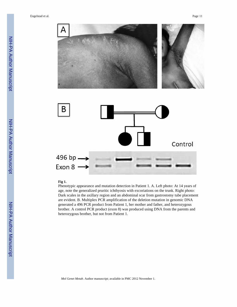
Fig 1.Phenotypic appearance and mutation detection in Patient 1. A. Left photo: At 14 years ofage, note the generalized pruritic ichthyosis with excoriations on the trunk. Right photo:Dark scales in the axillary region and an abdominal scar from gastrostomy tube placementare evident. B. Multiplex PCR amplification of the deletion mutation in genomic DNAgenerated a 496 PCR product from Patient 1, her mother and father, and heterozygousbrother. A control PCR product (exon 8) was produced using DNA from the parents andheterozygous brother, but not from Patient 1.
Engelstad et al. Page 11
Mol Genet Metab. Author manuscript; available in PMC 2012 November 1.
NIH
-PA Author Manuscript
NIH
-PA Author Manuscript
NIH
-PA Author Manuscript
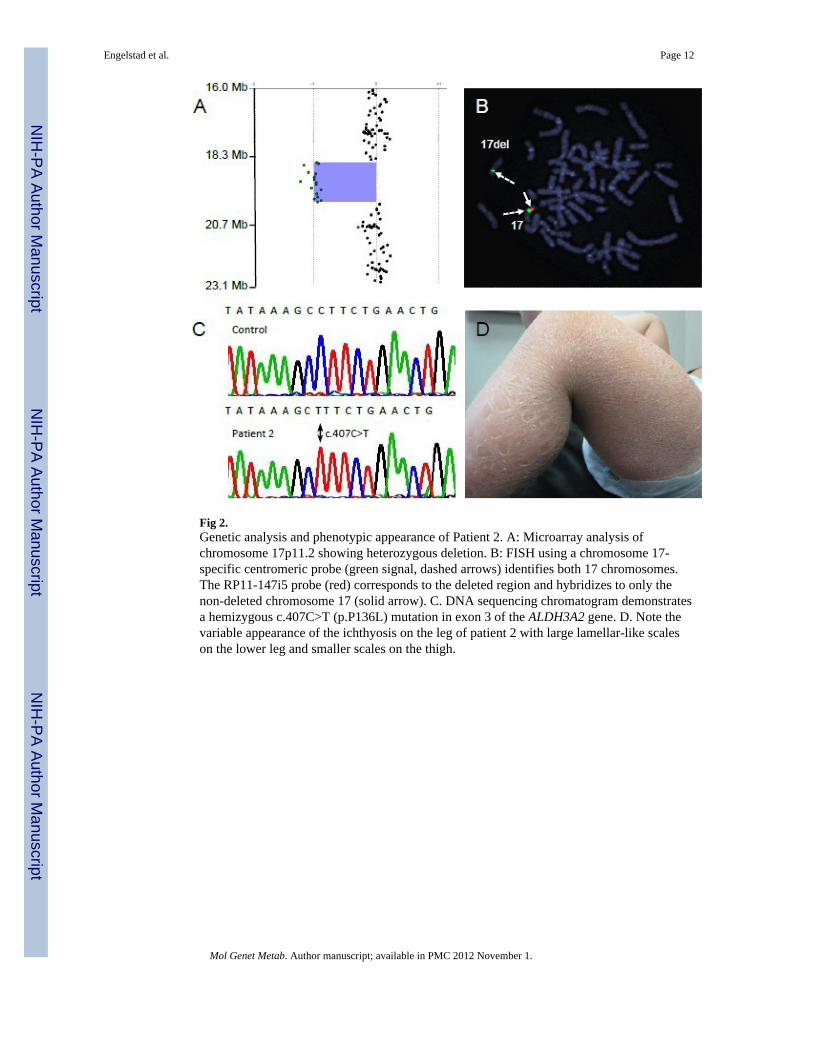
Fig 2.Genetic analysis and phenotypic appearance of Patient 2. A: Microarray analysis ofchromosome 17p11.2 showing heterozygous deletion. B: FISH using a chromosome 17-specific centromeric probe (green signal, dashed arrows) identifies both 17 chromosomes.The RP11-147i5 probe (red) corresponds to the deleted region and hybridizes to only thenon-deleted chromosome 17 (solid arrow). C. DNA sequencing chromatogram demonstratesa hemizygous c.407C>T (p.P136L) mutation in exon 3 of the ALDH3A2 gene. D. Note thevariable appearance of the ichthyosis on the leg of patient 2 with large lamellar-like scaleson the lower leg and smaller scales on the thigh.
Engelstad et al. Page 12
Mol Genet Metab. Author manuscript; available in PMC 2012 November 1.
NIH
-PA Author Manuscript
NIH
-PA Author Manuscript
NIH
-PA Author Manuscript

Fig 3.ALDH3A2 genetic locus at chromosome 17p11.2 showing the large deletions in the patients.The horizontal solid lines indicate chromosome DNA with an expanded region delimited bythe vertical dashed lines. Arrows represent orientation of genes from 5’ to 3’ direction.Horizontal dashed lines correspond to deleted regions in Patient 1 and Patient 2. Nucleotidepositions are numbered in megabases (M).
Engelstad et al. Page 13
Mol Genet Metab. Author manuscript; available in PMC 2012 November 1.
NIH
-PA Author Manuscript
NIH
-PA Author Manuscript
NIH
-PA Author Manuscript
NIH
-PA Author Manuscript
NIH
-PA Author Manuscript
NIH
-PA Author Manuscript
Engelstad et al. Page 14
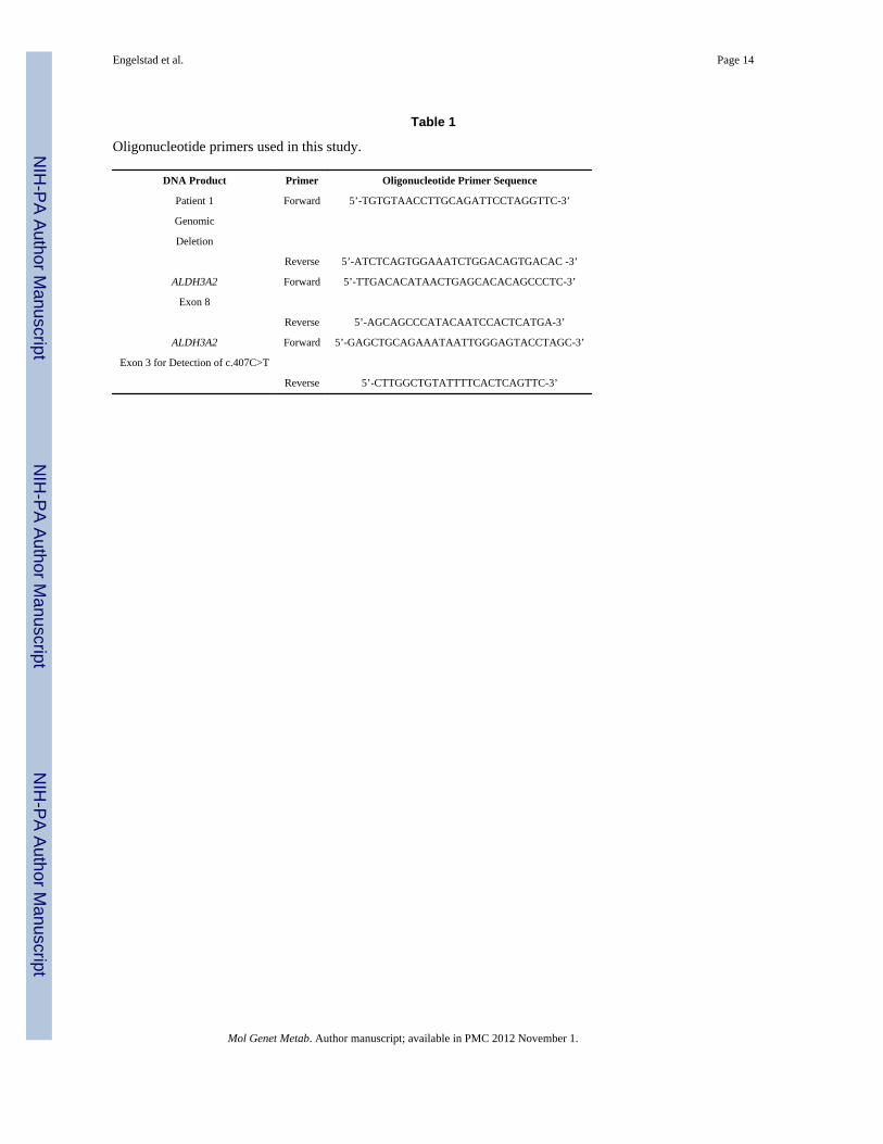
Table 1
Oligonucleotide primers used in this study.
DNA Product Primer Oligonucleotide Primer Sequence
Patient 1 Forward 5’-TGTGTAACCTTGCAGATTCCTAGGTTC-3’
Genomic
Deletion
Reverse 5’-ATCTCAGTGGAAATCTGGACAGTGACAC -3’
ALDH3A2 Forward 5’-TTGACACATAACTGAGCACACAGCCCTC-3’
Exon 8
Reverse 5’-AGCAGCCCATACAATCCACTCATGA-3’
ALDH3A2 Forward 5’-GAGCTGCAGAAATAATTGGGAGTACCTAGC-3’
Exon 3 for Detection of c.407C>T
Reverse 5’-CTTGGCTGTATTTTCACTCAGTTC-3’
Mol Genet Metab. Author manuscript; available in PMC 2012 November 1.