smash 2009 nmr conference
TRANSCRIPT

Conference Program September 22nd - 25th 2019
Porto, Portugal
SMASH 2019 NMR Conference Dear SMASH 2019 Attendees,
Firstly, thank you from everyone on the SMASH organising and executive committees for registering for this year’s meeting, our 20th anniversary event, and a special thank you from both of us for supporting the conference and helping to ensure that Small Molecules stay Hot!!
This year we are meeting in the wonderful city of Porto in northern Portugal, an historical city crossed by the Douro River and located on the Atlantic Ocean famed also for its port wine. We have tried to ensure that alongside the fantastic science you will hear about during SMASH 2019, there’s also time for you to be able to enjoy this great location throughout the duration of the meeting. If this is your first SMASH meeting, welcome, and if like many of us you are a SMASH veteran, welcome back - we hope you will all take away with you some great memories.
As ever, we have pulled together an excellent and diverse line of speakers for you from across both industry and academia – including world leading experts in their fields and the next generation of early career researchers who are hot on their heels. We’re delighted that this year we have 10 female speakers in the program, the most for a European SMASH meeting, all helping to build the open and engaged community of NMR spectroscopists that is the very heart of SMASH.
The formal program starts on Sunday evening with registration, followed by a mixer and dinner at the Palácio da Bolsa (Stock Exchange Palace), in the heart of the centre of old Porto, a wonderful historical building: you will be able to explore the interesting rooms including the Arab Room. This 300-metre sized room decorated in Moorish style, inspired by the Alhambra, is the Palace’s highlight while we will have our dinner in the large central courtyard called Pátio das Nações (Courtyard of the Nations), enclosed by a glass structure which lets in a beautiful light into the whole Palace.
This year there are seven oral sessions, two workshops and two poster sessions during which you will hear, learn and read about the latest advances in NMR research including; frontiers in sensitivity, analysis of dynamic systems, the latest NMR hot topics, NMR of small molecules in biological systems, industrial applications, advances in ssNMR, characterisation of pure and mixed materials, anisotropic NMR measurements and computational NMR.
On Monday evening we will celebrate 20 years of SMASH with an informal and light-hearted retrospective review of some of the scientific and social highlights of the last two decades and have dinner and wines at Casa Ferreirinha, at the Ferreira Cellars in the Historical Centre of Vila Nova de Gaia, by the Douro river. The Ferreira Cellar is one of the oldest Porto wine family (since 1751), producer of the famous Porto wine Barca Velha named in honour of its matriarch, Dona Antonia Adelaide Ferreira, who was affectionately nicknamed ‘Ferreirinha’ – or ‘little Ferreira’ in Portuguese.
Finally, thanks to all our sponsors for helping to support us financially – and please visit our exhibitors during the lunch and coffee breaks to hear about the latest and greatest developments in NMR hardware, software and consumables.
Here’s to another fantastic SMASH – cheers!! Steve and Carla Program co-chairs, SMASH 2019 conference
SMASH 2019 September 22nd-25th, 2019 Porto, Portugal 2 of 226

SMASH 2019 Conference Program
Sunday, September 22nd 09:00 AM - 05:00 PM Vendor User Meetings 05:00 PM - 06:30 PM Registration 06:45 PM - 07:00 PM Buses to dinner 07:30 PM - 11:00 PM Mixer & Dinner at Palacio da Bolsa 11:00 PM - 11:15 PM Buses to the Sheraton Porto Hotel
Monday, September 23rd 08:50 AM - 09:00 AM Welcome, Announcements and Opening Remarks 09:00 AM - 10:30 AM Frontiers in NMR Sensitivity
Session Organizer/Lead Speaker: Lyndon Emsley, EPFL Moderator: Christina Thiele, TU Darmstadt
DNP Enhanced NMR Crystallography Lyndon Emsley, EPFL
Zeno to the Rescue: Projective measurements to enhance cross peak intensities in NOESY and TOCSY based NMR correlations in peptides, nucleic acids, sugars and metabolites
Lucio Frydman, Weizmann Institute New sensitivity limits for small-volume NMR in combination with
hyperpolarization techniques Victoria Gómez, University of Castilla-la Mancha
Hitting the Jackpot: Forensic Hyperpolarisation of Fentalogues Thomas Robertson, Manchester Metropolitan University
10:30 AM - 11:00 AM Break
11:00 AM - 12:30 PM Workshop: Analysis of Dynamic Systems Co-Chairs: Michael Maiwald, Bundesanstalt für Materialforschung und -prüfung (BAM) David Foley, Pfizer
12:30 PM - 02:00 PM Lunch 02:00 PM - 03:30 PM Poster session 1 - Evens
Even numbered posters to be presented 03:30 PM - 04:00 PM Break
04:00 PM - 05:30 PM Hot Topics Session Organizer/Lead Speaker: Josep Sauri, Merck Moderator: Laura Castañar, University of Manchester
Modern approaches to the NMR characterization of complex natural products Josep Saurí, Merck
SEA XLOC: Distinguishing Two- and Three-bond Correlations in Heteronuclear NMR Spectroscopy Katalin Kövér, University of Debrecen, Hungary
Using Proton Residual Chemical Shift Anisotropy at Microgram Level for the Determination of the Relative Configuration in Marine Natural Products Juan Carlos C. Fuentes-Monteverde, Max Planck Institute for Biophysical Chemistry
Application of New NMR Methodologies in the Structural Characterization of a Novel Family of Alkaloids from the Marine Ascidian Polyandrocarpa sp. Kirk Gustafson, National Cancer Institute, NIH
15:30 - 16:00 Break
SMASH 2019 September 22nd-25th, 2019 Porto, Portugal 3 of 226

06:45 PM - 07:00 PM Buses to dinner
07:30 PM - 11:00 PM SMASH 20th Anniversary Celebration (inc. Dinner) at Casa Ferreirinha
11:00 PM - 11:15 PM Buses to the Sheraton Porto Hotel
Tuesday, September 24th
09:00 AM - 10:30 AM Small Molecules in Biological Systems Session Organizer/Lead Speaker: Maria Rangel, University of Porto Moderator: Amy Freund, Bruker Biospin
Insights on the Interaction of 3-hydroxy-4-pyridinones and their Metal Ion Chelates with Membrane Models from Biophysical Studies Maria Rangel, University of Porto
Novel Developments in Saturation Transfer Difference (STD) NMR Approaches to Investigate Weak Protein-Ligand Interactions Jesus Angulo, University of East Anglia
NMR Fragment-Based Discovery of Novel Potent Sepiapterin Reductase Inhibitors Markus Schade, Grunenthal GmbH
A Novel Method using NMR for Plasma Protein Binding Assessment in Drug Discovery Programs Mariana Gallo, IRBM
10:30 AM - 11:00 AM Break
11:00 AM - 12:30 PM Industrial Applications of NMR Session Organizer/Lead Speaker: Maria Victoria Silvia Elipe, Amgen Moderator: David Foley, Pfizer
Recent technology development and process understanding in high and low field NMR for small molecule pharmaceutics at Amgen Maria Victoria Silvia Elipe, Amgen
NMR of the Periodic Table, direct and indirect observation Bernd Diehl, Spectral Service AG
10 Things to do with an Open Access Proton NMR Spectrum of Classical and Emerging Drug Modalities Nichola Davies, AstraZeneca
A New Small Volume Online NMR Reaction Monitoring System Kathleen Farley, Pfizer
12:30 PM - 02:30 PM Buffet lunch and Poster session 2 - Odds Odd numbered posters to be presented
02:30 PM - 11:00 PM Free Time
SMASH 2019 September 22nd-25th, 2019 Porto, Portugal 4 of 226

Wednesday, September 25th 09:00 AM - 10:30 AM Advances in ssNMR
Session Organizer/Lead Speaker: Les Hughes, AstraZeneca Moderator: Ann-Christin Pöppler, University of Würzburg
Combining Solution and Solid-State NMR Data to Identify Tolfenamic Acid Polymorphs and Their Crystal Conformations Les Hughes, AstraZeneca
NMR Crystallography of Disorder in Molecular Organics Paul Hodgkinson, Durham University
Methods to Improve Resolution in 1H Solid State NMR at Ultra-Fast MAS Pinelopi Moutzouri, EPFL
43Ca Solid-State NMR Spectroscopy of Atorvastatin Calcium Steve Bai, University of Delaware
10:30 AM - 11:00 AM Break
11:00 AM - 12:30 PM Characterisation of Pure and Mixed Materials Session Organizer/Lead Speaker: Laura Castañar Acedo, University of Manchester Moderator: Mark Dixon, Mestrelab Research
New NMR Tools for Getting the Most Out of Complex Spectra Laura Castañar Acedo, University of Manchester
Measuring and Interpreting Couplings in Challenging Systems Davy Sinnaeve, CNRS
CSSF-CLIP-HSQMBC: The Measurement of Unresolved Heteronuclear Couplings Johan Isaksson, UiT the Arctic University of Norway
PSYCHE-EASY-ROESY Goes Quantitative: Extraction of 1H-1H Distance Restraints from Overcrowded Spectral Regions Julian Ilgen, Technical University of Darmstadt
12:30 PM - 02:00 PM Lunch
02:00 PM - 03:30 PM Workshop: All you Need to Know About Anisotropic NMR Measurements Chair: Christina Thiele, Technical University of Darmstadt
03:30 PM - 04:00 PM Break
04:00 PM - 05:30 PM Advances in Computational NMR Session Organizer/Lead Speaker: Armando Navarro-Vázquez, Universidade Federal de Pernambuco Moderator: Clark Ridge, FDA
CASE-3D. Advances and Perspectives Armando Navarro-Vázquez, Universidade Federal de Pernambuco
DFT and Beyond - Obtaining Accurate NMR Data Using Electronic Structure Methods Alexander A. Auer, Max-Planck-Institut für Kohlenforschung
IMPRESSION (Intelligent Machine PREdiction of Shift and Scalar Information Of Nuclei): Fast and accurate NMR Parameter Prediction with Machine Learning Will Gerrard, University of Bristol
BioMagResBank: Database and Tools for Bioactive Small Molecule NMR Analysis Pedro Romero, University of Wisconsin-Madison
05:30 PM - 06:00 PM Closing Remarks
SMASH 2019 September 22nd-25th, 2019 Porto, Portugal 5 of 226

Thursday, September 26th
09:00 AM - 04:30 PM NMReDATA Symposium Coordinator: Damien Jeannerat, University of Geneva
This event is not included in SMASH registration
Register Here
SMASH 2019 September 22nd-25th, 2019 Porto, Portugal 6 of 226

SMASH 2019 Scholarship Recipients
The following students received a scholarship to attend SMASH 2019
• Maria Beira, Faculdade de Ciências e Tecnologia, Portugal • Rachael Broomfield-Tagg, University of Bath, United Kingdom • Bhawna Chaubey, IIT JODHPUR, India • Matthew Davy, University of Bristol, United Kingdom • Lydia Dewis, University of Bristol, United Kingdom • Claire Dickson, University of Edinburgh, United Kingdom • Rafael Teixeira Freire, Institute for Bioengineering of Catalonia, Spain • Juan Carlos Fuentes Monteverde, Max Planck Institute for Biophysical Chemistry, Germany • Will Gerrard, University of Bristol, United Kingdom • Dariusz Golowicz, University of Warsaw, Poland • Dominik Herold, Technische Universität, Germany • Max Hirschmann, Technische Universität Darmstadt, Germany • Štepán Horník, ICPF CAS Prague, Czech Republic • Julian Ilgen, Technische Universität Darmstadt, Germany • Cristian Lepori, IFEG, FAMAF-CONICET, Argentina • Xiaolu Li, Leibniz-Forschungsinstitut für Molekulare Pharmakologie, Germany • Kumar Motiram Corral, Universitat Autònoma de Barcelona, Spain • Henry Nkabyo, Stellenbosch University, South Africa • Jens Nowag, TU Darmstadt, Germany • Elena Pisa, University of Cambridge, United Kingdom • Ann Christin Reiersølmoen, Norwegian University of Science and Technology, Norway • Thomas Robertson, Manchester Metropolitan University, United Kingdom • Alexandra Shchukina, University of Warsaw, Poland • Sebastian Spann, Karlsruhe Institute of Technology, Germany • Renan Ziemann Wilhelms, UFG, Brazil • Yanita Yankova, Miss, United Kingdom
Thanks to our scholarship sponsors for their generous support.
SMASH 2019 September 22nd-25th, 2019 Porto, Portugal 7 of 226

SMASH 2019 NMR Conference Acknowledgements
SMASH gratefully acknowledges the support from these companies:
Elite Sponsor
Premier Sponsor
Sponsors
SMASH 2019 September 22nd-25th, 2019 Porto, Portugal 8 of 226

Supporters
Fellowship Sponsors
SMASH 2019 September 22nd-25th, 2019 Porto, Portugal 9 of 226
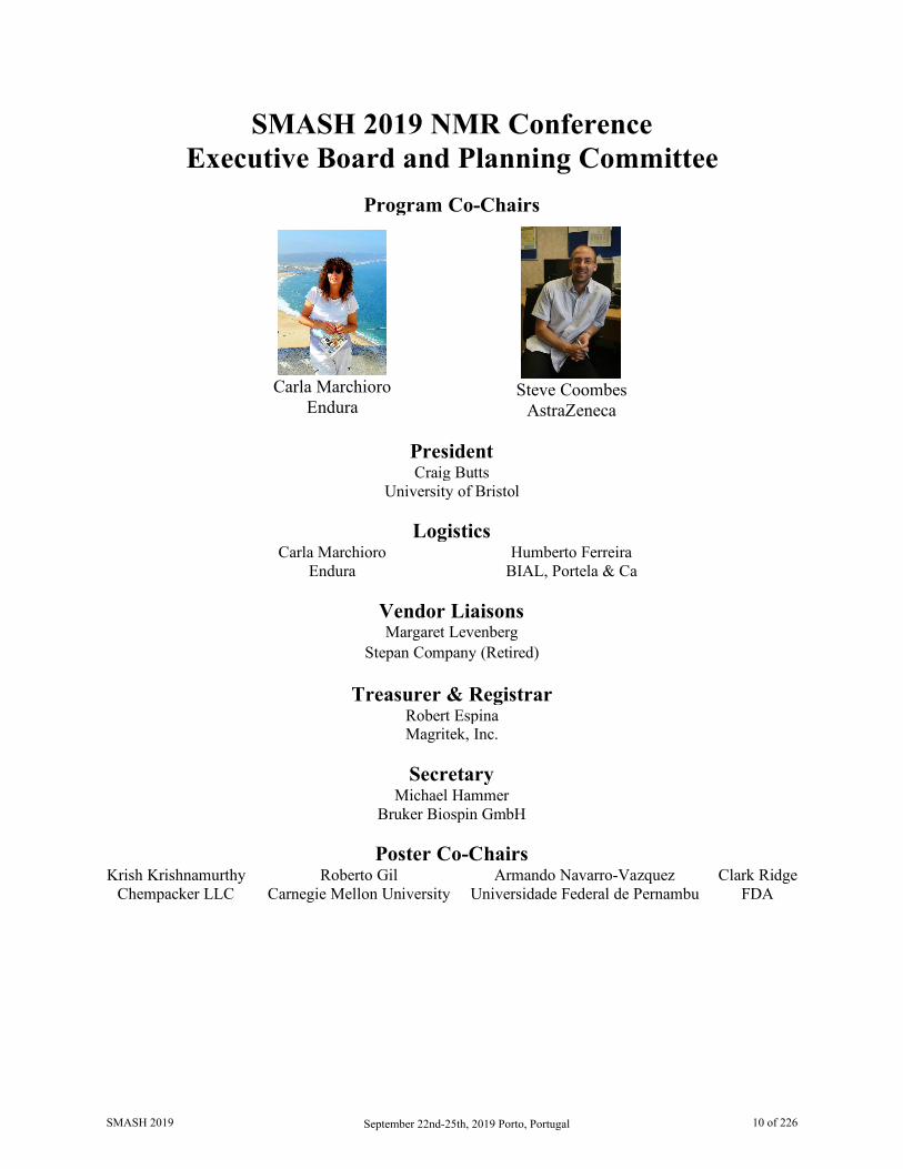
SMASH 2019 NMR Conference Executive Board and Planning Committee
Program Co-Chairs
Carla Marchioro
Endura
Steve Coombes
AstraZeneca
President Craig Butts
University of Bristol
Logistics Carla Marchioro Humberto Ferreira
Endura BIAL, Portela & Ca
Vendor Liaisons Margaret Levenberg
Stepan Company (Retired)
Treasurer & Registrar Robert Espina Magritek, Inc.
Secretary
Michael Hammer Bruker Biospin GmbH
Poster Co-Chairs
Krish Krishnamurthy Roberto Gil Armando Navarro-Vazquez Clark Ridge Chempacker LLC Carnegie Mellon University Universidade Federal de Pernambu FDA
SMASH 2019 September 22nd-25th, 2019 Porto, Portugal 10 of 226
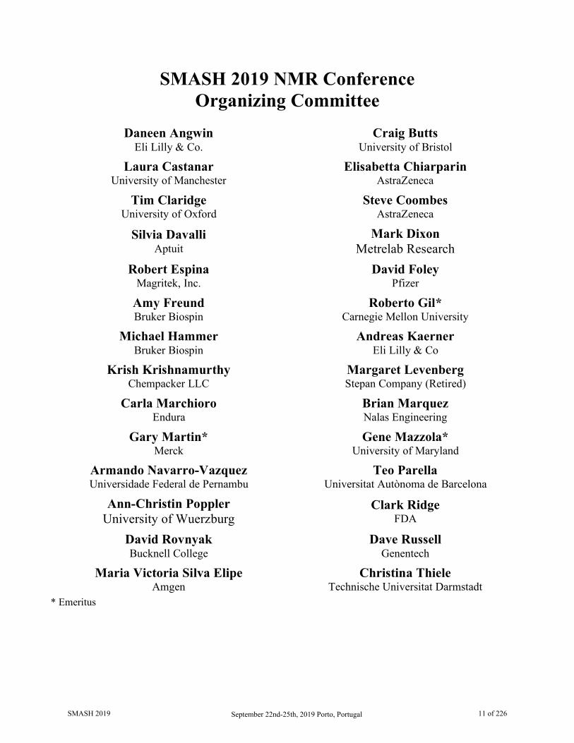
SMASH 2019 NMR Conference Organizing Committee
Daneen Angwin
Eli Lilly & Co. Craig Butts
University of Bristol
Laura Castanar University of Manchester
Elisabetta Chiarparin AstraZeneca
Tim Claridge University of Oxford
Steve Coombes AstraZeneca
Silvia Davalli Aptuit
Mark Dixon Metrelab Research
Robert Espina Magritek, Inc.
David Foley Pfizer
Amy Freund Bruker Biospin
Roberto Gil* Carnegie Mellon University
Michael Hammer Bruker Biospin
Andreas Kaerner Eli Lilly & Co
Krish Krishnamurthy Chempacker LLC
Margaret Levenberg Stepan Company (Retired)
Carla Marchioro Endura
Brian Marquez Nalas Engineering
Gary Martin* Merck
Gene Mazzola* University of Maryland
Armando Navarro-Vazquez Universidade Federal de Pernambu
Teo Parella Universitat Autònoma de Barcelona
Ann-Christin Poppler University of Wuerzburg
Clark Ridge FDA
David Rovnyak Bucknell College
Dave Russell Genentech
Maria Victoria Silva Elipe Amgen
Christina Thiele Technische Universitat Darmstadt
* Emeritus
SMASH 2019 September 22nd-25th, 2019 Porto, Portugal 11 of 226
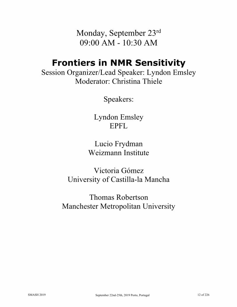
Monday, September 23rd 09:00 AM - 10:30 AM
Frontiers in NMR Sensitivity
Session Organizer/Lead Speaker: Lyndon Emsley Moderator: Christina Thiele
Speakers:
Lyndon Emsley
EPFL
Lucio Frydman Weizmann Institute
Victoria Gómez
University of Castilla-la Mancha
Thomas Robertson Manchester Metropolitan University
SMASH 2019 September 22nd-25th, 2019 Porto, Portugal 12 of 226

DNP Enhanced NMR Crystallography Lyndon Emsley
Institut des Sciences et Ingénierie Chimiques, Ecole Polytechnique Fédérale de Lausanne (EPFL), 1015 Lausanne, Switzerland
Structure elucidation of amorphous materials and microcrystalline solids presents one of the key challenges in chemistry today. While techniques such as single crystal diffraction and cryo-electron microscopy are generally not able to characterize such materials, we will show how an approach based on measured NMR chemical shifts in combination with DFT calculation of shifts from candidate structures calculated using crystal structure prediction (CSP) can rapidly determine full three-dimensional structures from powders.
Further, since the method does not requires any significant long-range order, we extend it to determine structures in non-crystalline and hierarchical or composite materials. It is not even known whether molecular fragments at surfaces form well defined structures or if they adopt disordered conformations. For example, specific metal-surface interactions have been proposed to be essential in stabilizing active site structures in many heterogeneous catalysts or nanoparticles, but so far it has not been possible to obtain three-dimensional structures to confirm such interactions.
We demonstrate how, combined with the sensitivity gains provided dynamic nuclear polarization (DNP) surface enhanced NMR spectroscopy (SENS) the above approach to NMR crystallography can determine the full atomic-level structure of supported active single site complexes and nanoparticle surfaces.
Full three-dimensional structure determined by NMR for a powder sample of Cocaine, The positional errors are visualized as ORTEP plots drawn at the 90% probability level.
SMASH 2019 September 22nd-25th, 2019 Porto, Portugal 13 of 226

Zeno to the Rescue: Projective measurements to enhance cross peak intensities in NOESY
and TOCSY based NMR correlations in peptides, nucleic acids, sugars and
metabolites
Mihajlo Novakovic and Lucio Frydman
Department of Chemical and Biological Physics, Weizmann Institute of Science, 7610001 Rehovot, Israel
TOCSY and NOESY are widely used blocks in the establishment of correlations between homonuclear systems – particularly 1Hs. The present study explores the possibilities to use projective measurements for enhancing signals in a majority of biomolecular systems. These gains arise from anti-Zeno effects speeding up these internuclear correlations. This led to the recently introduced Looped, PROjected SpectroscopY (L-PROSY, as in “leprosy”) approach, which enhances spin-diffusive processes by looping them repeatedly(1). As a result of this, the modulations imparted by J- or relaxation-driven processes involving labile protons can be enhanced by factors of ca. 200% for folded proteins and nucleic acids, 300% for unfolded biopolymers, 400% for polysaccharides, and variable gains for metabolites. These 2D NMR enhancements were also realized in novel homonuclear and heteronuclear 3D L-PROSY-based experiments. The field dependence of these gains will also be briefly discussed.
1. M. Novakovic, S. F. Cousin, M. J. Jaroszewicz, R. Rosenzweig and L. Frydman, J. Magn. Reson., 2018, 294, 169
Acknowledgement: This work was supported by the Israel Science Foundation (ISF 965/18), the Kimmel Institute of Magnetic Resonance (Weizmann Institute) and the EU Horizon 2020 programme (MC Grant 642773).
SMASH 2019 September 22nd-25th, 2019 Porto, Portugal 14 of 226
New sensivity limits for small-volume NMR in combination with hyperpolarization
techniques M. Victoria Gómez1, Miguel Mompeán1, Rosa Sanchez-Donoso1, Margarita Ruiz1, Ruben López-
Sánchez1, Antonio de la Hoz1, and Aldrik H. Velders1,2
1. IRICA, Universidad de Castilla-La Mancha, Ciudad Real, Spain 2. Wageningen University & Research, Wageningen, The Netherlands
Despite the power of NMR spectroscopy in a wide range of applications, it suffers from an intrinsically low sensitivity. In 1994 Sweedler and co-workers [1] launched the field of microcoil NMR spectroscopy as an approach to enhance NMR sensitivity, and the past two decades several groups have started to fabricate their own small-volume probe-heads and therefore, finding applications in different fields.
In the past few years, we have exploited mainly lower nanoliter planar spiral microcoils for their excellent mass sensitivity and their broad-band properties that allows homo- and heteronuclear multidimensinoal NMR experiments with a single coil in a non-tuned circuit [2]. These planar coils has also shown interesting properties for on-flow reaction monitoring [3], allowing the rapid determination of kinetic and thermodynamic parameters for reactions activated thermically, or photochemically.
Recently, we developed a simple method for the manufacturing of microfluidic devices in a single block of PDMS to include not only channels in the PDMS device, but also fully functional solenoidal NMR coils [4]. The fabrication method employs materials that are commonly present at chemistry laboratory, and does not required much knowledge on the technique, facilitating the access of microcoils to a wider public.
Herein, we present the combination of solenoidal microcoils with hyperpolarization schemes, based on photo-Chemically Induced Dynamic Nuclear Polarization (photo-CIDNP) experiments on just 1 µL of sample to boost the NMR signal detection down to sup-picomole detection limits in a 9.4T system (400 MHz 1H Larmor frequency) [5]. When this set up is brought to a 11.7 T spectrometer and planar spiral microcoils are used, the limit of detection is decreased even further, enabling the detection of a hundred of femtomol of material. In this experimental conditions, not only mass-sensivity is enhanced but also concentration sensitivity, as the new setup enable the detection of micromolar concentrations. This is really fascinating is we consider the low active volumes when working on microcoils and therefore the requirement of a relatively high sample concentration. It will certainly open the window for the structural determination of concentration limited samples.
Interestingly, this setup is unaffected by current major drawbacks of photo-CIDNP such as the use of high-power light sources to attempt uniform irradiation of the sample, and accumulation of degraded photosensitizer in the detection region. Optical density solutions can also be irradiated efficiently, resulting in very high signal enhancements. The issue related with the accumulation of degraded photosensitizer in the detection region is overcome with flow conditions, which in turn open avenues for complex applications
SMASH 2019 September 22nd-25th, 2019 Porto, Portugal 15 of 226
in i.e. Supramolecular Chemistry requiring rapid and efficient mixing that are not easily achievable on an NMR tube without resorting to complex hardware [5].
1. Olson DL.; Peck TL.; Webb AG.; Magin RL.; Sweedler JV. Science, 270, 1967-1970, 1995. 2. Fratila RM.; Gomez MV.; Sykora S.; Velders AH Nat. Commun. 5, 3025, 2014. 3. Gomez MV.; Rodriguez AM.; de la Hoz, A.; Jimenez F.; Fratila RM.; Barneveld PA.; Velders AH.;
Anal. Chem., 10547-10555, 2015. 4. Saggiomo V.; Velders AH.; Adv. Sci., 2, 1500125, 2015. 5. Mompeán, M.; Sanchez-Donoso, R.; de la Hoz, A.; Saggiomo, V., Velders, A.H., Gomez, M. V. Nat.
Commun., 9, 108, 2018.
SMASH 2019 September 22nd-25th, 2019 Porto, Portugal 16 of 226
Hitting the Jackpot: Forensic Hyperpolarisation of Fentalogues
Thomas B. R. Robertson1, Nicholas Gilbert1, Lysbeth H. Antonides1, Christopher J. Schofield2,3, Oliver
B. Sutcliffe1,3 and Ryan E. Mewis1
1. Manchester Metropolitan University, Manchester, Greater Manchester, UK 2. Greater Manchester Police, Manchester, Greater Manchester, UK
3. MANchester Drug Analysis & Knowledge Exchange (MANDRAKE), Manchester, Greater Manchester, UK
Signal Amplification By Reversible Exchange (SABRE), first reported in 2009 [1], allows for the creation of a hyperpolarised state in solution without chemical change to the substrate. For example, World Health Organisation essential medicines have been polarised without chemical modification [2]. This has made SABRE an attractive technique for medical imaging applications in the future. Despite also being a World Health Organisation essential medicine, fentanyl has recently become a drug of abuse with an estimated potency of ~500 times that of morphine. This poses a serious threat to public health with quantities as low as ~2 milligrams enough to induce overdose. In particular, the lacing of heroin samples with fentanyl for increased potency and decreased production cost has been a major contributing factor to thousands of deaths per year in the US and an increasing number across Europe. A range of fentanyl analogues (fentalogues), some of which are more potent than fentanyl, have recently appeared on the international drugs market however, due to the small quantities typically present, existing detection technology struggles to rapidly detect low concentrations of fentanyl or fentalogues. Given the range of fentalogues encountered three pyridyl fentalogues were prepared as this functionality has been the focus of multiple SABRE reports [3]. We present results for the hyperpolarisation of known biologically active fentalogues and unknown fentalogues produced through the systematic derivatisation of fentanyl. The successful SABRE hyperpolarisation of fentalogues is discussed in relation to concentrations commensurate with the lethal dose. The application of SABRE to a more realistic sample of fentalogue spiked heroin was tested and in a fentanyl/heroin mixture with 3% fentalogue, hyperpolarisation is observed. It is envisaged the application of SABRE could enable fentalogue detection on a benchtop instrument in a single scan, increasing the accessibility, and rapidity, of detection with a view towards a forensic application. 1. Adams R. W., Aguilar J. A., Atkinson K. D., Cowley M. J., Elliott P. I. P., Duckett S. B., Green G. G.
R., Khazal I. G., López-Serrano J. and Williamson D. C., Science, 323 (5922), 1708-1711, 2009 2. Olaru A. M., Robertson T. B. R., Lewis J. S., Anthony A., Iali W, Mewis R. E., Duckett S. B.,
ChemistryOpen, 7, 97-105, 2018 3. Ratajckyk T., Gutmann T., Bernatowicz P., Buntkowsky G., Frydel J., Fedorczyk B., Chemistry: A
European Journal, 21 (36), 12616-12619, 2015
SMASH 2019 September 22nd-25th, 2019 Porto, Portugal 17 of 226
Monday, September 23rd 11:00 AM - 12:30 PM
Workshop
Analysis of Dynamic Systems Coordinated by:
Michael Maiwald Bundesanstalt für Materialforschung und -prüfung (BAM)
David Foley Pfizer
SMASH 2019 September 22nd-25th, 2019 Porto, Portugal 18 of 226
Analysis of Dynamic Systems Coordinated by:
Michael Maiwald, Bundesanstalt für Materialforschung und -prüfung (BAM) David Foley, Pfizer
Monitoring specific information (i.e., physico-chemical properties, chemical reactions, etc.) is the key to chemical process control when looking at dynamic systems, and quantitative online NMR spectroscopy is the method of choice for the investigation and understanding of dynamic multi-component systems. NMR provides rapid and non-invasive information, and due to the inherent linearity between sample concentration and signal intensity, peak areas can be directly used for quantification of multiple components in a mixture (without the need for any further calibration). This is one of the most attractive features of quantitative NMR spectroscopy. With the launch of devices covering magnetic field strengths from 40 to 90 MHz, so called compact or benchtop NMR systems, this analytical method is now reaching a sufficient degree of compactness and operability for an application outside of very specialized laboratories.
Whilst there are also many other tools available to examine various analytical parameters from dynamic processes, such as mass spectrometry, (near) infrared or Raman spectroscopy, each of these tools can only really be used independently. How can we examine and compare all data describing a particular chemical reaction? How can we visualize information rich, specific, or direct methods together with less specific but established analytical methods? And how can we transfer calibration information to the most appropriate process analytical method or method combination? Quantitative NMR spectroscopy (qNMR) has the potential to substitute offline laboratory analysis for calibration purposes by delivering quantitative reference data as an online method.
The workshop briefly presents the current state of the art of the analysis of dynamic systems by online NMR spectroscopy and analytical data fusion, with the remaining time being used for questions and open discussion with the attendees.
SMASH 2019 September 22nd-25th, 2019 Porto, Portugal 19 of 226
Monday, September 23rd 04:00 PM - 05:30 PM
Hot Topics
Session Organizer/Lead Speaker: Josep Saurí Moderator: Laura Castañar
Speakers:
Josep Saurí
Merck
Katalin Kövér University of Debrecen, Hungary
Juan Carlos C. Fuentes-Monteverde
Max Planck Institute for Biophysical Chemistry
Kirk Gustafson National Cancer Institute, NIH
SMASH 2019 September 22nd-25th, 2019 Porto, Portugal 20 of 226
Modern Approaches to the NMR Characterization of Complex Natural Products
Josep Saurí1, Xiao Wang2, Mikhail Reibarkh2, Kirk R. Gustafson3, Yizhou Liu4, Gary E. Martin5 and R. Thomas Williamson6
1. Structure Elucidation Group, Analytical Research and Development, Merck & Co., Inc., Boston, MA, USA
2. Structure Elucidation Group, Analytical Research and Development, Merck & Co., Inc., Rahway, NJ, USA
3. Molecular Targets Laboratory, Center for Cancer Research, National Cancer Institute, Frederick, MD, USA
4. Analytical Research and Development, Pfizer Worldwide Research and Development, Groton, CT, USA
5. Department of Chemistry and Biochemistry, Seton Hall University, South Orange, NJ, USA 6. Department of Chemistry and Biochemistry, University of North Carolina at Wilmington,
Wilmington, NC, USA
Increasing natural product molecular complexity, correspondingly increases the probability of mistakes in assigning the constitution and configuration of the molecule in question. Standard 2D NMR experiments often prove insufficient, and there are cases where even computer-assisted structure elucidation (CASE) methods fail. These eventualities can, in part, explain the tide of mistakenly reported structures and the burgeoning number of structure revisions appearing in the literature.
Tremendous advances have been made over the last few years in the acquisition and analysis of anisotropic NMR data, the application of computational tools, and development of new pulse sequences. Combined, these methodologies provide a state-of-the-art road map for the unequivocal structure elucidation of complex natural products, synthetic organics and pharmaceuticals. This presentation will focus on the utilization of these techniques and their impact in solving challenging structural problems.
SMASH 2019 September 22nd-25th, 2019 Porto, Portugal 21 of 226
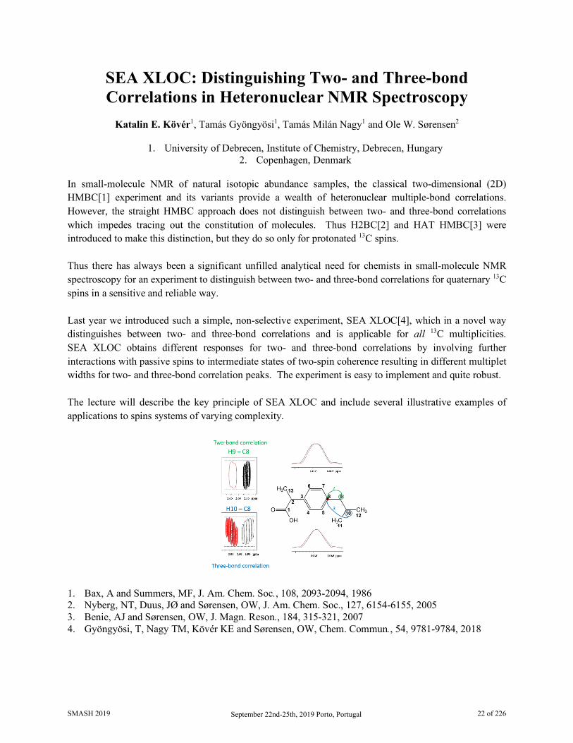
SEA XLOC: Distinguishing Two- and Three-bond Correlations in Heteronuclear NMR Spectroscopy
Katalin E. Kövér1, Tamás Gyöngyösi1, Tamás Milán Nagy1 and Ole W. Sørensen2
1. University of Debrecen, Institute of Chemistry, Debrecen, Hungary
2. Copenhagen, Denmark In small-molecule NMR of natural isotopic abundance samples, the classical two-dimensional (2D) HMBC[1] experiment and its variants provide a wealth of heteronuclear multiple-bond correlations. However, the straight HMBC approach does not distinguish between two- and three-bond correlations which impedes tracing out the constitution of molecules. Thus H2BC[2] and HAT HMBC[3] were introduced to make this distinction, but they do so only for protonated 13C spins. Thus there has always been a significant unfilled analytical need for chemists in small-molecule NMR spectroscopy for an experiment to distinguish between two- and three-bond correlations for quaternary 13C spins in a sensitive and reliable way. Last year we introduced such a simple, non-selective experiment, SEA XLOC[4], which in a novel way distinguishes between two- and three-bond correlations and is applicable for all 13C multiplicities. SEA XLOC obtains different responses for two- and three-bond correlations by involving further interactions with passive spins to intermediate states of two-spin coherence resulting in different multiplet widths for two- and three-bond correlation peaks. The experiment is easy to implement and quite robust. The lecture will describe the key principle of SEA XLOC and include several illustrative examples of applications to spins systems of varying complexity. 1. Bax, A and Summers, MF, J. Am. Chem. Soc., 108, 2093-2094, 1986 2. Nyberg, NT, Duus, JØ and Sørensen, OW, J. Am. Chem. Soc., 127, 6154-6155, 2005 3. Benie, AJ and Sørensen, OW, J. Magn. Reson., 184, 315-321, 2007 4. Gyöngyösi, T, Nagy TM, Kövér KE and Sørensen, OW, Chem. Commun., 54, 9781-9784, 2018
SMASH 2019 September 22nd-25th, 2019 Porto, Portugal 22 of 226
Using Proton Residual Chemical Shift Anisotropy at Microgram Level for the
Determination of the Relative Configuration in Marine Natural Products
Juan Carlos Fuentes-Monteverde,1,2 Nilamoni Nath,3 Dawrin Pech-Puch,2 Armando Navarro-Vázquez,4 Elena Balboa,5 Jaime Rodríguez,2 Carlos Jiménez,2 and Christian Griesinger1
1. NMR Based Structural Biology, MPI for Biophysical Chemistry, Göttingen, Germany 2. Centro de Investigacións Científicas Avanzadas (CICA) e Departamento de Química, Facultade
de Ciencias, Universidade da Coruña. A Coruña, Spain 3. Department of Chemistry, Gauhati University, Gopinath Bardoloi Nagar, Guwahati, India
4. Departamento de Química Fundamental, CCEN, Universidade Federal de Pernambuco, Cidade Universitária, Recife, Brazil
5. Department of Chemical Engineering, Faculty of Science, Campus Ourense, University of Vigo, Spain. CITI-University of Vigo, Parque Tecnolóxico de Galicia, Ourense, Spain
Determination of the three dimensional structure remains a challenging task for natural products when are available in minute amounts and cannot be crystallized. While the chemical constitution can be derived from proton/proton and proton/carbon correlations, J-couplings, NOEs and often 1H-13C RDCs or 13C RCSAs are used to determine the relative configuration. However, for compounds in the range of few microgram these RDC or 13C RCSAs are difficult to collect because of low sensitivity. [1,2] Herein, we demostrate that 1H RCSAs allows to determinte the orientation of different structural moieties within a molecule but with much less requirements on the amount of sample. An important issue that arises in RCSA measurements of small molecules in the range of micrograms, is very often the use of protonated polymeric gels to induce the alignment conditions present NMR signals that masks the actual sample. This problem was circumvented by the use of PMMA-d8 gel. The potential of the methodology was also demonstrated. 1H RCSAs were measured by using 3 mm devices (stretching and compression) manufactured by the glass company Hilgenberg, for microgram samples of marine natural products, isolated from Briareum asbestinum and Sargassum muticum, collected in Mexican Caribbean (Mexico) and Costa da Morte (Spain) respectively, for which relative configuration is still unknown [3,4]. Use of NOESY constraints restricted the number of possible relative configurations, from which 1H RCSA allowed us to select a single relative configuration. Determination of the absolute configuration could be accomplished by comparison of calculated (TD-DFT) and experimental electronic circular dichroism spectra [5]. In summary, we reported that 1H RCSAs as highly sensitive parameter that complemented J-couplings and NOEs, without the necessity to use any other anisotropic parameter at the microgram scale.
References: 1. Nath, N. et al., Journal of the American Chemical Society, 138(30), 9548-9556, 2016 2. Liu, Y. et al., Nature Protocol, 14, 217-247, 2019 3. Harvell, C. et al., Marine Ecology-Progress Series, 93, 165-173, 1993 4. Balboa, E. et al., J Photochem Photobiol B, 148, 51-58, 2015 5. Pescitelli, G. et al., Chirality, 28(6), 466-474, 2016
SMASH 2019 September 22nd-25th, 2019 Porto, Portugal 23 of 226

Application of New NMR Methodologies in the Structural Characterization of a Novel Family
of Alkaloids from the Marine Ascidian Polyandrocarpa sp
Kirk R. Gustafson1, Xiangrong Tian1,2, Dongdong Wang1, Heidi R. Bokesch1,3, Gary E. Martin4, R. Thomas Williamson5, and Josep Saurí6
1. Molecular Targets Program, National Cancer Institute, Frederick, MD, USA 2. Research & Development Center of Biorational Pesticide, Northwest A&F University,
Yangling, China 3. Basic Science Program, Leidos Biomedical Research, Inc., Frederick National Laboratory
for Cancer Research, Frederick, MD, USA 4. Department of Chemistry and Biochemistry, Seton Hall University, South Orange, NJ, USA 5. University of North Carolina-Wilmington, Department of Chemistry, Wilmington, NC, USA 6. Structure Elucidation Group, Process and Analytical Research and Development, Merck &
Co., Boston, MA, USA
A series of novel polyhalogenated pentacyclic alkaloids was isolated from an extract of the marine ascidian Polyandrocarpa sp. Assembly of the carbon-nitrogen framework was accomplished with the aid of heteronuclear correlation data measured using the 1,1-HD-ADEQUATE pulse sequence.1 This experiment is based on proton detection of one bond 13C-13C couplings, and its sensitivity is enhanced via BIRD-based homodecoupling of vicinal proton couplings. Thus, 1,1-HD-ADEQUATE provides correlations that are equivalent to 2-bond HMBC correlations, without the ambiguity over 2-bond versus 3-bond correlations in the HMBC experiment. This attribute is particularly useful for establishing the presence of nonprotonated carbons adjacent to protonated neighbors, that are not amenable to detection using H2BC. The new alkaloids contain both Cl and Br substituents, and assigning the sites of chlorination versus bromination is difficult based solely on 13C chemical shift comparisons or calculations. The recently described bs-CLIP-HSQMBC experiment allows detection of the 35,37Cl isotope effect on both protonated and nonprotonated carbons.2 This pulse sequence was crucial to the structure elucidation, as it unambiguously established the chlorination pattern in the new alkaloids, which have an unprecedented heterocyclic molecular scaffold. 1. Saurí, J.; Bermel, W.; Buevich, A. V.; Sherer, E. C.; Joyce, L. A.; Sharaf, M. H. M.; Schiff, P. L. Jr.;
Parella, T.; Williamson, R. T.; Martin, G. E. Angew. Chem. Int. Ed. 2015, 54, 10160-10164. 2. Saurí, J.; Reibarkh, M.; Zhang, T.; Cohen, R. D.; Wang, X.; Molinski, T. F.; Martin, G. E.;
Williamson, R. T. Org. Lett. 2016, 18, 4786-4789.
SMASH 2019 September 22nd-25th, 2019 Porto, Portugal 24 of 226

Tuesday, September 24th 09:00 AM - 10:30 AM
Small Molecules in Biological Systems
Session Organizer/Lead Speaker: Maria Rangel Moderator: Amy Freund
Speakers:
Maria Rangel
University of Porto
Jesus Angulo University of East Anglia
Markus Schade
Grunenthal GmbH
Mariana Gallo IRBM
SMASH 2019 September 22nd-25th, 2019 Porto, Portugal 25 of 226
Insights on the Interaction of 3-hydroxy-4-pyridinones and their Metal Ion Chelates with Membrane Models from Biophysical Studies
Maria Rangel
REQUIMTE-LAQV, Instituto de Ciências Biomédicas de Abel Salazar,
Universidade do Porto, Porto, Portugal Biophysical studies regarding the interaction of potential new drugs with biological membranes, in concert with establishment of structural activity relationships, constitute a fundamental part of the evaluation of the performance of new molecules in the implementation of novel therapeutic strategies. Moreover it is known that the introduction of spectroscopic labels, such as fluorophores, used to get insight on the cellular pathways and targets of a given molecule which exhibits biological activity may lead to a change in the membrane permeation properties of the molecule and result in a dramatic change in biological activity.
Our group has been interested in the design of 3-hydroxy-4-pyridinone ligands and their metal ion chelates to be applied in novel therapeutic strategies to fight Infection, Iron Overload and Diabetes.
An account of the work performed in our group regarding spectroscopic (NMR, EPR and steady-state fluorescence spectroscopy), molecular dynamics and confocal microscopy studies of molecules designed to be of use in biomedical applications will be given.
SMASH 2019 September 22nd-25th, 2019 Porto, Portugal 26 of 226
Novel Developments in Saturation Transfer Difference (STD) NMR Approaches to
Investigate Weak Protein-Ligand Interactions
Jesús Angulo
School of Pharmacy, University of East Anglia (UEA), Norwich Research Park, NR4 7LU, Norwich, U.K.
E-mail: [email protected] Saturation Transfer Difference (STD) NMR is a powerful ligand-based NMR technique for weak ligand screening of protein targets and to gain quantitative structural information from biologically relevant protein-ligand complexes.[1] The approach is appropriate for small/medium-sized molecule binders of medium-weak affinity (dissociation constants from high nM to low mM), there is no upper limit for protein size, and labelling is not required. The technique is highly versatile and popular in the context of hit identification in drug discovery.
In this talk the investigation by STD NMR of molecular recognition processes of ligands by biologically relevant protein receptors will be presented.[2-5] Although the technique has demonstrated its broad applicability to study many different protein-ligand systems, particularly in the field of Fragment Based Drug Design, the talk will keep a focus on the application to specific cases of recognition of glycans by proteins. Protein-glycan interactions are very relevant protein-ligand interactions in Nature and are processes typically falling within the range of fast chemical exchange (weak affinity) but still showing high specificity. [6] These protein-glycan systems will allow to introduce along the talk novel methodological developments in STD NMR produced in our lab, as the identification about how the fast ligand rebinding process can affect the determination of accurate dissociation constants by STD NMR,[7] as well as the development of the recent method “DiffErential EPitope mapping STD NMR (DEEP-STD NMR)” [8] that allows for the first time to identify the nature of the protein-ligand contacts in the bound state from STD NMR approaches.
References 1. M. Mayer and B. Meyer, Angew Chem Int Edit 1999, 38, 1784. 2. P. M. Enríquez-Navas, M. Marradi, D. Padro, J. Angulo and S. Penadés, Chem Eur J 2011, 17, 1547. 3. Thépaut, M.; Guzzi, C.; Sutkeviciute, I.; Sattin, S.; Ribeiro-Viana, R.; Varga, N.; Chabrol, E.; Rojo, J.;
Bernardi, A.; Angulo, J.; Nieto, P. M.; Fieschi, F. J Am Chem Soc 2013, 135, 2518. 4. Muñoz-García, J. C.; Chabrol, E.; Vives, R. R.; Thomas, A.; de Paz, J. L.; Rojo, J.; Imberty, A.; Fieschi,
F.; Nieto, P. M.; Angulo, J. J Am Chem Soc 2015., 137 (12), 4100. 5. C. D. Owen, L. E. Tailford, S. Monaco, T. Suligoj, L. Vaux, R. Lallement, Z. Khedri, H. Yu, K.
Lecointe, J. Walshaw, S. Tribolo, M. Horrex, A. Bell, X. Chen, G. L. Taylor, A. Varki, J. Angulo, N. Juge, Nat Commun 2017, 8.
6. Essentials of Glycobiology. Varki A, Cummings R, Esko J, et al., editors. Cold Spring Harbor (NY): Cold Spring Harbor Laboratory Press; 1999.
7. J. Angulo, Pedro M. Enríquez-Navas and Pedro M. Nieto, Chem Eur J 2010, 16, 7803. 8. S. Monaco, L. E. Tailford, N. Juge, J. Angulo, Angew Chem Int Edit 2017, 56, 15289
SMASH 2019 September 22nd-25th, 2019 Porto, Portugal 27 of 226
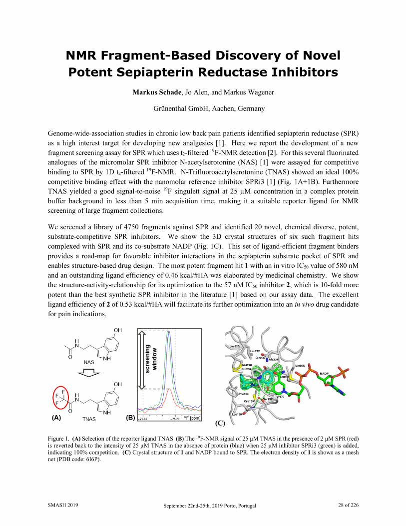
NMR Fragment-Based Discovery of Novel Potent Sepiapterin Reductase Inhibitors
Markus Schade, Jo Alen, and Markus Wagener
Grünenthal GmbH, Aachen, Germany
Genome-wide-association studies in chronic low back pain patients identified sepiapterin reductase (SPR) as a high interest target for developing new analgesics [1]. Here we report the development of a new fragment screening assay for SPR which uses t2-filtered 19F-NMR detection [2]. For this several fluorinated analogues of the micromolar SPR inhibitor N-acetylserotonine (NAS) [1] were assayed for competitive binding to SPR by 1D t2-filtered 19F-NMR. N-Trifluoroacetylserotonine (TNAS) showed an ideal 100% competitive binding effect with the nanomolar reference inhibitor SPRi3 [1] (Fig. 1A+1B). Furthermore TNAS yielded a good signal-to-noise 19F singulett signal at 25 µM concentration in a complex protein buffer background in less than 5 min acquisition time, making it a suitable reporter ligand for NMR screening of large fragment collections.
We screened a library of 4750 fragments against SPR and identified 20 novel, chemical diverse, potent, substrate-competitive SPR inhibitors. We show the 3D crystal structures of six such fragment hits complexed with SPR and its co-substrate NADP (Fig. 1C). This set of ligand-efficient fragment binders provides a road-map for favorable inhibitor interactions in the sepiapterin substrate pocket of SPR and enables structure-based drug design. The most potent fragment hit 1 with an in vitro IC50 value of 580 nM and an outstanding ligand efficiency of 0.46 kcal/#HA was elaborated by medicinal chemistry. We show the structure-activity-relationship for its optimization to the 57 nM IC50 inhibitor 2, which is 10-fold more potent than the best synthetic SPR inhibitor in the literature [1] based on our assay data. The excellent ligand efficiency of 2 of 0.53 kcal/#HA will facilitate its further optimization into an in vivo drug candidate for pain indications.
(C)
Figure 1. (A) Selection of the reporter ligand TNAS (B) The 19F-NMR signal of 25 µM TNAS in the presence of 2 µM SPR (red) is reverted back to the intensity of 25 µM TNAS in the absence of protein (blue) when 25 µM inhibitor SPRi3 (green) is added, indicating 100% competition. (C) Crystal structure of 1 and NADP bound to SPR. The electron density of 1 is shown as a mesh net (PDB code: 6I6P).
SMASH 2019 September 22nd-25th, 2019 Porto, Portugal 28 of 226

References:
1. Latremoliere, A., Latini, A., Andrews, N., Cronin, S. J., Fujita, M., Gorska, K., Neuron 86, 1393–1406, 2015
2. Dalvit, C., Vulpetti, A., J. Med. Chem. 62(5), 2218-2244, 2019
SMASH 2019 September 22nd-25th, 2019 Porto, Portugal 29 of 226
A Novel Method using NMR for Plasma Protein Binding Assessment in Drug Discovery
Programs Mariana Gallo,1 Sara Matteucci,2 Nadine Alaimo, 2 Erica Pitti, 2 Maria V. Orsale, 1 Vincenzo Summa,1
Daniel O. Cicero,1,2 and Edith Monteagudo1
1. IRBM Science Park S.p.A, Pomezia, Rome, Italy 2. Dipartimento di Scienze e Tecnologie Chimiche, Università di Roma “Tor Vergata”, Rome, Italy
The amount of plasma-bound drug influences compound dosing, efficacy, clearance rate, and potential for drug interactions. Therefore, having information about the extent to which a drug candidate binds to plasma proteins is a critical feature of drug development. A new methodology based on Nuclear Magnetic Resonance (NMR) was developed to determine plasma protein binding (PPB) of drug candidates in drug discovery programs [1].
Recognizing the utility of NMR as a very sensitive method for detecting binding, we have focused on developing an approach particularly fast and, if an NMR spectrometer is available, low cost alternative to those currently used for determining PPB. We have used the fastest and simplest NMR method for evaluating protein binding, the traditional 1D 1H line-broadening experiment, which additionally allows by a single-point measurement easily rank binding affinities [2]. A strong correlation was found between the attenuation of NMR signals of diverse drugs in the presence of different plasma concentrations and their fraction bound (fb) reported in the literature. Based on these results, a protocol for a rapid calculation of fb of small molecules was established. The advantage of using plasma instead of purified recombinant proteins and the possibility of pool analysis to increase throughput were also evaluated. This novel methodology proved to be very versatile, cost-effective, fast and suitable for automation. As a plus, it contemporarily provides a quality check and solubility of the compound.
1. Gallo M., Matteucci S., Alaimo N., Pitti E., Orsale M.V., Summa V., Cicero D.O., Monteagudo E., J. Pharm. Biomed. Anal., 167, 21-29 (2019)
2. Shortbridge M.D. Hage D.S., Harbison, G.S., Powers R., J. Comb. Chem., 10, 948-958 (2008)
SMASH 2019 September 22nd-25th, 2019 Porto, Portugal 30 of 226
Tuesday, September 24th 11:00 AM - 12:30 PM
Industrial Applications of NMR
Session Organizer/Lead Speaker: Maria Victoria Silvia Elipe
Moderator: David Foley
Speakers:
Maria Victoria Silvia Elipe Amgen
Bernd Diehl
Spectral Service AG
Nichola Davies AstraZeneca
Kathleen Farley
Pfizer
SMASH 2019 September 22nd-25th, 2019 Porto, Portugal 31 of 226
Recent Technology Development and Process Understanding in High and Low Field NMR for
Small Molecule Pharmaceutics at Amgen Maria Victoria Silva Elipe
Amgen, Inc., Thousand Oaks, CA US
Process understanding, characterization and quantitation are key areas for NMR in the pharmaceutical industry. In the case of small molecules, structure determination of substrates, products and in-process impurities are necessary to optimize the quality of the final products and their chemical processes. Typically, those characterizations are done off-line in high field NMR instruments (e.g., the determination of dimer in-process impurities for an investigational anti-inflammatory drug during the GMP campaign for FIH regulatory filing [1]). For the case of process understanding, online techniques are preferred. We explored low field NMR for reaction monitoring to determine the correct timing of a two-phase reaction with weak chromophores at 45 MHz with picoSpin-45 NMR spectrometer [2]. Ideally, the NMR instrument should be located in the chemistry laboratory where chemical reactions take place. We entered the area of technology innovation by testing a prototype 9.4 T (400 MHz for 1H observation) NMR instrument with a high temperature superconducting (HTS) power-driven magnet being cryogen-free and cooled by a standard helium cryocooler attached to commercial standard console and probe (Bruker Avance III HD NanoBay and BBFO probe). Our test indicated that 1D and 2D homonuclear and heteronuclear standard NMR experiments typically used for structure elucidation are comparable when using commercial NMR instruments with standard cryogen magnets [3,4]. We collocated the HTS NMR magnet system in the chemistry laboratory with the magnet in the hood of the reactor to perform online reaction monitoring and reducing the footprint of the NMR system by eliminating liquid cryogens needs. We demonstrated its usage for reaction monitoring with the synthesis of cycloalkenes through the catalytic reaction of ring closure metathesis (RCM) using Grubbs catalysts with protonated solvents and no field locking. We also explored the conversion of the pinacol arylboronic esters to triolborates by different nuclides detecting stable intermediates. In addition, we studied the thermodynamic process at different temperatures for the formation of a benzotriazole adduct in the NMR tube for process understanding. Furthermore, we have measured residual dipolar couplings (RDCs) to study relative configurations/conformations of molecules evaluating the HTS instrument performance. In addition, we have explored the application of low field NMR using an MQC-23 Oxford Instruments NMR (23 MHz for 1H and 22 MHz for 19F) for the determination of average drug content in formulated drug products by F-19 for fluorinated drugs. The measurement is done directly from tablets or capsules without sample manipulation and faster than the standard HPLC method [5,6]. We will present all those examples to demonstrate a series of application of NMR in drug development for the pharmaceutical industry.
1. Silva Elipe, M.V., Tan, Z.J., Ronk, M., Bostick, T., Int. J. Anal. Chem. 2009, article ID 768743, 12 pages, 2009, DOI: 10.1155/2009/76743.
2. Silva Elipe, M.V., Milburn, R., Magn. Reson. Chem. 2015, 54 (6), 437-443. DOI: 10.1002/mrc.4189. 3. Silva Elipe, M.V., Donovan, N., Krull, R., Pooke, D. Colson. K., Magn. Reson. Chem. 2018, 56 (9),
817-825. DOI: 10.1002/mrc.4740. 4. Silva Elipe, M.V., Donovan, N., Krull, R., Pooke, D. Colson. K., Instr. Sci. Technol. 2018. DOI:
10.1080/10739149.2018.1516226.
SMASH 2019 September 22nd-25th, 2019 Porto, Portugal 32 of 226

5. Silva Elipe, M.V., Li, L., Nagapudi, K., Kook, A., Cobas, C., Iglesias, I., Peng, C., Magn. Reson. Chem. 2015, 54 (6), 531-538. DOSI: 10.1002/mrc.4223.
6. Silva Elipe, M.V., Li, L., Nagapudi, K., Kook, A., Cobas, C., Iglesias, I., Peng, C., J. Vis. Exp. 2017 (126). DOI: 10.3791/55850.
SMASH 2019 September 22nd-25th, 2019 Porto, Portugal 33 of 226

NMR of the Periodic Table, Direct and Indirect Observation!
Bernd W.K. Diehl
Spectral Service AG, Cologne, Germany
A systematically Study and Outlook on heteronuclear quantitative NMR Spectroscopy.
In the routine operation of NMR laboratories quantitative methods are becoming more and more important. The focus, however, is on 1H NMR spectroscopy and, with some deductions, also on the phosphorus and fluorine nuclei, which are easy to handle due to their 100% natural abundance and the wide range of chemical shifts. However, if one looks more closely at the periodic table for NMR active nuclei, one finds that there are many more possibilities to perform qualitative and quantitative NMR spectroscopy on heteroatoms. The International Pharmacopoeia have a whole series of ionic compounds which, in addition to the organic molecules, have counterions accessible to direct or indirect NMR analysis. The direct observation is shown here, for example, on the anions Cl, Br, J and the cations Al, Na, K, Li, Zn. Indirect determinations of the various oxides of sulfur and nitrogen (NO2, NO3, SO3, SO4, S2O5) are carried out by 17O NMR. Another field of analysis for double charged metal ions such as Ca, Mg, Ba, Sr, Sn, Zn, Pb, Hg is enabled via the corresponding EDTA or DOTA complexes by means of 1H NMR. In addition, also classical manganometry is possible by 55Mn NMR on permanganate.
SMASH 2019 September 22nd-25th, 2019 Porto, Portugal 34 of 226
10 Things to do with an Open Access Proton NMR Spectrum of Classical and Emerging
Drug Modalities Nichola L. Davies1
, Rodrigo J. Carbajo1, Eva Lenz1, Amber Balazs2, Richard Lewis3, David Dias4 and Elisabetta Chiarparin1
1. Analytical and Structural Chemistry, Oncology R&D, AstraZeneca, Cambridge, UK 2. Oncology R&D, AstraZeneca, Boston, USA
3. RIA R&D, AstraZeneca, Gothenburgh, Sweden 4. Chemical Development, Pharmaceutical Technology & Development, Operations, AZ Macc, UK
Over the past fifty years NMR spectroscopy has become the preeminent technique for determining the structure of organic compounds. Within industry and academia a reference proton NMR spectrum (along with LC-MS) is required to establish structural identity and purity of organic substances. With advances in NMR automation, this data is generally acquired directly by the chemist in an open access environment.
This presentation will demonstrate the wealth of information we can obtain for a simple proton NMR spectrum, and how it can be utilised to inform both synthesis and design strategies within early stage drug discovery in the pharmaceutical industry. Case studies in the application of NMR from AstraZeneca will demonstrate 10 things that we can determine from a proton NMR spectrum, and of course Structural ID will be atop that list.
One of NMRs exquisite features is the inherent quantitative nature of the technique. This allows us to determine the Purity of samples without the requirement for method development. qNMR can also be utilised in automation to determine the Concentration of stock solutions to generate assay ready plates.
By monitoring the signals over a time course we can determine the Stability of our samples, and with the additional structural information characterise the degredation profiles to provide strategies to remove such liabilities in a series. Such a time course can also provide insight into Reaction Kinetics, for both chemical processes and biological events.
How a chemical shift changes with environment, such as temperature can indicate if a structural motif is involved in Intramolecular Hydrogen Bonding, a common design strategy to induce a bioactive conformation or improve passive permeability. Varying the solvent conditions such as the pH allows the determination of site specific pKas. The shape of an NMR peak can give insight into local Dynamic Behaviour in a molecule, such as flexibility.
And finally – tracking a diagnostic chemical shift from within a series can allow us to rapidly confirm the 3D Solution Conformation of a molecule, a critical parameter to improve potency. We will also demonstrate how chemical shift can be used as a predictor for relative Reactivity within the series, when developing a novel covalent inhibitor. These examples highlight the value of being able to readily access chemical shift information from a series, and the potential to develop predictive models for parameters which are critical to optimise during medicinal chemistry design strategies.
SMASH 2019 September 22nd-25th, 2019 Porto, Portugal 35 of 226

Examples from traditional small molecule, macrocycles, peptides and from some emerging drug classes such as proteolysis targeted chimeras (PROTACs) will be discussed.
SMASH 2019 September 22nd-25th, 2019 Porto, Portugal 36 of 226
A New Small Volume Online NMR Reaction Monitoring System
Kathleen A. Farley,1 David Foley1, Robert Krull2, Martin Hoffman3, and Anna Codina4
1. Pfizer, Groton, CT, US 2. Bruker BioSpin, Billerica, Massachusetts, US
3. Bruker BioSpin, Rheinstetten, Germany 4. Bruker UK Limited, Coventry, UK
A 5 mm NMR flow tube was previously developed that can be inserted into any standard 5 mm NMR probe converting a Bruker NMR spectrometer into a reaction monitoring system1. While this system is very useful for reactions volumes greater that 15 mL, the need for a flow tube which can accommodate smaller reaction volumes was desired. Towards this end, a 3 mm NMR flow tube with shorter transfer lines has been developed that can accommodate reaction mixtures from 2-20 mL. A standard pump is used to flow the reaction mixture from a reaction vial in an integrated heating block to the center of the magnet for detection and then return the mixture back to the reaction vial. Using a 3 mm glass tube and shorter transfer lines (volume ~1 mL) reduces the reaction volume to a minimum of ~2 mL. The transfer line contains the sample in and out tubing, surrounded by an outer chamber that circulates a temperature regulated fluid to maintain the reaction mixture at the desired temperature. The NMR instrument can easily be switched from regular NMR tubes to the flow tube mode in a few minutes allowing the instrument to be used for both open access and reaction monitoring as needs dictate. The new reaction monitoring system has been set up at Pfizer in Groton, CT and is being used to monitor small volume reactions. Several of these reactions will be described.
1. Foley, David A.; Bez, Eckhard; Codina, Anna; Colson, Kimberly L.; Fey, Michael; Krull, Robert; Piroli, Don; Zell, Mark T.; and Marquez, Brian L. Analytical Chemistry 2014, 86, 12008−12013.
SMASH 2019 September 22nd-25th, 2019 Porto, Portugal 37 of 226
Wednesday, September 25th 09:00 AM - 10:30 AM
Advances in ssNMR
Session Organizer/Lead Speaker: Les Hughes Moderator: Ann-Christin Pöppler
Speakers:
Les Hughes AstraZeneca
Paul Hodgkinson
Durham University
Pinelopi Moutzouri EPFL
Steve Bai
University of Delaware
SMASH 2019 September 22nd-25th, 2019 Porto, Portugal 38 of 226
Combining Solution and Solid-State NMR Data to Identify Tolfenamic Acid Polymorphs and
Their Crystal Conformations Leslie P. Hughes1, Helen Blade1, Charles D. Blundell2, Steven P. Brown3, Hugh R. W. Dannatt2,
Anjali K. Menakath3
1. Pharmaceutical Development, AstraZeneca, UK 2. C4X Discovery, Manchester, UK
3. Dept. Of Physics, University of Warwick, Poland
The NMR crystallography approach for solving\refining structural models requires a reasonably good model as a starting point. For those cases where a reasonable model cannot be generated from diffraction data then alternative approaches may be computationally intensive, i.e. crystal structure prediction (CSP). Conformational polymorphism, where different crystal structures exhibit different shapes of the molecule arising from different torsion angles, adds an additional complication as this cannot be easily modelled using CSP approaches.
We present a novel scoring function for assessing putative crystal structures of tolfenamic acid (TFA) using a combination of experimental solution- and solid-state NMR data and DFT based GIPAW calculations. The scoring function provides a method for assessing how well a suggested structure matches the experimental solid-state NMR chemical shifts. For TFA our approach can discriminate between four polymorphs of which Forms II, III and IV are structurally very similar. In particular, we believe that our new approach based on combining solution- and solid-state NMR offers a greater discriminating power than a comparison of GIPAW calculated and experimental solid-state chemical shifts alone.
We believe our approach has great potential for the many cases of solid-state chemistry where crystal structures are not available. We envisage that using our scoring function approach, CSP could now be used to solve intractable crystal structures. Specifically, before starting a CSP campaign the conformational search space can be dramatically reduced by using the experimental data with the scoring function to predict the most likely conformations present. After the CSP campaign has returned putative crystal structures, the scoring function can be used for a second time with the experimental data to reliably and unambiguously identify the correct structure. We can also envisage that this scoring function could be used in combination with structure determination by PXRD.
SMASH 2019 September 22nd-25th, 2019 Porto, Portugal 39 of 226

Dynamic 3D structure of TFA and plot of ∆δExperimental against ∆δCalculated for Form I
SMASH 2019 September 22nd-25th, 2019 Porto, Portugal 40 of 226
NMR Crystallography of Disorder in Molecular Organics Paul Hodgkinson
Department of Chemistry, Durham University, Durham, UK
Disorder in molecular materials is generally seen as a problem. This is understandable from the viewpoint of diffraction crystallography, where either static or dynamic disorder will disrupt Bragg diffraction and introduce uncertainties into the modelling of diffraction data. In many cases, however, a disordered state may represent the lowest (free) energy structure of material. By using solid-state NMR and computational chemistry to characterise disorder and understand its molecular origins, we can exploit materials that might be discarded as risky.
For example, the widely used drug valsartan is a marketed in an “amorphous” form, but DSC studies show that this form is distinct from a truly amorphous material produced by quench cooling from the melt. Powder diffraction, total scattering and 13C solid-state NMR measurements all imply that the “as received” material is more ordered, but neither material gives Bragg diffraction peaks. Counter-intuitively the more ordered material has a significantly higher solubility. A combination of time-modulated DSC, PXRD and solid-state NMR methods were used to understand the molecular origin of this “polyamorphic” behaviour in terms of conformational “defects”[1]. Other examples are shown using co-crystals and solvates of pharmaceutical actives where quantum chemical calculations are invaluable in determining whether a disordered or an ordered structure is adopted[2,3]. Recent work will also be presented showing how the complementary time and length scales of diffraction and NMR allow unexpected dynamic behaviour in apparently static strongly hydrogen-bonded structures to be uncovered.
1. Skotnicki, M., Apperley, D. C., Aguilar, J. A., Milanowski B., Pyda M., and Hodgkinson, P., Mol. Pharmaceutics, 13, 211–222, 2016
2. Kerr, H. E, Mason, H. E., Sparkes, H. A., and Hodgkinson, P., CrystEngComm, 18, 6700–6707, 2016 3. Bērziņš, A., and Hodgkinson, P., Solid State Nucl. Magn, Reson., 65, 12–20, 2015
SMASH 2019 September 22nd-25th, 2019 Porto, Portugal 41 of 226

Methods to Improve Resolution in 1H Solid State NMR at Ultra-Fast MAS Pinelopi Moutzouri, Federico M. Paruzzo, and Lyndon Emsley
Institut des Sciences et Ingénierie Chimiques, Ecole Polytechnique Fédérale de Lausanne (EPFL), CH-1015 Lausanne, Switzerland
In organic solids the homogeneous broadening caused by the strong 1H-1H dipolar coupling network yields 1H linewidths of few tens of kHz, orders of magnitude larger than those in solution and leads to broad and highly unresolved spectra from which very little information can be extracted.
Magic angle spinning (MAS)(1) can greatly improve spectral resolution. Recently, newly developed technology allows MAS rates greater than 100 kHz leading to 1H linewidths on the order of a few hundred Hz. However, in many applications and especially for complex systems, this is still not sufficient and limits the use of 1H in solid state NMR.
Here we explore new methods to further improve the spectral resolution of powdered organic solids in this fast spinning rate regime, and we show how to obtain experimental linewidths up to a factor of two narrower than those achieved with a conventional experiment at the same MAS rate.
1. Andrew, E.R., et al., Nature, 1958, 182, 1659-1659; Lowe, I.J., Phys. Rev. Lett., 1959, 2, 285-287
SMASH 2019 September 22nd-25th, 2019 Porto, Portugal 42 of 226
43Ca Solid-State NMR Spectroscopy of Atorvastatin Calcium Shi Bai1, Sean T. Holmes2, and Cecil Dybowski1
1. Department of Chemistry and Biochemistry, University of Delaware, DE, US 2. Department of Chemistry and Biochemistry, University of Windsor, Windsor, Canada
Form I of atorvastatin calcium trihydrate (ATC-I) is the active pharmaceutical ingradient (API) in Lipitor®, which is commonly prescribed for lowering blood cholesterol and preventing hypertension or other cardiovascular diseases. Lipitor® is arguably the best-selling drug in the history of modern Western medicine. Although the mechanism of the catalytic portion of complexes of 3-hydroxy-3-methylglutary-coenzme A (HMG-CoA) reductase with six statins, including atorvastatin, has been reported [1], the crystal structure of ATC-I, which determines the bioavailability, stability, safety, and efficacy of the medication [2], remains unknown to date.
Previously, we proposed a molecular structure of the atorvastatin ligands on the calcium ion in the ATC-I [3] based on13C, 19F, and 15N multinuclear solid-state NMR spectroscopic studies combined with extensive ab initio calculations. Although the model correctly predicted the two sets of 13C, 15N, and 19F isotropic chemical shifts observed for ATC-I under magic angle spinning conditions, the choice of the local calcium environment was arbitrary as it was derived from that of calcium dibenzoate·3H2O [4].
In this presentation, 43Ca solid-state NMR spectroscopy is employed to investigate local calcium structures in ATC-I and the ethylene glycol solvate of atorvastatin calcium (ATC-EG). We demonstrate that the 43Ca NMR spectral features are very sensitive to the local calcium environment in atorvastatin calcium and can be readily used as a unique spectroscopic index to differentiate solvomorphous forms of atorvastatin calcium.
We further discuss a crystallographic approach based on 43Ca NMR spectroscopy that includes an extensive survey of the Cambridge Structural Database (CSD) and a new symmetry benchmark to enhance the selectivity of structural screening [5]. In this method, experimental and computational 43Ca solid-state NMR parameters such as chemical-shift tensors, quadrupolar-coupling tensors, and Euler angles are compared to constrain the structure of the local calcium–ligand coordination environment in ATC-I. The structural search begins with an extensive survey of calcium-containing crystals in the CSD, followed by generation of a library of a large number of candidates for the local calcium environment in ATC-I that meet certain energetic criteria. The library is strictly screened for matches with experimental 43Ca NMR parameters using Hartree-Fock (HF) and density functional theory (DFT) calculations. While the majority of models in the library are eliminated by comparison between calculated and experimental 43Ca NMR chemical-shift and quadrupolar parameters, a number of structures cannot be differentiated with these comparisons alone. A complete analysis of the local symmetry of the calcium site demonstrates that the line shape of the 43Ca SSNMR powder pattern is very sensitive to β, the Euler angle between the most shielded CS principal axis (δ33) and the major axis (V33) of the EFG tensor. Simulation of the NMR spectra as a function of Euler angles reveals a unique relationship between the 43Ca quadrupolar line shape and the local symmetry at the calcium site. This observation demonstrates the Euler angle between these axes is a
SMASH 2019 September 22nd-25th, 2019 Porto, Portugal 43 of 226
particularly useful benchmark for screening structures to eliminate structural candidates remaining in the library that do not meet the calcium site symmetry criterion. As an example, only one candidate structure was ultimately retained from the remaining structures by the symmetry benchmark screening on the calcium coordination environment in ATC-I.
General implication of this structure-search protocol combining a survey of the Cambridge Structural Database with the Euler angle as a symmetry benchmark has been validated by applying it to identify the known calcium local structure of calcium acetate monohydrate [6].
References 1. Istvan, E. S. and Deisenhofer, J., Science, 292 (5519), 1160-4, 2001. 2. Brittain, H. G. and Grant, D. J. W., Effect of polymorphism and solid-state solvation on solubility and
dissolution rate. 1999, New York: Marcel Dekker, Inc. 3. Wang, W. D., Gao, X. D., Strohmeier, M., Wang, W., Bai, S., and Dybowski, C., Journal of Physical
Chemistry B, 116 (11), 3641-3649, 2012. 4. Senkovska, I. and Thewalt, U., Acta Crystallographica Section C-Crystal Structure Communications,
61, M448-M449, 2005. 5. Holmes, S. T., Wang, W. D., Hou, G., Dybowski, C., Wang, W., and Bai, S., Phys Chem Chem Phys,
21 (12), 6319-6326, 2019. 6. Widdifield, C. M., Moudrakovski, I., and Bryce, D. L., Physical Chemistry Chemical Physics, 16 (26),
13340-13359, 2014.
SMASH 2019 September 22nd-25th, 2019 Porto, Portugal 44 of 226
Wednesday, September 25th 11:00 AM – 12:30 PM
Characterisation of Pure and Mixed
Materials Session Organizer/Lead Speaker: Laura Castañar Acedo
Moderator: Mark Dixon
Speakers:
Laura Castañar Acedo University of Manchester
Davy Sinnaeve
CNRS
Johan Isaksson UiT the Arctic University of Norway
Julian Ilgen
Technical University of Darmstadt
SMASH 2019 September 22nd-25th, 2019 Porto, Portugal 45 of 226
New NMR Tools for Getting the Most Out of Complex Spectra
Laura Castañar Acedo
School of Chemistry, University of Manchester, Manchester, United Kingdom
Nuclear magnetic resonance (NMR) spectroscopy is a powerful and versatile tool for the structural and conformational analysis of molecules. However, conventional methods generally struggle to extract simple and clear information from the most challenging systems, such as macromolecules or mixtures. 1H NMR spectroscopy is the method most widely used, because of the proton’s high sensitivity and ubiquity in chemistry, and is by far the richest source of structural information. However, it has a major drawback: the narrow range of chemical shifts and the many interactions between spins make multiplets overlap, reducing spectral resolution drastically. As a result, 1H NMR spectra often show low resolution even for relatively simple species. In complex systems, signal overlap increases massively, making it extremely difficult (or even impossible) to extract and interpret structural information. Different strategies have been proposed for alleviating overlap in proton spectra and facilitating the extraction of valuable structural information, including the use of multidimensional NMR techniques, spectral editing to factorize the spectrum into subspectra, pure shift methods to suppress the effects of homonuclear couplings, and virtual separation of mixture components. Such methods are very efficient for the structural analysis of relatively simple systems, but they are still of limited use for solving the most complex structural problems. In this presentation some novel NMR methods for solving some of the most challenging problems encountered in pure and mixture systems will be presented.
The first family of experiments, REST (Relaxation-Encoded Selective TOCSY) [1], is based on the virtual separation of the components of a mixture by exploiting differences in the relaxation behaviour of spins. It combines selective excitation and isotropic mixing to label each spin in a given system with the same relaxation weighting (e.g. T1 or T2). REST experimental data can be subjected to univariate (e.g. ROSY) and/or bilinear (e.g. SCORE) analysis to extract the required structural information from the component subspectra. This family of experiments has proven to be useful in separating the spectra of spin systems from very similar molecules (e.g. disaccharides), cases in which conventional experiments fail.
In very challenging mixtures, univariate and bilinear analysis may be ineffective because of signal overlap, low signal-to-noise ratio, or large number of components. To overcome these limitations, a new family of experiments, SCALPEL (Spectral Component Acquisition by Localized PARAFAC Extraction of Linear components) [2], has been proposed. The SCALPEL approach consists in acquiring experimental data using a narrow bandwidth selective TOCSY pulse sequence in which signals are encoded with two or more types of species-selective labelling (e.g. diffusion, T1 or T2 relaxation, TOCSY-t1), and then using a multivariate analysis method such as PARAFAC to extract the subspectra of each of the different species present. The usefulness of the SCALPEL approach will be demonstrated on a natural fermented beverage (beer) and other carbohydrate mixtures.
The last type of method presented, FESTA (Fluorine-Edited Selective TOCSY Acquisition) [3], is designed for the structural analysis of fluorinated species (either pure or mixtures), for which interest has increased significantly in recent years, mainly due to their pharmacological activity (e.g. as anticancer, antidepressant,
SMASH 2019 September 22nd-25th, 2019 Porto, Portugal 46 of 226
or antiviral drugs). 1D FESTA exploits the high 19F spectral resolution to obtain clean 1H NMR subspectra for individual spin systems involving different 19F sites, facilitating structural analysis in complex fluorinated systems. Two different types of FESTA method will be presented, SRI (Selective Reverse INEPT) [3] and MODO (MODulated echO) [4], that differ in how the initial 19F → 1H heteronuclear editing is achieved.
When spectral overlap remains a problem, REST, SCALPEL and FESTA experiments and pure shift methodology [5,6] can be combined to provide further resolution.
The processing of REST and SCALPEL experimental data is straightforward using the free and open source software package GNAT (General NMR Analysis Toolbox) [7], which is a graphical user interface for analysis of NMR data from the major NMR manufacturers’ instruments. All of our pulse sequences and processing tools are free and can be downloaded from our homepage: https://nmr.chemistry.manchester.ac.uk
1. Dal Poggetto, G.; Castañar, L.; Adams, R. W.; Morris, G. A.; Nilsson, M.; Chem. Commun., 2017, 53, 7461-7464.
2. Dal Poggetto, G.; Castañar, L.; Adams, R. W.; Morris, G. A.; Nilsson, M.; J. Am. Chem. Soc., 2019, 141, 5766-5771.
3. Castañar, L.; Moutzouri, P.; Barbosa, T.; Tormena, C.; Rittner, R.; Phillips, A.; Coombes, S.; Nilsson, M.; Morris, G. A.; Anal. Chem., 2018, 90, 5445-5450.
4. Barbosa, T.; Castañar, L.; Moutzouri, P.; Nilsson, M.; Morris, G. A.; Rittner, R.; C. Tormena, C; (Unpublished)
5. Castañar, L.; Magn. Reson. Chem., 2017, 55, 47-53. 6. Castañar, L.; Parella T.; Magn. Reson. Chem., 2015, 53, 399-426. 7. Castañar, L.; Dal Poggetto, G.; Colbourne, A.; Morris, G. A.; Nilsson, M.; Magn. Reson. Chem.,
2018, 56, 546-558.
SMASH 2019 September 22nd-25th, 2019 Porto, Portugal 47 of 226

Measuring and Interpreting Couplings in Challenging Systems
Davy Sinnaeve
CNRS — Unité de Glycobiologie Structurale et Fonctionnelle (UGSF) UMR 8576, Lille, France
1H-1H couplings between spins provide valuable information for stereochemical or conformational analysis. However, the simultaneous presence of many couplings results in intricate and broad multiplets that easily overlap, preventing accurate coupling extraction. In recent years, several experiments have been proposed that deliver J-resolved spectra where individual couplings feature as simple doublets, alleviating the aforementioned issues [1,2]. These experiments include BSD-SERF [3], push-G-SERF, [4] PSYCHEDELIC [5] and several others, which are all variants of the progenitor SERF experiment [6] and exploit the principles of pure shift techniques [7]. It will be shown that these experiments make it possible to obtain nearly all 1H-1H couplings in most cases, even for challenging molecules like bile acids where a large network of coupled protons resonate in just a narrow part of the spectrum. However, even with the resolution issue overcome, another factor still limits the collection of all couplings in the molecule: strong coupling between protons. In 2D J-spectroscopy, this is well known to lead to additional peaks that complicate coupling measurement, as well as to peak splittings that deviate from the actual coupling value. Furthermore, measuring the coupling between strongly coupled protons is not feasible in SERF based experiments, as their multiplets typically overlap and cannot be easily distinguished using selective pulses. A number of new experiments will be presented based on the BIRD and PSYCHE pure shift elements that can relief the restrictions imposed by strong coupling.
Besides their measurement, also the accuracy of coupling interpretation can be challenging. This is particularly true for conformational analysis of five-membered rings based on vicinal scalar couplings. Saturated five-membered rings are generally flexible, sampling a distribution of conformers. In order to characterize this distribution experimentally, several vicinal couplings are required that encode dihedral angle information via Karplus relations. We wanted to assess the five-membered ring conformation of novel fluorinated proline variants [8,9] incorporated within peptides using 1H-1H and 1H-19F couplings. However, using established Karplus relations, the accuracy of this procedure turns out to be inadequate. No 1H-19F Karplus relations appropriate for these systems exist, while established general 1H-1H Karplus relations are not parametrized for every possible substituent on the HCCH fragments. Furthermore, the impact of non-bonding electronic interactions across the five-membered ring on the J-coupling are significant. We designed a strategy for J-coupling analysis of five-membered rings that is based on mapping the full conformational space of model compounds using DFT, followed by J-coupling calculation. This leads to accurate expressions of the J-coupling as a function of the global ring conformation rather than just a single dihedral angle.
1. Berger, S., Prog. Nucl. Magn. Reson. Spectrosc., 108, 74-114, 2018 2. Lin, Y.,Zeng, Q., Lin, L., Chen, Z., and Barker, P.B., Prog. Nucl. Magn. Reson. Spectrosc., 109, 135-159, 2018 3. Pucheta, J. E. H., Pitoux, D., Grison, C. M., Robin, S., Merlet, D., Aitken, D. J., Giraud, N., and Farjon, J., Chem. Commun. 51, 7939-7942, 2015
SMASH 2019 September 22nd-25th, 2019 Porto, Portugal 48 of 226
4. Pitoux, D., Plainchont, B., Merlet, D., Hu, Z. Y., Bonnaffe, D., Farjon, J., and Giraud, N., Chem. Eur. J., 21, 9044-9047, 2015 5. Sinnaeve, D., Foroozandeh, M., Nilsson, M., and Morris, G. A., Angew. Chem. Int. Edit., 55, 1090-1093, 2016 6. Fäcke, T., and Berger, S., J. Magn. Res. Ser. A, 113, 114-116, 1995 7. Zangger, K., Prog. Nucl. Magn. Reson. Spectrosc., 86-87, 1-20. 2015 8. Hofman, G. J., Ottoy, E., Light, M. E., Kieffer, B., Kuprov, I., Martins, J. C., Sinnaeve, D., and Linclau, B., Chem. Commun., 54, 5118-5121, 2018 9. Hofman, G. J., Ottoy, E., Light, M. E., Kieffer, B., Martins, J. C., Kuprov, I., Sinnaeve, D., and Linclau, B., J. Org. Chem., 84, 3100-3120, 2019
SMASH 2019 September 22nd-25th, 2019 Porto, Portugal 49 of 226
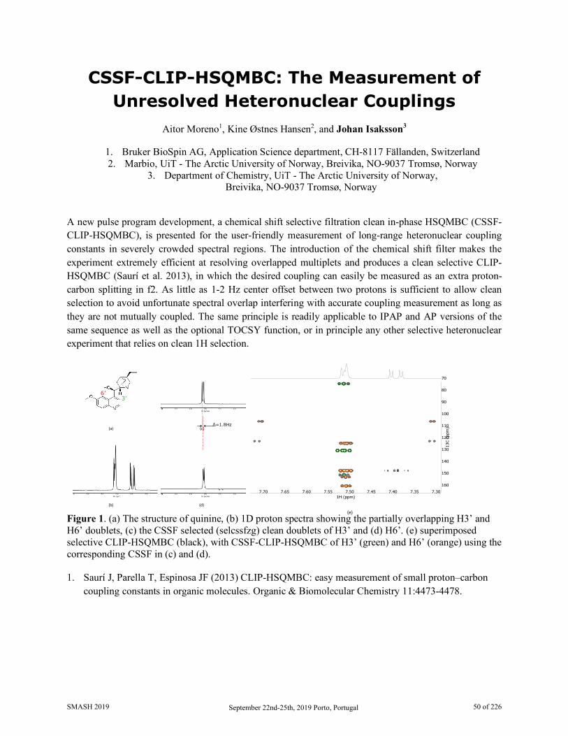
CSSF-CLIP-HSQMBC: The Measurement of Unresolved Heteronuclear Couplings
Aitor Moreno1, Kine Østnes Hansen2, and Johan Isaksson3
1. Bruker BioSpin AG, Application Science department, CH-8117 Fällanden, Switzerland 2. Marbio, UiT - The Arctic University of Norway, Breivika, NO-9037 Tromsø, Norway
3. Department of Chemistry, UiT - The Arctic University of Norway, Breivika, NO-9037 Tromsø, Norway
A new pulse program development, a chemical shift selective filtration clean in-phase HSQMBC (CSSF-CLIP-HSQMBC), is presented for the user-friendly measurement of long-range heteronuclear coupling constants in severely crowded spectral regions. The introduction of the chemical shift filter makes the experiment extremely efficient at resolving overlapped multiplets and produces a clean selective CLIP-HSQMBC (Saurí et al. 2013), in which the desired coupling can easily be measured as an extra proton-carbon splitting in f2. As little as 1-2 Hz center offset between two protons is sufficient to allow clean selection to avoid unfortunate spectral overlap interfering with accurate coupling measurement as long as they are not mutually coupled. The same principle is readily applicable to IPAP and AP versions of the same sequence as well as the optional TOCSY function, or in principle any other selective heteronuclear experiment that relies on clean 1H selection.
Figure 1. (a) The structure of quinine, (b) 1D proton spectra showing the partially overlapping H3’ and H6’ doublets, (c) the CSSF selected (selcssfzg) clean doublets of H3’ and (d) H6’. (e) superimposed selective CLIP-HSQMBC (black), with CSSF-CLIP-HSQMBC of H3’ (green) and H6’ (orange) using the corresponding CSSF in (c) and (d). 1. Saurí J, Parella T, Espinosa JF (2013) CLIP-HSQMBC: easy measurement of small proton–carbon
coupling constants in organic molecules. Organic & Biomolecular Chemistry 11:4473-4478.
(a) (c)
(b) (d)
6’3’
Δ=1.8Hz
(e)
7.307.357.407.457.507.557.607.657.701H (ppm)
70
80
90
100
110
120
130
140
150
160
13C
(ppm
)
SMASH 2019 September 22nd-25th, 2019 Porto, Portugal 50 of 226
PSYCHE-EASY-ROESY Goes Quantitative: Extraction of 1H-1H Distance Restraints from
Overcrowded Spectral Regions Julian Ilgen1, Jens Nowag1, Lukas Kaltschnee2,3 and Christina M. Thiele1
1. Clemens-Schöpf-Institut, Technische Universität Darmstadt, Germany 2. Max-Planck-Insitut für Biophysikalische Chemie, Göttingen, Germany
3. Center for Biostructural Imaging of Neurodegeneration (BIN), Göttingen, Germany
1H,1H-NOESY[1] and 1H,1H-ROESY[2] are valuable experiments for spatial structure elucidation of organic or bioorganic molecules of varying size. They provide access to proton-proton distances, frequently used as qualitative or quantitative measures of spatial proximity.
Since these experiments yield proton-proton correlation spectra, signal overlap caused by limited proton chemical shift dispersion becomes a major challenge with increasing molecular complexity. The incorporation of pure shift elements into NOESY and ROESY experiments represents a possible solution to improve spectral resolution in such cases [3-7].
We have used an EASY-ROESY [8] experiment with PSYCHE [9] homonuclear decoupling for the structural analysis of a peptide organocatalyst, which usually yields overcrowded spectra [7,10]. We show that the extraction of cross relaxation rates and distance restraints from PSYCHE-EASY-ROESY is accurate, and thus provides a valuable tool for studying overcrowded spectral regions. The enhanced resolution enabled us to extract the fourfold number of proton-proton distances, as compared to conventional techniques. These advances may lead to a significant improvement in the structural models also of the peptide catalyst in the future.
A notable advance over our earlier studies [7] is achieved, by using a gradient selected PSYCHE-EASY-ROESY instead of a phase-cycled experiment. This significantly attenuates “t1-noise” and thus enhances the spectral quality. Analysis of these “cleaned-up” PSYCHE-EASY-ROESY spectra with cyclosporine as model compound allows for the extraction of a number of distance restraints from overcrowded spectral regions, which were previously hidden under “t1-noise” and thus were not available for analysis.
1. Kumar, A., Ernst, R. R. and Wüthrich, K., Biochem. Biophys. Res. Commun., 95(1), 1-6, 1980 2. Bothner-By, A. A. et al., J. Am. Chem. Soc., 106(3), 811-813, 1984 3. Ying, J., Roche, J. and Bax, A., J. Magn. Reson., 241, 97-102, 2014 4. Kakita, V. M., Bharatam, J., Magn. Reson. Chem., 52(7), 389-394, 2014 5. Aguilar, J. A. et al., Angew. Chem. Int. Ed., 51(26), 6460-6463, 2012 6. Kaltschnee, L. et al., J. Magn. Reson., 271, 99-109, 2016 7. Procházková, E. et al., Angew. Chem. Int. Ed., 55(51), 15754-15759, 2016 8. Thiele, C. M., Petzold, K. and Schleucher, J., Chem. Eur. J., 15(3), 585-588, 2009 9. Foroozandeh, M. et al., Angew. Chem. Int. Ed. 53(27), 6990-6992, 2014 10. Müller, C. E., Wanka, L., Jewell, K. and Schreiner, P. R., Angew. Chem. Int. Ed., 120(33), 6275-
6278, 2008
SMASH 2019 September 22nd-25th, 2019 Porto, Portugal 51 of 226

Wednesday, September 25th 02:00 PM - 03:30 PM
Workshop
All you Need to Know About Anisotropic NMR Measurements
Coordinated by: Christina Thiele
SMASH 2019 September 22nd-25th, 2019 Porto, Portugal 52 of 226
All you Need to Know About Anisotropic NMR Measurements Coordinated by: Christina Thiele
Information about the three-dimensional structure of organic compounds can improve our understanding of their function. Isotropic NMR parameters as the 3J coupling providing angular information and NOE parameters providing distance information are very useful and used routinely. Residual dipolar couplings (RDCs) and residual chemical shift anisotropies (RCSAs) belong to the class of anisotropic NMR parameters. These two classes of anisotropic NMR parameters provide complementary information. Their use, however, is still not routine.
The steps necessary for a successful use of RDCs and RCSAs in structure determination of organic compounds involve sample preparation, measurement and data interpretation. These steps and the challenges associated will be discussed in detail in this workshop also highlighting the opportunities for structure determination.
SMASH 2019 September 22nd-25th, 2019 Porto, Portugal 53 of 226
Wednesday, September 25th 04:00 PM - 05:30 PM
Advances in Computational NMR
Session Organizer/Lead Speaker: Armando Navarro-Vázquez
Speakers:
Armando Navarro-Vázquez
Universidade Federal de Pernambuco
Alexander A. Auer Max-Planck-Institut für Kohlenforschung
Will Gerrard
University of Bristol
Pedro Romero University of Wisconsin-Madison
SMASH 2019 September 22nd-25th, 2019 Porto, Portugal 54 of 226
CASE-3D. Advances and Perspectives Armando Navarro-Vázquez
Universidade Federal de Pernambuco Departamento de Química Fundamental, CCEN, Recife, Pernambuco, Brazil, 50670-901
[email protected] The CASE-3D methodology[1,2] was introduced as an automatic multiparametric tool for the structural elucidation of the stereochemistry, configuration and conformation, of small molecules. We will present here recent applications to structural elucidation of natural products as well as advances in the methodology as the capability to use residual chemical shift anisotropies, determined from polymer gels[3] or liquid crystals, [4] or the use of chemical shift differences. Perspective in strategies for the speed-up of computations such as the use of RI-DFT methodologies[5] or the mixing of DFT[6] with machine learning delta methods will be also presented.
1. E. Troche-Pesqueira, C. Anklin, R. R. Gil, A. Navarro-Vázquez, Angew. Chem. Int. Ed. 2017, 56, 3660–3664.
2. A. Navarro-Vázquez, R. R. Gil, K. Blinov, J. Nat. Prod. 2018, 81, 203–210. 3. Y. Liu, A. Navarro-Vázquez, R. R. Gil, C. Griesinger, G. E. Martin, R. T. Williamson, Nat. Protoc.
2018, 44, 1012. 4. Y. Liu, R. D. Cohen, K. R. Gustafson, G. E. Martin, R. T. Williamson, Chem. Commun. 2018, 54,
4254–4257. 5. S. Grimme, C. Bannwarth, S. Dohm, A. Hansen, J. Pisarek, P. Pracht, J. Seibert, F. Neese,
Angewandte Chemie Int. Ed. 2017, 56, 14763–14769. 6. A. Navarro-Vázquez, Magn. Reson. Chem. 2017, 55, 29–32.
SMASH 2019 September 22nd-25th, 2019 Porto, Portugal 55 of 226
DFT and Beyond - Obtaining Accurate NMR Data Using Electronic Structure Methods
Alexander A. Auer, Georgi L. Stoychev, and Frank Neese
Max-Planck-Institut für Kohlenforschung, Kasier-Wilhelm-Platz 1, D-45470 Mülheim an der Ruhr, Germany
In this presentation I discuss the recent advances in electronic structure theory to compute NMR chemical shifts and spin-spin coupling constants. This includes Density Functional Theory (DFT) methods as well as perturbation theory, double hybrid DFT[1] and higher order post Hartree-Fock methods.
In addition to computations at equilibrium geometries, I will outline the role of zero-point vibrational contributions[2] and discuss recent work on the inclusion of the conformational degrees of freedom.[3]
In order to be able to compute NMR parameters for large and complex systems one especially promising development is the usage of local correlation methods[4], which has proven the potential of applying highly accurate methods like CCSD(T)[5] to systems with more than 100 atoms. Hence, current developments are aimed at putting powerful approximations in the hands of the users to help in the interpretation of spectra and the rationalization of structure-function relationships.
1. G. L. Stoychev, A. A. Auer and F. Neese, J. Chem. Theory Comput. 14 (9),4756- 4771 (2018); G. L. Stoychev , A. A. Auer, R. Izsák, and F. Neese, J. Chem. Theory Comput. 14 (2), 619-637 (2018)
2. A. A. Auer and J. Gauss, J. Chem. Phys. 118, 10407 (2003) 3. S. Grimme, C. Bannwarth, S. Dohm, A. Hansen, J. Pisarek, P. Pracht, J. Seibert and F.
Neese, Angewandte Chemie International Edition, 56 (46) 14763-14769 (2017) 4. S. Loibl and M. Schütz J. Chem. Phys. 137, 084107 (2012); J. Gauss and H.-J.Werner
Phys. Chem. Chem. Phys. ,2, 2083-2090 (2000) 5. D. G. Liakos and F. Neese, J. Chem. Theory Comput. 11 (9), 4054–4063 (2015)
SMASH 2019 September 22nd-25th, 2019 Porto, Portugal 56 of 226
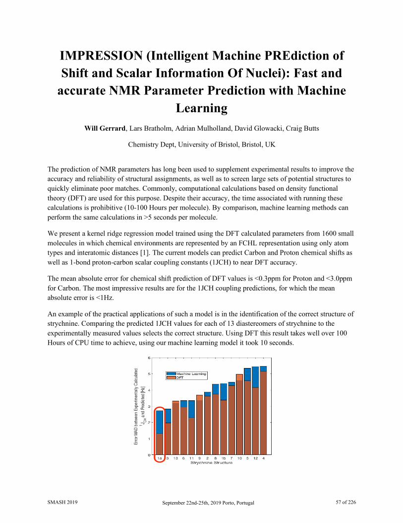
IMPRESSION (Intelligent Machine PREdiction of Shift and Scalar Information Of Nuclei): Fast and
accurate NMR Parameter Prediction with Machine Learning
Will Gerrard, Lars Bratholm, Adrian Mulholland, David Glowacki, Craig Butts
Chemistry Dept, University of Bristol, Bristol, UK
The prediction of NMR parameters has long been used to supplement experimental results to improve the accuracy and reliability of structural assignments, as well as to screen large sets of potential structures to quickly eliminate poor matches. Commonly, computational calculations based on density functional theory (DFT) are used for this purpose. Despite their accuracy, the time associated with running these calculations is prohibitive (10-100 Hours per molecule). By comparison, machine learning methods can perform the same calculations in >5 seconds per molecule.
We present a kernel ridge regression model trained using the DFT calculated parameters from 1600 small molecules in which chemical environments are represented by an FCHL representation using only atom types and interatomic distances [1]. The current models can predict Carbon and Proton chemical shifts as well as 1-bond proton-carbon scalar coupling constants (1JCH) to near DFT accuracy.
The mean absolute error for chemical shift prediction of DFT values is <0.3ppm for Proton and <3.0ppm for Carbon. The most impressive results are for the 1JCH coupling predictions, for which the mean absolute error is <1Hz.
An example of the practical applications of such a model is in the identification of the correct structure of strychnine. Comparing the predicted 1JCH values for each of 13 diastereomers of strychnine to the experimentally measured values selects the correct structure. Using DFT this result takes well over 100 Hours of CPU time to achieve, using our machine learning model it took 10 seconds.
SMASH 2019 September 22nd-25th, 2019 Porto, Portugal 57 of 226

1. Paruzzo, Federico and Hofstetter, Albert and Musil, Felix and De, Sandip and Ceriotti, Michele and Emsley, Lyndon, Nature Communications, 9, 4501, 2018.
SMASH 2019 September 22nd-25th, 2019 Porto, Portugal 58 of 226
BioMagResBank: Database and Tools for Bioactive Small Molecule NMR Analysis
Pedro R Romero1, Hamid Eghbalnia1, Hesam Dashti2, Naohiro Kobayashi3, Jonathan Wedell1, Kumaran Baskaran1, Takeshi Iwata3, Masashi Yokochi3, Dimitri Maziuk1, Hongyang Yao1, Toshimichi Fujiwara3,
Genji Kurusu3, Eldon L Ulrich1, Jeffrey C Hoch4, and John L Markley1*
1. BMRB, Biochemistry Department, University of Wisconsin-Madison, Madison, WI, USA
2. Brigham and Women's Hospital, Harvard Medical School, Boston, MA, USA 3. PDBj-BMRB, Institute for Protein Research, Osaka University, Suita, Osaka, Japan
4. BMRB, Department of Molecular Biology and Biophysics, UConn Health, Farmington, CT, USA
The Biological Magnetic Resonance Data Bank (BioMagResBank or BMRB) [1], founded in 1988, serves as the archive for data generated by Nuclear Magnetic Resonance (NMR) spectroscopy of biological systems. NMR spectroscopy is unique among biophysical approaches in its ability to provide a broad range of atomic and higher-level information relevant to the structural, dynamic, and chemical properties of biological macromolecules, as well as reporting on metabolite and natural product concentrations in complex mixtures and their chemical structures. BMRB stores experimental and derived data from biomolecular NMR studies on both biopolymers and bioactive small compounds. BMRB supports metabolomics NMR studies through a library of a variety of 1D and 2D NMR spectra of pure compounds (including metabolites, natural products, drugs, and compounds used for screening in drug discovery) and through its adoption of novel analytic tools, like the ALATIS unique atom identifiers [2], which are universal and based solely on the 3D structure of the compound and the InChI convention, and GISSMO spin matrices [3], which enable accurate simulation of compound and mixture spectra at any field strength. The combination of unique ALATIS naming and parameterized spectra offers the users of BMRB data a distinctive benefit in terms of robustness and reproducibility, as embodied in the FAIR principles for data resources, which are that data should be Findable, Accessible, Interoperable, and Reusable.
1. Ulrich, E.L., et al., “BioMagResBank.” Nucleic Acids Res, 2008. 36(Database issue): p.
D402-8. DOI: 10.1093/nar/gkm957. 2. Dashti, H., et al., “Unique identifiers for small molecules enable rigorous labeling of their
atoms.” Sci Data, 2017. 4: p. 170073. DOI: 10.1038/sdata.2017.73. 3. Dashti, H., et al., “Applications of Parametrized NMR Spin Systems of Small Molecules.”
Anal Chem, 2018. 90(18): p. 10646-10649. DOI: 10.1021/acs.analchem.8b02660.
SMASH 2019 September 22nd-25th, 2019 Porto, Portugal 59 of 226

Presenter Index to Posters
Ralph Adams 37 Simple Kinetics Analysis for Reactions with Catalyst (De)activation
Zahra Al-Aasmi 119 Acquisition of a quantitative 13C NMR spectra with reduced NOE by an EXACT Semi-Real-Time (ESRT) acquisition method.
Marcel Alcaraz Janßen 27 Influences of Chiral Sidechains in Polypeptide Based Alignment Media
Dmitry Aleshin 85 NMR Spectroscopy and Magnetometry of the Cobalt(II) Single Molecule Magnet with Unusual Coordianation Geometry
Khodzhaberdi Allaberdiev 74 Europium β-diketonate Complexes with Para-substituted Phenols
Dimitris Argyropoulos 117 Best Practices for the Use of NOESY and ROESY Data in Determining the 3D Structure of Compounds
Jose Ascenso 108 Slice Selective NMR Analysis of Lanthanum Picrate Distribution in Extraction Studies by Calixarene Ketone Derivatives
Shi Bai 19 43Ca Solid-State NMR Spectroscopy of Atorvastatin Calcium
Maria Beira 70 Molecular Dynamics Study of PEG-Based Mixtures with Gadolinium, Manganese and Cobalt Metallic Complexes by NMR Relaxometry, Diffusometry and X-ray Diffraction
David Bennett 41 Coldprobes, NUS & Automated Analysis: Revitalising INADEQUATE
Nele Berg 54 NMR Spectroscopic Analysis of the Hydrogen Bond Mediated Pre-Aggregates Responsible for Successful Photocatalytic PCET
Daniel Berry 58 Insights into Asymmetric Transfer Hydrogenation using Multi-technique Operando Reaction Monitoring
Dóra Bogdán 45 Enantiodiscriminative Self-Organization of β-Peptides - Structure Elucidation
Rachael Broomfield-Tagg 59 Optimisation of Multi-Nuclear FlowNMR for Monitoring a Bi-phasic Suzuki-Miyaura Cross Coupling
Julie Calahan 112 Quantitation of Multiple Crystal Forms of Magnesium Stearate in Tablet Formulations Using Variable Contact Time Experiments in CP/MAS SSNMR
Bhawna Chaubey 29 Solvation and Structural Transition of Melittin in Trifluoroethanol/Water: A NMR relaxometry and DNP Study
Rosa M Claramunt 81 Polymorphism and Tautomerism in Crystal Structures of Azoles and Benzazoles: SSNMR and GIPAW Calculations
Uenifer Couto 107 ³JCH Coupling in Unsaturated Compounds: The Effect of Neighboring Groups
Andreia E. S. Cunha 111 Towards the Discovery of Influenza NS1 Inhibitors: A Combined in Silico and NMR Fragment-based Approach
Ombeline Danton 79 Saponins from Saffron Corms Inhibit the Secretion of Pro-Inflammatory Cytokines at Both Protein and Gene Levels
Nichola Davies 46 10 Things to Do with an Open Access Proton NMR Spectrum of Classical and Emerging Drug Modalities
SMASH 2019 September 22nd-25th, 2019 Porto, Portugal 60 of 226
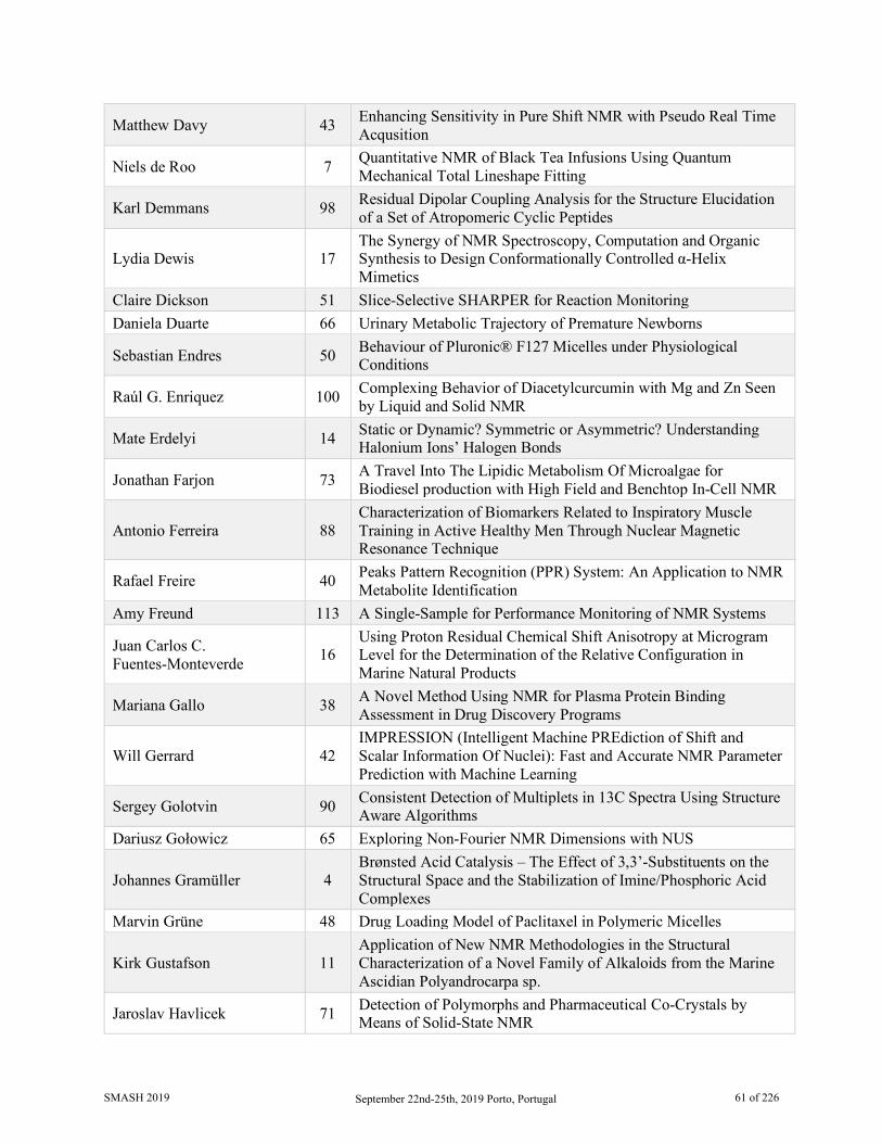
Matthew Davy 43 Enhancing Sensitivity in Pure Shift NMR with Pseudo Real Time Acqusition
Niels de Roo 7 Quantitative NMR of Black Tea Infusions Using Quantum Mechanical Total Lineshape Fitting
Karl Demmans 98 Residual Dipolar Coupling Analysis for the Structure Elucidation of a Set of Atropomeric Cyclic Peptides
Lydia Dewis 17 The Synergy of NMR Spectroscopy, Computation and Organic Synthesis to Design Conformationally Controlled α-Helix Mimetics
Claire Dickson 51 Slice-Selective SHARPER for Reaction Monitoring Daniela Duarte 66 Urinary Metabolic Trajectory of Premature Newborns
Sebastian Endres 50 Behaviour of Pluronic® F127 Micelles under Physiological Conditions
Raúl G. Enriquez 100 Complexing Behavior of Diacetylcurcumin with Mg and Zn Seen by Liquid and Solid NMR
Mate Erdelyi 14 Static or Dynamic? Symmetric or Asymmetric? Understanding Halonium Ions’ Halogen Bonds
Jonathan Farjon 73 A Travel Into The Lipidic Metabolism Of Microalgae for Biodiesel production with High Field and Benchtop In-Cell NMR
Antonio Ferreira 88 Characterization of Biomarkers Related to Inspiratory Muscle Training in Active Healthy Men Through Nuclear Magnetic Resonance Technique
Rafael Freire 40 Peaks Pattern Recognition (PPR) System: An Application to NMR Metabolite Identification
Amy Freund 113 A Single-Sample for Performance Monitoring of NMR Systems
Juan Carlos C. Fuentes-Monteverde 16
Using Proton Residual Chemical Shift Anisotropy at Microgram Level for the Determination of the Relative Configuration in Marine Natural Products
Mariana Gallo 38 A Novel Method Using NMR for Plasma Protein Binding Assessment in Drug Discovery Programs
Will Gerrard 42 IMPRESSION (Intelligent Machine PREdiction of Shift and Scalar Information Of Nuclei): Fast and Accurate NMR Parameter Prediction with Machine Learning
Sergey Golotvin 90 Consistent Detection of Multiplets in 13C Spectra Using Structure Aware Algorithms
Dariusz Gołowicz 65 Exploring Non-Fourier NMR Dimensions with NUS
Johannes Gramüller 4 Brønsted Acid Catalysis – The Effect of 3,3’-Substituents on the Structural Space and the Stabilization of Imine/Phosphoric Acid Complexes
Marvin Grüne 48 Drug Loading Model of Paclitaxel in Polymeric Micelles
Kirk Gustafson 11 Application of New NMR Methodologies in the Structural Characterization of a Novel Family of Alkaloids from the Marine Ascidian Polyandrocarpa sp.
Jaroslav Havlicek 71 Detection of Polymorphs and Pharmaceutical Co-Crystals by Means of Solid-State NMR
SMASH 2019 September 22nd-25th, 2019 Porto, Portugal 61 of 226
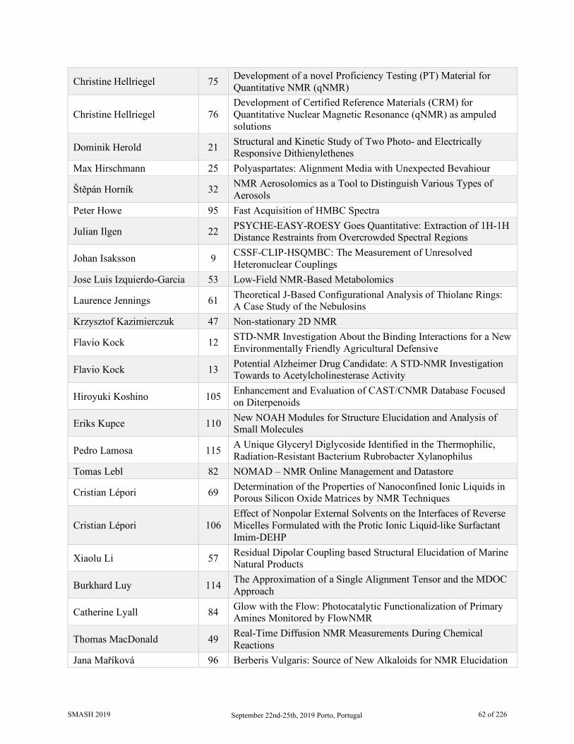
Christine Hellriegel 75 Development of a novel Proficiency Testing (PT) Material for Quantitative NMR (qNMR)
Christine Hellriegel 76 Development of Certified Reference Materials (CRM) for Quantitative Nuclear Magnetic Resonance (qNMR) as ampuled solutions
Dominik Herold 21 Structural and Kinetic Study of Two Photo- and Electrically Responsive Dithienylethenes
Max Hirschmann 25 Polyaspartates: Alignment Media with Unexpected Bevahiour
Štěpán Horník 32 NMR Aerosolomics as a Tool to Distinguish Various Types of Aerosols
Peter Howe 95 Fast Acquisition of HMBC Spectra
Julian Ilgen 22 PSYCHE-EASY-ROESY Goes Quantitative: Extraction of 1H-1H Distance Restraints from Overcrowded Spectral Regions
Johan Isaksson 9 CSSF-CLIP-HSQMBC: The Measurement of Unresolved Heteronuclear Couplings
Jose Luis Izquierdo-Garcia 53 Low-Field NMR-Based Metabolomics
Laurence Jennings 61 Theoretical J-Based Configurational Analysis of Thiolane Rings: A Case Study of the Nebulosins
Krzysztof Kazimierczuk 47 Non-stationary 2D NMR
Flavio Kock 12 STD-NMR Investigation About the Binding Interactions for a New Environmentally Friendly Agricultural Defensive
Flavio Kock 13 Potential Alzheimer Drug Candidate: A STD-NMR Investigation Towards to Acetylcholinesterase Activity
Hiroyuki Koshino 105 Enhancement and Evaluation of CAST/CNMR Database Focused on Diterpenoids
Eriks Kupce 110 New NOAH Modules for Structure Elucidation and Analysis of Small Molecules
Pedro Lamosa 115 A Unique Glyceryl Diglycoside Identified in the Thermophilic, Radiation-Resistant Bacterium Rubrobacter Xylanophilus
Tomas Lebl 82 NOMAD – NMR Online Management and Datastore
Cristian Lépori 69 Determination of the Properties of Nanoconfined Ionic Liquids in Porous Silicon Oxide Matrices by NMR Techniques
Cristian Lépori 106 Effect of Nonpolar External Solvents on the Interfaces of Reverse Micelles Formulated with the Protic Ionic Liquid-like Surfactant Imim-DEHP
Xiaolu Li 57 Residual Dipolar Coupling based Structural Elucidation of Marine Natural Products
Burkhard Luy 114 The Approximation of a Single Alignment Tensor and the MDOC Approach
Catherine Lyall 84 Glow with the Flow: Photocatalytic Functionalization of Primary Amines Monitored by FlowNMR
Thomas MacDonald 49 Real-Time Diffusion NMR Measurements During Chemical Reactions
Jana Maříková 96 Berberis Vulgaris: Source of New Alkaloids for NMR Elucidation
SMASH 2019 September 22nd-25th, 2019 Porto, Portugal 62 of 226
Lenka Michálková 34 Early Detection of Pancreatic Cancer: 1H NMR Metabolomics Study of its Connection with Diabetes Mellitus
Kumar Motiram-Corral 18 Implementing One-Shot Multiple-FID Acquisition into Homonuclear and Heteronuclear NMR Experiments
Pinelopi Moutzouri 36 Methods to Improve Resolution in 1H Solid State NMR at Ultra-Fast MAS
Juan Muñoz-García 104 Solid State NMR Characterisation of Novel Fluorinated Cellulose Materials
Suryaprakash Nagaraja rao 72 Intramolecular Hydrogen Bond Directed Distribution of Conformational Populations in Derivatives of N’-benzylidenebenzohydrazide
Henry Nkabyo 2 195Pt and 1D NOESY NMR Determination of E/Z Configurational and Geometric Isomers of Platinum(II) Acylthiourea Complexes
Predrag Novak 89 NMR and Docking Studies of Novel Macrolide Conjugates
Jens Nowag 23 Determining the Relative Configuration of 1,3 Diamides with Modern NMR Techniques
Jessyka Padilla 92 31P NMR Study of Trimethylphosphine Probe Adsorbed on Y Zeolite Modified with Boron
Yanina Pankratova 78 Electronic Structure of Bis[tris(pyrazolyl)]borate Co(II) by THz-EPR and Paramagnetic NMR Spectroscopy
Helena Pelantová 99 Treatment Response to Fiber in Diabetes: NMR-Based Metabolomic Approach
Ignacio Pérez-Victoria 94 Deconvoluting Compound Mixtures by Ratio Analysis NMR Spectroscopy (RANSY) of Low-Resolution Chromatographic Fractions
Tran Pham 87 Helium Recovery and Recycling at GSK Stevenage
Tran Pham 86 NMR Crystallography for the Acceleration of the Development of New Medicines
Elena Pisa 39 High Temperature NMR Studies of Diffusivity and Molecular Interactions in Pharmaceutical Hot-melt Extrusion Formulations
Ann-Christin Poeppler 31 Looking Beyond the Simplified Core Shell Arrangement - Structure and Properties of Loaded Polymer Micelles by Solid-State NMR
Rostislav Pol 102 Efficient Approaches for Addressing Spectral Ambiguities in Computer Assisted Structure Elucidation (CASE) Systems
Nicholas Rees 118 Conformational Exchange Processes in Lanthanide Complexes Ann Christin Reiersølmoen 6 Understanding Au(III) Catalysis by NMR
Clark Ridge 62 Examples of Complementary Small Molecule Mass Spectrometry and NMR: A Marine Toxin, a Food Dye and Tattoo Inks
Susanne Riegel 68 Quantitative Analysis of Cannabinoids using Benchtop NMR Spectrometers
Thomas Robertson 52 Hitting the Jackpot: Forensic Hyperpolarisation of Fentalogues
Pedro Romero 64 BioMagResBank: Database and Tools for Bioactive Small Molecule NMR Analysis
SMASH 2019 September 22nd-25th, 2019 Porto, Portugal 63 of 226
Jerk Rönnols 109 NMR Measurements on Lignin and Black Liquor: Towards Industrial Applications for the Pulp and Paper Industry
Federica Rossi 24 Determination of the 15N Chemical Shift Anisotropy in Natural Abundance Samples by Proton-Detected 3D Solid-state NMR Under Ultrafast MAS
Priscila Rubim 77 Towards Validatiton of Qualitative and Quantitative 1H NMR Method for the Quality Analysis of Drugs
Fredrik Garnås Rylandsholm 30 Determination of the Absolute Configuration of the Natural Product Commafric A by VCD, RDC, and RCSA
Antonio Salgado 83 ROESY Based Structural Studies of Anomalous High Order Complexes Between Daclatasvir and Some Cyclodextrins
Tatiana Santana Ribeiro 91 13C Solid State NMR Spectroscopy as a Method for Probing the Efficiency of Biomass Pretreatments for Second Generation Ethanol Production
Dominic Schirra 20 Investigations on the Alignment Process in LLC-phases of Poly-y-p-biphenylmethyl Glutamates
Denise Selegato 28 Improvement of Bioactive Metabolite Production in Microbial Cultures - A Systems Approach by OSMAC and Deconvolution-Based 1H NMR Quantification
Alexandra Shchukina 26 Faster Pure-Shift Spectra Acquisition: Utilizing the Prior Knowledge from 1H NMR
Ana Silva 60 1H-NMR Metabolomics Reveals Potential Biomarkers of Breast Cancer in an Endocrine-Related Mouse Model
Eduard Sistaré 97 2D ALICE Experiments: How to Resolve Chemical Shift Ambiguities in Aliased Spectra and Reconstruct Top-Resolution Spectra
Sebastian Spann 55 β-BPSE: Fast DOSY Acqusition of Small Molecules and Macromolecules
Thomas Stadelmann 15 Investigation of the Structure of Cyclic Depsipeptides PF1022A and Emodepside and Their Affinity to Cations
Ellen Steimers 101 Effective Bayesian Approach for Automated Quantitative Analysis of Benchtop NMR Data
Willibald Stockerl 33 NMR-Spectroscopic Investigations in Iminium Ion Catalysis
Verena Streitferdt 35 Mechanistic Investigations of the Direct Photocatalytic Transformation of P4 into Aryl Phosphines and Phosphonium Salts
Jan Sykora 103 A Construction of a Crystal Lattice from Intermolecular Interactions
Linette Twigge 1 Fischer Aminocarbene Conformers Containing a 2-thienyl or 2-furyl Ring: A Crystallographic, NMR, and DFT Study
Pavleta Tzvetkova 63 When Flexibility Meets with RDCs for Structure Elucidation
Tasneem Vaid 8 INPHARMA Based Determination of Ligand Binding Conformations at α1-Adrenergic Receptor Subtypes
Renan Vidal Viesser 93 How the Methyl Groups Affect the Spin-Orbit Component of 13C Chemical Shift
SMASH 2019 September 22nd-25th, 2019 Porto, Portugal 64 of 226
Gregory Walker 5 Oodels of Leads for the Price of One: Lead Diversification of Pharmaceutical Compounds and the Role of the Micro-Cryoprobe
Svenja Wesp 56 Helically Chiral Poly(isocyanides) and Poly(acetylenes) as NMR Alignment Media
Taichi Yamazaki 80 Method Optimization for Determination of Okadaic Acid in Solution by Quantitative NMR Spectroscopy with External Standard Method, and Validation by a Collaborative Study
Yanita Yankova 10 Forensic Analysis of Petrol Samples
Jonathan Yong 44 Computational Studies Towards the Stereochemical Assignment of Flexible Natural Products
Matej Zabka 3 Insights from NMR for Interactions and Processes in Phosphoramidite Pd(II) Complexes and Brønsted Acid Catalysis
Mark Zell 116 Recent Applications of Online NMR Reaction Monitoring in the Pharmaceutical Industry
Renan Ziemann Wilhelms 67 1H NMR and Fuzzy C-Means Analysis for Classifying and Adulteration Detection of Orange Drinks
SMASH 2019 September 22nd-25th, 2019 Porto, Portugal 65 of 226

Numerical Index to Posters
1 Linette Twigge Fischer Aminocarbene Conformers Containing a 2-thienyl or 2-furyl Ring: A Crystallographic, NMR, and DFT Study
2 Henry Nkabyo 195Pt and 1D NOESY NMR Determination of E/Z Configurational and Geometric Isomers of Platinum(II) Acylthiourea Complexes
3 Matej Zabka Insights from NMR for Interactions and Processes in Phosphoramidite Pd(II) Complexes and Brønsted Acid Catalysis
4 Johannes Gramüller Brønsted Acid Catalysis – The Effect of 3,3’-Substituents on the Structural Space and the Stabilization of Imine/Phosphoric Acid Complexes
5 Gregory Walker Oodels of Leads for the Price of One: Lead Diversification of Pharmaceutical Compounds and the Role of the Micro-Cryoprobe
6 Ann Christin Reiersølmoen Understanding Au(III) Catalysis by NMR
7 Niels de Roo Quantitative NMR of Black Tea Infusions Using Quantum Mechanical Total Lineshape Fitting
8 Tasneem Vaid INPHARMA Based Determination of Ligand Binding Conformations at α1-Adrenergic Receptor Subtypes
9 Johan Isaksson CSSF-CLIP-HSQMBC: The Measurement of Unresolved Heteronuclear Couplings
10 Yanita Yankova Forensic Analysis of Petrol Samples
11 Kirk Gustafson Application of New NMR Methodologies in the Structural Characterization of a Novel Family of Alkaloids from the Marine Ascidian Polyandrocarpa sp.
12 Flavio Kock STD-NMR Investigation About the Binding Interactions for a New Environmentally Friendly Agricultural Defensive
13 Flavio Kock Potential Alzheimer Drug Candidate: A STD-NMR Investigation Towards to Acetylcholinesterase Activity
14 Mate Erdelyi Static or Dynamic? Symmetric or Asymmetric? Understanding Halonium Ions’ Halogen Bonds
15 Thomas Stadelmann Investigation of the Structure of Cyclic Depsipeptides PF1022A and Emodepside and Their Affinity to Cations
16 Juan Carlos C. Fuentes-Monteverde
Using Proton Residual Chemical Shift Anisotropy at Microgram Level for the Determination of the Relative Configuration in Marine Natural Products
17 Lydia Dewis The Synergy of NMR Spectroscopy, Computation and Organic Synthesis to Design Conformationally Controlled α-Helix Mimetics
18 Kumar Motiram-Corral Implementing One-Shot Multiple-FID Acquisition into Homonuclear and Heteronuclear NMR Experiments
19 Shi Bai 43Ca Solid-State NMR Spectroscopy of Atorvastatin Calcium
20 Dominic Schirra Investigations on the Alignment Process in LLC-phases of Poly-y-p-biphenylmethyl Glutamates
SMASH 2019 September 22nd-25th, 2019 Porto, Portugal 66 of 226
21 Dominik Herold Structural and Kinetic Study of Two Photo- and Electrically Responsive Dithienylethenes
22 Julian Ilgen PSYCHE-EASY-ROESY Goes Quantitative: Extraction of 1H-1H Distance Restraints from Overcrowded Spectral Regions
23 Jens Nowag Determining the Relative Configuration of 1,3 Diamides with Modern NMR Techniques
24 Federica Rossi Determination of the 15N Chemical Shift Anisotropy in Natural Abundance Samples by Proton-Detected 3D Solid-state NMR Under Ultrafast MAS
25 Max Hirschmann Polyaspartates: Alignment Media with Unexpected Bevahiour
26 Alexandra Shchukina Faster Pure-Shift Spectra Acquisition: Utilizing the Prior Knowledge from 1H NMR
27 Marcel Alcaraz Janßen Influences of Chiral Sidechains in Polypeptide Based Alignment Media
28 Denise Selegato Improvement of Bioactive Metabolite Production in Microbial Cultures - A Systems Approach by OSMAC and Deconvolution-Based 1H NMR Quantification
29 Bhawna Chaubey Solvation and Structural Transition of Melittin in Trifluoroethanol/Water: A NMR relaxometry and DNP Study
30 Fredrik Garnås Rylandsholm
Determination of the Absolute Configuration of the Natural Product Commafric A by VCD, RDC, and RCSA
31 Ann-Christin Poeppler Looking Beyond the Simplified Core Shell Arrangement - Structure and Properties of Loaded Polymer Micelles by Solid-State NMR
32 Štěpán Horník NMR Aerosolomics as a Tool to Distinguish Various Types of Aerosols
33 Willibald Stockerl NMR-Spectroscopic Investigations in Iminium Ion Catalysis
34 Lenka Michálková Early Detection of Pancreatic Cancer: 1H NMR Metabolomics Study of its Connection with Diabetes Mellitus
35 Verena Streitferdt Mechanistic Investigations of the Direct Photocatalytic Transformation of P4 into Aryl Phosphines and Phosphonium Salts
36 Pinelopi Moutzouri Methods to Improve Resolution in 1H Solid State NMR at Ultra-Fast MAS
37 Ralph Adams Simple Kinetics Analysis for Reactions with Catalyst (De)activation
38 Mariana Gallo A Novel Method Using NMR for Plasma Protein Binding Assessment in Drug Discovery Programs
39 Elena Pisa High Temperature NMR Studies of Diffusivity and Molecular Interactions in Pharmaceutical Hot-melt Extrusion Formulations
40 Rafael Freire Peaks Pattern Recognition (PPR) System: An Application to NMR Metabolite Identification
41 David Bennett Coldprobes, NUS & Automated Analysis: Revitalising INADEQUATE
42 Will Gerrard IMPRESSION (Intelligent Machine PREdiction of Shift and Scalar Information Of Nuclei): Fast and Accurate NMR Parameter Prediction with Machine Learning
SMASH 2019 September 22nd-25th, 2019 Porto, Portugal 67 of 226

43 Matthew Davy Enhancing Sensitivity in Pure Shift NMR with Pseudo Real Time Acqusition
44 Jonathan Yong Computational Studies Towards the Stereochemical Assignment of Flexible Natural Products
45 Dóra Bogdán Enantiodiscriminative Self-Organization of β-Peptides - Structure Elucidation
46 Nichola Davies 10 Things to Do with an Open Access Proton NMR Spectrum of Classical and Emerging Drug Modalities
47 Krzysztof Kazimierczuk Non-stationary 2D NMR 48 Marvin Grüne Drug Loading Model of Paclitaxel in Polymeric Micelles
49 Thomas MacDonald Real-Time Diffusion NMR Measurements During Chemical Reactions
50 Sebastian Endres Behaviour of Pluronic® F127 Micelles under Physiological Conditions
51 Claire Dickson Slice-Selective SHARPER for Reaction Monitoring 52 Thomas Robertson Hitting the Jackpot: Forensic Hyperpolarisation of Fentalogues 53 Jose Luis Izquierdo-Garcia Low-Field NMR-Based Metabolomics
54 Nele Berg NMR Spectroscopic Analysis of the Hydrogen Bond Mediated Pre-Aggregates Responsible for Successful Photocatalytic PCET
55 Sebastian Spann β-BPSE: Fast DOSY Acqusition of Small Molecules and Macromolecules
56 Svenja Wesp Helically Chiral Poly(isocyanides) and Poly(acetylenes) as NMR Alignment Media
57 Xiaolu Li Residual Dipolar Coupling based Structural Elucidation of Marine Natural Products
58 Daniel Berry Insights into Asymmetric Transfer Hydrogenation using Multi-technique Operando Reaction Monitoring
59 Rachael Broomfield-Tagg Optimisation of Multi-Nuclear FlowNMR for Monitoring a Bi-phasic Suzuki-Miyaura Cross Coupling
60 Ana Silva 1H-NMR Metabolomics Reveals Potential Biomarkers of Breast Cancer in an Endocrine-Related Mouse Model
61 Laurence Jennings Theoretical J-Based Configurational Analysis of Thiolane Rings: A Case Study of the Nebulosins
62 Clark Ridge Examples of Complementary Small Molecule Mass Spectrometry and NMR: A Marine Toxin, a Food Dye and Tattoo Inks
63 Pavleta Tzvetkova When Flexibility Meets with RDCs for Structure Elucidation
64 Pedro Romero BioMagResBank: Database and Tools for Bioactive Small Molecule NMR Analysis
65 Dariusz Gołowicz Exploring Non-Fourier NMR Dimensions with NUS 66 Daniela Duarte Urinary Metabolic Trajectory of Premature Newborns
67 Renan Ziemann Wilhelms 1H NMR and Fuzzy C-Means Analysis for Classifying and Adulteration Detection of Orange Drinks
68 Susanne Riegel Quantitative Analysis of Cannabinoids using Benchtop NMR Spectrometers
SMASH 2019 September 22nd-25th, 2019 Porto, Portugal 68 of 226
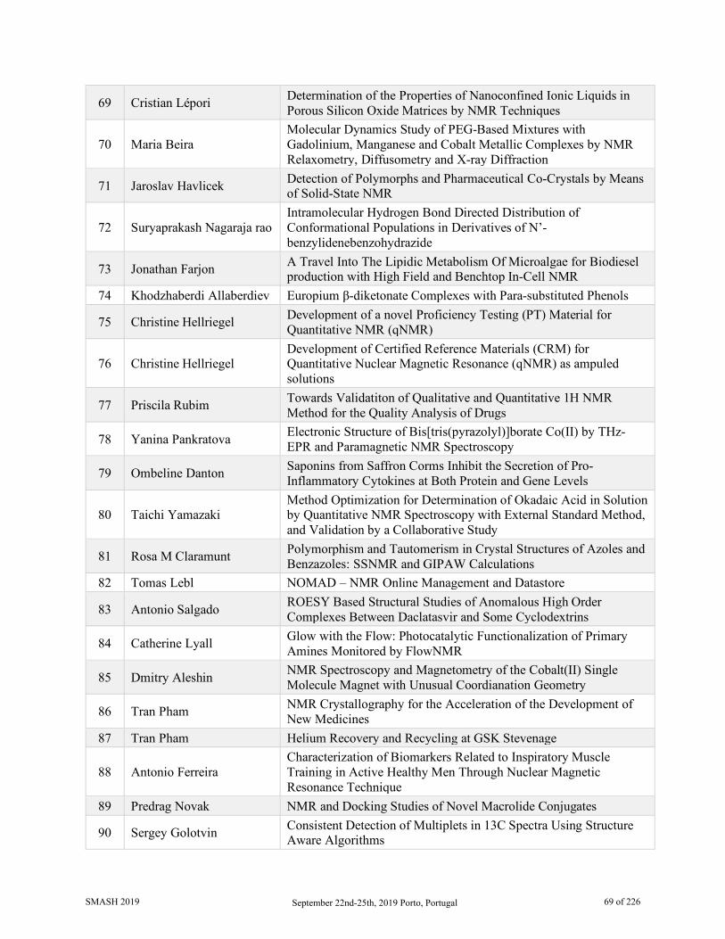
69 Cristian Lépori Determination of the Properties of Nanoconfined Ionic Liquids in Porous Silicon Oxide Matrices by NMR Techniques
70 Maria Beira Molecular Dynamics Study of PEG-Based Mixtures with Gadolinium, Manganese and Cobalt Metallic Complexes by NMR Relaxometry, Diffusometry and X-ray Diffraction
71 Jaroslav Havlicek Detection of Polymorphs and Pharmaceutical Co-Crystals by Means of Solid-State NMR
72 Suryaprakash Nagaraja rao Intramolecular Hydrogen Bond Directed Distribution of Conformational Populations in Derivatives of N’-benzylidenebenzohydrazide
73 Jonathan Farjon A Travel Into The Lipidic Metabolism Of Microalgae for Biodiesel production with High Field and Benchtop In-Cell NMR
74 Khodzhaberdi Allaberdiev Europium β-diketonate Complexes with Para-substituted Phenols
75 Christine Hellriegel Development of a novel Proficiency Testing (PT) Material for Quantitative NMR (qNMR)
76 Christine Hellriegel Development of Certified Reference Materials (CRM) for Quantitative Nuclear Magnetic Resonance (qNMR) as ampuled solutions
77 Priscila Rubim Towards Validatiton of Qualitative and Quantitative 1H NMR Method for the Quality Analysis of Drugs
78 Yanina Pankratova Electronic Structure of Bis[tris(pyrazolyl)]borate Co(II) by THz-EPR and Paramagnetic NMR Spectroscopy
79 Ombeline Danton Saponins from Saffron Corms Inhibit the Secretion of Pro-Inflammatory Cytokines at Both Protein and Gene Levels
80 Taichi Yamazaki Method Optimization for Determination of Okadaic Acid in Solution by Quantitative NMR Spectroscopy with External Standard Method, and Validation by a Collaborative Study
81 Rosa M Claramunt Polymorphism and Tautomerism in Crystal Structures of Azoles and Benzazoles: SSNMR and GIPAW Calculations
82 Tomas Lebl NOMAD – NMR Online Management and Datastore
83 Antonio Salgado ROESY Based Structural Studies of Anomalous High Order Complexes Between Daclatasvir and Some Cyclodextrins
84 Catherine Lyall Glow with the Flow: Photocatalytic Functionalization of Primary Amines Monitored by FlowNMR
85 Dmitry Aleshin NMR Spectroscopy and Magnetometry of the Cobalt(II) Single Molecule Magnet with Unusual Coordianation Geometry
86 Tran Pham NMR Crystallography for the Acceleration of the Development of New Medicines
87 Tran Pham Helium Recovery and Recycling at GSK Stevenage
88 Antonio Ferreira Characterization of Biomarkers Related to Inspiratory Muscle Training in Active Healthy Men Through Nuclear Magnetic Resonance Technique
89 Predrag Novak NMR and Docking Studies of Novel Macrolide Conjugates
90 Sergey Golotvin Consistent Detection of Multiplets in 13C Spectra Using Structure Aware Algorithms
SMASH 2019 September 22nd-25th, 2019 Porto, Portugal 69 of 226

91 Tatiana Santana Ribeiro 13C Solid State NMR Spectroscopy as a Method for Probing the Efficiency of Biomass Pretreatments for Second Generation Ethanol Production
92 Jessyka Padilla 31P NMR Study of Trimethylphosphine Probe Adsorbed on Y Zeolite Modified with Boron
93 Renan Vidal Viesser How the Methyl Groups Affect the Spin-Orbit Component of 13C Chemical Shift
94 Ignacio Pérez-Victoria Deconvoluting Compound Mixtures by Ratio Analysis NMR Spectroscopy (RANSY) of Low-Resolution Chromatographic Fractions
95 Peter Howe Fast Acquisition of HMBC Spectra 96 Jana Maříková Berberis Vulgaris: Source of New Alkaloids for NMR Elucidation
97 Eduard Sistaré 2D ALICE Experiments: How to Resolve Chemical Shift Ambiguities in Aliased Spectra and Reconstruct Top-Resolution Spectra
98 Karl Demmans Residual Dipolar Coupling Analysis for the Structure Elucidation of a Set of Atropomeric Cyclic Peptides
99 Helena Pelantová Treatment Response to Fiber in Diabetes: NMR-Based Metabolomic Approach
100 Raúl G. Enriquez Complexing Behavior of Diacetylcurcumin with Mg and Zn Seen by Liquid and Solid NMR
101 Ellen Steimers Effective Bayesian Approach for Automated Quantitative Analysis of Benchtop NMR Data
102 Rostislav Pol Efficient Approaches for Addressing Spectral Ambiguities in Computer Assisted Structure Elucidation (CASE) Systems
103 Jan Sykora A Construction of a Crystal Lattice from Intermolecular Interactions
104 Juan Muñoz-García Solid State NMR Characterisation of Novel Fluorinated Cellulose Materials
105 Hiroyuki Koshino Enhancement and Evaluation of CAST/CNMR Database Focused on Diterpenoids
106 Cristian Lépori Effect of Nonpolar External Solvents on the Interfaces of Reverse Micelles Formulated with the Protic Ionic Liquid-like Surfactant Imim-DEHP
107 Uenifer Couto ³JCH Coupling in Unsaturated Compounds: The Effect of Neighboring Groups
108 Jose Ascenso Slice Selective NMR Analysis of Lanthanum Picrate Distribution in Extraction Studies by Calixarene Ketone Derivatives
109 Jerk Rönnols NMR Measurements on Lignin and Black Liquor: Towards Industrial Applications for the Pulp and Paper Industry
110 Eriks Kupce New NOAH Modules for Structure Elucidation and Analysis of Small Molecules
111 Andreia E. S. Cunha Towards the Discovery of Influenza NS1 Inhibitors: A Combined in Silico and NMR Fragment-based Approach
SMASH 2019 September 22nd-25th, 2019 Porto, Portugal 70 of 226

112 Julie Calahan Quantitation of Multiple Crystal Forms of Magnesium Stearate in Tablet Formulations Using Variable Contact Time Experiments in CP/MAS SSNMR
113 Amy Freund A Single-Sample for Performance Monitoring of NMR Systems
114 Burkhard Luy The Approximation of a Single Alignment Tensor and the MDOC Approach
115 Pedro Lamosa A Unique Glyceryl Diglycoside Identified in the Thermophilic, Radiation-Resistant Bacterium Rubrobacter Xylanophilus
116 Mark Zell Recent Applications of Online NMR Reaction Monitoring in the Pharmaceutical Industry
117 Dimitris Argyropoulos Best Practices for the Use of NOESY and ROESY Data in Determining the 3D Structure of Compounds
118 Nicholas Rees Conformational Exchange Processes in Lanthanide Complexes
119 Zahra Al-Aasmi Acquisition of a quantitative 13C NMR spectra with reduced NOE by an EXACT Semi-Real-Time (ESRT) acquisition method.
SMASH 2019 September 22nd-25th, 2019 Porto, Portugal 71 of 226

1 Fischer Aminocarbene Conformers Containing a 2-thienyl or 2-furyl
Ring: A Crystallographic, NMR, and DFT Study
Marilé Landman1, Roan Fraser1, Linette Twigge2 and Jeanet Conradie2
4. University of Pretoria, Chemistry Department, Hatfield, Gauteng, South Africa 5. University of the Free State, Chemistry Department, Bloemfontein, Free State, South Africa
Fischer aminocarbene complexes [(CO)5M=C(NHR)Y] (M=Cr or W; R=H, Cy or C2H4NH2; Y=2-thienyl or 2-furyl) containing an amino group exist as two isomers in solution, the E and Z isomers[1]. The two isomers arise from restricted rotation about the N–Ccarbene bond that exhibits double bond character due to π-donation from nitrogen to the carbene carbon. Each isomer exists as two conformers in fast equilibrium with each other. The conformers arise from the rotation of the aryl ring around the Ccarbene–Caryl single bond with a DFT calculated rotation barrier of 0.1–0.5 eV. The main isomer, isolated in the solid state, generally exhibits a syn orientation of the aryl ring relative to the amino substituent and a Z configuration of the amino substituent relative to the metal.
The Z configuration of the main isomers was confirmed by 1H NMR shifts of the α-hydrogens of the alkyl group on the nitrogen that are downfield for the Z-isomers relative to those for the E-isomers[2]. These assignments were further proven by the 1D nuclear overhauser enhancement (NOE) spectra of the [(CO)5Cr=C(NHCy)(2-thienyl)] and [(CO)5Cr=C(NHCy)(2-furyl)] complexes.
The ratio of interproton distances in rigid molecules can be determined by the ratio of the intensities of a pair of NOE signals η1/η2 within the 1D NOE spectrum of the complex by the equation
η1/η2 = r26/r1
6
where η1 = integral of proton 1, η2 = integral of proton 2, r1 = distance between proton 1 and irradiated peak for which the integral was arbitrarily assigned a value of 1000 and r2 = distance between proton 2 and irradiated peak. If one of the distances r1 or r2 is known, the other distance can thus be determined[3]. Various interproton distances within the molecules were calculated from the 1D NOE spectra using this ratio and compared to those obtained from DFT calculations. It was found that the NOE integral ratio differed largely from the DFT calculated values which was contributed to the molecules exhibiting multiple conformations in solution that are interconverting rapidly on the NMR time-scale. This conformational exchange leads to an ensemble-averaging of the observed NOEs for each corresponding interproton distance in each contributing conformer.
8. Landman, M., Fraser, R., Twigge, L., Conradie, J., Journal of Coordination Chemistry, 68 (14), 2388–2408, 2015
9. Fischer, E.O., Leupold, M., Chemische Berichte, 105, 599-608, 1972 10. Butts, C.P., Jones, C.R., Towers, E.C., Flynn, J.L., Appleby, L., Barron, N.J, Organic and
Biomolecular Chemistry., 9, 177-184, 2011
SMASH 2019 September 22nd-25th, 2019 Porto, Portugal 72 of 226
2 195Pt and 1D NOESY NMR
Determination of E/Z Configurational and Geometric
Isomers of Platinum(II) Acylthiourea Complexes
Henry A. Nkabyo1 and Klaus R. Koch2
Department of Chemistry and polymer Science, Stellenbosch University, Matieland, South Africa
Platinum(II) complexes of the asymmetrically substituted N,N-dialkyl-N’-acylthioureas exhibit configurational E,Z isomerism, resulting to two separate sets of 1H and 13C{1H} resonances[1]. These configurational E,Z isomers exist as cis-[Pt(ZZ-L-S,O)2], cis-[Pt(EZ-L-S,O)2] and cis-[Pt(EE-L-S,O)2] isomers in chloroform-d and give rise to three well-resolved 195Pt{1H} resonances which were assinged by 1H detected 1H-195Pt-13C correlation[2]. The cis-[Pt(L-S,O)2] complexes were characterized using HSQC and HMBC, while unambigous assignment of the cis-[Pt(ZZ-L-S,O)2], cis-[Pt(EZ-L-S,O)2], cis-[Pt(EE-L-S,O)2] isomers was achieved by 1D NOESY.
The relative distributions of the configurational E,Z isomers obtained from deconvultion of the 195Pt{1H} resonances reveal that the C-N rotational barrier in the cis-[Pt(L-S,O)2] complexes is largely dependent on the ligand structure, while no significant changes were observed upon variation of temperature. The results obtained were consistent with the isolation of first examples of cis-[Pt(EE-L-S,O)2] and cis-[Pt(ZZ-L-S,O)2] complexes from chloroform solutions for bis(N-methyl,N-ethyl-N’-benzoylthioureato)platinum(II) and bis(N-isopropyl,N-4-methoxy-phenyl-N’-(2,2-dimethylpropanoyl)thioureato)platinum(II) respectively. From the single-crystal XRD structure of the isolated cis-[Pt(EE-L-S,O)2] isomer for bis(N-methyl,N-ethyl-N’-benzoylthioureato)platinum(II), H-H bond distances were measured for atoms in space and these were consistent with the 1D NOESY results.
The irradiation of a chloroform-d solution of the cis-[Pt(L-S,O)2] complexes with either polychromatic light from a 5 Watt LED lamp or with monochromatic light from a 405 nm laser leads to photo-induced cis-trans isomerization[3]. This results in the formation of respective trans-[Pt(L-S,O)2] geometric isomers as reflected by the formation of an additional 195Pt{1H} resonance shifted relatively downfield by ca 745 ppm relative to the 195Pt{1H} resonance for the cis-[Pt(L-S,O)2] complex. For complexes with asymmetrically substituted ligands, photo-induced isomerization of the configurational cis-[Pt(ZZ-L-S,O)2], cis-[Pt(EZ-L-S,O)2], cis-[Pt(EE-L-S,O)2] isomers results in the formation of a set of three additional 195Pt{1H} resonances assigned to the trans-[Pt(ZZ-L-S,O)2], trans-[Pt(EZ-L-S,O)2], trans-[Pt(EE-L-S,O)2] geometric isomers.
1. Koch K. R., Sacht C., Grimmbacher T., Bourne S., S. Afr. J. Chem., 48, 71, 1995. 2. Argyropoulus D., Hoffmann E., Mtongana S., Koch K. R., Magn. Reson. Chem., 41, 102, 2003. 3. H. A. Nkabyo, D. Hannekom, J. McKenzie, K. R. Koch, J. Coord. Chem., 4039, 76, 2014.
SMASH 2019 September 22nd-25th, 2019 Porto, Portugal 73 of 226
3 Insights from NMR for Interactions and Processes in Phosphoramidite
Pd(II) Complexes and Brønsted Acid Catalysis
Matej Zabka and Ruth M. Gschwind
Institut für Organische Chemie, Universität Regensburg, Regensburg, Germany
Standard 1D and 2D NMR techniques provide an incredible amount of information about structures and processes in catalytic cycles. We have investigated weak, non-covalent intermolecular interactions as well as strong, charge-assisted hydrogen bonds, and their role in asymmetric catalysis by NMR spectroscopy.
The dispersion energy donors, weak non-covalent interactions, and flexibility of chiral phosphoramidite ligands play a key role in enantioselective metal-catalyzed reactions. The interligand interactions in phosphoramidite cis-Pd(II)L2Cl2 complexes1 were investigated mainly through NOESY and ROESY sequences at room and cryogenic temperatures. Moreover, heterocomplexes with two different ligands can have a significant impact on enantioselectivity. We have screened a library of chiral binaphthyl and more flexible biphenyl ligands for a high preference for heterocomplex formation using 1H chemical shifts anisotropy, together with 1H- and 31P-correlated spectra. These ligand combinations were also exploited to evaluate the aryl substituent effect for CH-π and π-π interactions in these complexes by NMR spectroscopy.
BINOL-based chiral phosphoric acids (CPA), and structurally similar disulfonylimides and disulfonic acids, form either strong hydrogen bonds or ion pairs with aromatic imines. Interestingly, neither the reported acidities nor the developed internal acidity scale,2 based on hydrogen bond 1H and 15N chemical shifts, correlate with the observed reactivity in a transfer hydrogenation reaction. This outcome hints at the importance of non-covalent interactions between different catalysts and substrate in the rate-determining step.
As another example, in a formal 6π-cyclization reaction of E-hydrazones catalyzed by CPA, our LED-illumination NMR setup3 allowed us to observe the direct effect of hydrazone E/Z photoswitching on the reaction rates at room temperature. The underlying principle is known as ‘DTS’.4 Moreover, chemical exchange saturation transfer (CEST)5 indicated an equilibrium between the products and the intermediates, thus providing complementary information about another step in the catalytic cycle.
1. Hartmann, E.; Gschwind, R. M. Angew. Chem. Int. Ed., 52, 2350-2354, 2013; Hartmann, E.; Hammer, M. M.; Gschwind, R. M. Chem. Eur. J., 19, 10551-10562, 2013.
2. Rothermel, K., Gschwind, R. M. et al. submitted, 2019. 3. Feldmeier, C.; Bartling, H.; Riedle, E.; Gschwind, R. M. J. Magn. Reson., 232, 39-44, 2013. 4. Renzi, P.; Hioe, J.; Gschwind, R. M. J. Am. Chem. Soc., 139, 6752-6760, 2017. 5. Lokesh, N.; Seegerer, A.; Hioe, J.; Gschwind, R. M. J. Am. Chem. Soc., 140, 1855-1862, 2018.
SMASH 2019 September 22nd-25th, 2019 Porto, Portugal 74 of 226

4 Brønsted Acid Catalysis – The Effect of 3,3’-Substituents on the Structural Space and the
Stabilization of Imine/Phosphoric Acid Complexes
J. Gramüller, M. Melikian, J. Hioe and R. M. Gschwind
Institute for Organic Chemistry, University of Regensburg, Regensburg, Germany
BINOL derived chiral phosphoric acids (CPAs) are widely known for their high selectivity. Numerous 3,3’-substituents are used for a variety of stereoselective reactions and theoretical models of their effects are provided. However, experimental insights about the structural space of CPA complexes in solution are extremely rare and so far restricted to NMR investigations of binary CPA/imine complexes with TRIP (3,3ʹ-bis(2,4,6-triisopropylphenyl)-1,1ʹ-binaphthyl-2,2ʹ-diyl hydrogenphosphate), featuring two E- and two Z-imine conformations.[1,2] Therefore, in this work[3] the structural space of three additional CPAs was screened and CPA/imine complexes with TRIM (3,3ʹ-bis(2,4,6-trimethylphenyl)-1,1ʹ-binaphthyl-2,2ʹ-diyl hydrogenphosphat were investigated in detail by NMR. For the first time dimers of CPA/imine complexes in solution were experimentally identified, which show an imine position similar to the transition state in transfer hydrogenations. Furthermore, our experimental and computational studies revealed an astonishing invariance of the four core structures regardless of the different steric and electronic properties of the 3,3’-substituent. However, a significant variation of E- and Z-imine populations is observed, demonstrating a strong influence of the 3,3’-substituents on the stabilization of the imine in the complexes. Moreover, studies on the stereoselective transfer hydrogenation of the investigated CPA/imine complexes revealed, that unlike presumed in theoretical models[3] not exclusively steric properties, but also electrostatic interactions modulate the enantioselective outcome.
1. J. Greindl, J. Hioe, N. Sorgenfrei, F. D. Morana and R. M. Gschwind, J. Am. Chem. Soc., 2016, 49, 15965–15971.
2. N. Sorgenfrei, J. Hioe, J. Greindl, K. Rothermel, F. Morana, N. Lokesh and R. M. Gschwind, J. Am. Chem. Soc., 2016, 138, 16345–16354
3. The presented work is part of the publication: M. Melikian, J. Gramüller, J. Hioe, J. Greindl and R. M. Gschwind, Chem. Sci., 2019, DOI: 10.1039/c9sc01044k; J. P. Reid and J. M. Goodman, J. Am. Chem. Soc., 2016, 138, 7910–7917.
SMASH 2019 September 22nd-25th, 2019 Porto, Portugal 75 of 226
5 Oodels of Leads for the Price of One: Lead Diversification of
Pharmaceutical Compounds and the Role of the Micro-Cryoprobe
Gregory S. Walker, Manjinder Lall, R. Scott Obach, Usa Reilly, Doug Spracklin, Raman Sharma and
Bo Liu
Pfizer, Medicine Design, Groton, Connecticut USA Lead compound diversification and late stage functionalization add molecular diversity to compounds with already established pharmacological activity. The goal of this process is to enhance the already proven pharmacology of a molecule or to address other compound properties such as solubility or ADME related issues. One of the hindrances in late stage functionalization is the availability of initial starting material to screen. Classical approaches to lead diversification usually involve multi-step synthesis and require tens to hundreds of milligrams of starting material to ensure weighable amounts of a single end product. These amounts of parent material are often not available in early discovery programs. We have previously published a lead diversification workflow using oxidative enzyme incubations on a micro-scale [1,2,3]. Recently, we have implemented a platform for radical based chemistries on the same micro-scale to further expand the structural space accessible to chemists, while still minimizing the initial compound investment. By design, both approaches are promiscuous and produce multiple products at low levels (micrograms) which are then isolated via preparative HPLC, characterized by MS and NMR using a 1.7 mm micro-cryoprobe. Because of the low amount of product generated, gravimetric analysis is not possible. Instead, we have used qNMR, in conjunction with the enhanced mass sensitivity of the 1.7 mm cryoprobe, to determine the solution concentration of the isolated material in the NMR sample. The levels of these isolated products preclude the routine use of room temperature and larger volume cryo-NMR probes, hence, key to this entire process is the enhanced sensitivity of the micro-cryoprobe. These NMR samples are then used as stock solutions for pharmacology and ADME assays. Using this process, we have registered 400 to 500 new leads per year.
To demonstrate the effectiveness of this method we have used nevirapine, quinidine and imatinib as tool compounds under Minisci/Baran conditions [4]. Less than two milligrams of each tool compound was used per reaction, resulting in a total of sixteen radical based derivatives from trifluoromethylation and seven products from cyclobutylation based chemistry. Using qNMR we determined that products from these reactions ranged from 0.05 to 0.2 mg and were of sufficient quality and quantity for pharmacological and ADME assays.
1. Michael A. Brodney et al, Bioorgan Med Chem Lett. 2018, 28, 2068-2073. 2. Obach RS et al, J Med Chem. 2018, 61, 3626-3640. 3. Antonia F. Stepan et al, ACS Med Chem Lett. 2018, 9, 68-72. 4. Duncton, M. A. J. Med. Chem. Commun. 2011, 2, 1135-1161. 5. Kuttruff, C.; Haile, M.; Kraml, J.; Tautermann, C. S. ChemMedChem, 2018, 13, 983-987.
SMASH 2019 September 22nd-25th, 2019 Porto, Portugal 76 of 226

6 Understanding Au(III) Catalysis by NMR
Ann Christin Reiersølmoen1, Imre Pápai2, Anne Fiksdahl1 and Máté Erdélyi3
1. Department of Chemistry, Norwegian University of Science and Technology, Trondheim, Norway
2. Institute of Organic Chemistry, Research Centre for Natural Sciences, Hungarian Academy of Sciences, Budapest, Hungary
3. Department of Chemistry – BMC, Uppsala University, Uppsala, Sweden Gold catalysis has in the last decade been a rapidly developing field in synthetic organic chemistry with the main focus being on the development and application of ligated Au(I) complexes [1]. Being more challenging, Au(III) chemistry has so far received less attention, and has been limited to reactions with inorganic salts, e.g. AuCl3, AuBr3 [2]. A variety of gold catalyzed reactions are recently being developed and efforts have been made to establish the details of Au(I) mediated reaction pathways. However, the understanding of the mechanism of Au(III) catalyzed reactions is lagging behind, with the reactive intermediates not yet been elucidated [3]. Consequently, Au(III) remains barely understood and explored.
In this project we used multinuclear NMR spectroscopy to gain a better understanding of Au(III) chemistry. Using 15N NMR, we monitored the ligand coordination to Au(III) and halogen exchange throughout the reaction. We used diffusion NMR experiments to evaluate the behavior of counterions in three solvents with different polarity and coordination ability. Further on, T1 and T2 measurements were used to understand the possible interactions of the solvent with the Au(III) complex. The experimental results were corroborated by and interpreted with the help of DFT calculations. We will present the combined NMR spectroscopic and computational investigation of the mechanism of the Au(III) catalyzed reaction.
1. Hashmi, A. Stephen K., Chem. Rev., 107, 3180-3211, 2007. 2. Joost, Maximilian, Amgoune, Abderrahmane, Bourissou, Dider, Angew. Chem. Int. Ed., 55, 15022-
15045, 2015. 3. Schmidbaur, Hubert, Schier, Annette, Arb. J. Sci. Eng., 37, 1187-1225. 2012. 4. Kumar, Roopender, Nevado, Christina, Angew. Chem. Int. Ed., 56, 1994-2015, 2007.
SMASH 2019 September 22nd-25th, 2019 Porto, Portugal 77 of 226
7 Quantitative NMR of Black Tea Infusions Using Quantum
Mechanical Total Lineshape Fitting Niels de Roo1, Velitchka V. Mihaleva1, Marion Maravat1, Hafsa Hassan1, John P.M. van Duynhoven1,2,
Ewoud J.J. van Velzen1 and Doris M. Jacobs1
1. Unilever Research & Development Vlaardingen, Spectroscopy & Imaging, Vlaardingen, The Netherlands
2. Wageningen University & Research, Laboratory of Biophysics, Wageningen, The Netherlands
Black tea is one of the most consumed beverages in the world after water. More than 91 % of the total world tea production is produced in China, India and Sri Lanka. Infusions of black tea are complex samples comprising of a wide range of different types of compounds. Typical non-volatiles are theanine, caffeine, theobromine, amino acids, sugars, organic acids, catechins, gallic acids, theaflavins and flavonol glycosides. High resolution one-dimensional (1D) 1H NMR spectroscopy is a very powerful technique for the simulteanous detection of a wide range of different classes of low molecular weight, non-volatile compounds. Compositional analysis using NMR spectroscopy is fast, robust and reproducible and directly quantitative. NMR-based metabolite profiling has already been applied to simultaneously analyse multiple metabolites from green tea [1]. However, the more complex composition of black tea infusions result in crowded 1D 1H NMR spectra, with high signal overlap, varying line-widths, non-flat baselines and chemical shift perturbations. This hampers the use of simple quantification methods, based on (manual) signal integration, where isolated signals and flat baselines are required for reliable and accurate quantification. This bottleneck can nowadays be overcome by building models employing quantum mechanical total line shape (QMTLS) fitting. These models use libraries of spectral parameters and rely on quantum mechanical calculations of chemical shifts, J-coupling and splitting patterns [2]. We present a semi-automated approach, using a QMTLS fitting model for black tea infusions which was implemented in PERCH NMR software (Perch Solutions Ltd., Finland). Using this approach, we are able to quantify 30 non-volatiles from black tea infusion samples. Additionally, the method also gives a measure for total thearubigins present in the black tea infusions. Here, the broad signal envelope in the aromatic region between δ 6.3 – 5.7 ppm is fitted using several broad signals to carefully describe this part of the NMR spectrum. In this way, total thearubigins could be determined in a semi-quantitative way. The model is controlled by Python scripts and can be run in semi-automatic mode, resulting in a processing time of approximately three minutes per sample. The entire method was validated with respect to accuracy and reproducibility.
1. Le Gall, Gwénaëlle; Colquhoun, Ian J.; Defernez, Marianne; J. Agric. Food. Chem., 52, 692-700, 2004
2. Mihaleva, Velitchka V.; Korhonen, Samuli-Petrus; van Duynhoven, John P.M.; Niemitz, Mathias; Vervoort, Jacques; Jacobs, Doris M.; Anal. and Bioanal. Chem., 406(13), 3091-3102, 2014
SMASH 2019 September 22nd-25th, 2019 Porto, Portugal 78 of 226
8 INPHARMA Based Determination of Ligand Binding Conformations at α1-Adrenergic Receptor Subtypes
Tasneem M. Vaid1,2,3, David K. Chalmers4, Daniel J. Scott1,3, and Paul Gooley1,2
1. Department of Biochemistry and Molecular Biology, University of Melbourne
Parkville, VIC, Australia 2. Bio21 Molecular Science & Biotechnology Institute, University of Melbourne
Parkville, VIC, Australia 3. The Florey Institute of Neuroscience & Mental Health, University of Melbourne
Parkville, VIC, Australia 4. Monash Institute of Pharmaceutical Sciences, Monash University, Parkville, VIC, Australia
Adrenergic receptor (AR) subtypes are G-Protein Coupled Receptors (GPCRs) activated by the same endogenous catecholamines, adrenaline and noradrenaline. The two subtypes α1A- and α1B-AR maintain a complex balance in modulating the functions of the sympathetic and central nervous systems, whereby chronic activity can be either detrimental or protective for both heart and brain function. Regulation is believed to be mediated through the distinct activation of individual α1-AR subtypes [1] and thus, subtype selective activation or blocking may have major clinical implications. Despite such physiological and pharmacological importance, there are no clinically approved selective α1A- or α1B-AR drugs, most likely due to conservation of sequence and possibly structure of the orthosteric binding site hampering molecular design. This is further exacerbated by a general lack of structural and dynamic knowledge on α1-ARs and the ligands that bind and modulate the activity of these receptors.
In this study, we have used thermostabilized mutants of α1A- and α1B-AR to assess the ligand binding conformations by employing solution-based ligand-observed Nuclear Magnetic Resonance methods including INPHARMA (Interligand Noes for PHARmacophore Mapping) supported by Saturation Transfer Difference NMR (STD-NMR) [2,3] and Tr-NOESY (Transferred Nuclear Overhauser Effect Spectroscopy) experiments. The INPHARMA experiment takes advantage of known or expected orientations of a ligand, for example adrenaline, to determine the respective orientation of novel ligands. We obtained experimental NMR data for the ligand A-61603 (α1A-AR selective agonist) with respect to adrenaline for both α1A- and α1B-AR. These data were compared to the back-calculated spectra obtained from molecular dynamics simulations. The results helped mechanistically explain the selectivity of A-61603 towards α1A-AR. Overall, we have shown that this solution-based methodology provides valuable information on ligand-binding poses inside the highly conserved orthosteric binding site of ARs, dissecting out subtle structural variations across the subtypes and thereby may aid in future subtype selective drug development.
1. Perez DM & Doze VA. Cardiac and neuroprotection regulated by alpha(1)-adrenergic receptor subtypes. J Recept Signal Transduct Res 31, 98–110 (2011).
2. Yong KJ, Vaid TM et al. Determinants of Ligand Subtype-Selectivity at α1A-Adrenoceptor Revealed Using Saturation Transfer Difference (STD) NMR. ACS Chem. Biol. 13, 1090–1102 (2018).
3. Williams LM, He X, Vaid TM et al. Diazepam is not a direct allosteric modulator of α1‐adrenoceptors, but modulates receptor signaling by inhibiting phosphodiesterase‐4. Pharmacol Res Perspect 7, (2018).
SMASH 2019 September 22nd-25th, 2019 Porto, Portugal 79 of 226

9 CSSF-CLIP-HSQMBC: The Measurement of Unresolved
Heteronuclear Couplings
Aitor Moreno, Kine Østnes Hansen, and Johan Isaksson
1. Bruker BioSpin AG, Application Science department, CH-8117 Fällanden, Switzerland 2. Marbio, UiT - The Arctic University of Norway, Breivika, NO-9037 Tromsø, Norway
3. Department of Chemistry, UiT - The Arctic University of Norway, Breivika, NO-9037 Tromsø, N A new pulse program development, a chemical shift selective filtration clean in-phase HSQMBC (CSSF-CLIP-HSQMBC), is presented for the user-friendly measurement of long-range heteronuclear coupling constants in severely crowded spectral regions. The introduction of the chemical shift filter makes the experiment extremely efficient at resolving overlapped multiplets and produces a clean selective CLIP-HSQMBC (Saurí et al. 2013), in which the desired coupling can easily be measured as an extra proton-carbon splitting in f2. As little as 1-2 Hz center offset is sufficient to allow clean selection to avoid dispersive line shapes or unfortunate spectral overlap interfering with accurate coupling measurement. The same principle is readily applicable to IPAP and AP versions of the same sequence as well as the optional TOCSY function, or in principle any other selective heteronuclear experiment that relies on clean 1H selection.
1. Moreno, A., Hansen, K. Ø. and Isaksson, J. (2019) CSSF-CLIP-HSQMBC: The measurement of unresolved heteronuclear couplings, manuscript submitted.
2. Saurí J, Parella T, Espinosa JF (2013) CLIP-HSQMBC: easy measurement of small proton–carbon coupling constants in organic molecules. Organic & Biomolecular Chemistry 11:4473-4478.
SMASH 2019 September 22nd-25th, 2019 Porto, Portugal 80 of 226
10 Forensic Analysis of Petrol Samples
Yanita Yankova1, John Warren2, and Prof. Michael Cole3
1. Eurofins Forensic Services, London, UK
2. Laboratory of Government Chemist, London, UK 3. Faculty of Science and Engineering, Anglia Ruskin University, Cambridge, UK
Petrol plays an important role in forensic science cases as a key evidence especially in fire investigation, environmental spillage and pollution, fuel smuggling and motor vehicle cases. The ability to chemically ‘fingerprint’ petrol samples and link them to or differentiate them from a source related to a suspect or a crime scene is still an analytical challenge. In order to address the analytical technology gap, the understanding of the chemical and physical properties of petroleum as a complex mixture is of great importance.
Petrol is a refined petroleum product consisting of a mixture of hydrocarbons (alkanes (paraffins), alkenes (olefins), iso-alkanes, cycloalkanes (naphthenes), cycloalkenes and aromatics [Nasser et. al., 2016]), additives and blending agents. The blends of these latter that can be added to petrol have not been defined [Nasser et. al., 2016] and are usually added to fuels after refinery in small amounts (<1%) to improve the combustion behaviour of the base fuels. The final composition of the petrol depends upon the crude oils used, the refinery processing and/or the distributors’ product specifications and these are not made public by the refineries [Nasser et. al., 2016].
In order to individualize petrol samples, and hence relate them to an individual(s) or an event(s), researchers have investigated petrol ‘fingerprinting’ using additives as individual markers. A method based on gas chromatography (GC) and neural network (NN) algorithms was developed and applied to achieve rapid identification and classification of different brands of native fuels based on their chromatography fingerprints [Ugena et. al.,2016]. The previously established gas chromatography methods have not achieved an identification of ‘source-related’ chemical markers in weathered or/and fire residue debris liquid petrol samples [Ugena et.al., 2016]. At present, the identification and classification of ignitable liquid residues in fire debris is made according to ASTM E1618 methodology based on GC-MS pattern recognition procedures [Stauffer & Lentini, 2002] namely (i) use of an extracted ion profile visually matching to the profile of known petroleum distillates and (ii) visual target compound recognition for the identification of unknown accelerants [Tan, 2000].
NMR spectroscopy has been used to provide detailed information on the hydrocarbon chemistry of petroleum and its blending agents for quality control purposes [Flumnigan et. al., 2010; Meusinger, 1996] or petrol types [Pinto et. al., 2016]. None of these methods can be used to individualise a petrol sample. In this study a novel approach is taken in developing a system of individualization and discrimination of native, weathered and burned petrol samples using TOCSY based 1H NMR spectroscopy and intelligent decision-making algorithm.
SMASH 2019 September 22nd-25th, 2019 Porto, Portugal 81 of 226

The NMR method is optimized for the purpose of analyzing aliquots of fresh and naturally weathered(aged) petroleum samples from various sources (brands and locations) collected systematically across various petrol stations, across various locations from different pumps or different IBC’s (intermediate bulk containers) and at different times. 1H NMR profiling clearly shows that each petrol source has a diagnostic fingerprint that can be used for identification, classification and ultimately linking unknown samples to their sources where samples can have different evaporation and degradation levels. The region of spectrum where the greatest variations are observed is between ∂=4.00 ppm to 6.00ppm. To overcome superimposition of the signals observed the 1H TOCSY experiment has been chosen to enhance the selectivity of the method. Ultimately, a 1H TOCSY NMR method is developed which allowed successful identification and discrimination of petrol sources based on ‘source-related’ chemical markers such as 1-butene and 2-methyl-butene with different classification learner training models.
The current work is assessing the robustness of the methodology to weathering, burning and extraction from crime scene debris. The principle goal is to be able to compare crime scene recovered sample with unadulterated bulk material sample (i.e. from petrol station) to establish common origin. In addition, the study is looking for any additional chemical markers that may provide information regarding the timeline of the petrol sample.
1. Flumignan, D., Boralle, N. and Oliveira, J. (2010). Screening Brazilian commercial gasoline quality by hydrogen nuclear magnetic resonance spectroscopic fingerprinting and pattern-recognition multivariate chemometric analysis. Talanta, 82(1), pp.99-105.
2. Meusinger, R. (1996). Gasoline analysis by 1H nuclear magnetic resonance spectroscopy. Fuel, 75(10), pp.1235-1243.
3. Nassar, A., Ahmed, N., Abdel-Hameed, H. and El-Kafrawy, A. (2016). Synthesis and utilization of non-metallic detergent/dispersant and antioxidant additives for lubricating engine oil. Tribology International, 93, pp.297-305.
4. Pinto, V., Gambarra-Neto, F., Flores, I., Monteiro, M. and Lião, L. (2016). Use of 1 H NMR and chemometrics to detect additives present in the Brazilian commercial gasoline. Fuel, 182, pp.27-33.
5. Stauffer, É. and Lentini, J. (2003). ASTM standards for fire debris analysis: a review. Forensic Science International, 132(1), pp.63-67.
6. Tan, B., Hardy, J. and Snavely, R. (2000). Accelerant classification by gas chromatography/mass spectrometry and multivariate pattern recognition. Analytica Chemical Acta, 422(1), pp.37-46.
7. Ugena, L., Moncayo, S., Manzoor, S., Rosales, D. and Cáceres, J. (2016). Identification and Discrimination of Brands of Fuels by Gas Chromatography and Neural Networks Algorithm in Forensic Research. Journal of Analytical Methods in Chemistry, 2016, pp.1-7.
SMASH 2019 September 22nd-25th, 2019 Porto, Portugal 82 of 226
11 Application of New NMR Methodologies in the Structural
Characterization of a Novel Family of Alkaloids from the Marine Ascidian
Polyandrocarpa sp.
Kirk R. Gustafson1, Xiangrong Tian1,2, Dongdong Wang1, Heidi R. Bokesch1,3, Gary E. Martin4, R. Thomas Williamson5, and Josep Saurí6
1. Molecular Targets Program, National Cancer Institute, Frederick, MD, USA 2. Research & Development Center of Biorational Pesticide, Northwest A&F University,
Yangling, China 3. Basic Science Program, Leidos Biomedical Research, Inc., Frederick National Laboratory
for Cancer Research, Frederick, MD, USA 4. Department of Chemistry and Biochemistry, Seton Hall University, South Orange, NJ, USA 5. University of North Carolina-Wilmington, Department of Chemistry, Wilmington, NC, USA 6. Structure Elucidation Group, Process and Analytical Research and Development, Merck &
Co., Boston, MA, USA A series of novel polyhalogenated pentacyclic alkaloids was isolated from an extract of the marine ascidian Polyandrocarpa sp. Assembly of the carbon-nitrogen framework was accomplished with the aid of heteronuclear correlation data measured using the 1,1-HD-ADEQUATE pulse sequence.1 This experiment is based on proton detection of one bond 13C-13C couplings, and its sensitivity is enhanced via BIRD-based homodecoupling of vicinal proton couplings. Thus, 1,1-HD-ADEQUATE provides correlations that are equivalent to 2-bond HMBC correlations, without the ambiguity over 2-bond versus 3-bond correlations in the HMBC experiment. This attribute is particularly useful for establishing the presence of nonprotonated carbons adjacent to protonated neighbors, that are not amenable to detection using H2BC. The new alkaloids contain both Cl and Br substituents and assigning the sites of chlorination versus bromination is difficult based solely on 13C chemical shift comparisons or calculations. The recently described bs-CLIP-HSQMBC experiment allows detection of the 35,37Cl isotope effect on both protonated and nonprotonated carbons.2 This pulse sequence was crucial to the structure elucidation, as it unambiguously established the chlorination pattern in the new alkaloids, which have an unprecedented heterocyclic molecular scaffold. 1. Saurí, J.; Bermel, W.; Buevich, A. V.; Sherer, E. C.; Joyce, L. A.; Sharaf, M. H. M.; Schiff, P. L. Jr.;
Parella, T.; Williamson, R. T.; Martin, G. E. Angew. Chem. Int. Ed. 2015, 54, 10160-10164. 2. Saurí, J.; Reibarkh, M.; Zhang, T.; Cohen, R. D.; Wang, X.; Molinski, T. F.; Martin, G. E.;
Williamson, R. T. Org. Lett. 2016, 18, 4786-4789.
SMASH 2019 September 22nd-25th, 2019 Porto, Portugal 83 of 226
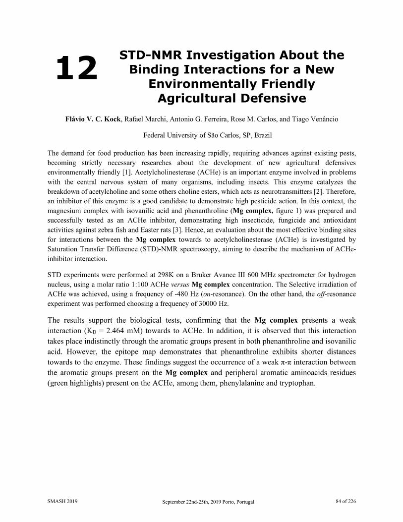
12 STD-NMR Investigation About the Binding Interactions for a New
Environmentally Friendly Agricultural Defensive
Flávio V. C. Kock, Rafael Marchi, Antonio G. Ferreira, Rose M. Carlos, and Tiago Venâncio
Federal University of São Carlos, SP, Brazil The demand for food production has been increasing rapidly, requiring advances against existing pests, becoming strictly necessary researches about the development of new agricultural defensives environmentally friendly [1]. Acetylcholinesterase (ACHe) is an important enzyme involved in problems with the central nervous system of many organisms, including insects. This enzyme catalyzes the breakdown of acetylcholine and some others choline esters, which acts as neurotransmitters [2]. Therefore, an inhibitor of this enzyme is a good candidate to demonstrate high pesticide action. In this context, the magnesium complex with isovanilic acid and phenanthroline (Mg complex, figure 1) was prepared and successfully tested as an ACHe inhibitor, demonstrating high insecticide, fungicide and antioxidant activities against zebra fish and Easter rats [3]. Hence, an evaluation about the most effective binding sites for interactions between the Mg complex towards to acetylcholinesterase (ACHe) is investigated by Saturation Transfer Difference (STD)-NMR spectroscopy, aiming to describe the mechanism of ACHe-inhibitor interaction.
STD experiments were performed at 298K on a Bruker Avance III 600 MHz spectrometer for hydrogen nucleus, using a molar ratio 1:100 ACHe versus Mg complex concentration. The Selective irradiation of ACHe was achieved, using a frequency of -480 Hz (on-resonance). On the other hand, the off-resonance experiment was performed choosing a frequency of 30000 Hz.
The results support the biological tests, confirming that the Mg complex presents a weak interaction (KD = 2.464 mM) towards to ACHe. In addition, it is observed that this interaction takes place indistinctly through the aromatic groups present in both phenanthroline and isovanilic acid. However, the epitope map demonstrates that phenanthroline exhibits shorter distances towards to the enzyme. These findings suggest the occurrence of a weak π-π interaction between the aromatic groups present on the Mg complex and peripheral aromatic aminoacids residues (green highlights) present on the ACHe, among them, phenylalanine and tryptophan.
SMASH 2019 September 22nd-25th, 2019 Porto, Portugal 84 of 226
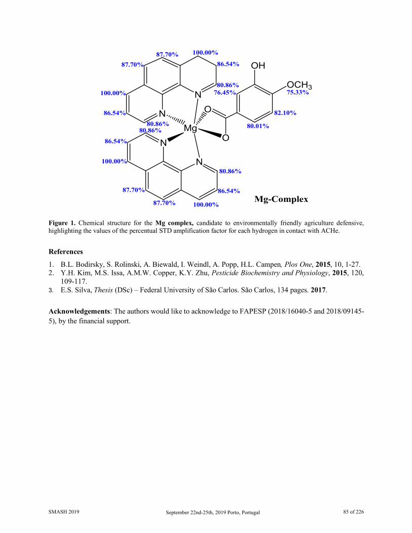
Figure 1. Chemical structure for the Mg complex, candidate to environmentally friendly agriculture defensive, highlighting the values of the percentual STD amplification factor for each hydrogen in contact with ACHe.
References
1. B.L. Bodirsky, S. Rolinski, A. Biewald, I. Weindl, A. Popp, H.L. Campen, Plos One, 2015, 10, 1-27. 2. Y.H. Kim, M.S. Issa, A.M.W. Copper, K.Y. Zhu, Pesticide Biochemistry and Physiology, 2015, 120,
109-117. 3. E.S. Silva, Thesis (DSc) – Federal University of São Carlos. São Carlos, 134 pages. 2017.
Acknowledgements: The authors would like to acknowledge to FAPESP (2018/16040-5 and 2018/09145-5), by the financial support.
N
NMg
N
N
OH
OCH3
O
O
80.86%
80.86%80.86%
80.86%
80.01%
100.00%
100.00%
100.00%
100.00%
87.70%
87.70%
87.70%87.70%
86.54%
86.54%
86.54%
86.54%
76.45%
82.10%
75.33%
Mg-Complex
SMASH 2019 September 22nd-25th, 2019 Porto, Portugal 85 of 226
13 Potential Alzheimer Drug Candidate: A STD-NMR Investigation Towards to
Acetylcholinesterase Activity
Flávio V. C. Kock, Marlon Profeta, Antonio G. Ferreira, Rose M. Carlos, and Tiago Venâncio
Federal University of São Carlos, SP, Brazil Alzheimer disease (AD) is one of the main causes of dementia in elderly and will affect 88 million people worldwide in 2050 [1]. One of the mechanisms to reach this neurologic disorder is the reduction of the activity of cholinergic neurons. This problem can be solved increasing the amount of the neurotransmitter acetylcholine (ACh). However, the acetylcholine hydrolysis in AD patients is promoted by the higher amount of acetylcholinesterase (ACHe). Therefore, which can be reached through an efficient interaction between the drug candidate and ACHe, causing it inhibition [2]. The intermolecular interaction between drugs candidates and ACHe has deep implications on the research about Alzheimer.
The Ru-Complex (figure 1) is a noteworthy candidate for acting as an ACHe inhibitor (IC50= 17.78µM). This complex has three phenanthroline rings able to efficiently interact towards to ACHe and a polysaccharide branch responsible to overcome the blood-brain barrier (BBB). Therefore, in this work, the molecular interaction at atomic level between this candidate and ACHe will be investigated by Saturation Transfer Difference (STD)-NMR spectroscopy, aiming to elucidate the main interactions sites present on this Ru-complex responsible by the ACHe inhibition.
The STD-NMR experiments were performed at 298K on a Bruker Avance III 600 MHz spectrometer for hydrogen nucleus, using a molar ratio 1:100 ACHe versus Ru-Complex concentration. The Selective irradiation (saturation) of ACHe was achieved, using a frequency of –480 Hz (on-resonance). On the other hand, the off-resonance experiment was realized choosing a frequency of 30000 Hz.
The results suggests that the presence of a glycosidic ring outside to aromatic core (figure 1), supplies an additonal and important interaction site, having shorter distances towards to ACHe. This results suggests that the major chemical mobility from this group can be associated to it major intermolecular interaction. In addition, the results confirms that this Ru-complex candidate have an efficient interaction (KD = 0.83 mM) towards to ACHe, and therefore, could be acts as an strong inhibitor for this enzyme. Similar candidates have been successfully tested, and besides to demonstrates a high permeability in the BBB, also shows a concomitant efficient interact towards to ACHe [3]. Lastly, the set of these results suggests the occurrence of an efficiente interaction between the glycosidic ring present on the Ru-complex and polar aminoacids residues (cyan highlights) present on the ACHe, among them, serine and histidine in the active sites from ACHe.
SMASH 2019 September 22nd-25th, 2019 Porto, Portugal 86 of 226

Figure 1: On the left-hand side, the chemical structure of the Ru-complex highlighting the values of the percentual STD amplification factor for each hydrogen in contact with ACHe. On the right-hand side, the structure of ACHe, highlighting the some polar domains (cyan).
Acknowledgements
The authors of this manuscript are gratefully acknowledging the financial support from FAPESP (2018/16040-5 and 2018/09145-5). References
1. Estrada, L.D., Oliveira-Cruz, L., Cabrera, D. Current Protein and Peptide Science, 2018, 19, 1180-1188.
2. Talesa, V.N. Mechanism of Ageing and development, 2001, 122, 1961-1969. 3. Sweeney, M.D., Sagare, A.P., Zlokovic, B.V. Nature Reviews Neurology, 2018, 14, 133-150.
NN
N
N
N
N
OS
OHHO
HO
HO
Ru61.41%
61.41%
38.51%40.81%39.06%
39.06%
40.81%
38.51%
45.89%
95.81%
100.00% 67.23%
77.08%
69.78%
Ru-Complex
SMASH 2019 September 22nd-25th, 2019 Porto, Portugal 87 of 226
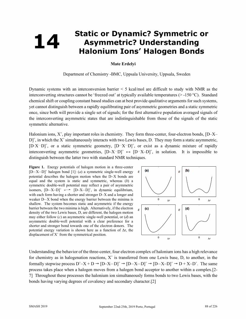
14 Static or Dynamic? Symmetric or Asymmetric? Understanding
Halonium Ions’ Halogen Bonds
Mate Erdelyi
Department of Chemistry -BMC, Uppsala University, Uppsala, Sweden
Dynamic systems with an interconversion barrier < 5 kcal/mol are difficult to study with NMR as the interconverting structures cannot be ‘freezed out’ at typically available temperatures (> -150 °C). Standard chemical shift or coupling constant based studies can at best provide qaulitative arguments for such systems, yet cannot distinguish between a rapidly equilibrating pair of asymmetric geometries and a static symmetric once, since both will provide a single set of signals; for the first alternative population averaged signals of the interconverting asymmetric states that are indistinguishable from those of the signals of the static symmetric alternative.
Halonium ions, X+, play important roles in chemistry. They form three-center, four-electron bonds, [D–X–D]+, in which the X+ simultaneously interacts with two Lewis bases, D. They may form a static asymmetric, [D_X…D]+, or a static symmetric geometry, [D…X…D]+, or exist as a dynamic mixture of rapidly interconverting asymmetric geometries, [D–X…D]+ ↔ [D…X–D]+, in solution. It is impossible to distinguish between the latter two with standard NMR techniques.
Figure 1. Energy potentials of halogen motion in a three-center [D⋅⋅⋅X⋅⋅⋅D]+ halogen bond [1]: (a) a symmetric single-well energy potential describes the halogen motion when the D−X bonds are equal and the system is static and symmetric, whereas (b) a symmetric double-well potential may reflect a pair of asymmetric isomers, [D⋅⋅⋅X−D]+ [D−X⋅⋅⋅D]+, in dynamic equilibrium, with each form having a shorter and stronger D−X and a longer and weaker D⋅⋅⋅X bond when the energy barrier between the minima is shallow. The system becomes static and asymmetric if the energy barrier between the two minima is high. Alternatively, if the electron density of the two Lewis bases, D, are different, the halogen motion may either follow (c) an asymmetric single-well potential, or (d) an asymmetric double-well potential with a clear preference for a shorter and stronger bond towards one of the electron donors. The potential energy variation is shown here as a function of Δr, the displacement of X+ from the symmetrical position.
Understanding the behavior of the three-center, four electron complex of halonium ions has a high relevance for chemistry as in halogenation reactions, X+ is transferred from one Lewis base, D, to another, in the formally stepwise process D+−X + D [D−X⋅⋅⋅D]+ [D⋅⋅⋅X⋅⋅⋅D]+ [D⋅⋅⋅X−D]+ D + X−D+. The same process takes place when a halogen moves from a halogen bond acceptor to another within a complex.[2-7] Throughout these processes the halonium ion simultaneously forms bonds to two Lewis bases, with the bonds having varying degrees of covalency and secondary character.[2]
SMASH 2019 September 22nd-25th, 2019 Porto, Portugal 88 of 226
We have used selective deuterium labeling in combination with the isotope perturbation of equilibrium technique to distinguish between tapidly interconverting asymmetric structures from static symmetric ones. Relaxation measurements were applied on highly reactive systems only stable < -70 °C. 15N NMR chemical shift measurements were applied to distiguish between static symmetric and static asymmetric geometries, whereas diffusion NMR to evaluate the association of counterions and its impact on the geometry on the [D–X–D]+ complexes.
Here, the NMR investigation of the influence of electronic and steric factors, solvent polarity and counterions, and of the type of the halogen on the geometry and reactivity of [D⋅⋅⋅X⋅⋅⋅D]+ halogen bond complexes will be discussed. The symmetric state, [D⋅⋅⋅X⋅⋅⋅D]+, is demonstrated to be strongly preferred over the alternative asymmetric arrangements [D⋅⋅⋅X−D]+.[2-6]
References
1. Halogen bonding is the noncovalent interaction of halogen in which they act as electron acceptors. 2. Karim, A.; Reitti, M.; Carlsson, A.-C.C.; Gräfenstein, J.; Erdelyi, M., Chem. Sci., 5, 3226, 2014. 3. Carlsson, A.-C.C.; Mehmeti, K.; Uhrbom, M.; Karim, A.; Bedin, M.; Puttreddy, R.; Kleinmaier, R.;
Neverov, A.; Nekoueishahraki, B.; Gräfenstein, J.; Rissanen, K.; Erdelyi, M., J. Am. Chem. Soc., 138, 9853, 2016.
4. Carlsson, A.-C.C.; Gräfenstein,J.; Budnjo, A.; Bergquist, J.; Karim, A.; Kleinmaier, R.; Brath, U.; Erdelyi, M. J. Am. Chem. Soc., 134, 5706, 2012.
5. Hakkert, S.B.; Erdelyi, M., J. Phys. Org. Chem., 28, 226, 2015. 6. Lindblad, S.; Mehmeti, K.; Veiga, A.; Nekoueishahraki, B.; Gräfenstein, J.; Erdelyi, M., J. Am.
Chem. Soc., 140, 13503, 2018. 7. Vanderkooy, A.; Gupta, A.K.; Földes, T.; Lindblad, S.; Orthaber, A.; Pápai, I.; Erdélyi, M., Angew.
Chem. Int. Ed. 2019, in press.
SMASH 2019 September 22nd-25th, 2019 Porto, Portugal 89 of 226
15 Investigation of the Structure of Cyclic Depsipeptides PF1022A and Emodepside and Their Affinity to
Cations
Thomas Stadelmann, Sereina Riniker, and Marc-Olivier Ebert
Department of Chemistry and Applied Biosciences, ETH Zurich, Switzerland The cyclooctadepsipeptide PF1022A is a natural product with activity against nematodes [1]. Its synthetic derivative emodepside shows increased activity and is used as a commerical drug [2]. In NMR spectra of PF1022A in CD3OH and CDCl3, two main conformations can be observed, which interconvert slowly on the NMR time-scale. The major conformation is characterized by a cis-amide bond and is thus termed “asymmetric”, whereas all amide bonds are trans in the minor conformation, thus termed “symmetric” [1,3]. The exchange rate between the two conformers was calculated by analyzing the exchange peaks in the ROESY spectra in CDCl3.
PF1022A and derivatives have been found to be ionophores with a selectivity for potassium over sodium [4]. To test whether ions have an impact on the conformational ensemble, titrations with various monovalent thiocyanate salts in methanol were performed. It could be observed that the symmetric conformations of PF1022A and emodepside are able to bind cations. In addition, a shift in the equilibrium between the asymmetric and the symmetric conformations was observed. For example, in presence of 200 mM KSCN the ratio between the asymmetric and symmetric conformation changed from 5:1 to 1:16 in the case of PF1022A.
Furthermore, molecular dynamics (MD) simulations of PF1022A and emodepside in chloroform with a K+ ion show a strong binding affinity of the ion to the peptides and the adoption of a cavitand-like structure was observed. In addition, the presence of the K+ ion appears to increase the likelihood of the cis-trans isomerization of the backbone amide.
1. Sasaki, T. et al., J. Antibiot. (Tokyo). 45, 692–697, 1992. 2. Harder, A., Holden-Dye, L., Walker, R. & Wunderlich, F., Parasitol. Res. 97, S1–S10, 2005. 3. Scherkenbeck, J., Jeschke, P. & Harder, A., Curr. Top. Med. Chem. 2, 759–77 2002. 4. Gessner, G., Meder, S., Rink, T. & Boheim, G., Pestic. Sci. 48, 399–407 1996.
SMASH 2019 September 22nd-25th, 2019 Porto, Portugal 90 of 226
16 Using Proton Residual Chemical Shift Anisotropy at Microgram Level for the Determination of the Relative Configuration in Marine Natural
Products
Juan Carlos Fuentes-Monteverde,1,2 Nilamoni Nath,3 Dawrin Pech-Puch,2 Armando Navarro-Vázquez,4 Elena Balboa,5 Jaime Rodríguez,2 Carlos Jiménez,2 and Christian Griesinger1
6. NMR Based Structural Biology, MPI for Biophysical Chemistry, Göttingen, Germany 7. Centro de Investigacións Científicas Avanzadas (CICA) e Departamento de Química, Facultade
de Ciencias, Universidade da Coruña. A Coruña, Spain 8. Department of Chemistry, Gauhati University, Gopinath Bardoloi Nagar, Guwahati, India
9. Departamento de Química Fundamental, CCEN, Universidade Federal de Pernambuco, Cidade Universitária, Recife, Brazil
10. Department of Chemical Engineering, Faculty of Science, Campus Ourense, University of Vigo, Spain. CITI-University of Vigo, Parque Tecnolóxico de Galicia, Ourense, Spain
Determination of the three dimensional structure remains a challenging task for natural products when are available in minute amounts and cannot be crystallized. While the chemical constitution can be derived from proton/proton and proton/carbon correlations, J-couplings, NOEs and often 1H-13C RDCs or 13C RCSAs are used to determine the relative configuration. However, for compounds in the range of few microgram these RDC or 13C RCSAs are difficult to collect because of low sensitivity. [1,2] Herein, we demostrate that 1H RCSAs allows to determinte the orientation of different structural moieties within a molecule but with much less requirements on the amount of sample. An important issue that arises in RCSA measurements of small molecules in the range of micrograms, is very often the use of protonated polymeric gels to induce the alignment conditions present NMR signals that masks the actual sample. This problem was circumvented by the use of PMMA-d8 gel. The potential of the methodology was also demonstrated. 1H RCSAs were measured by using 3 mm devices (stretching and compression) manufactured by the glass company Hilgenberg, for microgram samples of marine natural products, isolated from Briareum asbestinum and Sargassum muticum, collected in Mexican Caribbean (Mexico) and Costa da Morte (Spain) respectively, for which relative configuration is still unknown [3,4]. Use of NOESY constraints restricted the number of possible relative configurations, from which 1H RCSA allowed us to select a single relative configuration. Determination of the absolute configuration could be accomplished by comparison of calculated (TD-DFT) and experimental electronic circular dichroism spectra [5]. In summary, we reported that 1H RCSAs as highly sensitive parameter that complemented J-couplings and NOEs, without the necessity to use any other anisotropic parameter at the microgram scale.
11. Nath, N. et al., Journal of the American Chemical Society, 138(30), 9548-9556, 2016 12. Liu, Y. et al., Nature Protocol, 14, 217-247, 2019 13. Harvell, C. et al., Marine Ecology-Progress Series, 93, 165-173, 1993 14. Balboa, E. et al., J Photochem Photobiol B, 148, 51-58, 2015 15. Pescitelli, G. et al., Chirality, 28(6), 466-474, 2016
SMASH 2019 September 22nd-25th, 2019 Porto, Portugal 91 of 226
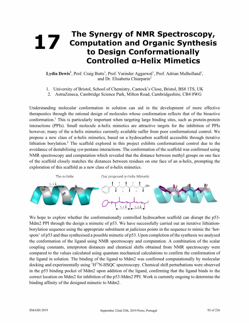
17 The Synergy of NMR Spectroscopy, Computation and Organic Synthesis
to Design Conformationally Controlled α-Helix Mimetics
Lydia Dewis1, Prof. Craig Butts1, Prof. Varinder Aggarwal1, Prof. Adrian Mulholland1,
and Dr. Elisabetta Chiarparin2
1. University of Bristol, School of Chemistry, Cantock’s Close, Bristol, BS8 1TS, UK 2. AstraZeneca, Cambridge Science Park, Milton Road, Cambridgeshire, CB4 0WG
Understanding molecular conformation in solution can aid in the development of more effective therapeutics through the rational design of molecules whose conformation reflects that of the bioactive conformation.1 This is particularly important when targeting large binding sites, such as protein-protein interactions (PPIs). Small molecule α-helix mimetics are attractive targets for the inhibition of PPIs however, many of the α-helix mimetics currently available suffer from poor conformational control. We propose a new class of α-helix mimetics, based on a hydrocarbon scaffold accessible through iterative lithiation borylation.2 The scaffold explored in this project exhibits conformational control due to the avoidance of destabilising syn-pentane interactions. The conformation of the scaffold was confirmed using NMR spectroscopy and computation which revealed that the distance between methyl groups on one face of the scaffold closely matches the distances between residues on one face of an α-helix, prompting the exploration of this scaffold as a new class of α-helix mimetics.
We hope to explore whether the conformationally controlled hydrocarbon scaffold can disrupt the p53-Mdm2 PPI through the design a mimetic of p53. We have successfully carried out an iterative lithiation-borylation sequence using the appropriate substituent at judicious points in the sequence to mimic the ‘hot-spots’ of p53 and thus synthesised a possible mimetic of p53. Upon completion of the synthesis we analysed the conformation of the ligand using NMR spectroscopy and computation. A combination of the scalar coupling constants, interproton distances and chemical shifts obtained from NMR spectroscopy were compared to the values calculated using quantum mechanical calculations to confirm the conformation of the ligand in solution. The binding of the ligand to Mdm2 was confirmed computationally by molecular docking and experimentally using 1H15N-HSQC spectroscopy. Chemical shift perturbations were observed in the p53 binding pocket of Mdm2 upon addition of the ligand, confirming that the ligand binds to the correct location on Mdm2 for inhibition of the p53-Mdm2 PPI. Work is currently ongoing to determine the binding affinity of the designed mimetic to Mdm2.
SMASH 2019 September 22nd-25th, 2019 Porto, Portugal 92 of 226
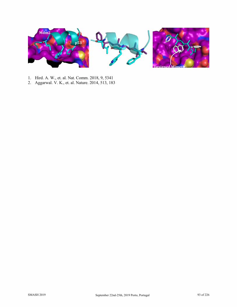
1. Hird. A. W., et. al. Nat. Comm. 2018, 9, 5341 2. Aggarwal. V. K., et. al. Nature. 2014, 513, 183
p53
Mdm2
Designed Mimetic
SMASH 2019 September 22nd-25th, 2019 Porto, Portugal 93 of 226

18 Implementing One-Shot Multiple-FID Acquisition into Homonuclear
and Heteronuclear NMR Experiments
Kumar Motiram-Corral, Míriam Pérez-Trujillo, Pau Nolis, and Teodor Parella
Servei de RMN, Universitat Autónoma de Barcelona. Barcelona, Spain. To date, time-efficient NMR approaches are a challenging task for spectroscopists. The goal is to obtain chemical information reducing experimental time and without considerably losing sensitivity. Different time-efficient methods have been described over the years. For instance, time sharing techniques acquire 15N and 13C data in the same spectrum in spectrometers having a triple channel hardware configuration [1]. Non-Uniform Sampling (NUS) algorithms afford a substantial reduction of experimental times by reducing the number of t1 increments in multidimensional experiments. [2] Recently, the interleaved acquisition of multiple experiments by the NOAH (NMR by ordered Acquisition using 1H detection) strategy allow the reduction of long recycle delays in consecutive experiments. [3]
On the other hand, the MFA (Multiple FID Acquisition) strategy was initially proposed many years ago with the COCONOSY experiment [4-6], which collected 2D COSY and NOESY data with a single pulse scheme. We show here how MFA can be implemented in several applications, offering a new novel proof of concept to obtain COSY, TOCSY and HMBC datasets in small molecules. For instance, it is shown how the simultaneous recording of four different NMR experiments in a single-shot acquisition, decreasing close to 60% of experiment time. [7] This approach is based on the recovery of the remaining transverse magnetization after an acquisition period. Magnetization components that usually are lost by relaxation to its original z-position can be manipulated by an appropriate additional mixing process and recorded again to obtain a second or third NMR data provided that T2 relaxation times are long enough. It is shown how MFA is a powerful strategy for the sequential structural assignment of a whole spin system without ambiguities using a multiple-step RELAY experiment.
1. Nolis, P., Pérez, M., & Parella, T. (2006). Magnetic Resonance in Chemistry, 44, 11, 1031-1036, 2006.
2. K. Kazimierczuk and V. Y. Orekhov , Angew. Chem., Int. Ed., 2011, 50 , 5556 -5559 3. Kupče, E., & Claridge, T. D. W. (2018). Chemical Communications, 54, 7139-7142, 2018. 4. A. Z. Gurevich , I. L. Barsukov , A. S. Arseniev and V. F. Bystrov , J. Magn. Reson., 1984, 56 , 471 -
478 5. C. A. G. Haasnoot , F. J. M. van de Ven and C. W. Hilbers , J. Magn. Reson., 1984, 56 , 343 -349 6. J. Cavanagh and M. Rance , J. Magn. Reson., 1990, 14 , 408 -414 7. Motiram-Corral, K., Pérez-Trujillo, M., Nolis, P., & Parella, T. (2018). Chemical Communications,
54(96), 13507–13510, 2018.
SMASH 2019 September 22nd-25th, 2019 Porto, Portugal 94 of 226

19 43Ca Solid-State NMR Spectroscopy
of Atorvastatin Calcium
Shi Bai1, Sean T. Holmes2, and Cecil Dybowski1
1. Department of Chemistry and Biochemistry, University of Delaware, DE, US 2. Department of Chemistry and Biochemistry, University of Windsor, Windsor, Canada
Form I of atorvastatin calcium trihydrate (ATC-I) is the active pharmaceutical ingradient (API) in Lipitor®, which is commonly prescribed for lowering blood cholesterol and preventing hypertension or other cardiovascular diseases. Lipitor® is arguably the best-selling drug in the history of modern Western medicine. Although the mechanism of the catalytic portion of complexes of 3-hydroxy-3-methylglutary-coenzme A (HMG-CoA) reductase with six statins, including atorvastatin, has been reported [1], the crystal structure of ATC-I, which determines the bioavailability, stability, safety, and efficacy of the medication [2], remains unknown to date.
Previously, we proposed a molecular structure of the atorvastatin ligands on the calcium ion in the ATC-I [3] based on13C, 19F, and 15N multinuclear solid-state NMR spectroscopic studies combined with extensive ab initio calculations. Although the model correctly predicted the two sets of 13C, 15N, and 19F isotropic chemical shifts observed for ATC-I under magic angle spinning conditions, the choice of the local calcium environment was arbitrary as it was derived from that of calcium dibenzoate·3H2O [4].
In this presentation, 43Ca solid-state NMR spectroscopy is employed to investigate local calcium structures in ATC-I and the ethylene glycol solvate of atorvastatin calcium (ATC-EG). We demonstrate that the 43Ca NMR spectral features are very sensitive to the local calcium environment in atorvastatin calcium and can be readily used as a unique spectroscopic index to differentiate solvomorphous forms of atorvastatin calcium.
We further discuss a crystallographic approach based on 43Ca NMR spectroscopy that includes an extensive survey of the Cambridge Structural Database (CSD) and a new symmetry benchmark to enhance the selectivity of structural screening [5]. In this method, experimental and computational 43Ca solid-state NMR parameters such as chemical-shift tensors, quadrupolar-coupling tensors, and Euler angles are compared to constrain the structure of the local calcium–ligand coordination environment in ATC-I. The structural search begins with an extensive survey of calcium-containing crystals in the CSD, followed by generation of a library of a large number of candidates for the local calcium environment in ATC-I that meet certain energetic criteria. The library is strictly screened for matches with experimental 43Ca NMR parameters using Hartree-Fock (HF) and density functional theory (DFT) calculations. While the majority of models in the library are eliminated by comparison between calculated and experimental 43Ca NMR chemical-shift and quadrupolar parameters, a number of structures cannot be differentiated with these comparisons alone. A complete analysis of the local symmetry of the calcium site demonstrates that the line shape of the 43Ca SSNMR powder pattern is very sensitive to β, the Euler angle between the most shielded CS principal axis (δ33) and the major axis (V33) of the EFG tensor. Simulation of the NMR spectra as a function of Euler angles reveals a unique relationship between the 43Ca quadrupolar line shape and the local symmetry at the calcium site. This observation demonstrates the Euler angle between these axes is a
SMASH 2019 September 22nd-25th, 2019 Porto, Portugal 95 of 226

particularly useful benchmark for screening structures to eliminate structural candidates remaining in the library that do not meet the calcium site symmetry criterion. As an example, only one candidate structure was ultimately retained from the remaining structures by the symmetry benchmark screening on the calcium coordination environment in ATC-I.
General implication of this structure-search protocol combining a survey of the Cambridge Structural Database with the Euler angle as a symmetry benchmark has been validated by applying it to identify the known calcium local structure of calcium acetate monohydrate [6].
1. Istvan, E. S. and Deisenhofer, J., Science, 292 (5519), 1160-4, 2001. 2. Brittain, H. G. and Grant, D. J. W., Effect of polymorphism and solid-state solvation on solubility and
dissolution rate. 1999, New York: Marcel Dekker, Inc. 3. Wang, W. D., Gao, X. D., Strohmeier, M., Wang, W., Bai, S., and Dybowski, C., Journal of Physical
Chemistry B, 116 (11), 3641-3649, 2012. 4. Senkovska, I. and Thewalt, U., Acta Crystallographica Section C-Crystal Structure Communications,
61, M448-M449, 2005. 5. Holmes, S. T., Wang, W. D., Hou, G., Dybowski, C., Wang, W., and Bai, S., Phys Chem Chem Phys,
21 (12), 6319-6326, 2019. 6. Widdifield, C. M., Moudrakovski, I., and Bryce, D. L., Physical Chemistry Chemical Physics, 16
(26), 13340-13359, 2014.
SMASH 2019 September 22nd-25th, 2019 Porto, Portugal 96 of 226
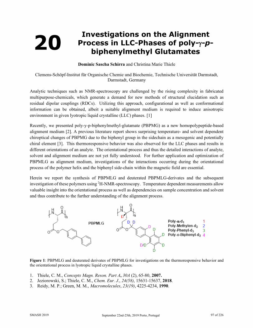
20 Investigations on the Alignment Process in LLC-Phases of poly-γ-p-
biphenylmethyl Glutamates
Dominic Sascha Schirra and Christina Marie Thiele
Clemens-Schöpf-Institut für Organische Chemie und Biochemie, Technische Universität Darmstadt, Darmstadt, Germany
Analytic techniques such as NMR-spectroscopy are challenged by the rising complexity in fabricated multipurpose-chemicals, which generate a demand for new methods of structural elucidation such as residual dipolar couplings (RDCs). Utilizing this approach, configurational as well as conformational information can be obtained, albeit a suitable alignment medium is required to induce anisotropic environment in given lyotropic liquid crystalline (LLC) phases. [1]
Recently, we presented poly-y-p-biphenylmethyl-glutamate (PBPMG) as a new homopolypeptide-based alignment medium [2]. A previous literature report shows surprising temperature- and solvent dependent chiroptical changes of PBPMG due to the biphenyl group in the sidechain as a mesogenic and potentially chiral element [3]. This thermoresponsive behavior was also observed for the LLC phases and results in different orientations of an analyte. The orientational process and thus the detailed interactions of analyte, solvent and alignment medium are not yet fully understood. For further application and optimization of PBPMLG as alignment medium, investigations of the interactions occurring during the orientational process of the polymer helix and the biphenyl side-chain within the magnetic field are essential.
Herein we report the synthesis of PBPMLG and deuterated PBPMLG-derivates and the subsequent investigation of these polymers using 2H-NMR-spectroscopy. Temperature dependent measurements allow valuable insight into the orientational process as well as dependencies on sample concentration and solvent and thus contribute to the further understanding of the alignment process.
Figure 1: PBPMLG and deuterated derivates of PBPMLG for investigations on the thermoresponsive behavior and the orientational process in lyotropic liquid crystalline phases. 1. Thiele, C. M., Concepts Magn. Reson. Part A, 30A (2), 65-80, 2007. 2. Jeziorowski, S.; Thiele, C. M., Chem. Eur. J., 24(58), 15631-15637, 2018. 3. Reidy, M. P.; Green, M. M., Macromolecules, 23(19), 4225-4234, 1990.
SMASH 2019 September 22nd-25th, 2019 Porto, Portugal 97 of 226

21 Structural and Kinetic Study of Two Photo- and Electrically Responsive
Dithienylethenes
D. Herold1, J. Kind1, F. Frieß1, J. von Irmer2, M. Gallei3, and C. M. Thiele1
1. Clemens Schöpf Institut, Technische Universität Darmstadt, Germany 2. Ernst-Berl-Institut, Technische Universität Darmstadt, Germany
3. Organische und Makromolekulare Chemie, Universität des Saarlandes, Germany Smart materials are one of the most innovative and versatile developments during the last decades. These chemical compounds utilize an external stimulus like pH, temperature, light or electric fields to change the physical and chemical properties. Especially light is a fascinating stimulus due to its spatio-temporal controllability[1]. Nevertheless monitoring of photoreactions can be rather challenging, which implicates the necessity for a setup enabling in situ irradiation for NMR-spectroscopy [2].
The GALLEI group is investigating stimulus-responsive properties of surface-modified materials[3,4]. Recently, two photo- and electrically responsive dithienylethenes, attached to a gold surface, were synthesized and investigated. It could be shown that switchable conductivity for the use in plasmon resonance is possible; however, the rates of forming the reversible and irreversible states (scheme below) were still unknown.
In order to obtain a deeper insight into the reaction schemes not only UV/Vis- but also in situ irradiation NMR-spectroscopy was executed. With this powerful combination we were able to determine reaction constants for the reversible and irreversible reactions as well as extinction coefficients. This allowed conclusions on possible reaction schemes for both compounds. These photochromic systems of the dithienylethene class show effectively no thermal relaxation and we present their structural and kinetic characteristics.
1. Kathan, M., Hecht S., Chem. Soc. Rev. 46, 5536-5550, 2017 2. Feldmeier, C. et al., J. Magn. Reson., 232, 39-44, 2013 3. Gallei, M., Rüttiger C., Chem. Eur. J., 24, 10006-10021, 2018 4. Gallei, M., Macromol. Rapid Commun., 39, 1700648, 2018
SMASH 2019 September 22nd-25th, 2019 Porto, Portugal 98 of 226
22 PSYCHE-EASY-ROESY Goes Quantitative: Extraction of 1H-1H
Distance Restraints from Overcrowded Spectral Regions
Julian Ilgen1, Jens Nowag1, Lukas Kaltschnee2,3 and Christina M. Thiele1
1. Clemens-Schöpf-Institut, Technische Universität Darmstadt, Germany 2. Max-Planck-Insitut für Biophysikalische Chemie, Göttingen, Germany
3. Center for Biostructural Imaging of Neurodegeneration (BIN), Göttingen, Germany 1H,1H-NOESY[1] and 1H,1H-ROESY[2] are valuable experiments for spatial structure elucidation of organic or bioorganic molecules of varying size. They provide access to proton-proton distances, frequently used as qualitative or quantitative measures of spatial proximity.
Since these experiments yield proton-proton correlation spectra, signal overlap caused by limited proton chemical shift dispersion becomes a major challenge with increasing molecular complexity. The incorporation of pure shift elements into NOESY and ROESY experiments represents a possible solution to improve spectral resolution in such cases [3-7].
We have used an EASY-ROESY [8] experiment with PSYCHE [9] homonuclear decoupling for the structural analysis of a peptide organocatalyst, which usually yields overcrowded spectra [7,10]. We show that the extraction of cross relaxation rates and distance restraints from PSYCHE-EASY-ROESY is accurate, and thus provides a valuable tool for studying overcrowded spectral regions. The enhanced resolution enabled us to extract the fourfold number of proton-proton distances, as compared to conventional techniques. These advances may lead to a significant improvement in the structural models also of the peptide catalyst in the future.
A notable advance over our earlier studies [7] is achieved, by using a gradient selected PSYCHE-EASY-ROESY instead of a phase-cycled experiment. This significantly attenuates “t1-noise” and thus enhances the spectral quality. Analysis of these “cleaned-up” PSYCHE-EASY-ROESY spectra with cyclosporine as model compound allows for the extraction of a number of distance restraints from overcrowded spectral regions, which were previously hidden under “t1-noise” and thus were not available for analysis.
1. Kumar, A., Ernst, R. R. and Wüthrich, K., Biochem. Biophys. Res. Commun., 95(1), 1-6, 1980 2. Bothner-By, A. A. et al., J. Am. Chem. Soc., 106(3), 811-813, 1984 3. Ying, J., Roche, J. and Bax, A., J. Magn. Reson., 241, 97-102, 2014 4. Kakita, V. M., Bharatam, J., Magn. Reson. Chem., 52(7), 389-394, 2014 5. Aguilar, J. A. et al., Angew. Chem. Int. Ed., 51(26), 6460-6463, 2012 6. Kaltschnee, L. et al., J. Magn. Reson., 271, 99-109, 2016 7. Procházková, E. et al., Angew. Chem. Int. Ed., 55(51), 15754-15759, 2016 8. Thiele, C. M., Petzold, K. and Schleucher, J., Chem. Eur. J., 15(3), 585-588, 2009 9. Foroozandeh, M. et al., Angew. Chem. Int. Ed. 53(27), 6990-6992, 2014 10. Müller, C. E., Wanka, L., Jewell, K. and Schreiner, P. R., Angew. Chem. Int. Ed., 120(33), 6275-
6278, 2008
SMASH 2019 September 22nd-25th, 2019 Porto, Portugal 99 of 226
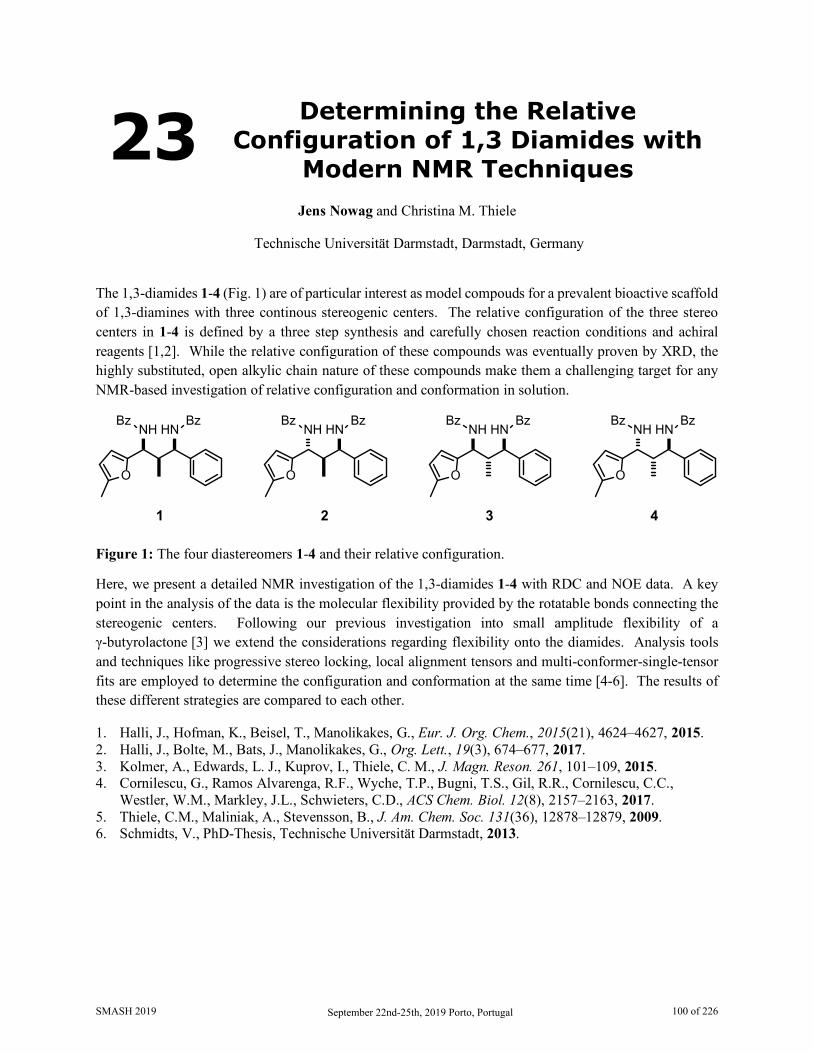
23 Determining the Relative Configuration of 1,3 Diamides with
Modern NMR Techniques
Jens Nowag and Christina M. Thiele
Technische Universität Darmstadt, Darmstadt, Germany
The 1,3-diamides 1-4 (Fig. 1) are of particular interest as model compouds for a prevalent bioactive scaffold of 1,3-diamines with three continous stereogenic centers. The relative configuration of the three stereo centers in 1-4 is defined by a three step synthesis and carefully chosen reaction conditions and achiral reagents [1,2]. While the relative configuration of these compounds was eventually proven by XRD, the highly substituted, open alkylic chain nature of these compounds make them a challenging target for any NMR-based investigation of relative configuration and conformation in solution.
BzNH HN
Bz
O
BzNH HN
Bz
O
BzNH HN
Bz
O
BzNH HN
Bz
O
1 2 3 4
Figure 1: The four diastereomers 1-4 and their relative configuration.
Here, we present a detailed NMR investigation of the 1,3-diamides 1-4 with RDC and NOE data. A key point in the analysis of the data is the molecular flexibility provided by the rotatable bonds connecting the stereogenic centers. Following our previous investigation into small amplitude flexibility of a γ-butyrolactone [3] we extend the considerations regarding flexibility onto the diamides. Analysis tools and techniques like progressive stereo locking, local alignment tensors and multi-conformer-single-tensor fits are employed to determine the configuration and conformation at the same time [4-6]. The results of these different strategies are compared to each other.
1. Halli, J., Hofman, K., Beisel, T., Manolikakes, G., Eur. J. Org. Chem., 2015(21), 4624–4627, 2015. 2. Halli, J., Bolte, M., Bats, J., Manolikakes, G., Org. Lett., 19(3), 674–677, 2017. 3. Kolmer, A., Edwards, L. J., Kuprov, I., Thiele, C. M., J. Magn. Reson. 261, 101–109, 2015. 4. Cornilescu, G., Ramos Alvarenga, R.F., Wyche, T.P., Bugni, T.S., Gil, R.R., Cornilescu, C.C.,
Westler, W.M., Markley, J.L., Schwieters, C.D., ACS Chem. Biol. 12(8), 2157–2163, 2017. 5. Thiele, C.M., Maliniak, A., Stevensson, B., J. Am. Chem. Soc. 131(36), 12878–12879, 2009. 6. Schmidts, V., PhD-Thesis, Technische Universität Darmstadt, 2013.
SMASH 2019 September 22nd-25th, 2019 Porto, Portugal 100 of 226
24 Determination of the 15N Chemical Shift Anisotropy in Natural
Abundance Samples by Proton-detected 3D Solid-State NMR Under
Ultrafast MAS
F. Rossi,1 N. Tuan Duong,2 M. K. Pandey,3 M. R. Chierotti,1 R. Gobetto,1 and Y. Nishiyama2,4
1. Department of Chemistry and NIS Centre, University of Torino, Italy 2. RIKEN CLST-JEOL Collaboration Center, Yokohama, Kanagawa, Japan
3. Indian Institute of Technology Ropar, Rupnagar, Punjab, India 4. JEOL RESONANCE Inc., Musashino, Akishima, Tokyo, Japan
Chemical shift anisotropy (CSA) represents a sensitive probe of electronic nuclear environment and thus, it offers deeper insights into detailed structural and dynamics properties of different chemical and biological systems. Over the years, massive efforts have been made to develop recoupling methods that reintroduce CSA interaction under MAS condition.[1-3] Among these techniques, one method presented by M. Levitt makes use of rotor-synchronized symmetry-based pulse sequences.[4]
We present here a proton-detected 3D 1H/15N/15N CS/CS/CSA correlation experiment which employs a γ-encoded RNn
ν-symmetry based CSA recoupling scheme. The advantage of such schemes is that they can be exploited under a wide range of MAS rates within the practically attainable rf field strength. In this work, two different symmetries, i.e. R87
3 and R1094, have been tested on [U-15N]-L-histidine·HCl·H2O as a model
sample and then successfully employed to retrieve 15N CSA information in natural abundance glycyl-L-alanine (GlyAla) sample under 70 kHz MAS. We demonstrate that this 3D R-symmetry based pulse sequence is robust with respect to offset wide-range mismatches and to rf inhomogeneity within mis-sets of ±10% from the theoretical value. The application of the 3D experiment on GlyAla sample results in site-resolved 15N CSA lineshapes, from which CSA parameters have been retrieved by numerical fittings using SIMPSON program.
The advantages of this proton-detected 3D correlation experiment are manifold: efficient and robust 15N CSA recoupling is achievable; low-power rf fields can be employed to avoid sample and probe heating; the sensitivity is strongly enhanced by 1H indirect detection approach which thus permits to retrieve CSA for natural abundance samples; by incorporating a 15N isotropic chemical shift dimension into the 3D sequence, the experiment becomes applicable for simultaneous determination of multiple sites; the small rotor volume essential for the ultrafast MAS requires a tiny amount of sample, extending the method to systems that need isotope enrichment or for which only small amounts are feasible.
1. Nishiyama Y., Yamazaki T., Terao T., J. Chem. Phys., 124(6), 64304, 2006 2. Orr R.M., Duer M.J., Solid State Nucl. Magn. Reson. 30(1), 1–8, 2006 3. Hung I., Gan Z., J. Magn. Reson., 213(1), 196, 2011 4. Levitt M.H., in Encyclopedia of Nuclear Magnetic Resonance. Wiley, Chichester, pp. 165-196, 2002
SMASH 2019 September 22nd-25th, 2019 Porto, Portugal 101 of 226
25 Polyaspartates: Alignment Media with Unexpected Bevahiour
Max Hirschmann and Christina M. Thiele
Technische Universität Darmstadt, Clemens-Schöpf-Institut für Organische Chemie und Biochemie, Darmstadt, Germany
Polyaspartates are known to form helical secondary structures and therewith possess axial chirality additionally to the point chirality of aspartic acid. In general poly-L-peptides form right-handed helices and poly-D-peptides form left-handed helices. Interestingly, this conjunction between configuration of amino acid and helical sense is not observed for polyaspartates: Poly-L-aspartates are known to form either right-handed or left-handed helices depending on ester group [1], temperature [2], light [3] or pH [1].
Due to the helical, rod-like structure, polypeptides are capable of forming lyotropic liquid crystalline phases at appropriate concentrations in organic solvents. These phases are cholesteric in nature and have a helical sense themselves. In addition to the change in helical sense of the backbone, some polyaspartates can further change the helical sense of the cholesteric pitch by temperature [2, 4]. Inside a magnetic field (e.g. used in NMR spectroscopy) the cholesteric phase is converted into a nematic phase, which is equivalent with the cholesteric pitch becoming infinitely [5].
We recently synthesized and made use of a copolymer of polybenzylaspartate and polyphenethylaspartate as alignment medium to partially align small organic molecules during NMR spectroscopic measurements [6]. In this study we observed three regimes in a temperature range between 300 K and 330 and investigated the different orientational / enantiodifferentiating properties of the alignment medium at these regimes: When comparing the regime at 300 K with the regime at 312 K a sign change of all the meausred RDCs (of various analytes) and a sign change of the quadrupolar splitting of the deuterated solvent was observed while the enantiodifferentiation remains almost unaffected. Further increasing the temperature leads to regime three, for which a change of RDCs was observed as well. This change seems to follow no trend and the enantiodifferentatiation is reduced compared to regimes one and two.
However, only two of the temperature regimes can be explained by the literature-known helix reversal - from right to left-handed helix or vice versa - of the copolymer used. The nature of the third regime is unknown. Therefore, we synthesized various partially deuterated polymers and investigated the phenomenon by 2H-NMR spectroscopy and circular dichroism spectroscopy. Herein we report our latest results on this matter and which implications this has on the use as (enantiodifferentiating) alignment medium.
1. De Zotti, M., Formaggio, F., Crisma, M., Peggion, C., Morettoa, A., Toniolo, C., J. Pept. Sci., 20, 307-322, 2014.
2. Abe, A., Furuya, H., Okamoto, S., Pept. Sci., 43, 405-412, 1997. 3. Ueno, A., Anzai, J., Osa, T., J. Polym. Sci., Polym. Lett. Ed., 15, 407-410, 1977. 4. Watanabe, J., Okamoto, S., Abe, A., Liq. Cryst., 15, 259-263, 1993. 5. Abe, A., in Macromolecules 1992 (Ed.: J. Kahovec), pp. 221-228, 1993. 6. Schwab, M., Schmidts, V., Thiele, C., Chem. - Eur. J., 24, 14373-13477, 2018.
SMASH 2019 September 22nd-25th, 2019 Porto, Portugal 102 of 226
26 Faster Pure-Shift Spectra Acquisition: Utilizing the Prior
Knowledge from 1H NMR Alexandra Shchukina1, Magdalena Kaźmierczak2, Paweł Kasprzak1, Matthew Davy3, Geoffrey Akien4,
Craig P. Butts3,and Krzysztof Kazimierczuk1
1. University of Warsaw, Poland 2. Warsaw University of Technology, Poland
3. University of Bristol, UK 4. University of Lancaster, UK
Pure-shift NMR [1] allows the suppression of homonuclear J-couplings in an NMR spectrum. Thus, multiplets in a spectrum collapse into singlets, which enhances the spectral resolution. Pure-shift techniques are based on echo-type pulse sequences with selective pulses. In between the pulses, a short chunk of an FID is acquired, and later the whole FID is constructed from these chunks.
The acquisition can be performed in different ways. Among them, so called pseudo-2D acquisition yields spectra of better resolution and fewer artifacts. This method resembles a standard two-dimensional measurement, where each chunk is measured as a separate pseudo-F1 increment. The one-dimensional FID is then assembled from these increments. A widely used PSYCHE pulse sequence [2] is an example of such an acquisition method. The drawback of such experiments is that they are time-consuming. This leads to an idea to accelerate them with non-uniform sampling (NUS).
Normally, NUS in NMR means omitting some measurement points and reconstructing them mathematically afterwards. Here, one can only omit whole chunks, not separate points. We call this type of sampling “burst sampling”. This situation is potentially more challenging, as any regularity in omittions can increase undersampling artifacts and hamper the consequent reconstruction. However, this procedure still works, as was demonstrated in [3].
On the other hand, some prior information on the spectrum is at or disposal: the conventional 1H spectrum can tell us, within some tolerance, where to anticipate peaks in a pure-shift spectrum. In this work, we show how to optimize the burst sampling scheme taking the advantage of this prior knowledge. Our aim is to minimize the risk of high undersampling artifacts and thus assist the reconstruction. We demonstrate on several examples that the optimization procedure eliminates the risk of the reconstruction failure, which is otherwise present. We also provide a step-by-step workflow for this optimization. An automated version of the prior information extraction, optimization, NUS acquisition and reconstruction is being developed as well.
The procedure is also suitable for pseudo-3D (actual 2D) pure-shift spectra, where the homodecoupling is performed in the direct dimension. In this case, the measurement time saving is really beneficial: the full acquisition can last for several hours, while the NUS acquisition is a few times shorter. We demonstrate that the reconstructed spectrum is, however, of almost the same quality.
1. L.Castanar, T.Parella, MRC, 53 (2015)
SMASH 2019 September 22nd-25th, 2019 Porto, Portugal 103 of 226

2. M.Foroozandeh, R.W.Adams, N.J.Meharry, D.Jeannerat, M.Nilsson, G.A.Morris, Angewandte Chemie, 53 (2014)
3. I.E.Ndukwe, A.Shchukina, V.Zorin, C.Cobas, K.Kazimierczuk, C.P.Butts, ChemPhysChem 18 (2017)
SMASH 2019 September 22nd-25th, 2019 Porto, Portugal 104 of 226
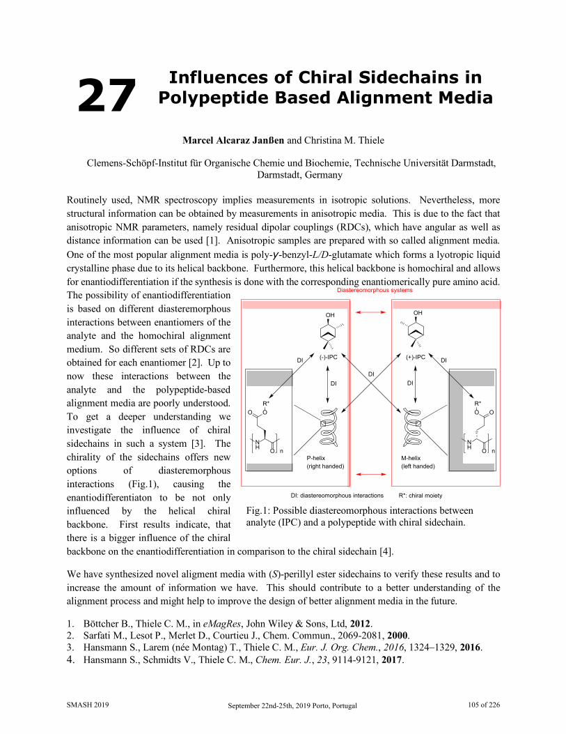
27 Influences of Chiral Sidechains in Polypeptide Based Alignment Media
Marcel Alcaraz Janßen and Christina M. Thiele
Clemens-Schöpf-Institut für Organische Chemie und Biochemie, Technische Universität Darmstadt, Darmstadt, Germany
Routinely used, NMR spectroscopy implies measurements in isotropic solutions. Nevertheless, more structural information can be obtained by measurements in anisotropic media. This is due to the fact that anisotropic NMR parameters, namely residual dipolar couplings (RDCs), which have angular as well as distance information can be used [1]. Anisotropic samples are prepared with so called alignment media. One of the most popular alignment media is poly-y-benzyl-L/D-glutamate which forms a lyotropic liquid crystalline phase due to its helical backbone. Furthermore, this helical backbone is homochiral and allows for enantiodifferentiation if the synthesis is done with the corresponding enantiomerically pure amino acid. The possibility of enantiodifferentiation is based on different diasteremorphous interactions between enantiomers of the analyte and the homochiral alignment medium. So different sets of RDCs are obtained for each enantiomer [2]. Up to now these interactions between the analyte and the polypeptide-based alignment media are poorly understood. To get a deeper understanding we investigate the influence of chiral sidechains in such a system [3]. The chirality of the sidechains offers new options of diasteremorphous interactions (Fig.1), causing the enantiodifferentiaton to be not only influenced by the helical chiral backbone. First results indicate, that there is a bigger influence of the chiral backbone on the enantiodifferentiation in comparison to the chiral sidechain [4].
We have synthesized novel aligment media with (S)-perillyl ester sidechains to verify these results and to increase the amount of information we have. This should contribute to a better understanding of the alignment process and might help to improve the design of better alignment media in the future.
1. Böttcher B., Thiele C. M., in eMagRes, John Wiley & Sons, Ltd, 2012. 2. Sarfati M., Lesot P., Merlet D., Courtieu J., Chem. Commun., 2069-2081, 2000. 3. Hansmann S., Larem (née Montag) T., Thiele C. M., Eur. J. Org. Chem., 2016, 1324–1329, 2016. 4. Hansmann S., Schmidts V., Thiele C. M., Chem. Eur. J., 23, 9114-9121, 2017.
Fig.1: Possible diastereomorphous interactions between analyte (IPC) and a polypeptide with chiral sidechain.
(-)-IPC
OH
(+)-IPC
OH
Diastereomorphous systems
DI: diastereomorphous interactions
NH O n
O
DIDIDI
DIDI
M-helix(left handed)
P-helix(right handed)
OR*
R*: chiral moiety
NH O n
OOR*
SMASH 2019 September 22nd-25th, 2019 Porto, Portugal 105 of 226
28 Improvement of Bioactive Metabolite Production in Microbial Cultures - A Systems Approach by
OSMAC and Deconvolution-Based 1H NMR Quantification
Denise Medeiros Selegato1, Rafael Teixeira Freire2, Alan César Pilon3, Carolina Rabal Biasetto1,
Haroldo Cesar de Oliveira4, Lucas Magalhães de Abreu5, Angela Regina Araujo1, Vanderlan da Silva Bolzani1, and Ian Castro-Gamboa1
1. Nucleus of Bioassays, Biosynthesis and Ecophysiology of natural products (NuBBE), Organic
Chemistry Depart., Institute of Chemistry, São Paulo State University (UNESP), Brazil 2. Medway Metabonomics Research Group, University of Greenwich, UK
3. Núcleo de Pesquisa em Produtos Naturais e Sintéticos (NPPNS), Faculdade de Ciências Farmacêuticas, São Paulo University (USP), Ribeirão Preto, SP, Brazil
4. Laboratório de Micologia Clínica, Núcleo de Proteômica, Faculdade de Ciências Farmacêuticas de Araraquara, São Paulo State University (UNESP), Araraquara, SP, Brazil
5. Departamento de Fisiopatologia, Universidade de Viçosa (UFV), Viçosa, MG, Brazil Traditionally, the screening of metabolites in microbial matrices is performed by monocultures [1]. Nonetheless, the absence of biotic and abiotic interactions generally observed in nature still limit the chemical diversity and leads to “poorer” chemical profiles. Nowadays, several methods have been developed to determine the conditions under which cryptic genes are activated, in an attempt to induce these silenced biosynthetic pathways. Among those, the One Strain Many Compounds (OSMAC) strategy has been applied to enhance metabolic production by a systematic variation of growth parameters [2-6]. The complexity of the chemical profiles from OSMAC experiments has required increasingly robust and accurate techniques. In this sense, deconvolution-based 1H NMR quantification have emerged as a promising methodology to decrease complexity and provide a comprehensive perspective for metabolomics studies [7-15]. Our present work shows an integrated strategy for the increased production and rapid quantification of compounds from microbial sources. Specifically, an OSMAC-Design of Experiments (DoE) was used to optimize the microbial production of bioactive fusaric acid, cytochalasin D and 3-nitropropionic acid, and Global Spectral Deconvolution (GSD)-based 1HNMR quantification was carried out for their measurement. The results showed that OSMAC increased the production of the metabolites by up to 33% and that GSD was able to extract accurate NMR integrals even in heavily coalescence spectral regions. Moreover, GSD-1HNMR quantification was reproducible for all species and exhibited validated results that were more selective and accurate than comparative methods. Overall, this strategy up-regulated the production of important metabolites using a reduced number of experiments and provided fast analyte monitor directly in raw extracts.
1. Schroeckh et al. Proc. Natl. Acad. Sci. (2009) 106, 14558–14563. 2. Bode et al. Chembiochem (2002) 3, 619–627. 3. Wei et al. Wei Sheng Wu Xue Bao (2010) 50, 701–709. 4. Romano et al. Mar. Drugs (2018) 16, 1–29. 5. Hussain et al. Bioorganic Med. Chem. Lett. (2017) 27, 2579–2582.
SMASH 2019 September 22nd-25th, 2019 Porto, Portugal 106 of 226

6. Bracarense and. Takahashi, Brazilian J. Microbiol. (2014) 45, 313–321. 7. Pauli et al. J. Nat. Prod. (2012) 75, 834–851. 8. Markley et al. Curr. Opin. Biotechnol. (2017) 43, 34–40. 9. Dagnino and Schripsema, Toxicon (2005) 46, 236–240. 10. Mehr et al. Anal. Chem. (2008) 80, 8320–8323. 11. Farrant et al. Magn. Reson. Chem. (2010) 48, 753–762. 12. Watanabe et al. Toxins (Basel). (2016) 8, 1–9. 13. Hong et al. J. Korean Magn. Reson. Soc. (2013) 17, 98–104. 14. Cobas et al., in:, SMASH Conf., Santa Fe, 2008. 15. Cobas et al., in:, 50th ENC Conf., Asilomar, 2009.
SMASH 2019 September 22nd-25th, 2019 Porto, Portugal 107 of 226
29 Solvation and Structural Transition of Melittin in
Trifluoroethanol/Water: A NMR Relaxometry and DNP Study
Bhawna Chaubey1, Arnab Dey2, Abhishek Banerjee2, N. Chandrakumar2, and Samanwita Pal1
1. Department of Chemistry, Indian Institute of Technology Jodhpur, Rajasthan, 342037, India
2. MRI-MRS Centre, Indian Institute of Technology Madras, Tamil Nadu, 600036, India Over the past several decades the solution state structural transition exhibited by Melittin (MLT), the bee venome peptide with potential antimicrobial and anticancer property [1] has been studied extensively with varying physical conditions such as temperature, pH and concentration using different analytical techniques [2-5]. However, the effect of solvent on such structural dynamics is yet to be explored cogently. In this context we present a brief account of a set of experiments based on NMR relaxometry and Dynamic Nuclear Polarization (DNP) studies to decipher the effect of trifluoroethanol (TFE) as a cosolvent for aqueous MLT solution. In accordance with the literature reports, Circular Dichroism (CD) experiments performed for aqueous MLT solution with TFE as cosolvent at two different physiological conditions demonstrated reversible monomer-tetramer transition [4-5]. This observation was complemented by 1H NMR spectra of MLT performed with same set of samples in terms of considerable linewidth and chemical shift changes of MLT protons. To analyze further, MLT structural transitions were characterized by observing the changes in the dynamical properties of TFE by employing 19F spin-lattice relaxation at 11.7 T and 0.35 T. Further, the steady state ODNP experiments using TEMPOL with large dynamical range [6] allowed us to comment on the change in TFE diffusion coefficient (D) calculated using TFE correlation time measured at 0.35T considering Hubbard’s isotropic interaction model. The choice of experiments performed at low field (0.35T) at one hand enabled us to mitigate the strong contribution of 19F CSA at high magnetic field while the ODNP experiments offered a window of probing molecular dynamics of the order of 10-1000 picoseconds timescale regime. The changes observed in diffusion coefficient of MLT in presence of TEMPOL as a function of TFE concentration can definitely be attributed to the gradual structural transition of MLT.
1. Liao, C. et al., J. Phys. Chem. B, 119, 10390–10398, 2015 2. Miuara, Y. Eur Biophys J, 41, 629–636, 2012 3. Neuman, R.C. et al., Magn. Reson. Chem., 47, 925–931, 2009 4. Roccatano, D.et al., PNAS, 99, 12179-12184, 2002 5. Othon, C. M. et al., PNAS, 106, 12593-12598, 2009 6. Banerjee, A.et al., Angew. Chem. Int. Ed., 55, 1-7, 2016
SMASH 2019 September 22nd-25th, 2019 Porto, Portugal 108 of 226

30 Determination of the Absolute Configuration of the Natural Product Commafric A by VCD, RDC, and RCSA
Fredrik Garnås Rylandsholm*1, Petr Bouř,2 Eva Brichtová2, 3, Aman Dekeboa4, and Johan Isaksson1.
1. Department of Chemistry, UiT The Arctic University of Norway, Tromsø, Norway
2. Institute of Organic Chemistry and Biochemistry, Academy of Sciences, Prague, Dejvice, Czech Republic
3. Department of Analytical Chemistry, University of Chemistry and Technology, Prague, Czech Republic
4. Department of Applied Chemistry, Adama Science and Technology University, Ethiopia
The structure of Commafric A (Fig. 1), a natural product extracted from the resin of the plant Commiphora africana, was structure elucidated by traditional NMR methods [1]. The absolute configuration could be determined relative to the biosynthetic precursor for all stereocenters except C-14, which could not be conclusively decided due to local flexibility. A combination of VCD, NOESY build-up, RDC and RCSA were successfully used to determine the absolute configuration of C-14.
In this work, the PBLG polymer system in chloroform recently proposed by Liu et al. [2] was used to create a liquid crystal that could increase the anisotropy with high precision and ease of use. The result allowed us to reject the 14R configuration in favour of the 14S configuration.
Fig. 1: Stereochemical differentiation between the two possible configurations of C-14 in Commafric A using RDC and RCSA, showing that S is the preferred conformer.
HOH
OHO
H(R)
H2COH
Commafric AHO
H
OHO
H(S)
H2COH
Commafric A
-60-40-20
020406080
100120
-60 -40 -20 0 20 40 60 80 100 120
Back
-cal
cula
ted
(Hz)
Experimental (Hz)
RDC
RCSA
Q-fac: 0.33
-60-40-20
020406080
100120
-60 -40 -20 0 20 40 60 80 100 120
Back
-cal
cula
ted
(Hz)
Experimental (Hz)
RDC
RCSA
Q-fac: 0.19
SMASH 2019 September 22nd-25th, 2019 Porto, Portugal 109 of 226

1. Dinku, W., Isaksson, J., Rylandsholm, F. G., Bouř, P., Brichtová, E., Choi, S. U., Lee, S. H., Jung, Y. S., Sung, Z., Svendsen, J. S. M., Aasen, A. J., & Dekebo, A. Commafric A, (3S,4S,14S,7E,17E,21Z)-3-hydroxypodida-7,17,21-trien-4-carboxylic acid, a new triterpenoid with moderate cytostatic activity isolated from the resin of Commiphora Africana. Submitted manuscript, J. Nat. Prod.
2. Liu, Y., Cohen, R. D., Gustafson, K. R., Martin, G. E., & Williamson, R. T. Enhanced measurement of residual chemical shift anisotropy for small molecule structure elucidation. Chem. Commun., 54, 4254-4257, 2018.
SMASH 2019 September 22nd-25th, 2019 Porto, Portugal 110 of 226
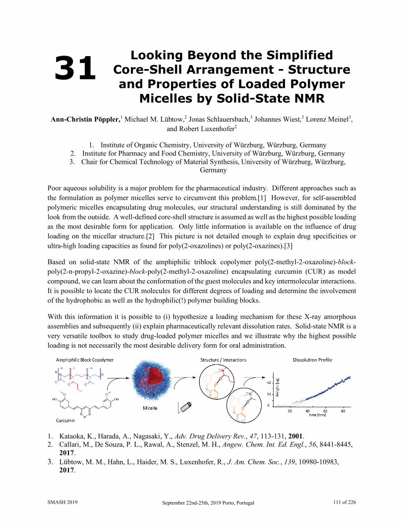
31 Looking Beyond the Simplified Core-Shell Arrangement - Structure and Properties of Loaded Polymer
Micelles by Solid-State NMR Ann-Christin Pöppler,1 Michael M. Lübtow,2 Jonas Schlauersbach,3 Johannes Wiest,3 Lorenz Meinel3,
and Robert Luxenhofer2
1. Institute of Organic Chemistry, University of Würzburg, Würzburg, Germany 2. Institute for Pharmacy and Food Chemistry, University of Würzburg, Würzburg, Germany 3. Chair for Chemical Technology of Material Synthesis, University of Würzburg, Würzburg,
Germany Poor aqueous solubility is a major problem for the pharmaceutical industry. Different approaches such as the formulation as polymer micelles serve to circumvent this problem.[1] However, for self-assembled polymeric micelles encapsulating drug molecules, our structural understanding is still dominated by the look from the outside. A well-defined core-shell structure is assumed as well as the highest possible loading as the most desirable form for application. Only little information is available on the influence of drug loading on the micellar structure.[2] This picture is not detailed enough to explain drug specificities or ultra-high loading capacities as found for poly(2-oxazolines) or poly(2-oxazines).[3]
Based on solid-state NMR of the amphiphilic triblock copolymer poly(2-methyl-2-oxazoline)-block-poly(2-n-propyl-2-oxazine)-block-poly(2-methyl-2-oxazoline) encapsulating curcumin (CUR) as model compound, we can learn about the conformation of the guest molecules and key intermolecular interactions. It is possible to locate the CUR molecules for different degrees of loading and determine the involvement of the hydrophobic as well as the hydrophilic(!) polymer building blocks.
With this information it is possible to (i) hypothesize a loading mechanism for these X-ray amorphous assemblies and subsequently (ii) explain pharmaceutically relevant dissolution rates. Solid-state NMR is a very versatile toolbox to study drug-loaded polymer micelles and we illustrate why the highest possible loading is not necessarily the most desirable delivery form for oral administration.
1. Kataoka, K., Harada, A., Nagasaki, Y., Adv. Drug Delivery Rev., 47, 113-131, 2001. 2. Callari, M., De Souza, P. L., Rawal, A., Stenzel, M. H., Angew. Chem. Int. Ed. Engl., 56, 8441-8445,
2017. 3. Lübtow, M. M., Hahn, L., Haider, M. S., Luxenhofer, R., J. Am. Chem. Soc., 139, 10980-10983,
2017.
SMASH 2019 September 22nd-25th, 2019 Porto, Portugal 111 of 226

32 NMR Aerosolomics as a Tool to Distinguish Various Types of
Aerosols
Štěpán Horník1,2, Jaroslav Schwarz1, Vladimír Ždímal1, and Jan Sýkora1
1. Institute of Chemical Process Fundamentals of the CAS, v. v. i., Prague, Czech Republic 2. University of Chemistry and Technology, Department of Analytical Chemistry, Prague, Czech
Republic Atmospheric aerosols are a small but very important part of the Earth’s atmosphere. They have an important influence on climate forcing through direct, indirect and semi-direct effects [1]. At the same time, aerosol particles have various detrimental effects on human health [2]. Polluted air can cause many diseases, especially those of respiratory tract and cardiovascular system. The proportion of inorganic and organic compounds in aerosol particles seems to be equal on average [3]. While the inorganic composition of aerosols is well explored, knowledge about the organic part is still very limited. It is well known that the major part of organic aerosol compounds is represented by polar, water-soluble organic compounds (WSOC) [3]. NMR spectroscopy was for the purpose of aerosol chemistry “discovered“ only recently [4], but its potential has not yet been fully exploited.
Aerosolomics provides complex evaluation of aerosol composition and compound concentration. It is exploiting metabolomic approach applied to aerosol samples. In NMR aerosolomics, the assignment of dominant signals is based on precise chemical shift of the compound which enables identification of organic compounds in given aerosol sample. For this purpose, a comprehensive library of 1H NMR spectra of organic compounds known to be present in aerosol particles is essential. The database of the ChenomX NMR Suite program contains about 70 compounds that have also been found in aerosol samples according to the literature. Up to now, 50 new compounds attributed to aerosol have been added to the database; the largest gap was found in aromatic carboxylic acids (12), compounds with sulphur (11) and amines (8). Additionally, about 30 new organic compounds (mainly hydroxyl carboxylic acids) were found in aerosol samples. These compounds were present in the original ChenomX library and have not been identified in aerosol samples yet.
In the recent study, the summer and winter aerosol samples were analyzed using NMR aerosolomics approach. The samples were collected in Prague-Suchdol during summer 2008 and winter 2009 in two different particle size fractions – PM2.5 and PM10. Around 50 compounds were identified in each aerosol spectrum owing to the comprehensive library. The profile of 86 compounds identified in the samples altogether served as an input data for statistical analysis. Multivariate statistical analysis clearly discriminates the two groups studied. Furthermore, the most significant compounds varying between seasons were determined by the means of univariate statistical analysis. These compounds were subjected to further analysis of their possible sources.
This work was supported by Technology Agency of the Czech Republic project (TK02010035) and by Large Research Infrastructures project of the MEYS of the Czech Republic ACTRIS-CZ, project No. LM2015037.
SMASH 2019 September 22nd-25th, 2019 Porto, Portugal 112 of 226

1. Charlson, R.J., Schwartz, S.E., Hales, J.M., et al., Science 256, 598-599, 1992. 2. Dockery, D.W., Pope, C.A., Xu, X., et al., N. Engl. J. Med. 329, 1753-1759, 1993. 3. Saxena, P., Hildemann, L.M., J. Atmos. Chem., 24(1), 57-109, 1996. 4. Decesari, S., Facchini, M.C., Fuzzi, S., Tagliavini, E., J. Geophys. Res., [Atmos.], 105, 1481–1489,
2000.
SMASH 2019 September 22nd-25th, 2019 Porto, Portugal 113 of 226

33 NMR-Spectroscopic Investigations in Iminium Ion Catalysis
Willibald Stockerl and Ruth M. Gschwind*
Department of Organic Chemistry, University of Regensburg, Regensburg, Germany Iminium ion catalysis stands out on account of highly stereoselective reactions using uncomplicated procedures.[1] Starting from α,β-unsaturated carbonyl compounds, a lot of different addition reactions, for example cycloadditions [2], epoxidations [3] and 1,4-addition reactions with a large number of different nucleophiles [4,5,6,7] and in high enantioselectivity were elaborated in the last decades.
To the best of our knowledge, most studies on the origin of stereoselectivity are based on theoretical calculations. These arrive at the conclusion that a configurational preference of the iminium ion double bonds combined with a fixed approach pathway of the reactant is responsible for the stereochemical outcome.[8,9] In 2017, our group developed a method to gain experimental insight in enantioselective addition reactions by decrypting transition states by light (DTS-hν).[10] Here, illumination with light shifts the population of differently configurated isomers respectively configurational preferences. The different isomer-ratios result in a characteristic change of ee and/or reaction rate, hence an experimental verification of the theoertical calculations is possible.
Isomerization of the two double bonds of α,β-unsaturated iminium ions yields up to four different configuration isomers, (E)(E), (Z)(E), (E)(Z) and (Z)(Z). Simultaneous photoinduced isomerization and structural assignment of the different isomers is possible by using an LED based in situ NMR illumination device.[11] We herein present a method for selective isomerization of the two double bonds by using different temperatures and illumination modes. Illuminating at low temperature can decelerate the thermal backisomerization of certain isomers to shift the ratio of the isomers with respect to each other.
1. Bertelsen, S. & Jørgensen, K. A., Chem. Soc. Rev. 38, 2178–2189 (2009). 2. Ahrendt, K. A., Borths, C. J. & MacMillan, D. W., J. Am. Chem. Soc. 122, 4243–4244 (2000). 3. Tanzer, E. M., Zimmer, L. E., Schweizer, W. B. & Gilmour, R., Chem. - A Eur. J. 18, 11334–
11342 (2012). 4. Bertelsen, S., Dine, P., Johansen, R. L. & Jørgensen, K. A., J. Am. Chem. Soc. 129, 1536–1537
(2007). 5. Brandau, S., Landa, A., Franzøn, J., Marigo, M. & Jørgensen, K. A., Angew. Chemie Int. Ed. 45,
4305–4309 (2006). 6. Marigo, M., Schulte, T., Franzen, J. & Jørgensen, K. A., J. Am. Chem. Soc. 127, 15710–15711
(2005). 7. Li, T. et al., Chem. - A Eur. J. 19, 9147–9150 (2013). 8. Dinér, P., Nielsen, M., Marigo, M. & Jørgensen, K. A., Angew. Chemie - Int. Ed. 46, 1983–1987
(2007). 9. Seebach, D. et al., Helv. Chim. Acta 93, 603–634 (2010). 10. Renzi, P., Hioe, J. & Gschwind, R. M., J. Am. Chem. Soc. 139, 6752–6760 (2017). 11. Feldmeier, C., Bartling, H., Riedle, E. & Gschwind, R. M., J. Magn. Reson. 232, 39–44 (2013).
SMASH 2019 September 22nd-25th, 2019 Porto, Portugal 114 of 226
34 Early Detection of Pancreatic Cancer: 1H NMR Metabolomics Study of its Connection with Diabetes Mellitus
Lenka Michálková1,2, Štěpán Horník1,2, Lucie Habartová1, Jan Sýkora2 and Vladimír Setnička1
1. Department of Analytical Chemistry, University of Chemistry and Technology Prague, Prague 6, Czech Republic
2. Department of Analytical Chemistry, Institute of Chemical Process and Fundamentals of the CAS v. v. i., Prague 6, Czech Republic
High mortality places pancreatic cancer (PC) as the fourth cause of cancer-associated death; more than 95% of all patients will succumb to PC within 2 years of diagnosis [1,2]. PC is expected to become the second most common cause of death by 2030, and the first by 2050 [3]. PC diagnosis is often late due to nonspecific symptoms, and radical surgery is then performed in only 15–20% of cases [1].
Epidemiological studies [1,4] clearly show that patients with diabetes mellitus (DM) are at increased risk of PC development (75% of patients are diagnosed with DM), but the relationship is much more complex. The highest risk of PC development afflicts patients suffering from the so-called pancreatogenic diabetes, which is classified as type 3c diabetes mellitus (T3cDM). Pancreatogenic diabetes is a secondary form of diabetes and, as such, it is a consequence of pancreatic exocrine pathology [1]. According to recent data [1], the prevalence of T3cDM can be around 30% among individuals with new-onset DM. The interval between new-onset diabetes and PC manifestation is 2–3 years [1]. Thus, distinguishing T3cDM in new-onset diabetics may facilitate the identification of patients with tumour at the curable stage (resectable tumour), thereby improving the prognosis of this fatal disease [1]. However, there is a problem with T3cDM identification among new-onset diabetics, up to 90% of which are misdiagnosed with type 2 diabetes mellitus (T2DM) [5].
In this work, we tested the strategy for the detection of T3cDM and early PC based on NMR metabolomics. 1H NMR spectroscopy was used to detect changes in the concentration of low molecular metabolites in the blood plasma of four groups: PC patients, long-term T2DM (lasting more than 5 years), new-onset diabetic patients (lasting less than 3 years, potential T3cDM) and healthy controls. The ChenomX software was used to identify 65 metabolites across all samples. Based on the concentrations of the identified metabolites, methods of pattern recognition (primarily PCA and OPLS-DA) and statistical tests (t-test and Wilcoxon rank sum test) were used to differentiate between the studied groups. The obtained results suggested a panel of biomarkers for the detection of PC. Within the group of new-onset diabetics, our proposed OPLS-DA model, (verified by Monte Carlo cross-validation), was able to predict possible T3cDM patients at risk of developing PC. Regarding such results, the suggested model might serve as a screening method for the early detection of PC.
Acknowledgment: The authors thank Prof. Miroslav Zavoral and Dr. Bohuš Bunganič from the Department of Gastrointestinal Endoscopy at the Internal Clinic of the Military University Hospital and the First Faculty of Medicine of the Charles University in Prague, Czech Republic for providing the plasma
SMASH 2019 September 22nd-25th, 2019 Porto, Portugal 115 of 226
samples. The work was realized within grant no. 16-31028A provided by the Ministry of Health of the Czech Republic and Specific University Research MSMT no. 21-SVV/2019.
1. Cui, Y.; Andersen, D. K., Endocrine Related Cancer, 19 (5), F9-F26, 2012. 2. Edwards, B. K.; Brown, M. L.; Wingo, P. A.; Howe, H. L.; Ward, E.; Ries, L. A. G.; Schrag, D.;
Jamison, P. M.; Jemal, A.; Wu, X. C.; Friedman, C.; Harlan, L.; Warren, J.; Anderson, R. N.; Pickle, L. W., JNCI: Journal of the National Cancer Institute, 97 (19), 1407-1427, 2005.
3. Siegel, R. L.; Miller, K. D.; Jemal, A., CA: A Cancer Journal for Clinicians, 67 (1), 7-30, 2017. 4. Pannala, R.; Basu, A.; Petersen, G. M.; Chari, S. T., The Lancet Oncology, 10 (1), 88-95, 2009. 5. Ewald, N.; Bretzel, R. G., European Journal of Internal Medicine, 24 (3), 203-206, 2013.
SMASH 2019 September 22nd-25th, 2019 Porto, Portugal 116 of 226
35 Mechanistic Investigations of the Direct Photocatalytic Transformation
of P4 into Aryl Phosphines and Phosphonium Salts
Verena Streitferdt, Ulrich Lennert, Percia Beatrice Arockiam, Daniel J. Scott, Christian Rödl, Robert
Wolf, and Ruth M. Gschwind*
Department of Chemistry and Pharmacy, University of Regensburg, Regensburg, Germany Organophosphorus compounds find extensive uses across the chemical industry. White phosphorus (P4) is the common precursor in the synthesis of nearly all these valuable and important molecules.[1] Yet current state-of-the-art processes for P4 functionalisation rely on chlorinated intermediates such as PCl3, PCl5, and POCl3, whose production requires oxidation of P4 with toxic chlorine gas.[2] Displacement of chloride must then be performed in a subsequent step.[2] New methods for the direct functionalization of P4 avoiding this chlorination step are therefore highly desirable. Although P4 activation and functionalization by main group elements and transition metal (TM) compounds has been studied for decades,[3,4] catalytic TM-mediated P4 functionalization reactions have remained elusive.
We herein present mechanistic investigations of the first efficient catalytic process being capable of forming P—C bonds from P4. Using an iridium catalyst and blue LED light, this visible-light-driven procedure affords useful tetraarylphosphonium salts and triarylphosphanes in good to moderate yields. Fluorescence quenching, UV-VIS, spectro electro chemistry, cyclic voltammetry experiments and in situ 1H, 2H and 31P NMR reaction monitoring helped to reveal key parts of the mechanism of this highly valuable catalytic functionalization of P4.
1. Borger, J. E., Ehlers, A. W., Slootweg, J. C. & Lammertsma, K. Chem. - A Eur. J. 23, 11738–11746 (2017).
2. Corbridge, D. E. C., Phosphorus 2000. Chemistry, Biochemistry and Technology (Elsevier: Amsterdam. 2000).
3. Caporali, M., Gonsalvi, L., Rossin, A. & Peruzzini, M. P 4 Chem. Rev. 110, 4178–4235 (2010). 4. Scheer, M., Balázs, G. & Seitz, A. Chem. Rev. 110, 4236–4256 (2010).
SMASH 2019 September 22nd-25th, 2019 Porto, Portugal 117 of 226
36 Methods to Improve Resolution in 1H Solid State NMR at Ultra-Fast MAS
Pinelopi Moutzouri, Federico M. Paruzzo, Lyndon Emsley
Institut des Sciences et Ingénierie Chimiques, Ecole Polytechnique Fédérale de Lausanne (EPFL),
CH-1015 Lausanne, Switzerland In organic solids the homogeneous broadening caused by the strong 1H-1H dipolar coupling network yields 1H linewidths of few tens of kHz, orders of magnitude larger than those in solution and leads to broad and highly unresolved spectra from which very little information can be extracted.
Magic angle spinning (MAS)(1) can greatly improve spectral resolution. Recently, newly developed technology allows MAS rates greater than 100 kHz leading to 1H linewidths on the order of a few hundred Hz. However, in many applications and especially for complex systems, this is still not sufficient and limits the use of 1H in solid state NMR.
Here we explore new methods to further improve the spectral resolution of powdered organic solids in this fast spinning rate regime, and we show how to obtain experimental linewidths up to a factor of two narrower than those achieved with a conventional experiment at the same MAS rate.
1. Andrew, E.R., et al., Nature, 1958, 182, 1659-1659; Lowe, I.J., Phys. Rev. Lett., 1959, 2, 285-287
SMASH 2019 September 22nd-25th, 2019 Porto, Portugal 118 of 226
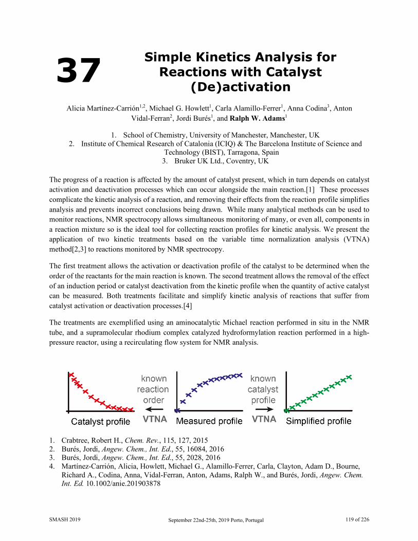
37 Simple Kinetics Analysis for Reactions with Catalyst
(De)activation
Alicia Martínez-Carrión1,2, Michael G. Howlett1, Carla Alamillo-Ferrer1, Anna Codina3, Anton Vidal-Ferran2, Jordi Burés1, and Ralph W. Adams1
1. School of Chemistry, University of Manchester, Manchester, UK 2. Institute of Chemical Research of Catalonia (ICIQ) & The Barcelona Institute of Science and
Technology (BIST), Tarragona, Spain 3. Bruker UK Ltd., Coventry, UK
The progress of a reaction is affected by the amount of catalyst present, which in turn depends on catalyst activation and deactivation processes which can occur alongside the main reaction.[1] These processes complicate the kinetic analysis of a reaction, and removing their effects from the reaction profile simplifies analysis and prevents incorrect conclusions being drawn. While many analytical methods can be used to monitor reactions, NMR spectrocopy allows simultaneous monitoring of many, or even all, components in a reaction mixture so is the ideal tool for collecting reaction profiles for kinetic analysis. We present the application of two kinetic treatments based on the variable time normalization analysis (VTNA) method[2,3] to reactions monitored by NMR spectrocopy.
The first treatment allows the activation or deactivation profile of the catalyst to be determined when the order of the reactants for the main reaction is known. The second treatment allows the removal of the effect of an induction period or catalyst deactivation from the kinetic profile when the quantity of active catalyst can be measured. Both treatments facilitate and simplify kinetic analysis of reactions that suffer from catalyst activation or deactivation processes.[4]
The treatments are exemplified using an aminocatalytic Michael reaction performed in situ in the NMR tube, and a supramolecular rhodium complex catalyzed hydroformylation reaction performed in a high-pressure reactor, using a recirculating flow system for NMR analysis.
1. Crabtree, Robert H., Chem. Rev., 115, 127, 2015 2. Burés, Jordi, Angew. Chem., Int. Ed., 55, 16084, 2016 3. Burés, Jordi, Angew. Chem., Int. Ed., 55, 2028, 2016 4. Martínez-Carrión, Alicia, Howlett, Michael G., Alamillo-Ferrer, Carla, Clayton, Adam D., Bourne,
Richard A., Codina, Anna, Vidal-Ferran, Anton, Adams, Ralph W., and Burés, Jordi, Angew. Chem. Int. Ed. 10.1002/anie.201903878
SMASH 2019 September 22nd-25th, 2019 Porto, Portugal 119 of 226
38 A Novel Method Using NMR for Plasma Protein Binding Assessment
in Drug Discovery Programs Mariana Gallo1, Sara Matteucci2, Nadine Alaimo2, Erica Pitti2, Maria V. Orsale1, Vincenzo Summa1,
Daniel O. Cicero1,2, and Edith Monteagudo1
1. IRBM Science Park S.p.A, Pomezia, Rome, Italy 2. Dipartimento di Scienze e Tecnologie Chimiche, Università di Roma “Tor Vergata”, Rome, Italy
The amount of plasma-bound drug influences compound dosing, efficacy, clearance rate, and potential for drug interactions. Therefore, having information about the extent to which a drug candidate binds to plasma proteins is a critical feature of drug development. A new methodology based on Nuclear Magnetic Resonance (NMR) was developed to determine plasma protein binding (PPB) of drug candidates in drug discovery programs [1].
Recognizing the utility of NMR as a very sensitive method for detecting binding, we have focused on developing an approach particularly fast and, if an NMR spectrometer is available, low cost alternative to those currently used for determining PPB. We have used the fastest and simplest NMR method for evaluating protein binding, the traditional 1D 1H line-broadening experiment, which additionally allows by a single-point measurement easily rank binding affinities [2]. A strong correlation was found between the attenuation of NMR signals of diverse drugs in the presence of different plasma concentrations and their fraction bound (fb) reported in the literature. Based on these results, a protocol for a rapid calculation of fb of small molecules was established. The advantage of using plasma instead of purified recombinant proteins and the possibility of pool analysis to increase throughput were also evaluated. This novel methodology proved to be very versatile, cost-effective, fast and suitable for automation. As a plus, it contemporarily provides a quality check and solubility of the compound.
1. Gallo M., Matteucci S., Alaimo N., Pitti E., Orsale M.V., Summa V., Cicero D.O., Monteagudo E., J. Pharm. Biomed. Anal., 167, 21-29 (2019)
2. Shortbridge M.D. Hage D.S., Harbison, G.S., Powers R., J. Comb. Chem., 10, 948-958 (2008)
SMASH 2019 September 22nd-25th, 2019 Porto, Portugal 120 of 226

39 High Temperature NMR Studies of Diffusivity and Molecular
Interactions in Pharmaceutical Hot-melt Extrusion Formulations
Elena Pisa1, Mick D. Mantle1, and Leslie P. Hughes2
1. Department of Chemical Engineering and Biotechnology, University of Cambridge, UK 2. Global Product Development, AstraZeneca, Macclesfield, UK
Formulation of drug/polymer amorphous solid dispersions (ASDs) is one successful strategy for improving dissolution and absorption of poorly soluble active pharmaceutical ingredients (APIs). ASDs lack the long range ordering present in crystalline solids, and thus do not suffer from having to overcome high lattice energies or poor solvation effects during dissolution. Hot-melt extrusion (HME), which relies on high temperature and high shear environments is one of the most popular methods of ASD preparation and it is the focus of this work.
This work presents several novel applications of NMR at high temperature and fast-MAS to improve our understanding of API (paracetamol) behavior in polymer (copovidone) melts. Pulsed-field gradient NMR and 1D NMR imaging techniques provide information on the mass transport at a microscopic and macroscopic scale respectively, at temperatures relevant to HME. Solid state NMR (ssNMR) is used to probe the amorphous state of the API and the microscopic structure of the extruded product (phase separation). Preliminary results from high-field (850 MHz) fast-MAS (60 kHz) 1H-1H ssNMR (BABA) and high-tempeature (170°C) solution-state 2D spectroscopy (COSY), give an insight into intermolecular interactions.
1D NMR imaging shows that there is a significant depression in the melting point of paracetamol (>50°C) in the presence of copovidone, as is typical in miscible systems. There is also a positive correlation between paracetamol’s diffusivity in molten copovidone with both temperature and drug loading (Fig. 1A). The API molecular self-diffusivity in systems of 20% drug loading is two orders of magnitude slower than pure molten paracetamol at 170°C. This suggests that polymer behavior and paracetamol/copovidone interactions restrict API mobility. The quantitative information from 1H T1 and T1ρ relaxation experiments shows that paracetamol/copovidone extruded products (at different temperature, screw speed and drug loading) all have API domain sizes smaller than the limit of detection (3.5 nm). 2D 1H-1H BABA (Fig. 1B) as well as high-temperature COSY (Fig. 1C) experiments indicate the presence of intermolecular hydrogen bonds.
SMASH 2019 September 22nd-25th, 2019 Porto, Portugal 121 of 226
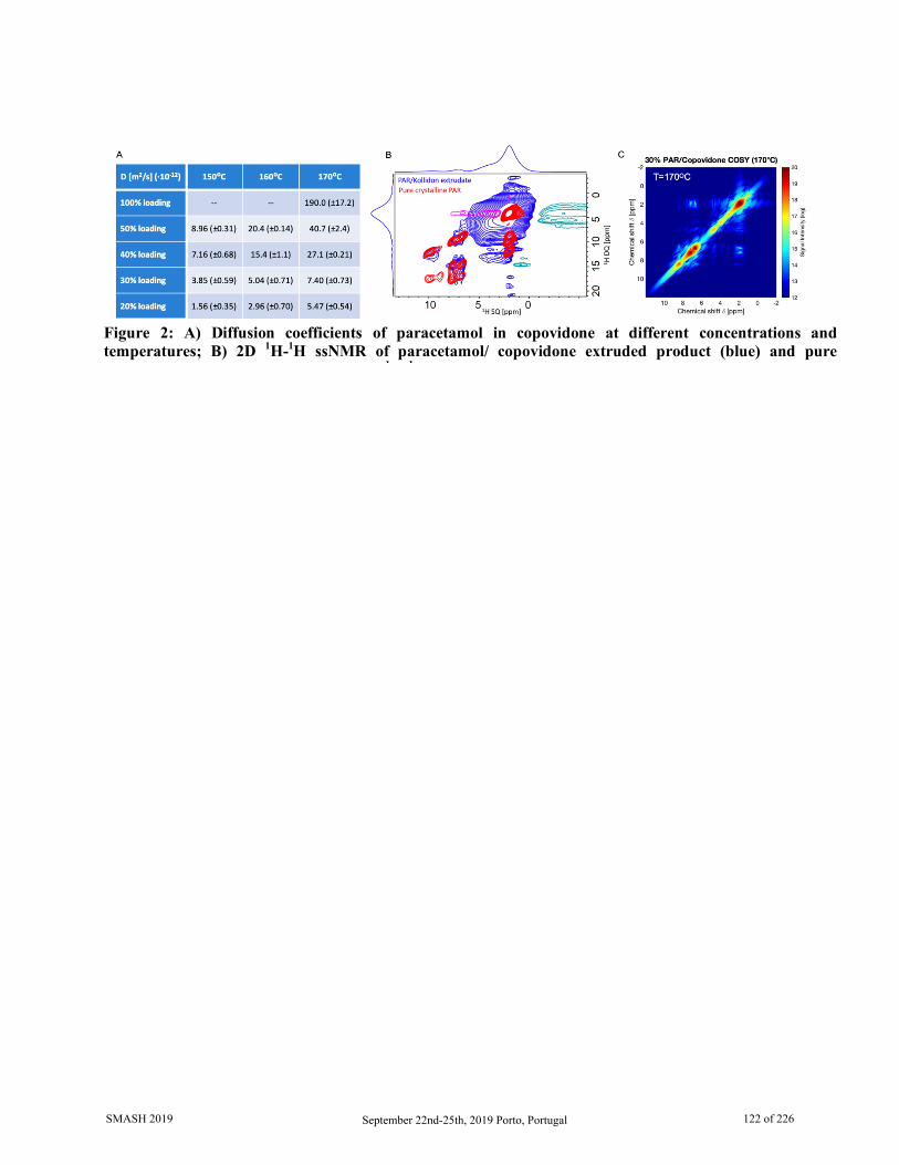
Figure 2: A) Diffusion coefficients of paracetamol in copovidone at different concentrations and temperatures; B) 2D 1H-1H ssNMR of paracetamol/ copovidone extruded product (blue) and pure
1 1
SMASH 2019 September 22nd-25th, 2019 Porto, Portugal 122 of 226
40 Peaks Pattern Recognition (PPR) System: An Application to NMR
Metabolite Identification
Rafael T. Freire,1 Denise M. Selegato2 and Santiago Marco1
1. Signal and Information Processing for Sensing Systems. Institute for BioEngineering of Catalonia. Barcelona, Catalonia, Spain
2. Nucleus of Bioassays, Biosynthesis and Ecophysiology of natural products, Organic Chemistry Depart., Institute of Chemistry
Abstract: In science, peaks values are used to interpret a specific information given by its dimensions. The interpretation of peaks is used in any field, from economics to physics, medicine and chemistry. In chemistry, depending on the choice of the experiment, peaks may be related to chemical structures. The NMR analysis is one of the most important techniques to identify a compound and its complexity increases with the complexity of the chemical structure. One of the most exhaustive and difficult steps in structural elucidation and identification of compounds is to correlate the peaks with a molecule in order to obtain candidates that matches to the observed peaks in the spectra. There are many techniques to help with rapid characterization of known compounds (dereplication), but the lack of organized data is one of the major drawbacks in natural products and medicinal chemistry research. In addition, if a compound is not presented in the database, erroneous information is retrieved by the user in which makes the task even more difficult. Based on this bottleneck, we developed a new fingerprint and patented method of peak analysis and pattern recognition [1] which takes into consideration the intuitive idea of how close the peaks from different samples are, more similar the information provided is. This new technique is divided in four stages: 1) Identify similar peaks between analysis, 2) Score a similarity value between two analysis; 3) Creates a direct and an inverse similarity profile and 4) Creates a similarity matrix to be statistically and mathematically treated. We demonstrate the power of the algorithm on nuclear magnetic resonance spectroscopy data. The data was download from online database, such as BMRB, MMCD and HMDB comprising a total of 15.496 analysis divided in 1H, 13C and 13C-HSQC with an information of more than 500.000 peaks. The result of this algorithm, applied to NMR analyses presented in the database, allowed us to group molecules according to their chemical structures in clusters (HCA), networks and in multidimensional spaces (PCA) facilitating the elucidation and identification process of chemical compounds even if it is not presented in the database. In the future, it will be able to incorporate analysis from any kind of detector, once this algorithm can be applied to any analysis that generate peaks (NMR, Mass spectroscopy, IV, UV, LC, and etc). This will make the results more robust and reliable by integrating all the spectral information into a single matrix. Finally, the objectives proposed in this research were achieved by the interdisciplinary collaboration in the areas of computational, statistical, spectroscopy, and chemistry.
1. Freire R. T., inventor; Gamboa-Castro I., inventor; Universidade Estadual Julio deMesquita Filho-UNESP., tranferee. Method of analysis' pattern recognition and computer program. World Intellectual Property Organization (WIPO) WO2016183647A1 PCT/BR2015/000075 Mar 18 2015.
SMASH 2019 September 22nd-25th, 2019 Porto, Portugal 123 of 226

41 Coldprobes, NUS & Automated Analysis: Revitalising INADEQUATE
David A. Bennett1, Claire Dickson2, Craig P. Butts2 and Iain J. Day1
1. School of Life Sciences, University of Sussex, Falmer, Brighton, UK
2. School of Chemistry, University of Bristol, Bristol, UK The Incredible Natural Abundance DoublE QUAntum Transfer Experiment (INADEQUATE [1,2]) is an elegant and powerful tool for structural determination in small molecules. In principle, all carbon-carbon bonds can be found and identified in a single experiment! However, INADEQUATE is rarely used as it requires both the carbons in any given bond to be 13C. This means that, at natural abundance, only around 1 in 8000 bonded carbon-carbon pairs in the sample actually contribute to the final signal. Even at 1M concentrations, the experiment can take days using routine spectrometer hardware.
Here, we demonstrate the effectiveness of modern techniques at overcoming this difficulty: the signal-to-noise ratio was boosted by a carbon-detection optimised coldprobe, the experiment time was reduced with Non-Uniform Sampling (NUS), and the final results were analysed using a modern implementation of the automated analysis strategy by Grant and coworkers [3,4]. This allows a computer to search only the meaningful areas of the spectrum (using the full hypercomlex data-set instead of just the real-real part which is traditionally displayed) and from this determine which carbon sites are bonded, as well as returning the corresponding J-couplings and secondary isotope shifts.
Combined, this allowed the full, unsupervised identification of all carbon-carbon bonds in our test molecule (Strychnine) in under 12 hours (10 mg in a standard 5mm NMR tube) or 48 hours when testing a mass-limited case (4 mg in a susceptibility-matched Shigemi tube).
1. A. Bax, R. Freeman and S. P. Kempsell, J. Am. Chem. Soc. 102, 4849-4851, 1980 2. A. Bax, R. Freeman and T. A. Frenkiel, J. Am. Chem. Soc. 103, 2102-2104, 1981 3. R. Dunkel, C. L. Mayne, J. Curtis, R. J. Pugmire and D. M. Grant, J. Magn. Reson. 90, 290-302, 1990 4. R. Dunkel, C. L. Mayne, R. J. Pugmire and D. M. Grant, Anal. Chem. 64, 3133-3149, 1992
SMASH 2019 September 22nd-25th, 2019 Porto, Portugal 124 of 226
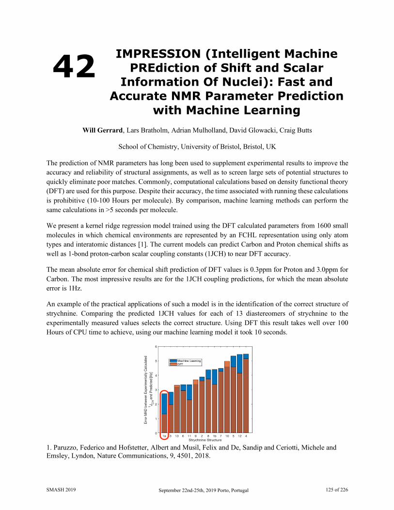
42 IMPRESSION (Intelligent Machine PREdiction of Shift and Scalar
Information Of Nuclei): Fast and Accurate NMR Parameter Prediction
with Machine Learning
Will Gerrard, Lars Bratholm, Adrian Mulholland, David Glowacki, Craig Butts
School of Chemistry, University of Bristol, Bristol, UK The prediction of NMR parameters has long been used to supplement experimental results to improve the accuracy and reliability of structural assignments, as well as to screen large sets of potential structures to quickly eliminate poor matches. Commonly, computational calculations based on density functional theory (DFT) are used for this purpose. Despite their accuracy, the time associated with running these calculations is prohibitive (10-100 Hours per molecule). By comparison, machine learning methods can perform the same calculations in >5 seconds per molecule.
We present a kernel ridge regression model trained using the DFT calculated parameters from 1600 small molecules in which chemical environments are represented by an FCHL representation using only atom types and interatomic distances [1]. The current models can predict Carbon and Proton chemical shifts as well as 1-bond proton-carbon scalar coupling constants (1JCH) to near DFT accuracy.
The mean absolute error for chemical shift prediction of DFT values is 0.3ppm for Proton and 3.0ppm for Carbon. The most impressive results are for the 1JCH coupling predictions, for which the mean absolute error is 1Hz.
An example of the practical applications of such a model is in the identification of the correct structure of strychnine. Comparing the predicted 1JCH values for each of 13 diastereomers of strychnine to the experimentally measured values selects the correct structure. Using DFT this result takes well over 100 Hours of CPU time to achieve, using our machine learning model it took 10 seconds.
1. Paruzzo, Federico and Hofstetter, Albert and Musil, Felix and De, Sandip and Ceriotti, Michele and Emsley, Lyndon, Nature Communications, 9, 4501, 2018.
SMASH 2019 September 22nd-25th, 2019 Porto, Portugal 125 of 226
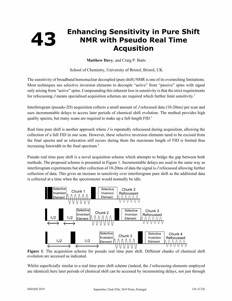
43 Enhancing Sensitivity in Pure Shift NMR with Pseudo Real Time
Acqusition
Matthew Davy, and Craig P. Butts
School of Chemistry, University of Bristol, Bristol, UK The sensitivity of broadband homonuclear decoupled (pure shift) NMR is one of its overarching limitations. Most techniques use selective inversion elements to decouple “active” from “passive” spins with signal only arising from “active” spins. Compounding this inherent loss in sensitivity is that the strict requirements for refocussing J means specialised acquisition schemes are required which further limit sensitivity.1 Interferogram (pseudo-2D) acquisition collects a small amount of J refocused data (10-20ms) per scan and uses incrementable delays to access later periods of chemical shift evolution. The method provides high quality spectra, but many scans are required to make up a full-length FID.1 Real time pure shift is another approach where J is repeatedly refocussed during acquisition, allowing the collection of a full FID in one scan. However, these selective inversion elements need to be excised from the final spectra and as relaxation still occurs during them the maximum length of FID is limited thus increasing linewidth in the final spectrum.2 Pseudo real time pure shift is a novel acquisition scheme which attempts to bridge the gap between both methods. The proposed scheme is presented in Figure 1. Incrementable delays are used in the same way as interferogram experiments but after collection of 10-20ms of data the signal is J refocussed allowing further collection of data. This gives an increase in sensitivity over interferogram pure shift as the additional data is collected at a time when the spectrometer would normally be idle.
Figure 1: The acquisition scheme for pseudo real time pure shift. Different chunks of chemical shift evolution are accessed as indicated. Whilst superficially similar to a real time pure shift scheme (indeed, the J refocussing elements employed are identical) here later periods of chemical shift can be accessed by incrementing delays, not just through
SMASH 2019 September 22nd-25th, 2019 Porto, Portugal 126 of 226
use of more J refocussing elements which require excision. This allows later periods of chemical shift evolution to be accessed, meaning that narrow linewidths are retained. Relaxation does occur during the selective inversion elements so refocussed chunks are weaker than the original collections. This means signal averaging is less favourable than the typical square root of the number of scans would suggest. However, preliminary testing suggests that signal to noise gains can be substantial when compared to a standard Zangger Sterk interferogram approach, allowing interferogram quality data to be acquired in a time closer to that of a real time spectrum. 1. Castañar, L, Magnetic Resonance in Chemistry, 55, 47-53, 2017 2. Moutzori, P, Chen Y, Foroozandeh, M, Kiraly, P, Phollips, A, Commes, S, Nilsson, M and Morris, G,
Chemical Communications, 53, 10188-10191. 2017
SMASH 2019 September 22nd-25th, 2019 Porto, Portugal 127 of 226

44 Computational Studies Towards the Stereochemical Assignment of
Flexible Natural Products
Jonathan R. J. Yong, and Fernanda Duarte
Department of Chemistry, University of Oxford, Chemistry Research Laboratory, Mansfield Road, Oxford, OX1 3TA, UK
Despite the vast array of techniques in nuclear magnetic resonance (NMR) spectroscopy available to the modern chemist, the structural assignment of large and highly flexible natural products remains a difficult task, as evidenced by the frequency of misassignments. Computational methods have emerged as a powerful approach, complementary to total synthesis, to address this challenge.[1]
In this work, we investigate various computational techniques for assignment of stereochemistry in acyclic polyhydroxylated systems in methanol, employing density functional theory (DFT) for the calculation of chemical shifts and coupling constants. We demonstrate that, in line with previous studies,[2] DFT alone is not sufficiently accurate in the calculation of chemical shifts or conformer energies to confidently distinguish between diastereomers.
To this end, we introduce a new cpdDP4 parameter, an extension of Goodman’s DP4[3] methodology; while DP4 only allows for assignment of one experimental spectrum at a time, cpdDP4 enables the assignment of multiple different experimental spectra simultaneously. We show that, in cases where sufficient experimental data is available, this has the potential to correct for erroneous individual predictions made by DP4 due to the aforementioned inaccuracies in DFT-based calculations. This analysis is especially effective when using computed 1H shifts, which prove to generally be better stereochemical reporters than 13C shifts even in conformationally flexible molecules. We anticipate that these results will ultimately lead to a general method for stereochemical assignment of complex natural products in protic solvents.
1. (a) Lodewyk, M. W.; Siebert, M. R.; Tantillo, D. J. Chem. Rev. 2012, 112 (3), 1839–1862. (b) Bifulco, G.; Dambruoso, P.; Gomez-Paloma, L.; Riccio, R. Chem. Rev. 2007, 107 (9), 3744–3779. 2. (a) Wu, J.; Lorenzo, P.; Zhong, S.; Ali, M.; Butts, C. P.; Myers, E. L.; Aggarwal, V. K. Nature 2017, 547, 436–440. (b) Grimblat, N.; Gavín, J. A.; Daranas, A. H.; Sarotti, A. M. Org. Lett. 2019, ASAP. DOI: 10.1021/acs.orglett.9b01193. 3. Smith, S. G.; Goodman, J. M. J. Am. Chem. Soc. 2010, 132 (37), 12946–12959.
SMASH 2019 September 22nd-25th, 2019 Porto, Portugal 128 of 226
45 Enantiodiscriminative Self-Organization of β-Peptides -
Structure Elucidation
Dóra Bogdán1,2, Tamás Gáti3 and István Mándity1,2
1. Institute of Materials and Environmental Chemistry, Research Centre for Natural Sciences, Hungarian Academy of Sciences, Budapest, Hungary
2. Department of Organic Chemistry, Semmelweis University, Budapest, Hungary 3. Servier Research Institute of Medicinal Chemistry, Budapest, Hungary
β-peptides are important peptidomimetic compounds. Here the formation of ordered backbone conformation of new pentamer β-peptides was studied by NMR (1H, ROESY, TOCSY, COSY NMR techniques) and ECD spectroscopy.
The investigated pentamers include trans-[R,R]-2-aminocyclohexanecarboxylic acid (trans-[R,R]-ACHC) building blocks interrupted by different elements in the middle of the chain in order to study the effect of new structural elements on conformation. Trans-[R,R]-ACHC residues support H14 helix formation. The following β-amino acids were used as 3rd builing blocks in the peptides: β-alanine, Z-dehydro-β-alanine and two different types of bicyclic elements, as diexo-3-aminobicyclo[2.2.1]hept-5-ene-2-carboxylic acid (diexo-ABHEC) enantiomers or diexo-3-amino-7-oxabicyclo[2.2.1]hept-5-ene-2-carboxylic acid (diexo-AOBHEC) enantiomers.
In case of the flexible β-alanine building block, [1R,2R,3S,4S]-diexo-ABHEC and [1R,2S,3R,4S]-diexo-AOBHEC residues stable helices were discovered. Characteristic NOE-interactions for H14 helices were found and amide NH-ND exchange occurred over 10 hours. In ECD spectra intensive maximum and minimum bands were shown at 215 and 195 nm, which are also typical for H14 helices. Differences were found for [1S,2S,3R,4R]-diexo-ABHEC, [1S,2R,3S,4R]-diexo-AOBHEC and Z-β-dehydroalanine as 3rd unit, stable self-organization could not be observed.
In three peptides stable H14 helix was formed, in further three peptides less stable structures were identified. In case of bicyclic elements an effect was found on folding regarding the configuration. The O-atom in this residue has no effect on self-organization. A double bond in the middle unit (Z-dehydro-β-alanine) rules out the helix formation.
SMASH 2019 September 22nd-25th, 2019 Porto, Portugal 129 of 226
46 10 Things to Do with an Open Access Proton NMR Spectrum of Classical
and Emerging Drug Modalities
Nichola L. Davies1
, Rodrigo J. Carbajo1, Eva Lenz1, Amber Balazs2, Richard Lewis3, David Dias4 and Elisabetta Chiarparin1
1. Analytical and Structural Chemistry, Oncology R&D, AstraZeneca, Cambridge, UK 2. Oncology R&D, AstraZeneca, Boston, USA
3. RIA R&D, AstraZeneca, Gothenburgh, Sweden 4. Chemical Development, Pharmaceutical Technology & Development, Operations, AZ
Macclesfield, UK
Over the past fifty years NMR spectroscopy has become the preeminent technique for determining the structure of organic compounds. Within industry and academia a reference proton NMR spectrum (along with LC-MS) is required to establish structural identity and purity of organic substances. With advances in NMR automation, this data is generally acquired directly by the chemist in an open access environment.
This presentation will demonstrate the wealth of information we can obtain for a simple proton NMR spectrum, and how it can be utilised to inform both synthesis and design strategies within early stage drug discovery in the pharmaceutical industry. Case studies in the application of NMR from AstraZeneca will demonstrate 10 things that we can determine from a proton NMR spectrum, and of course Structural ID will be atop that list.
One of NMRs exquisite features is the inherent quantitative nature of the technique. This allows us to determine the Purity of samples without the requirement for method development. qNMR can also be utilised in automation to determine the Concentration of stock solutions to generate assay ready plates.
By monitoring the signals over a time course we can determine the Stability of our samples, and with the additional structural information characterise the degredation profiles to provide strategies to remove such liabilities in a series. Such a time course can also provide insight into Reaction Kinetics, for both chemical processes and biological events.
How a chemical shift changes with environment, such as temperature can indicate if a structural motif is involved in Intramolecular Hydrogen Bonding, a common design strategy to induce a bioactive conformation or improve passive permeability. Varying the solvent conditions such as the pH allows the determination of site specific pKas. The shape of an NMR peak can give insight into local Dynamic Behaviour in a molecule, such as flexibility.
And finally – tracking a diagnostic chemical shift from within a series can allow us to rapidly confirm the 3D Solution Conformation of a molecule, a critical parameter to improve potency. We will also demonstrate how chemical shift can be used as a predictor for relative Reactivity within the series, when developing a novel covalent inhibitor. These examples highlight the value of being able to readily access
SMASH 2019 September 22nd-25th, 2019 Porto, Portugal 130 of 226

chemical shift information from a series, and the potential to develop predictive models for parameters which are critical to optimise during medicinal chemistry design strategies.
Examples from traditional small molecule, macrocycles, peptides and from some emerging drug classes such as proteolysis targeted chimeras (PROTACs) will be discussed.
SMASH 2019 September 22nd-25th, 2019 Porto, Portugal 131 of 226
47 Non-Stationary 2D NMR
Krzysztof Kazimierczuk
University of Warsaw, Centre of New Technologies, Warsaw, Poland
Among various NMR tools designed for the analysis of small molecules, two-dimensional (2D) experiments are particularly informative. Unfortunately, they are usually too time-consuming to serve as a "snapshot" technique for in situ reaction monitoring. The process occurring in the sample and altering the spectrum will not stop just because spectroscopist needs more time to acquire the data. In fact, even if one can control the speed of the process, e.g. by controlling the temperature, the spectrometer time is often too precious to allow the measurement of a series of conventional 2D experiments [1]. Although various techniques of fast or non-uniform sampling are helpful, they can still suffer from artifacts caused by non-stationarity of an FID signal. Typical example of non-stationarity are resonance frequencies shifting in the course of a studied process. This leads to sensitivity and resolution drop, sometimes hindering the detection of transient products. In the presentation I will discuss alternative approaches to acquiring and processing non-stationary 2D NMR signals. The examples will include: a) The application of Radon transform[2] and its variants[3] to deal with non-stationary 2D and pseudo-2D pure-shift [5] data. Examples of controlled and uncontrolled changes will be given.
b) A correction for non-stationarity based on interleaved acquisition of 2D and 1D data. An interesting multi-way reaction involving dynamic transient products will be shown.
The potentially important areas of small-molecular 2D NMR where non-stationary signals are observed involve: titration experiments, variable-temperature studies, pure-shift spectroscopy, reaction monitoring and others. The aim of the talk is to inspire chemists using these tools to acquire and process signals in a non-standard way.
1. Shchukina, A.; Urbańczyk, M.; Kasprzak, P.; Kazimierczuk, K. Alternative Data Processing Techniques for Serial NMR Experiments. Concepts Magn. Reson. Part A 46A (2), e21429.
2. Kupče, E.; Freeman, R. Mapping Molecular Perturbations by a New Form of Two-Dimensional Spectroscopy. J. Am. Chem. Soc., 135 (8), 2871–2874, 2013
3. Dass, R.; Kasprzak, P.; Kazimierczuk, K. Quick, Sensitive Serial NMR Experiments with Radon Transform. J. Magn. Reson. 282, 114–118, 2017
4. Nawrocka, E.; Kasprzak, P.; Zawada, K.; Sadło, J.; Grochala, W.; Kazimierczuk, K.; Leszczyński P. Non-stationary 2D NMR - a novel method for studying reaction mechanism in situ. submitted;
5. Rytel, M.; Kasprzak, P.; Setny, P.; Kazimierczuk, K. Quick temperature-swept pure-shift NMR: case of solvent effects in atorvastatin, in preparation;
SMASH 2019 September 22nd-25th, 2019 Porto, Portugal 132 of 226

48 Drug Loading Model of Paclitaxel in Polymeric Micelles
Marvin Grüne1, Michael Lübtow2, Dinu Iuga3, S. P. Brown3, Robert Luxenhofer2 and Ann-Christin Pöppler1
1. University of Würzburg, Institute of Organic Chemistry, Würzburg, Germany 2. University of Würzburg, Chair for Functional Polymer Materials, Würzburg, Germany
3. University of Warwick, Department of Physics, Coventry, United Kingdom During the last decades, the scarce natural product paclitaxel (PTX) became one of the market-leading anti-cancer drugs. It is one of the most important drugs in the fight against ovarian and breast cancers, yet the activity is hampered by its poor aqueous solubility.[1] One possibility to overcome this solubility issue is the use of polymeric micelles as transport systems. In our case, we are using the block copolymer poly(2-oxazoline).[2] Beside the pure components, we
investigated three corresponding formulations (20, 30 wt% & 50 wt% PTX). Since the analysis of the drug-polymer and thus also drug-drug interactions is not possible in solution, we investigated these micellar formulations using solid-state NMR at high magnetic fields and fast MAS. Among others, the different 1H-14N interactions of the hydrophilic and hydrophobic polymer blocks as well as from the paclitaxel give information on the arrangement of the paclitaxel within the micelles. Together with the change of the 1H-13C – cross polarization spectra throughout the formulations we were able to establish a structural model for the incorporation of paclitaxel into the polymeric micelles. This hypothesized model is interesting in view of further studies, where we found that the choice of the drug as well as subtle changes in the
polymer affect this structural model of the polymeric micelle and loading differently.
The NMR experiments in this study were complemented by GIPAW (CASTEP)[3] calculations
of different hydrated structures (~ 950 atoms each in the crystallographic unit cell) and other experimental techniques like PXRD, DSC as well as elementary analysis.
1. Qiao N. Li M., Schlindwein W., Malek N., Davies A. and Trappitt G., Int. J. Pharm, 419, 1-11, 2011 2. Schulz A., Schubel R., Wegener E., Di Z, Han Y., Meister A., Kressler J., Kabanov A., Papadakis C.
and Jordan R., ACS Nano, 8(3), 2686-2696, 2014 3. a) Clark S. J., Segall M. D., Pickard C. J., Hasnip P. J., Probert M. I. J., Refson K., Payne M. C., Z.
Kristallogr., Vol. 220, 567, 2005; b) Pickard C. J., Mauri F., Phys. Rev. B, 63, 245101, 2001; c) Yates J. R., Pickard C. J., Mauri F., Phys. Rev. B, 76, 024401, 2007
1H(DQ)-1H(SQ) NMR spectrum of pure paclitaxel recorded at 850 MHz and 60 kHz
SMASH 2019 September 22nd-25th, 2019 Porto, Portugal 133 of 226

49 Real-Time Diffusion NMR Measurements During Chemical
Reactions
Thomas MacDonald,1 Bill Price,2 and Jonathon Beves1
1. School of Chemistry, UNSW Sydney, Sydney, NSW, Australia 2. School of Science and Health, Western Sydney University, Penrith, NSW, Australia
Recent reports of chemotaxis and enhanced diffusion of active enzymes and molecular catalysts have drawn interest to the potential of active propulsion at the nanoscale[1]. Theoretical models describing these phenomena are the subject of active and ongoing debate but have suffer from a lack of comprehensive data. Concerns have also been raised over the use of fluorescence correlation spectroscopy (FCS) to measure changes in the diffusion coefficients D of enzymes, with claims that the chemical modifications and low substrate concentrations required for FCS may lead to artifacts in measured D for these systems [2].
Mindful of these challenges in studying the enhanced diffusion of catalytically active enzymes, we have chosen to investigate the behaviour of active small molecular catalyst by diffusion NMR. Advantages of this approach include the use of more physically-relevant concentrations (mM – M), the well-defined nature of small transition metal catalysts, and the ability to conduct diffusion NMR experiments on unmodified substrates. While diffusion NMR is generally slow and unsuited for kinetics-like experiments, we have developed a new method of rapidly acquiring time-resolved diffusion data through the use of continuous random gradient lists while simultaneously following internal sample temperatures using a methanol capillary chemical shift thermometer (Figure 1)[3].
Figure 1. a) Continuous rapid acquisition of spectra using a random gradient list (grey bars) generates an array of NMR spectra over time from which peaks can be integrated (red diamonds). Fitting the Stejskal-Tanner equation to a moving subset of these datapoints (b) gives access to (c) time-dependent diffusion information with sub-minute resolution.3
SMASH 2019 September 22nd-25th, 2019 Porto, Portugal 134 of 226
We have now begun to use these techniques to investigate the enhanced diffusion of small molecular species during transition-metal catalysed reactions. In unpublished work, we have reproduced previous measurements of enhanced diffusion [1b] while using our enhanced NMR techniques to make new observations incompatible with current theoretical models describing this curious phenomenon.
1. a) C. Riedel, R. Gabizon, C. A. M. Wilson, K. Hamadani, K. Tsekouras, S. Marqusee, S. Presse, C. Bustamante, Nature 2015, 517, 227-288; b) K. K. Dey, F. Y. Pong, J. Breffke, R. Pavlick, E. Hatzakis, C. Pacheco, A. Sen, Angew. Chem. Int. Ed. 2016, 55, 1113-1117.
2. J.-P. Günther, M. Börsch, P. Fischer, Acc. Chem. Res. 2018, 51, 1911-1920. 3. T. S. C. MacDonald, W. S. Price, J. E. Beves, ChemPhysChem 2019, 20, 926-930.
SMASH 2019 September 22nd-25th, 2019 Porto, Portugal 135 of 226
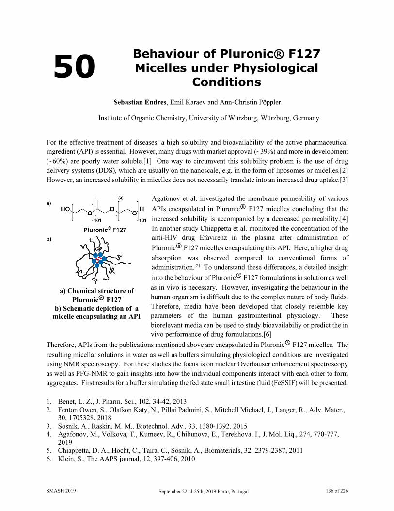
50 Behaviour of Pluronic® F127 Micelles under Physiological
Conditions
Sebastian Endres, Emil Karaev and Ann-Christin Pöppler
Institute of Organic Chemistry, University of Würzburg, Würzburg, Germany
For the effective treatment of diseases, a high solubility and bioavailability of the active pharmaceutical ingredient (API) is essential. However, many drugs with market approval (~39%) and more in development (~60%) are poorly water soluble.[1] One way to circumvent this solubility problem is the use of drug delivery systems (DDS), which are usually on the nanoscale, e.g. in the form of liposomes or micelles.[2] However, an increased solubility in micelles does not necessarily translate into an increased drug uptake.[3]
Agafonov et al. investigated the membrane permeability of various APIs encapsulated in Pluronic® F127 micelles concluding that the increased solubility is accompanied by a decreased permeability.[4] In another study Chiappetta et al. monitored the concentration of the anti-HIV drug Efavirenz in the plasma after administration of Pluronic® F127 micelles encapsulating this API. Here, a higher drug absorption was observed compared to conventional forms of administration.[5] To understand these differences, a detailed insight into the behaviour of Pluronic® F127 formulations in solution as well as in vivo is necessary. However, investigating the behaviour in the human organism is difficult due to the complex nature of body fluids. Therefore, media have been developed that closely resemble key parameters of the human gastrointestinal physiology. These biorelevant media can be used to study bioavailabiliy or predict the in vivo performance of drug formulations.[6]
Therefore, APIs from the publications mentioned above are encapsulated in Pluronic® F127 micelles. The resulting micellar solutions in water as well as buffers simulating physiological conditions are investigated using NMR spectroscopy. For these studies the focus is on nuclear Overhauser enhancement spectroscopy as well as PFG-NMR to gain insights into how the individual components interact with each other to form aggregates. First results for a buffer simulating the fed state small intestine fluid (FeSSIF) will be presented. 1. Benet, L. Z., J. Pharm. Sci., 102, 34-42, 2013 2. Fenton Owen, S., Olafson Katy, N., Pillai Padmini, S., Mitchell Michael, J., Langer, R., Adv. Mater.,
30, 1705328, 2018 3. Sosnik, A., Raskin, M. M., Biotechnol. Adv., 33, 1380-1392, 2015 4. Agafonov, M., Volkova, T., Kumeev, R., Chibunova, E., Terekhova, I., J. Mol. Liq., 274, 770-777,
2019 5. Chiappetta, D. A., Hocht, C., Taira, C., Sosnik, A., Biomaterials, 32, 2379-2387, 2011 6. Klein, S., The AAPS journal, 12, 397-406, 2010
a) Chemical structure of Pluronic® F127
b) Schematic depiction of a micelle encapsulating an API
SMASH 2019 September 22nd-25th, 2019 Porto, Portugal 136 of 226
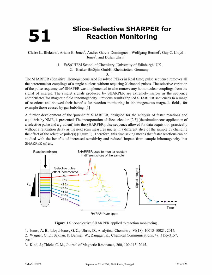
51 Slice-Selective SHARPER for Reaction Monitoring
Claire L. Dickson1, Ariana B. Jones1, Andres Garcia-Dominguez1, Wolfgang Bermel2, Guy C. Lloyd-
Jones1, and Dušan Uhrín1
1. EaStCHEM School of Chemistry, University of Edinburgh, UK 2. Bruker BioSpin GmbH, Rheinstetten, Germany
3. The SHARPER (Sensitive, Homogeneous And Resolved PEaks in Real time) pulse sequence removes all the heteronuclear couplings of a single nucleus without requiring X channel pulses. The selective variation of the pulse sequence, sel-SHAPER was implemented to also remove any homonuclear couplings from the signal of interest. The singlet signals produced by SHARPER are extremely narrow as the sequence compensates for magnetic field inhomogeneity. Previous results applied SHARPER sequences to a range of reactions and showed their benefits for reaction monitoring in inhomogeneous magnetic fields, for example those caused by gas bubbling. [1]
A further development of the 'pure-shift' SHARPER, designed for the analysis of faster reactions and equilibria by NMR, is presented. The incorporation of slice-selection [2,3] (the simultaneous application of a selective pulse and a gradient) into the SHARPER pulse sequence allowed for data acquisition practically without a relaxation delay as the next scan measures nuclei in a different slice of the sample by changing the offset of the selective pulse(s) (Figure 1). Therefore, this time saving means that faster reactions can be studied with the benefits of increased sensitivity and reduced impact from sample inhomogeneity that SHARPER offers.
Figure 1 Slice-selective SHARPER applied to reaction monitoring.
1. Jones, A. B.; Lloyd-Jones, G. C.; Uhrín, D., Analytical Chemistry, 89(18), 10013-10021, 2017. 2. Wagner, G. E.; Sakhaii, P; Bermel, W.; Zangger, K., Chemical Communications, 49, 3155-3157, 2013. 3. Kind, J.; Thiele, C. M., Journal of Magnetic Resonance, 260, 109-115, 2015.
SMASH 2019 September 22nd-25th, 2019 Porto, Portugal 137 of 226
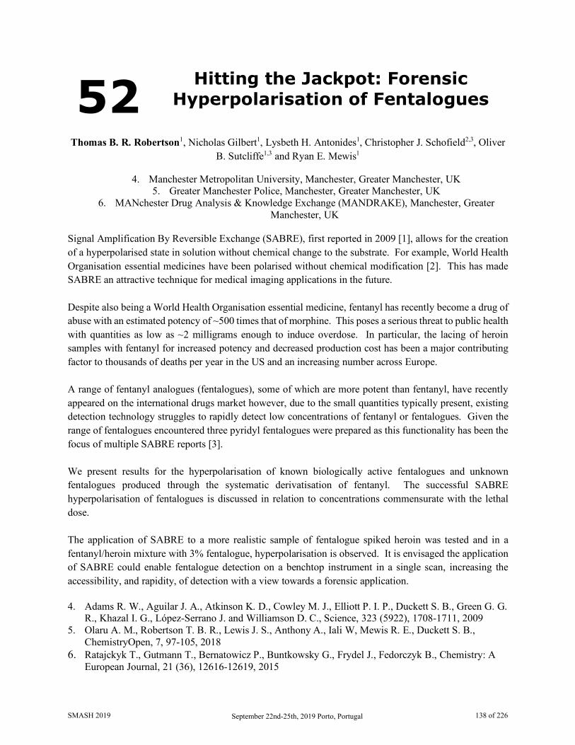
52 Hitting the Jackpot: Forensic Hyperpolarisation of Fentalogues
Thomas B. R. Robertson1, Nicholas Gilbert1, Lysbeth H. Antonides1, Christopher J. Schofield2,3, Oliver
B. Sutcliffe1,3 and Ryan E. Mewis1
4. Manchester Metropolitan University, Manchester, Greater Manchester, UK 5. Greater Manchester Police, Manchester, Greater Manchester, UK
6. MANchester Drug Analysis & Knowledge Exchange (MANDRAKE), Manchester, Greater Manchester, UK
Signal Amplification By Reversible Exchange (SABRE), first reported in 2009 [1], allows for the creation of a hyperpolarised state in solution without chemical change to the substrate. For example, World Health Organisation essential medicines have been polarised without chemical modification [2]. This has made SABRE an attractive technique for medical imaging applications in the future. Despite also being a World Health Organisation essential medicine, fentanyl has recently become a drug of abuse with an estimated potency of ~500 times that of morphine. This poses a serious threat to public health with quantities as low as ~2 milligrams enough to induce overdose. In particular, the lacing of heroin samples with fentanyl for increased potency and decreased production cost has been a major contributing factor to thousands of deaths per year in the US and an increasing number across Europe. A range of fentanyl analogues (fentalogues), some of which are more potent than fentanyl, have recently appeared on the international drugs market however, due to the small quantities typically present, existing detection technology struggles to rapidly detect low concentrations of fentanyl or fentalogues. Given the range of fentalogues encountered three pyridyl fentalogues were prepared as this functionality has been the focus of multiple SABRE reports [3]. We present results for the hyperpolarisation of known biologically active fentalogues and unknown fentalogues produced through the systematic derivatisation of fentanyl. The successful SABRE hyperpolarisation of fentalogues is discussed in relation to concentrations commensurate with the lethal dose. The application of SABRE to a more realistic sample of fentalogue spiked heroin was tested and in a fentanyl/heroin mixture with 3% fentalogue, hyperpolarisation is observed. It is envisaged the application of SABRE could enable fentalogue detection on a benchtop instrument in a single scan, increasing the accessibility, and rapidity, of detection with a view towards a forensic application. 4. Adams R. W., Aguilar J. A., Atkinson K. D., Cowley M. J., Elliott P. I. P., Duckett S. B., Green G. G.
R., Khazal I. G., López-Serrano J. and Williamson D. C., Science, 323 (5922), 1708-1711, 2009 5. Olaru A. M., Robertson T. B. R., Lewis J. S., Anthony A., Iali W, Mewis R. E., Duckett S. B.,
ChemistryOpen, 7, 97-105, 2018 6. Ratajckyk T., Gutmann T., Bernatowicz P., Buntkowsky G., Frydel J., Fedorczyk B., Chemistry: A
European Journal, 21 (36), 12616-12619, 2015
SMASH 2019 September 22nd-25th, 2019 Porto, Portugal 138 of 226

53 Low-Field NMR-Based Metabolomics
Jose L. Izquierdo-Garcia1,2, Patricia Comella-del-Barrio3 , Ramon Campos-Olivas4, Federico Casanova5,
Jose Dominguez3,2 and Jesus Ruiz-Cabello1,2
1. CIC biomaGUNE, San Sebastian, Spain. 2. CIBER de Enfermedades Respiratorias (CIBERES), Madrid, Spain.
3. Servicio de Microbiologia, Institut d'Investigació Germans Trias i Pujol, Badalona, Spain. 4. Centro Nacional de Investigaciones Oncologicas (CNIO), Madrid, Spain.
5. Magritek GmbH, Aachen, Germany.
Introduction: Classical metabolomic approaches based on Nuclear Magnetic Resonance (NMR) spectroscopy or Mass Spectrometry have been successfully used in the past to identify metabolic biomarkers of several diseases in preclinical and pilot studies. However, their use as a routine tool in clinic has been limited due to the delay between sample collection and analysis, the need for highly skilled scientists, and the expensive maintenance cost of these equipment. Here, we introduce benchtop low-field (LF) NMR spectrometers as an affordable in situ metabolomic approach for clinical samples. Until recently, the magnetic field homogeneity of benchtop LF NMR spectrometers did not reach the subparts per million needed to record spectra with sufficient resolution for metabolomic analysis. However, manufacturers now propose benchtop cryogen-free magnets working at either 42.5, 60, or 82 MHz for 1H NMR. In paricular, one of these models offers the possibility to achieve the homogeneity required to apply solvent suppression methods and resolve the metabolites in urine without any sample preparation. Here, we have tested the resolution of LF NMR spectrometer as metabolomic tool in a proof-of-concept study with tuberculosis patients. The objective was to compare the sensibility and specificity in the diagnosis of tuberculosis using a LF NMR-based metabolomic approach versus a classical high field (HF) NMR-based metabolomic approach.
Material and Methods: Urine samples from adult patients diagnosed of tuberculosis (n=19), other respiratory infection (n=25), and healthy controls (n=29) were examined using a Bruker Avance (16.4T) spectrometer and a Magritek Spinsolve ULTRA (1.4T) spectrometer. NMR spectra were phase and base-line corrected and 0.01ppm binned. Binning (or bucketing) is widely used in metabolomics studies to reduce the influence of pH variability between biofluid samples such as urine [1]. Principal Component Analysis (PCA) was applied to identify metabolic differences between groups. Classificatory models of partial least squares discriminant analysis (PLS-DA) was developed for the diagnosis of Tuberculosis.
Results: The resolution of 0.01ppm binned HF and LF NMR spectra are comparable for metabolic profiling. Both urine HF and LF NMR spectra provide a high discrimination between the three groups (Fig. 1). We identified 31 NMR regions corresponding to 8 metabolites significantly diferent between groups. The 8 metabolites were indentified in both HF and LF NMR spectra (Fig. 2). HF NMR PLS-DA classification models, tuberculosis vs control and tuberculosis vs respiratory infection, provided a diagnosis accuracy of about 100% by test samples. A similar PLS-DA classification models was developed based on LF NMR spectra, providing a diagnosis accuracy of over 90% by test samples.
SMASH 2019 September 22nd-25th, 2019 Porto, Portugal 139 of 226
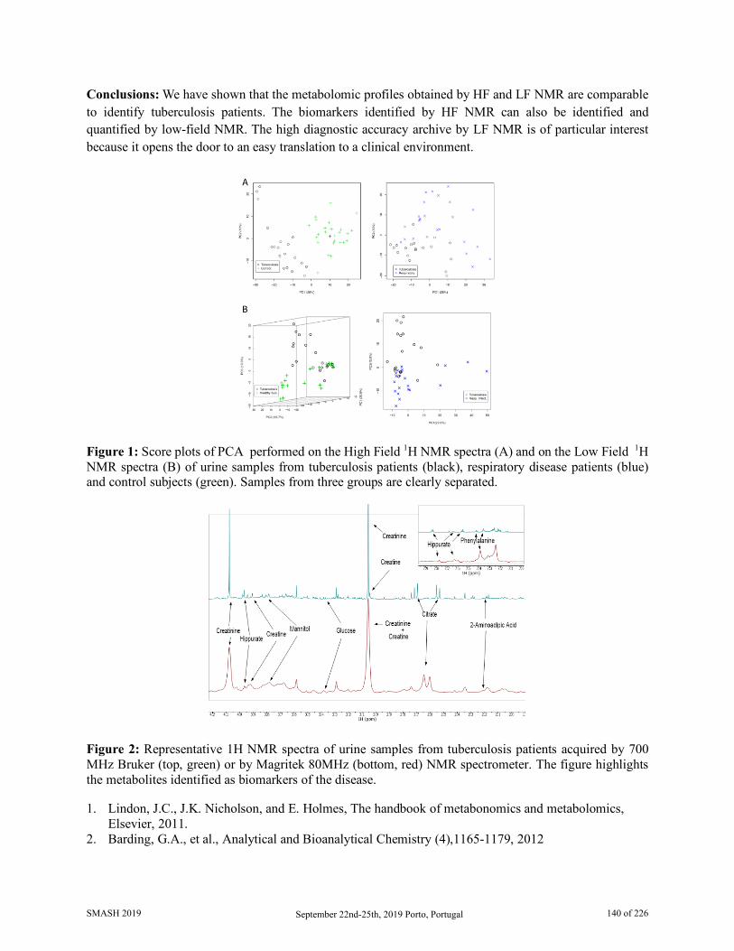
Conclusions: We have shown that the metabolomic profiles obtained by HF and LF NMR are comparable to identify tuberculosis patients. The biomarkers identified by HF NMR can also be identified and quantified by low-field NMR. The high diagnostic accuracy archive by LF NMR is of particular interest because it opens the door to an easy translation to a clinical environment.
Figure 1: Score plots of PCA performed on the High Field 1H NMR spectra (A) and on the Low Field 1H NMR spectra (B) of urine samples from tuberculosis patients (black), respiratory disease patients (blue) and control subjects (green). Samples from three groups are clearly separated.
Figure 2: Representative 1H NMR spectra of urine samples from tuberculosis patients acquired by 700 MHz Bruker (top, green) or by Magritek 80MHz (bottom, red) NMR spectrometer. The figure highlights the metabolites identified as biomarkers of the disease. 1. Lindon, J.C., J.K. Nicholson, and E. Holmes, The handbook of metabonomics and metabolomics,
Elsevier, 2011. 2. Barding, G.A., et al., Analytical and Bioanalytical Chemistry (4),1165-1179, 2012
SMASH 2019 September 22nd-25th, 2019 Porto, Portugal 140 of 226
54 NMR Spectroscopic Analysis of the Hydrogen Bond Mediated Pre-Aggregates Responsible for
Successful Photocatalytic PCET
Nele Berg and Ruth M. Gschwind
Department of Organic Chemistry, University of Regensburg, Regensburg, Germany The catalytic olefin hydroamidation is an efficient process to generate biologically active nitrogen-containing compounds without prefunctionalization of the amide substrate.[1] In 2015, Knowles et al. published an optimized catalyst system for the photocatalytic intramolecular hydroamidation of unactivated aryl amide derivatives.[2] In this report, the amide was transferred into an amidyl radical via oxidative proton-coupled electron transfer (PCET) by using an excited iridium photocatalyst and a Brønsted base. Subsequent olefin addition using thiophenol as the most effective hydrogen-atom donor generates the cyclic product. Surprisingly, the amide substrate was selectively transferred into the radical in presence of thiophenol although the N-H-bond (99 kcal/mol) is much stronger than the S-H-bond (79 kcal/mol). In literature, the light driven oxidative PCET is assumed to occur via a hydrogen bond between substrate and base.[3,4] Hence, a potential explanation is a difference in the hydrogen bond donor abilities of the amide and thiol with the N-H functionality being a better donor. Using DFT, the group of Knowles calculated the amide/base aggregate to be more favorable than the thiophenol/base complex by 5.2 kcal/mol.[2,5,6] In order to investigate the entire hydrogen bond situation and aggregation trends of this photocatalytic PCET model system, a detailed NMR spectroscopic investigation of 15N-labeled N-phenylpent-4-enamide, thiophenol and tetrabutylammonium di-tert-butyl phosphate was performed. Using 1H, 15N, 31P chemical shifts and 1JNH scalar couplings for the analysis of the central hydrogen bond, the donor (amide) and acceptor (base) sides were addressed separately.[7] Moreover, diffusion measurements revealed an unexpected modulation of the hydrogen bonded aggregates by thiophenol. Thus, for the first time, not the intrinsic hydrogen bond strengths but an increased electron density of the acceptor aggregate was found to be crucial for photocatalytic PCET reactivity. 1. Banerjee, Debasis, Junge, Kathrin, Beller, Matthias, Angew. Chem. Int. Ed. Engl., 53, 1630-5, 2014 2. Miller, David C., Choi, Gilbert J., Orbe, Hudson S., Knowles, Robert R., J. Am. Chem. Soc., 137,
13492-5, 2015 3. Weinberg, David R., Gagliardi, Christopher J., Hull, Johathan F., Murphy, Christine F., Kent, Caleb
A., Westlake, Brittany C., Paul, Amit, Ess, Daniel H., McCafferty, Dewey G., Meyer, Thomas J., Chem. Rev., 112, 4016–93, 2012
4. Huynh, My Hang V., Meyer, Thomas J., Chem. Rev., 107, 5004–5064, 2007 5. Choi, Gilbert J., Knowles, Robert R., J. Am. Chem. Soc., 137, 9226-9, 2015 6. Miller, David C., Tarantino, Kyle T., Knowles, Robert R., Top. Curr. Chem., 374, 1–59, 2016 7. Sharif, Shasad, Denisov, Gleb S., Toney, Michael D., Limbach, Hans-Heinrich, J. Am. Chem. Soc.,
129, 6313–27, 2007
SMASH 2019 September 22nd-25th, 2019 Porto, Portugal 141 of 226

55 β-BPSE: Fast DOSY Acqusition of Small Molecules and Macromolecules
Sebastian Spann1, Katharina S. Wetzel1, Michael A. R. Meier1 and Burkhard Luy2
1. Institute or Organic Chemistry, KIT Karlsruhe, Germany 2. Institute for Biological Interfaces 4, KIT Eggenstein-Leopoldshafen, Germany
The application of Diffusion Ordered NMR Spectroscopy has vastly increased in the last years, which illustrates the benefits of monitoring diffusion in solution NMR. Consequntly much effort has been ventured to improve the application of diffusion measurements in NMR [1]. In this work we introduce a pulse sequence that enables the spectroscopist to perform DOSY experiments in a matter of tens of seconds, providing the opportunity to perform reaction monitoring via diffusion. The newly derived sequence utilisies β-angle excitation, a well established method for deacreasing the overall experimental time in NMR spectroscopy [2]. Additionally, the following spin echo is realised with bipolar gradients, therefore the sequence is called β-Bipolar Spin Echo (β-BPSE). To suppress the signal loss due to J-modulation during the diffusion time, the mixing sequence DIPSI2 is implemented during Δ. The isotropic mixing during the β-BPSE sequence increases the signal by the factor of 2 compared to the conventional stimulated echo sequence. The β-BPSE sequence was applied to small molecules with measurement times of 9 seconds. Moreover, extending the β-BPSE on larger molecules such as a sequence-defined decamer [3], illustrates the sequence’s applicability on macromolecules.
1. Cohen Y., Avram L., Frish L., Angew. Chem. Int. Ed. 44, 520-554, 2005 2. Ross A., Salzmann M., Senn H., J-Bio NMR 10, 389-396, 1997 3. Solleder S. C., Zengel D., Wetzel K. S., Meier M.A.R., Angew. Chem. Int. Ed. 55, 1204-1207, 2016
SMASH 2019 September 22nd-25th, 2019 Porto, Portugal 142 of 226

56 Helically Chiral Poly(isocyanides) and Poly(acetylenes) as NMR
Alignment Media
Svenja Wesp and Michael Reggelin
Clemens-Schöpf-Institut, Technische Universität Darmstadt, Darmstadt, Germany
The measurement of residual dipolar couplings (RDCs) has become an important tool for solving conformational and configurational problems in structural analysis of small molecules. One reason for the increasing interest in RDCs is the fact that, unlike NOE- or J-coupling based methods, RDCs are global parameters being applicable without prior parametrization. However, RDCs are anisotropic NMR parameters which requires that the analyte molecule is oriented anisotropically by an alignment medium [1,2]. The model-free analysis of configurational problems using the recently developed software ConArch+ [3] benefits from measurements in as many as possible independent alignment media. Beyond that, we are interested in enantiodifferentiating orientation and therefore, we try to combine these goals by the development of helically chiral polyacetylenes [4] (PPAs) and polyisocyanides [5] (PICs). We synthesized different PICs based on various amino acids to investigate the influence of the side chain in relation to the strength of alignment and the enantiodifferentiation. The LLC phases prepared yield RDCs in the range of up to ± 40 Hz and show excellent orienting properties for a broad range of analytes bearing various functional groups. Additionally, we observed outstanding enantiodifferentiation for alcohols, ketones and carboxylic acids. Additionally, we developed a strategy to functionalise achiral grapheneoxide (GO), which was first introduced as alignment medium by LEI [6], with helically chiral PPA. Thus, we generated a new chiral alignment medium which combines the positive aspects of both media (GO + PPA).
1. Böttcher, Benjamin, Thiele, Christina M., eMagRes, 1(1), 169-180, 2012 2. Kummerlöwe, Grit, Luy, Burkhard, Annu. Rep. NMR Spectrosc., 68, 193-230, 2009 3. Immel, Stefan, Köck, Matthias, Reggelin, Michael, Chem. Eur. J., 24(52), 13918-13930, 2018 4. Meyer, Nils-Christopher, Krupp, Alexis, Schmidts, Volker, Thiele, Christina M., Reggelin, Michael,
Angew. Chem., 124(33), 8459-8463, 2012 5. Reller, Malin, Wesp, Svenja, Koos, Martin R.M., Reggelin, Michael, Luy, Burkhard, Chem. Eur. J.,
23(54), 13351-13359, 2017 6. Lei, Xinxiang, Xu, Zhen, Sun, Han, Wang, Shun, Griesinger, Christian, Peng, Li, Gao, Chao, Tan,
Ren X., J. Am. Chem. Soc., 136(32), 11280-11283, 2014
SMASH 2019 September 22nd-25th, 2019 Porto, Portugal 143 of 226

57 Residual Dipolar Coupling based Structural Elucidation of Marine
Natural Products
Xiao-Lu Li1, Min Yang2, Qi-Hao Wu2, Yue-Wei Guo2 and Han Sun1
1. Structural Biology, Leibniz-Forschungsinstitut für Molekulare Pharmakologie, Berlin, Germany 2. State Key Laboratory of Drug Research, Shanghai Institute of Materia Medica, CAS, Shanghai, China
Cembranoids are main metabolites of genus Sarcophyton soft corals. They have been proven with intriguing structural features and promising biological activities ranging from antitumoral, antibacterial, to enzyme inhibitory effects. Sarcomililate A(1) is a novel cembranoid isolated from Hainan Sarcophyton soft corals, which possesses five stereocenters. Determination of the relative and absolute configuration of 1 was particularly challenging as it was non-crystallizable and two stereochemical domains cannot be connected by NOEs and J-couplings. We employed in this study RDC-based anisotropic NMR spectroscopy to determine its relative configuration. The absolute configuration was established by comparing the experimental and computed electronic circular dichroism (ECD) spectra [1].
Figure 1. Structural elucidation of Sarcomililate A.
Soft corals of the genus Lemnalia are rich sources for ses-quiterpenoids and diterpenoids with different carbon skeletons. Many of these secondary metabolites are highly interesting because of their potent bioactivities, such as neuroprotective and anti-inflammatory properties. Xishaflavalin C (2) is a
SMASH 2019 September 22nd-25th, 2019 Porto, Portugal 144 of 226
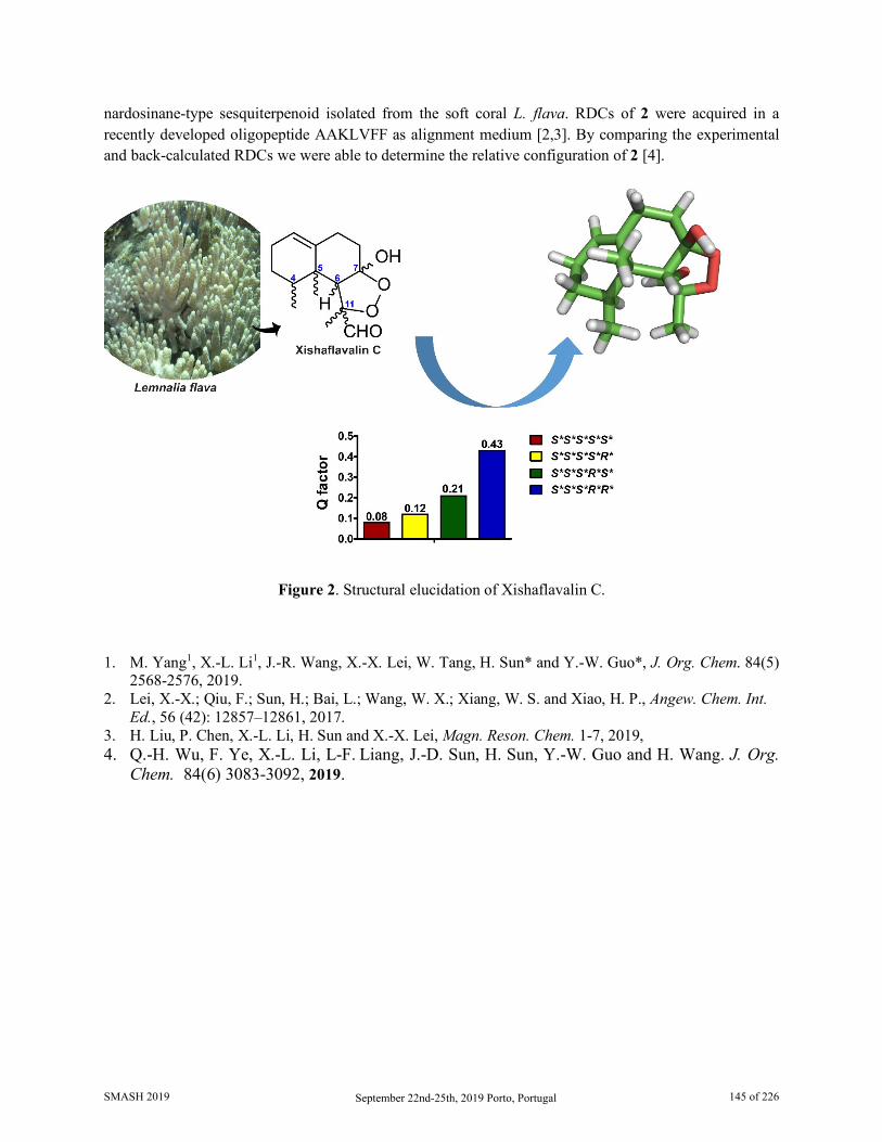
nardosinane-type sesquiterpenoid isolated from the soft coral L. flava. RDCs of 2 were acquired in a recently developed oligopeptide AAKLVFF as alignment medium [2,3]. By comparing the experimental and back-calculated RDCs we were able to determine the relative configuration of 2 [4].
Figure 2. Structural elucidation of Xishaflavalin C.
1. M. Yang1, X.-L. Li1, J.-R. Wang, X.-X. Lei, W. Tang, H. Sun* and Y.-W. Guo*, J. Org. Chem. 84(5) 2568-2576, 2019.
2. Lei, X.-X.; Qiu, F.; Sun, H.; Bai, L.; Wang, W. X.; Xiang, W. S. and Xiao, H. P., Angew. Chem. Int. Ed., 56 (42): 12857–12861, 2017.
3. H. Liu, P. Chen, X.-L. Li, H. Sun and X.-X. Lei, Magn. Reson. Chem. 1-7, 2019, 4. Q.-H. Wu, F. Ye, X.-L. Li, L-F. Liang, J.-D. Sun, H. Sun, Y.-W. Guo and H. Wang. J. Org.
Chem. 84(6) 3083-3092, 2019.
SMASH 2019 September 22nd-25th, 2019 Porto, Portugal 145 of 226

58 Insights into Asymmetric Transfer Hydrogenation using Multi-technique
Operando Reaction Monitoring
Daniel B. G. Berry,1 Ian Clegg,2 Anna Codina,2 Catherine L. Lyall,1,3 John P. Lowe,1,3 and Ulrich
Hintermair*1,3,4
1. Department of Chemistry, University of Bath, Bath, UK 2. Bruker UK Ltd, Coventry, UK
3. Dynamic Reaction Monitoring Facility, University of Bath, Bath, UK 4. Centre for Sustainable Chemical Technologies, University of Bath, Bath, UK
Nuclear magnetic resonance spectroscopy (NMR) has been noted as limited by sensitivity and bulk representability [1,2]. In recent years these limitations have been overcome by the development of FlowNMR (Figure 1 left) in conjunction with techniques such as selective excitation [3,4]. This technique allows for quantitative analysis that is under both realistic conditions and representative of the bulk reaction. It has been particularly useful as a method of acquiring mechanistic information and intermediate speciation in catalysis.
Enantioselective transfer hydrogenation using isopropanol/potassium hydroxide at room temperature has been previously studied in depth using FlowNMR [3]. This work has been developed further to investigate the heated reaction using a formic acid/triethylamine mixture as more commonly used in industry (Figure 1 right). While this process has been well studied offline [5], unanswered questions about the reaction including catalyst intermediates and pH dependence remain.
Figure 1: (left) Basic FlowNMR schematic.
Figure 2: (right) General reaction mechanism for catalytic transfer hydrogenation of acetophenone using formic acid/triethylamine.
N
RuN
HPh
Ph
Ts
OOH
CO2
NEt3-HCl
21 4
N
RuN
Ph
Ph
Ts
NH2
RuN
ClPh
Ph
Ts
O
OHN
RuN
Ph
Ph
Ts
3
OH
O
H HH
H HH
(R)-1-phenylethanol Acetophenone
SMASH 2019 September 22nd-25th, 2019 Porto, Portugal 146 of 226
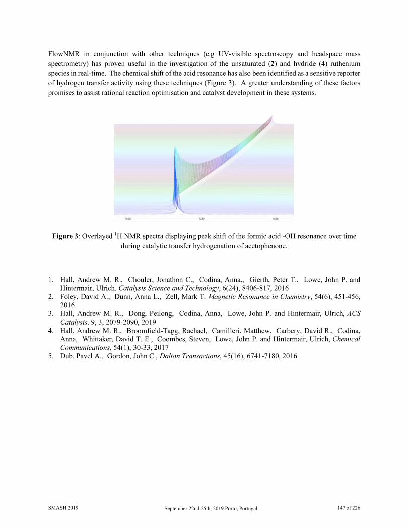
FlowNMR in conjunction with other techniques (e.g UV-visible spectroscopy and headspace mass spectrometry) has proven useful in the investigation of the unsaturated (2) and hydride (4) ruthenium species in real-time. The chemical shift of the acid resonance has also been identified as a sensitive reporter of hydrogen transfer activity using these techniques (Figure 3). A greater understanding of these factors promises to assist rational reaction optimisation and catalyst development in these systems.
Figure 3: Overlayed 1H NMR spectra displaying peak shift of the formic acid -OH resonance over time during catalytic transfer hydrogenation of acetophenone.
1. Hall, Andrew M. R., Chouler, Jonathon C., Codina, Anna., Gierth, Peter T., Lowe, John P. and Hintermair, Ulrich. Catalysis Science and Technology, 6(24), 8406-817, 2016
2. Foley, David A., Dunn, Anna L., Zell, Mark T. Magnetic Resonance in Chemistry, 54(6), 451-456, 2016
3. Hall, Andrew M. R., Dong, Peilong, Codina, Anna, Lowe, John P. and Hintermair, Ulrich, ACS Catalysis. 9, 3, 2079-2090, 2019
4. Hall, Andrew M. R., Broomfield-Tagg, Rachael, Camilleri, Matthew, Carbery, David R., Codina, Anna, Whittaker, David T. E., Coombes, Steven, Lowe, John P. and Hintermair, Ulrich, Chemical Communications, 54(1), 30-33, 2017
5. Dub, Pavel A., Gordon, John C., Dalton Transactions, 45(16), 6741-7180, 2016
SMASH 2019 September 22nd-25th, 2019 Porto, Portugal 147 of 226
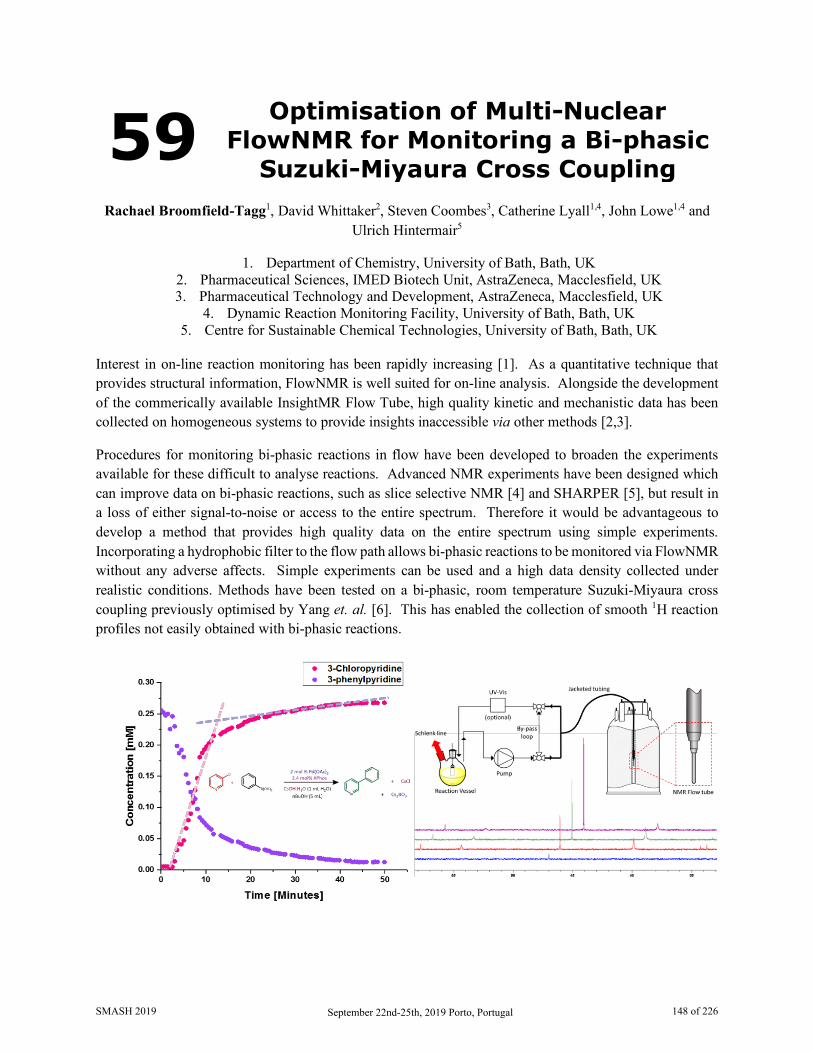
59 Optimisation of Multi-Nuclear FlowNMR for Monitoring a Bi-phasic
Suzuki-Miyaura Cross Coupling Rachael Broomfield-Tagg1, David Whittaker2, Steven Coombes3, Catherine Lyall1,4, John Lowe1,4 and
Ulrich Hintermair5
1. Department of Chemistry, University of Bath, Bath, UK 2. Pharmaceutical Sciences, IMED Biotech Unit, AstraZeneca, Macclesfield, UK 3. Pharmaceutical Technology and Development, AstraZeneca, Macclesfield, UK
4. Dynamic Reaction Monitoring Facility, University of Bath, Bath, UK 5. Centre for Sustainable Chemical Technologies, University of Bath, Bath, UK
Interest in on-line reaction monitoring has been rapidly increasing [1]. As a quantitative technique that provides structural information, FlowNMR is well suited for on-line analysis. Alongside the development of the commerically available InsightMR Flow Tube, high quality kinetic and mechanistic data has been collected on homogeneous systems to provide insights inaccessible via other methods [2,3].
Procedures for monitoring bi-phasic reactions in flow have been developed to broaden the experiments available for these difficult to analyse reactions. Advanced NMR experiments have been designed which can improve data on bi-phasic reactions, such as slice selective NMR [4] and SHARPER [5], but result in a loss of either signal-to-noise or access to the entire spectrum. Therefore it would be advantageous to develop a method that provides high quality data on the entire spectrum using simple experiments. Incorporating a hydrophobic filter to the flow path allows bi-phasic reactions to be monitored via FlowNMR without any adverse affects. Simple experiments can be used and a high data density collected under realistic conditions. Methods have been tested on a bi-phasic, room temperature Suzuki-Miyaura cross coupling previously optimised by Yang et. al. [6]. This has enabled the collection of smooth 1H reaction profiles not easily obtained with bi-phasic reactions.
SMASH 2019 September 22nd-25th, 2019 Porto, Portugal 148 of 226
Acquisition parameters have been optimised for monitoring catalytic species using 31P FlowNMR. The combination of insensitive nuclei, low concentration species and rapid reaction kinetics required experiments to optimised for signal-to-noise per time (S/N min-1). Experiment type, relaxation delays, acquisition time and flip angle have all been altered to provide a robust experiment which allows short lived intermediates to be seen over the entire spectral region. This has provided insight into the activation of Pd(OAc)2 to the catalytic species, interactions and speciation with XPhos and the effect of ligand concentration.
1. Chung, Ryan; Hein, Jason; E., Top. Catal., 60(8), 594-608, 2017 2. Hall, Andrew M. R.; Broomfield-Tagg, Rachael; Camilleri, Matthew; Carbery, David R; Codina,
Anna; Whittaker, David. T. E.; Coombes, Steven; Lowe, John P.; Hintermair, Ulrich; Chem. Commun., 54(1), 30-33, 2018
3. Hall, Andrew M. R; Dong, Peilong; Codina, Anna; Lowe, John P.; Hintermair, Ulrich; ACS Catal., 9(3), 2079-2090, 2019
4. Evans, Robert; Sandhu, Aran; Bridgwater, Anthony V.; Chong, Katie J.; Energy Fuels, 31(4), 4135-4142, 2017
5. Jones, Ariana B.; Lloyd-Jones, Guy C.; Uhrín, Dušan, Anal. Chem., 89(18), 10013-10021, 2017 6. Yang, Junfeng; Liu, Sijia; Zheng, Jian-Feng, Zhou, Jianrong (Steve), Eur. J. Org. Chem., 2012(31),
6248-6259, 2012
SMASH 2019 September 22nd-25th, 2019 Porto, Portugal 149 of 226
60 1H-NMR Metabolomics Reveals Potential Biomarkers of Breast Cancer in an Endocrine-Related
Mouse Model Rita Araújo1, Daniela Bispo2, Caroline Lamb2, Victoria Fabris2, Claudia Lanari2, Luisa Helguero2, and
Ana M. Gil1
1. Department of Chemistry and CICECO - Aveiro Institute of Materials (CICECO/UA), University of Aveiro, Aveiro, Portugal
2. iBIMED—Institute for Biomedicine, Department of Medical Sciences, Universidade de Aveiro, Aveiro, Portugal
3. Instituto de Biología y Medicina Experimental (IBYME), CONICET, Buenos Aires, Argentina
Breast cancer (BC) is the most common and deadliest type of cancer in women. In most cases, breast tumors are hormone-dependent (HD) and rely on steroid hormones (estrogen and progesterone) to activate intracellular receptors and stimulate tumor growth. However, with time, almost half of breast cancer patients develop resistance to antagonist therapy. This process involves the progression of HD tumors to hormone-independent tumors (HI) and ultimately to tumor resistance to therapy (HIR). The full mechanism and metabolic pathways involved in this progression remain unknown. Metabolic alterations are considered a hallmark of cancer progression and the altered metabolism in breast cancer has been significantly investigated by metabolomics seeking understanding of the interplay between tumor progression/aggressiveness and metabolic adaptations [1,2]
In this work, we aimed to understand the metabolic interplay associated with the transition from HD to HI and HIR phenotypes and identify metabolic markers potentially useful to anticipate response to endocrine therapy. For this purpose, we applied an untargeted metabolomics strategy to characterize metabolic changes in the medroxyprogesterone acetate (MPA) mouse mammary carcinoma model, which has been refined to grow infiltrating mammary carcinomas that evolve through different stages of hormone dependence and therefore mimic human endocrine breast cancer progression (HD which grows only with MPA, HI and HIR) [3]. Polar metabolites were extracted from tumor tissue samples and analyzed by 1H-NMR (500 MHz) [4]. The principal component analysis (PCA) and partial least-squares discriminant analysis (PLS-DA) models derived from our data showed robust separations between HD, HI and HIR tumors and PLS-DA loading plots helped to identify lower levels of metabolites such as glutamic acid, glutamine, glycine, taurine, choline and glycerophosphocholine (among others) as specific for HI and HIR tumors, compared to HD. In addition, a set of metabolites was identified as potentially indicative of resistance to therapy (high in HIR compared to HI), including a marked lactate increase. To assess the effect of MPA we also analyzed normal mammary tissue with and without the hormone. This analysis also helped understand if MPA influenced the differences found between HD and HI tumors.
The observed distinguishing metabolic signatures of HD, HI and HIR tumors show that endocrine breast cancer exhibits distinct metabolomic patterns depending on the stage of aggressiveness and the altered
SMASH 2019 September 22nd-25th, 2019 Porto, Portugal 150 of 226

metabolites found here may be the basis for the definition of biomarkers of endocrine-related breast tumors at different stages of progression and prognostic indicators of response to antagonist therapy.
This study demonstrates the applicability of 1H-NMR metabolomics for understanding important metabolic alterations occurring during breast cancer progression and simultaneously determine direct effects of drug/tumor interactions.
1. Suman S, Sharma R K, Kumar V,et al., Journal of Pharmaceutical and Biomedical Analysis, 160(1),
38-45, 2018 2. Dai C, Arceo J, Arnold J, et al., Cancer & Metabolism, 6(5), 1-17, 2018 3. Lanari C L M, Lamb C, Fabris V, et al., Endocrine Related Cancer, 16(2), 333-350, 2009 4. Beckonert O, Keun HC, Ebbeks TMD, Bundy J, Holmes E, Lindon JC, Nicholson JK, Nature
Protocols, 2(11), 2692 -2703, 2007
SMASH 2019 September 22nd-25th, 2019 Porto, Portugal 151 of 226
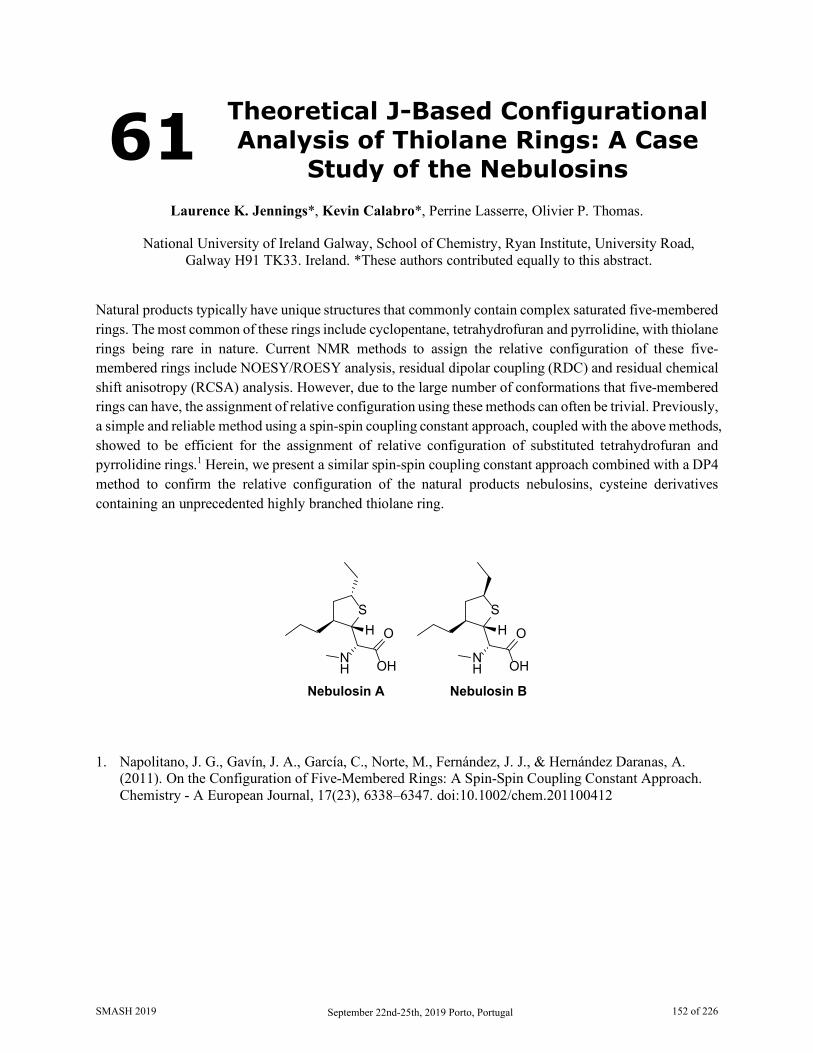
61 Theoretical J-Based Configurational Analysis of Thiolane Rings: A Case
Study of the Nebulosins
Laurence K. Jennings*, Kevin Calabro*, Perrine Lasserre, Olivier P. Thomas.
National University of Ireland Galway, School of Chemistry, Ryan Institute, University Road, Galway H91 TK33. Ireland. *These authors contributed equally to this abstract.
Natural products typically have unique structures that commonly contain complex saturated five-membered rings. The most common of these rings include cyclopentane, tetrahydrofuran and pyrrolidine, with thiolane rings being rare in nature. Current NMR methods to assign the relative configuration of these five-membered rings include NOESY/ROESY analysis, residual dipolar coupling (RDC) and residual chemical shift anisotropy (RCSA) analysis. However, due to the large number of conformations that five-membered rings can have, the assignment of relative configuration using these methods can often be trivial. Previously, a simple and reliable method using a spin-spin coupling constant approach, coupled with the above methods, showed to be efficient for the assignment of relative configuration of substituted tetrahydrofuran and pyrrolidine rings.1 Herein, we present a similar spin-spin coupling constant approach combined with a DP4 method to confirm the relative configuration of the natural products nebulosins, cysteine derivatives containing an unprecedented highly branched thiolane ring.
S
NH
O
OH
H
Nebulosin A
S
NH
O
OH
H
Nebulosin B
1. Napolitano, J. G., Gavín, J. A., García, C., Norte, M., Fernández, J. J., & Hernández Daranas, A. (2011). On the Configuration of Five-Membered Rings: A Spin-Spin Coupling Constant Approach. Chemistry - A European Journal, 17(23), 6338–6347. doi:10.1002/chem.201100412
SMASH 2019 September 22nd-25th, 2019 Porto, Portugal 152 of 226

62 Examples of Complementary Small Molecule Mass Spectrometry and NMR: A Marine Toxin, a Food Dye
and Tattoo Inks
Clark D. Ridge1, Eugene P. Mazzola1, Whitney L. Stutts2, Adrian Weisz3, Christine M. Fisher1, Sarah E. Donnelly1, Jonathan R. Deeds1, R. Thomas Williamson4, Gary E. Martin5, Douglas P. Ridge6, and
Timothy R. Croley1
1. Office of Regulatory Science, Center for Food Safety and Applied Nutrition, U.S. Food and Drug Administration, College Park, MD, USA
2. Molecular Education, Technology and Research Innovation Center, North Carolina State University, Raleigh, NC, USA.
3. Office of Cosmetics and Colors, Center for Food Safety and Applied Nutrition, U.S. Food and Drug Administration, College Park, MD, USA
4. Department of Chemistry and Biochemistry, University of North Carolina Wilmington, Wilmington, NC, USA.
5. Department of Chemistry and Biochemistry, Seton Hall University, South Orange, NJ, USA. 6. Department of Chemistry and Biochemistry, University of Delaware, Newark, DE, USA.
Small molecule analysis usually involves mass spectrometry (MS) and NMR spectroscopy performed in relative isolation. The NMR spectroscopist may get little more than the chemical formula from the MS analysis and in return provide a structure. There is usually far more information in both analyses that can be useful. In many other cases, more collaboration is needed to elucidate structure or solve the problem at hand. Three instructive cases that highlight the complementary nature of the techniques are presented here.
1. Maitotoxin (MTX) is a large polyketide marine toxin. With a molecular weight upwards of 3000 Da, it is the largest known non-biopolymer marine toxin. Several compounds similar to MTX were isolated and found to be deficient in mass by either one sulfate group or a sulfate group, a methyl, and one degree of unsaturation. With only 50 nmol of material, a complete assignment was not possible. However, MTX has only two sulfate groups with one near the end of the molecular chain and accessible by NMR techniques. The combined high-resolution MS and NMR analysis were able to partially assign and distinguish which of these sulfate groups is missing in this new set of maitotoxins. [1]
2. The FDA has an ongoing effort to identify and characterize minor components of food, drug, and cosmetic dyes. One such component, a low concentration byproduct of the synthesis of D&C Red No. 33 (R33), had MS and NMR data that were seemingly irreconcilable. Other components were readily isolated and identified [2], but efforts on this one were stalled. Further inquiry by both techniques indicate that the problem is likely due to a charge-remote process prior to detection. This presents a rare case in which the NMR structure is used to correct the molecular formula determined by MS.
3. There are few studies into the composition of tattoo inks and dyes, but there is increased interest in determining what compounds are generally used and whether any potentially harmful chemicals might be present. As part of this effort at the FDA, a combined LCMS and NMR analysis was done on a number of
SMASH 2019 September 22nd-25th, 2019 Porto, Portugal 153 of 226
commonly used and commercially available tattoo inks. The separate techniques provide distinct and complementary data about the inks with NMR primarily determining concentrations of the major components of the carrier solution and LCMS the minor ones. However, there was some overlap. A minor, possibly carcinogenic [3] compound, 2-methoxy-4-nitroanline, was indicated in one of the inks by LCMS in a relatively low concentration of less than 10 uM. This is usually not a high enough concentration to see in NMR spectra of mixtures, but this case proved to be an exception. With the combined analysis the structure was confirmed with near certainty and the concentration was determined with higher accuracy and precision.
1. Mazzola, Eugene P., Deeds, Jonathan R, Stutts, Whitney L., Ridge, Clark D., Dickey, Robert W., White, Kevin D., Williamson, R. Thomas, Martin, Gary E., Toxicon, 164, 44-50, 2019
2. Weisz, Adrian, Ridge, Clark D., Mazzola, Eugene P., Ito, Yoichiro, J. Chromatogr. A, 1380, 120–129, 2015
3. Chemie, BG, Toxicological Evaluations, Vol.14, 59-68, 1999
SMASH 2019 September 22nd-25th, 2019 Porto, Portugal 154 of 226
63 When Flexibility Meets with RDCs for Structure Elucidation
Pavleta Tzvetkova1 and Burkhard Luy1,2
1. Karlsruhe Institute of Technology, Institute for Biological Interfaces 4 – Magnetic Resonance, Karlsruhe, Germany
2. Karlsruhe Institute of Technology, Institute of Organic Chemistry, Karlsruhe, Germany
The three dimensional structure of the molecule impacts some of its physical and chemical properties. The most widely spread method to obtain 3D structures is single crystal X-Ray crystallography, however, suffering from the disadvantage of being performed in solid state. High resolution NMR spectroscopy has the main advantage of accessing 3D structures in liquid state. In classical NMR approaches chemical shifts, scalar couplings and NOEs are used to determine the relative configuration and conformation of the molecules. However, in the case of conformationally flexible molecules this information is averaged and can be insufficient to solve all ambiguities in the structure. Thus, it is of crucial importance to measure and use as many as possible NMR parameters in the structure elucidation procedure. The measurement of anisotropic parameters in partially alignment samples, such as Residual Dipolar Couplings (RDCs) are nowadays an established technique which could provide this important additional information. To today RDCs have been used for the analysis of constitution, configuration and conformation of molecules in number of cases.
The most straightforward approach to use RDCs for structure evaluation uses sterically fixed orientational models as implemented for example in the programs Pales [1,2] or MSpin [3], which become considerably inaccurate in the case of conformational flexibility as they don’t take into the account conformational averaging. Currently further developments to use RDCs for structure elucidation on flexible models are emerging in the literature, as in the CASE-3D, where a set of preferred conformations are used to represent the conformational flexibility [4,5].
Here, on an example of flexible molecule with ring systems, where various number of measurable NMR parameters are available, is demonstrated how the introduction of flexibility could hamper the unambiguous structure elucidation. Alongside with the comparison with some of the existing approaches the use of time averaged molecular dynamics simulations with orientational constraints (MDOC) [6] is evaluated. RDCs are used as orientational constraints, where the tensorial properties are computed as time averages over the MD trajectory and pseudo forces are derived after comparison with experimental values which allow fast sampling of the global rotation and internal dynamics. The full tensorial calculation performed allows improved characterisation of the orientation of the system. The MDOC protocol is implemented in the program COSMOS [7]. The reproducibility of the experimental parameters is monitored with a quality factor for the correspondence of experimental (imported) and calculated data. The quality measure for the RDCs is defined as the summed squared difference of the experimental and calculated RDC divided by the respective experimental error. High quality measure during the simulation serves as an indication of configurational assignment being correct.
SMASH 2019 September 22nd-25th, 2019 Porto, Portugal 155 of 226

1. Zweckstetter, M, Bax, A., J. Am. Chem. Soc., 122, 3791-3792, 2000. 2. Zweckstetter, M, Nature Prot., 3, 679-690, 2008. 3. Navarro-Vazquez, A, Magn. Reson. Chem., 50, S73-S79, 2012. 4. Troche-Pesqueira, E, Anklin, C, Gil, R.R, Navarro-Vázquez A, Angew. Chem. Int. Ed., 56 (13),
3660-3664, 2017. 5. Navarro-Vázquez, A, Gil, R.R, Blinov, K, J. Nat. Prod., 81 (1), 203-210, 2018. 6. Sternberg, U, Witter, R, Ulrich, A, J. Biomol. NMR, 38, 23-39, 2007. 7. Sternberg, U, Koch, F-Th., Losso, P, COSMOS 6.0 Pro, www.cosmos-software.de
SMASH 2019 September 22nd-25th, 2019 Porto, Portugal 156 of 226
64 BioMagResBank: Database and Tools for Bioactive Small Molecule NMR
Analysis
Pedro R Romero1, Hamid Eghbalnia1, Hesam Dashti2, Naohiro Kobayashi3, Jonathan Wedell1, Kumaran Baskaran1, Takeshi Iwata3, Masashi Yokochi3, Dimitri Maziuk1, Hongyang Yao1, Toshimichi Fujiwara3,
Genji Kurusu3, Eldon L Ulrich1, Jeffrey C Hoch4, and John L Markley1*
5. BMRB, Biochemistry Department, University of Wisconsin-Madison, Madison, WI, USA
6. Brigham and Women's Hospital, Harvard Medical School, Boston, MA, USA 7. PDBj-BMRB, Institute for Protein Research, Osaka University, Suita, Osaka, Japan
8. BMRB, Department of Molecular Biology and Biophysics, UConn Health, Farmington, CT, USA The Biological Magnetic Resonance Data Bank (BioMagResBank or BMRB) [1], founded in 1988, serves as the archive for data generated by Nuclear Magnetic Resonance (NMR) spectroscopy of biological systems. NMR spectroscopy is unique among biophysical approaches in its ability to provide a broad range of atomic and higher-level information relevant to the structural, dynamic, and chemical properties of biological macromolecules, as well as reporting on metabolite and natural product concentrations in complex mixtures and their chemical structures. BMRB stores experimental and derived data from biomolecular NMR studies on both biopolymers and bioactive small compounds. BMRB supports metabolomics NMR studies through a library of a variety of 1D and 2D NMR spectra of pure compounds (including metabolites, natural products, drugs, and compounds used for screening in drug discovery) and through its adoption of novel analytic tools, like the ALATIS unique atom identifiers [2], which are universal and based solely on the 3D structure of the compound and the InChI convention, and GISSMO spin matrices [3], which enable accurate simulation of compound and mixture spectra at any field strength. The combination of unique ALATIS naming and parameterized spectra offers the users of BMRB data a distinctive benefit in terms of robustness and reproducibility, as embodied in the FAIR principles for data resources, which are that data should be Findable, Accessible, Interoperable, and Reusable. 1. Ulrich, E.L., et al., “BioMagResBank.” Nucleic Acids Res, 2008. 36(Database issue): p. D402-8.
DOI: 10.1093/nar/gkm957. 2. Dashti, H., et al., “Unique identifiers for small molecules enable rigorous labeling of their atoms.” Sci
Data, 2017. 4: p. 170073. DOI: 10.1038/sdata.2017.73. 3. Dashti, H., et al., “Applications of Parametrized NMR Spin Systems of Small Molecules.” Anal
Chem, 2018. 90(18): p. 10646-10649. DOI: 10.1021/acs.analchem.8b02660.
SMASH 2019 September 22nd-25th, 2019 Porto, Portugal 157 of 226
65 Exploring Non-Fourier NMR Dimensions with NUS
Dariusz Gołowicz1,2, Mateusz Urbańczyk3, Alexandra Shchukina2, and Krzysztof Kazimierczuk2
1. University of Warsaw, Faculty of Chemistry, Warsaw, Poland 2. University of Warsaw, Centre of New Technologies, Warsaw, Poland
3. University of Oulu, NMR Research Centre, Oulu, Finland
Nuclear magnetic resonance spectroscopy (NMR) of small and medium size molecules usually does not require high-dimensional experiments because of sufficient peak resolution. However, new NMR dimensions do not necessarily have to be frequency dimensions. A special dimension can be introduced to yield task-specific information in various conventional N-dimensional experiments.
One can imagine an extra 3rd dimension which serves a new information without the use of Fourier Transform. We can call it a non-Fourier dimension. A traditional approach to explore such pseudo-dimensions is to record a series of spectra with different acquisition parameters. In this work, we describe a quick method for simultaneous sampling of Fourier and non-Fourier dimensions. We also show two examples of applications for INEPT-transfer and TOCSY-transfer non-Fourier dimensions.
The method is a modification of time-resolved non uniform sampling technique (TR-NUS)[1]. Originally, TR-NUS employs uninterrupted non-uniform sampling acquisition of evolution time points in parallel to occurring process, e.g. chemical reaction. The missing data is then reconstructed using dedicated algorithms[2,3] according to the moving frame processing method. The result of such experiment is a stack of 2D (or nD) spectra that correspond to different time points of a chemical reaction.
In order to create a new non-Fourier dimension, a signal intensity change has to be induced during NUS acquisition. Therefore, the selected parameter in a pulse sequence (e.g. delay, offset) is co-incremented linearly (or not) with the index of NUS-schedule.
We show that the method may be used for improving quantitativeness of 13C HSQC spectra by introducing INEPT-transfer dimension[4] as well as for identification of saccharides in mixtures using 1H TOCSY-transfer dimension as a fingerprint.
1. Mayzel M, Rosenlöw J, Isaksson L, Orekhov VY, J. Biomol. NMR, 58(2), 129-39, 2014 2. Kazimierczuk K, Orekhov VY, Angew. Chem. Int. Ed., 50, 5556–5559, 2011 3. Orekhov VY, Jaravine VA, Prog. Nucl. Magn. Reson. Spectrosc, 59, 271-292, 2011 4. Gołowicz D, Urbańczyk M, Shchukina A, Kazimierczuk K, J. Magn. Reson, 294, 1-6, 2018
SMASH 2019 September 22nd-25th, 2019 Porto, Portugal 158 of 226

66 Urinary Metabolic Trajectory of Premature Newborns
Daniela Duarte1, Cristina Pita2, Fátima Negrão2, Isabel M. Carreira3, Maria do Céu Almeida2,
and Ana M. Gil1
1. CICECO, Aveiro Institute of Materials, Department of Chemistry, University of Aveiro, Aveiro,
Portugal 2. Maternidade Bissaya Barreto, Centro Hospitalar e Universitário de Coimbra, CHUC, Coimbra,
Portugal 3. Cytogenetics and Genomics Laboratory, Faculty of Medicine, University of Coimbra, and
CIMAGO - Center for Research in Environment, Genetics and Oncobiology, Faculty of Medicine, University of Coimbra, Coimbra, Portugal
Preterm birth (PTB) is defined as birth before 37 gestational weeks [1]. It was estimated that 14.8 million infants were born prematurely in 2014 [2]. Actually, complications related to prematurity are the leading cause of neonatal deaths causing ca. 1 million infant deaths per year [3]. Moreover, US medical costs associated with PTB infants in the first year are 10 times greater compared to term infants [4]. Metabolomics, defined as the qualitative and quantitative analysis of metabolites present within organism, cell, or tissue [5], can aid in the detection and possible identification of biomarkers for this condition and related complications. Aiming to evaluate the metabolic trajectory of PTB newborns during probation, newborn urine samples were collected at Maternity Bissaya Barreto, University Hospital Center of Coimbra (CHUC), with parental informed consents obtained for each infant and samples were analyzed by Nuclear Magnetic Resonance (NMR) spectroscopy on a Bruker Avance III spectrometer, operating at 500 MHz. Multivariate analysis was applied to the 1H-NMR urine spectra to define putative metabolic markers of prematurity in the first days of life. PTB newborns with different gestational ages were also followed during probation, so we could identify urinary metabolites varying during that period as possible markers of deviant behaviors indicative of health complications. 1. Blencowe, H., Cousens, S., Chou, D., Oestergaard, M., Say, L., Moller, A., Kinney, M., Lawn, J.,
Reproductive Health 10(Suppl 1), S2, 2013. 2. Chawanpaiboon, S., Moller, A-B., Lumbiganon, P., Petzold, M., Hogan, D., Landoulsi, S.,
Jampathong, N., Kongwattanakul, K., Laopaiboon, M., Lewis, C., Rattanakanokchai, S., Teng, D. N., Thinkhamrop, J., Watananirun, K., Zhang, J., Zhou, W., Gülmezoglu, A.M., Lancet Global Health, 7, e37-46, 2019.
3. Liu, L., Oza, S., Hogan, D., Perin, J., Rudan, I., Lawn, J.E., Cousens, S., Mathers, C., Black, R.E., The Lancet, 385(9966), 430-440, 2015.
4. Howson, C.P., Kinney, M.V., McDougall, L., Lawn, J.E., Reproductive Health, 10(Suppl 1), S1, 2013. 5. Nicholson, J.K., Lindon, J.C., Holmes, E., Xenobiotica 29(9), 1181-1189, 1999.
SMASH 2019 September 22nd-25th, 2019 Porto, Portugal 159 of 226
67 1H NMR and Fuzzy C-Means Analysis
for Classifying and Adulteration Detection of Orange Drinks
Renan Ziemann Wilhelms and Luiz Henrique Keng Queiroz Júnior
Instituto de Química, Universidade Federal de Goias, Goiânia-GO,Brazil
Most of the population considers fruit juices as a healthy drinks, compared to soft drinks, and this fact is used by food industry for marketing purposes, what moves billions dollars every year. The high consumption of these juices demands a growing concern with quality control, in order to prevent production errors or adulteration.1,2 Nowadays, one of the most preferred flavors for the aforementioned drinks is orange, and it is mainly consumed as nectar and integral juices and soft drinks. In this context, we evaluated in this study commercial samples of nectar and integral juices and soft drinks, all of orange flavor, in order to investigate some possible adulteration and the differences between these beverages. We performed 1H NMR quantitative experiments and traced the chemical profiling for each sample and used statistical tools to better explore the experimental data. The NMR measurements were made on a Bruker Avance III 500 (11.74 T). An aliquot of 100.0 µL of the sample was diluted in 400.0 µL D2O. The statistical analyses were performed by home made routines in the R software. The main compounds characterized by 1H NMR data were sucrose, glucose, fructose, citric acid and ethanol. The ethanol contents were higher for nectar juices, what indicates a relevant fermentation process compared to the other drinks. Applying the non hard clustering method Fuzzy C-Means3 to the NMR data, four groups were discriminated, what could sound strange because we have only three kinds of drinks. Indeed, the additional group is explained by apple juice addition in some nectar juice samples. One group consists in soft drinks and one synthetic juice, another group consists in a mix of integral and nectar juices, with and without added apple juice. A third group is constituted by nectar juice with and without apple added, and the fourth one has only one juice without addition of apple and the others have apple juice added. We evaluated the chemical profile of this unique sample without apple juice addition, in the fourth group, and concluded that indeed it has apple juice added. Then, this indicates the mentioned sample was adulterated, by its inconsistency between the label and the real chemical profile. Other important observation was that sugar content for one nectar juice was similar to the high sugar content founded for the soft drinks samples. So, by NMR and statistical combined analyses it was possible to discriminate the types of beverage and also detect one adulterated sample, highlighting the potential of this conjunct approach. 1. Ogrinc, N; Kosir, I, J; Spangenberg, J, E; Kidric, J., ABC – Analytical and Bioanalytical Chemistry,
376, 424-430, 2003. 2. Vogels, J, T, W, E; Terwel, L; Tas, A, C; Berg, F, V, D; Dukel, F; Greef, J, V, D., J Agric. Food
Chem. – Journal of Agricultural and Food Chemistry, 44, 175-180, 1996. 3. Bezdek, J, C; Ehrlich, R; Full, W., Computers & Geosciences, 10, 191-203, 1984. 4. Cusano, E; Simonato, B.; Consonni, R., LTW – Food Science and Technology, 96, 147-151, 2018.
SMASH 2019 September 22nd-25th, 2019 Porto, Portugal 160 of 226

68 Quantitative Analysis of Cannabinoids using Benchtop NMR
Spectrometers
Susanne D. Riegel1 Juan F. Araneda1, Terry Chu,1 and Neil Spingarn2
1. Nanalysis Corp. Calgary, AB, Canada 2. S&N Labs, Santa Ana, CA, USA
While liquid chromatography is the gold standard in the majority of mixture analyses, depending on the mixture components, time of separation, the cost of the solvent, both in purchasing and disposal, it does not always represent the ideal solution. Nuclear Magnetic Resonance (NMR) Spectroscopy has proven to be an attractive alternative for quantitative analysis (qNMR) given its non-targeted nature and the fact that all resonances give the same response factor, allowing them to be compared directly without calibration. Regardless, the capital and operating cost associated with high-field instruments have left them excluded from many quantitative quality control and quality assurance initiatives. The emergence of a new class of instrument: high-resolution benchtop NMR spectrometers offer a easy-to-use, low-maintenance alternative to quantify some key components of cannabis samples. Herein we will discuss our most recent results using 60 MHz NMR spectrometers to determine the relative and/or absolute concentration of tetrahydrocannabinol (THC) and cannabidiol (CBD) in a series of cannabis products. We will highlight the analytical characteristics to determine the feasibility and outlook of using benchtop NMR for quantification of the primary cannabinoids required rapid buy or no buy decisions for producers in both cannabis and hemp markets. We will discuss developments to make this technique suitable for operation by non-expert while maintaining the data trail and integrity.
SMASH 2019 September 22nd-25th, 2019 Porto, Portugal 161 of 226
69 Determination of the Properties of Nanoconfined Ionic Liquids in
Porous Silicon Oxide Matrices by NMR Techniques
Cristian Lépori,1 Horacio Corti2 and Fábian Vaca Chávez1,3
1. Instituto de Física Enrique Gaviola, CONICET, Universidad Nacional de Córdoba, Córdoba, Argentina
2. Departamento de Física de la Materia Condensada, Instituto de Nanociencia y Nanotecnologia, CONICET, Comisión Nacional de Energía Atómica, Buenos Aires, Argentina
3. Facultad de Matemática, Astronomía, Física y Computación, Universidad Nacional de Córdoba, Córdoba, Argentina
Ionic liquids (ILs) are liquid salts formed by organic cations and organic or inorganic anions. They are used in replacement of traditional organic solvents as a reaction medium or in electrochemistry as a conductive solvent, among other applications [1]. But this liquid nature causes difficulties, such as packaging, leakage and portability. For this reason, the study of the confinement of LIs within porous matrices acquired great importance in recent times, since it maintains its liquid dynamics but with a behavior of solid material, such that in lithium batteries they function effectively as solid electrolytes [2]. On the other hand, it is already known that the NMR technique is sensitive to order and molecular dynamics. In particular, measurements of spin-network (T1) and spin-spin (T2) relaxation times provide detailed information on intramolecular and/or intermolecular interactions and molecular reorientation movements [3]. The purpose of this work was to study the dynamic characteristics of the protic hydrophilic IL ethylammonium (EAN) and of the aprotic hydrophobic IL 1-ethyl-3-methylimidazolium bis(trifluoromethylsulfonyl)imide (emimTFSI) confined in silicon oxide matrices with pores of the order of nanometers, as well as molecular organization and fluid-pore interaction.
Measurements of T1 and T2 were carried out as a function of temperature in a range between Tm and Tm + 100 °C, where Tm is the IL melting temperature. In addition, the relaxation profiles of T1 were measured, that is, the dependence on the Larmor frequency of T1 (NMRD, its acronym in English). The systems studied were the ILs EAN and emimTFSI confined in porous matrices with pores between 3 nm and 60 nm. With these results it is possible to obtain information about molecular dynamics at different time scales between 10-10 and 10-5 sec.
It was observed that relaxation times change significantly reflecting the effect of confinement in both ILs due to the interaction of these with the pore surface, regardless of the hydrophobicity characteristics of each LI. This effect is more noticeable when the pore size is below 5 nm, which is reflected in the relaxation profiles where the obtained curves reflect movements much slower than in bulk.
1. Komatsue, L., J. Chem. Eng. Japan, 1977, 10, 200-205 2. Zhang S., Zhang J., Zhang Y., Deng Y., Chem. Rev., 2017, 117, 6755-6833 3. Ordikhani-Seyedlar A., de Almeida Martins J., Sebastiao P. J., Jardim Beira M. J., Stapf S., Vaca
Chávez F. and Mattea C., Magn. Res. Chem., 2018, 56(2), 108
SMASH 2019 September 22nd-25th, 2019 Porto, Portugal 162 of 226
70 Molecular Dynamics Study of PEG-Based Mixtures with Gadolinium, Manganese and Cobalt Metallic
Complexes by NMR Relaxometry, Diffusometry and X-ray Diffraction
M. Beira1, Ricardo Martins3, Andreia A. Rosatella3 ,Pedro L. Almeida2, Marta C. Corvo2, Carlos M. A. Afonso3, Teodor Parella5, Eurico J. Cabrita4, P J. Sebastião,1
1. Center of Physics and Engineering of Advanced Materials and Departamento de Física, Instituto Superior Técnico, Universidade de Lisboa, Lisbon, Portugal
2. CENIMAT-Faculdade de Ciencias e Tecnologia, Universidade Nova de Lisboa, Portugal 3. Research Institute for Medicines (iMed.ULisboa), Faculty of Pharmacy, Universidade de Lisboa,
Lisbon, Portugal 4. REQUIMTE, CQFB, Departamento de Química, Faculdade de Ciências e Tecnologia,
Universidade Nova de Lisboa, Monte de Caparica, Portugal 5. Autonomous University of Barcelona, Catalonia, Spain
We present a study of the molecular dynamics in PEG-based non-ionic magnetic complexes by means of 1H NMR relaxometry and diffusometry. This study was complemented with x-ray diffraction measurements to characterize the local molecular organization. Previous NMR relaxometry studies on Aliquat and P66614-based magnetic ionic liquid mixtures have put in evidence a strong paramagnetic relaxation enhancement as well as a considerable diffusion coefficient and viscosity response to the applied external magnetic fields [1-3]. In order to complement those studies and enable the unveiling of the mechanisms which are behind the observed enhanced paramagnetic properties, a new set of PEG-based non-ionic magnetic complexes were analyzed. The Manganese and Gadolinium-based metallic complexes clearly present enhanced paramagnetic properties for concentrations below 1% (m/m). The analogous cobalt-based complex, however, did not show the same properties for similar concentrations. In the case of cobalt it was even possible to study the pure metallic complex, which is generally impossible due to hardware limitations of FFC spectrometers, namely, the switching time, which do not allow for the measurement of T1 values smaller than approximately 5 milliseconds. Diffusometry was performed at 300 and 400MHz and allowed the conclusion that, in these non-ionic mixtures, magnetic field does not have an appreciable effect on the diffusion coefficient, unlike what was observed for the previously studied ionic systems [1-3]. The small angle x-ray diffraction results are discussed in terms of the local order degree evaluation in comparison with other systems. 1. Carla Daniel et al., J. Phys. Chem. B, 117 (39), pp 11877–11884, 2013 2. Carla Daniel et al., J. Phys. Chem. B, 119 (35), pp 11740–11747, 2015 3. Maria Beira et al., J. Phys. Chem. B, 121 (51), pp 11472-11484, 2017
SMASH 2019 September 22nd-25th, 2019 Porto, Portugal 163 of 226

71 Detection of Polymorphs and Pharmaceutical Co-Crystals by
Means of Solid-State NMR
Jaroslav Havlicek1 and Gerhard Althoff- Ospelt 2
1. Laboratory of Physico-Chemical Methods, Zentiva k.s., Prague, Czech Republic 2. Bruker BioSpin GmbH, Silberstreifen, 76287 Rheinstetten, Germany
Polymorphs are chemically identical substances that differ in their structure, e. g. in their crystal forms. Often, these forms have significantly different chemical, physical, spectroscopic and pharmaceutical properties. Co-crystals are solids that are crystalline single phase materials composed of two or more different molecular or ionic compounds generally in a stoichiometric ratio which are neither solvates nor simple salts. A broader definition is that co-crystals consist of two or more components that form a unique crystalline structure having unique properties. Solid-state NMR spectroscopy is one of the few techniques that allow unequivocal identification of polymorphs as well as the detection of polymorphic (crystalline) purity. It is suited for the detection and characterization of co-crystals, too. 13C solid-state NMR is a routine technique for characterizing organic solids. It is complemented by 19F solid-state NMR which offers the advantage of selectivity and sensitivity: 19F usually is present in the active pharmaceutical ingredient (API), only, its 100% natural abundance allows the detection of low API concentrations, and its chemical shift strongly depends on the local environment. Discrimination of physical mixtures, salts, and co-crystals may be achieved by 1H or 19F relaxation time measurements. 15N solid state NMR is rather sensitive to the nitrogen protonation state and, therefore, can be utilized to characterize salt formation and hydrogen bonding.
SMASH 2019 September 22nd-25th, 2019 Porto, Portugal 164 of 226
72 Intramolecular Hydrogen Bond Directed Distribution of
Conformational Populations in Derivatives of N’-
benzylidenebenzohydrazide
Neeru Arya, Sandeep Kumar Mishra and N Suryaprakash
NMR Research Centre and Solid State and Structural Chemistry Unit, Indian Institute of Science,
Bangalore 560012, India.
Extensive investigation by 1D and various 2D NMR techniques revealed the presence of only E isomers with respect to C=N bond and the existence of cis/trans conformations in the synthesized N’-benzylidenebenzohydrazide and its derivatives. Stable conformations of these molecules are attributed to the rotation of molecular fragment around C(O)-N bond. Interestingly the conformational rigidity and the populations of the conformers are governed by the strengths of intramolecular hydrogen bonds (HBs) between the ortho substituent on the benzoyl ring and the proton of the amide group, thereby permitting the architectural design of the preferred conformation of the molecules. The temperature perturbation studies and dilution studies by solvents of different polarities aided in the interpretation of inter- and intra- molecular HB interactions. The engagement of organic fluorine in intramolecular HB is indubitably ascertained by the detection of interaction strengths of significant magnitude between organic fluorine and the NH proton where the only mode of magnetization transfer between the interacting nuclei is HB (1hJFH). This is further endorsed by physical parameter dependent perceivable variation in the strength of 1hJFH. The weak molecular interactions are further ascertained by DFT based computations. 1. A. Lakshmipriya, C. Madhusudan, Santosh Mogurampelly, Michael L Klein and N. Suryaprakash, J. Phys. Chem. A, 122, 2703-2710, 2018 2. P. Dhanishta, S. K. Mishra, and N. Suryaprakash, J. Phys. Chem. A. 122, 199-208, 2018 3. P. Dhanishta, P. Sai Siva Kumar, Sandeep Kumar Mishra and N. Suryaprakash, RSC Advances, 8, 11230-11240, 2018 4. A. Lakshmipriya and N. Suryaprakash, J. Phys. Chem. A, 120, 7810-7816, 2016 5. S. K. Mishra and N. Suryaprakash, Phys. Chem. Chem. Phys., 2015, 17, 15226
SMASH 2019 September 22nd-25th, 2019 Porto, Portugal 165 of 226
73 A Travel Into The Lipidic Metabolism Of Microalgae for Biodiesel
production with High Field and Benchtop In-Cell NMR
Dylan Bouillaud1,2, Thomas Castaing-Cordier1, Arturo Vladimir Heredia Marquez2, Delphine Drouin2, Benoît Charrier1, Olivier Gonçalves2, Patrick Giraudeau1,3 and Jonathan Farjon1
1. Université de Nantes, CEISAM, UMR CNRS 6230, BP 92208, Nantes, France 2. Université de Nantes, GEPEA, UMR CNRS 6144, Saint-Nazaire Cedex France
3. Institut Universitaire de France, Paris Cedex 05, France
Microalgae are photosynthetic cells with a high biochemical diversity and a great potential to produce valuable biomass. Only a minor part of the millions of species living on earth are currently known, therefore microalgae form a subject with a strong investigation ability. Some microalgae such as sweet water Parachlorella Kessleri and sea water Nannochloropsis Gaditana are well known to naturaly accumulate lipids under nitrogen starving stress conditions [1]. This behavior is essential because it forms the basis of the third generation biofuel production, a promising alternative energy. Microalgae are generally detected by cytometry at the cell scale, or by Infra-red and Raman spectroscopies but these techniques unfortunately fail to probe intra-cellular biochemicals due to the interfering signal of the culture water. In this context, the potential of multiscale NMR as a non destructive approach is explored for the analysis of entire microalgae cells and their lipidic extracts. The main challenge is to identify and quantify lipids with advanced techniques in microalgae aqueous cultures. To reach this goal, we will show that high field NMR provides the best sensitivity and resolution to decipher complex lipidic extracts, in particular through quantitative multi-dimensional tools that we recently developed [2]. In a complementary approach, benchtop NMR has a great ability for real time online in vivo monitoring of bioprocesses [3] such as the accumulation of lipids in microalgae during a nitrogen starvation in photobioreactor. Recent advances in compact NMR tools, especially in solvent suppression [4] and pure-shift [5] NMR are promising for profiling the lipidic metabolism of entire microalgae. We recently performed a first proof of concept on Nannochloropsis Gaditana showing that the total lipidic content can be accessed in vivo and in real time through flow measurements on differently nitrogen starved microalgae cultures [6]. Work under progress is dedicated to follow lipids accumulation during a nitrogen starvation in Parachlorella Kessleri by benchtop NMR coupled to a photobioreactor. 1. Li, Xiuling, Přibyl, Pavel, Bišová, Kateřina, Kawano, Shigeyuki, Cepák, Vladislav, Zachleder, Vilém,
Čížková, Mária, Brányiková, Irena, Vítová, Milada, Biotechnol. Bioeng., 110(1), 97-107, 2012. 2. Farjon, Jonathan, Milande, Clément, Martineau, Estelle, Akoka, Serge, Giraudeau, Patrick, Anal.
Chem., 90(3), 1845-1851, 2018 3. Bouillaud, Dylan, Farjon, Jonathan, Gonçalves, Olivier, Giraudeau, Patrick, Magn. Reson. Chem. in
press, 2019, doi: 10.1002/mrc.4821 4. Gouilleux, Boris, Charrier, Benoît, Akoka, Serge, Giraudeau, Serge, Magn. Reson. Chem. 55(2), 91-
98, 2017
SMASH 2019 September 22nd-25th, 2019 Porto, Portugal 166 of 226

5. Castaing-Cordier, Thomas, Bouillaud, Dylan, Bowyer, Paul, Gonçalves, Olivier, Giraudeau, Patrick, Farjon, Jonathan, ChemPhysChem, in press, 2019, doi: 10.1002/cphc.201801116
6. Bouillaud, Dylan, Cordier-Castaing, Thomas, Heredia Marquez, Arturo Vladimir, Drouin, Delphine, Gonçalves, Olivier, Farjon, Jonathan, Giraudeau, Patrick, under review, 2019
SMASH 2019 September 22nd-25th, 2019 Porto, Portugal 167 of 226
74 Europium β-diketonate Complexes with Para-substituted Phenols
Khodzhaberdi Allaberdiev1 and Vladimir V.Trachevski2
1. Ukraine State Scientific Research Institute for Plastics, Donetsk,Ukraine 2. Institute for Metal Physics N.A.S. of Ukraine, Kyev,Ukraine
The association of para-substituted phenols with europium tris(6,6,7,7,8,8,8-heptafluoro-2,2-dimethyl-3,5-octanedionate), Eu(fod)3 was studied by NMR spectroscopy in the temperature range 25-55°C. The 1:1stoichiometry of the complexes are identified. The binding constants (K), enthalpy (-∆H°) and entropy (-∆S°) of complexes were calculated. The para-substituted phenols were classified in two groups in complex formation, from plots of the relative binding constants, lnK against the Hammett substituent parameters,σp: one- and two the binding sites. It is found an excellent linear correlation the relative binding constants, lnK with para-substituent constants, σp: lnK=0.951088-1.21546 σp( r2=0.9966). Moreover, a linear correlation between the lnK and ionization constants, РKa of p-substituted phenols has been revealed (r2=0.9874) for phenols of the one the binding site with Eu(fod)3. The magnitudes of K and -∆H° of the phenols can be taken to indicate that the interaction occurs through the oxygen atom of the europium diketonate. These results the FTIR data also confirmed. The linearly relationship between -∆H° and -∆S° (r2=0.9901) was established. It is known that a similar relationship is characteristic to complexes of phenols with hydrogen bonds. The belonging of the studied objects to the charge transfer complexes is discussed.
SMASH 2019 September 22nd-25th, 2019 Porto, Portugal 168 of 226

75 Development of a novel Proficiency Testing (PT) Material for
Quantitative NMR (qNMR) Christine Hellriegel1, Alexander Rueck1, Romana Rigger1, Kathrin Breitruck1, Markus Obkircher1 and
Nicolas J. Hauser2
1. Sigma-Aldrich Production GmbH (a subsidiary of Merck KGaA, Darmstadt, Germany), Buchs SG, Switzerland
2. Sigma-Aldrich Corporation (a subsidiary of Merck KGaA, Darmstadt, Germany), Laramie, WY, USA
Quantitative NMR (qNMR) spectroscopy has evolved to become one of the most important tools for content determination of organic substances and for the quantitative evaluation of impurities in various industries such as chemistry, food and pharmacy. Since the signal intensity is directly proportional to the number of protons contributing to the resonance, qNMR is considered as a relative primary method [1-3]. Some years ago, we demonstrated the validity, robustness and precision of the 1H qNMR technique by optimization of High-Performance qNMR (HP-qNMR) to maximum metrological accuracy [4].
While there is a comprehensive portfolio of (certified) reference materials available for qNMR calibration and method validation from various producers and vendors, up to now, there have only been limited possibilities for laboratories to participate in proficiency testing (PT) schemes. While there have been interlaboratory comparison studies conducted by various organizations, these were either only available for a regionally restricted participant circle or only for certain labs. In order to ensure easy worldwide availability of interlaboratory comparisons in the field of qNMR, a PT material for performance evaluation of qNMR analysis has been developed [5].
The main component, which needs to be determined by a qNMR experiment, has a ≥90% content of the analyte. This covers a typical range in the determination of pure neat materials. The product is set up as a quick-turn study material, which means that it can be ordered any time. This leads to the advantage that the laboratory does not have to wait for a Proficiency Testing campaign to start. Analytical result can be submitted online by the user, and the result (given as Acceptable or Not Acceptable according to the statistical z-scores) will be provided shortly thereafter. The material is available worldwide to ensure that a better comparability of all the labs performing qNMR is made possible. The PT material is produced and sold under as well as ISO/IEC 17043, ISO/IEC 17025 and ISO 17034 (formerly ISO Guide 34) accreditations.
1. Malz F, Jancke H, Journal of Pharmaceutical and Biomedical Analysis, 38(5), 813-823, 2005 2. Saito T, Ihara T, Koike M, Kinugasa S, Fujimine Y, Nose K, Hirai T, Accreditation and Quality Assurance, 14(2), 79-86, 2009 3. De Bievre P, Dybkaer R, Fajgelj A, Hibbert BD, Pure and Applied Chemistry, 83(10), 1873-1935, 2011 4. Weber M, Hellriegel C, Rueck A, Sauermoser R, Wuethrich J, Accreditation and Quality Assurance, 18(2), 91–98, 2013 5. https://www.sigmaaldrich.com/catalog/product/sial/pe5000
SMASH 2019 September 22nd-25th, 2019 Porto, Portugal 169 of 226
76 Development of Certified Reference Materials (CRM) for Quantitative
Nuclear Magnetic Resonance (qNMR) as Ampuled Solutions
Christine Hellriegel, Alex Rueck, Romana Rigger, Kathrin Breitruck, Markus Obkircher
Sigma-Aldrich Production GmbH (a subsidiary of Merck KGaA, Darmstadt, Germany), Industriestrasse
25, 9471 Buchs, Switzerland Quantitative NMR (qNMR) spectroscopy is considered as a relative primary method [1-3], since the signal intensity is directly proportional to the number of nuclei contributing to the resonance. Therefore, the method depends on the availability of suitable reference materials e.g. Certified Reference Material (CRM). CRM are characterized by specific requirements, such as traceability, measurement uncertainty, homogeneity-assessment, stress- and stability–tests, expiry date, certificate and others. All these items are defined in ISO/IEC 17025 and ISO 17034 [4,5]. Through the last years, we have been working on the development of neat CRM for use in 1H-qNMR, as well as 31P and 19F [6,7]. Here we present the development of CRM for qNMR dissolved in different deuterated solvents with a certain concentration and filled into ampules. Traceability to the SI was achieved using primary reference material from the National Institute of Standards and Technology (NIST) or the National Metrology Institute of Japan (NMIJ). Working with ready-to-use solutions can moreover be time saving for particular qNMR applications working either with internal or external standard.
But not only the certified reference material is of importance. Especially in regulated environments, it is also the performance of the NMR instrument itself that has to be demonstrated continuously according to requirements given e.g. by the authorities or guidelines. Through a collaboration between Bruker and Merck R&D in Buchs, Switzerland a new two-component mixture has been developed for its use as qNMR performance qualification test (qPQ). The mixture consists of two common CRM for qNMR in DMSO-d6
delivered in ampules. The certified mass fraction values result from a combination of gravimetry and qNMR according to ISO/IEC 17025 and ISO 17034 [8].
1. Malz F, Jancke H, Journal of Pharmaceutical and Biomedical Analysis, 38(5), 813-823, 2005 2. Saito T, Ihara T, Koike M, Kinugasa S, Fujimine Y, Nose K, Hirai T, Accreditation and Quality Assurance, 14(2), 79-86, 2009 3. De Bievre P, Dybkaer R, Fajgelj A, Hibbert BD, Pure and Applied Chemistry, 83(10), 1873-1935, 2011 4. ISO/IEC 17025:2005, “General requirements for the competence of testing and calibration laboratories” 5. ISO 17034:2016, “General requirements for the competence of reference material producers” 6. Weber M, Hellriegel C, Rueck A, Wuethrich J, Jenks P, Obkircher M, Analytical and Bioanalytical Chemistry, 407(11), 3115-3123, 2015 7. Rigger R, Hellriegel C, Rueck A, Sauermoser R, Morf F, Breitruck K, Obkircher M, Journal of AOAC International, 100(5), 1365-1375, 2017 8. https://www.bruker.com/fileadmin/user_upload/8-PDF-Docs/MagneticResonance/NMR/brochures/nmr-pharma-qPQ_apps.pdf
SMASH 2019 September 22nd-25th, 2019 Porto, Portugal 170 of 226
77 Towards Validatiton of Qualitative and Quantitative 1H NMR Method for
the Quality Analysis of Drugs
Priscila Rubim1, Mychelle Monteiro2, Filipe Silva2, Tanos França1, Jochen Junker3
1. Instituto Militar de Engenharia, Rio de janeiro, RJ, Brazil 2. Instituto Nacional de Controle de Qualidade em Saúde, Fiocruz, Rio de Janeiro, RJ, Brazil
3. Centro de Desenvolvimento Tecnológico em Saúde, Fiocruz, Rio de Janeiro, RJ Brazil
Reliable drug quality tests are essential for ensuring the health of consumers of farmaceutical preparations. Legally binding instructions for quality tests have been complied into the so-called Pharmacopeias, which tends to be specific for each country. Most of the methods for qualitative and quantitative caracterization described are derived from quality control methods that were already used by the industry. Therefore, when those methods use equipments, they use equipment that was readily availble, mainly cromatography. Over the past decades the demand for quality control has increased a lot, whereas the methods have remained unchanged. In some cases, the Pharmacopeia method takes 2 days until delivering the result, making it difficult to keep up with the increasing demand. Motivated by this bottleneck (among other reasons), in 2016 ANVISA (the brazilian FDA) has initiated an effort to include Nuclear Magnetic Ressonance (NMR) into the brazilian Pharmacopeia. In the present study we have put together a set of NMR methods for qualification and quantification of farmaceutical preparations. Together with the Instituto Nacional de Controle de Qualidade em Saúde (INCQS) we have selected we have selected 11 reference drugs, which we then run through the NMR methods for quantification and compared the results to Pharmacopeia methods, as executed by INCQS. Afterwards we have explored the use of software for metabolomics in order to automatically qualify and quantify our samples.
SMASH 2019 September 22nd-25th, 2019 Porto, Portugal 171 of 226
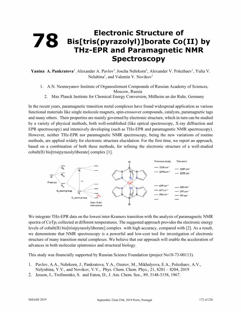
78 Electronic Structure of Bis[tris(pyrazolyl)]borate Co(II) by
THz-EPR and Paramagnetic NMR Spectroscopy
Yanina A. Pankratova1
, Alexander A. Pavlov1, Joscha Nehrkorn2, Alexander V. Polezhaev1, Yulia V.
Nelubina1, and Valentin V. Novikov1
1. A.N. Nesmeyanov Institute of Organoelement Compounds of Russian Academy of Sciences, Moscow, Russia
2. Max Planck Institute for Chemical Energy Conversion, Mülheim an der Ruhr, Germany
In the recent years, paramagnetic transition metal complexes have found widespread application as various functional materials like single molecule magnets, spin-crossover compounds, catalysts, paramagnetic tags and many others. Their properties are mainly governed by electronic structure, which in turn can be studied by a variety of physical methods, both well-established (like optical spectroscopy, X-ray diffraction and EPR spectroscopy) and intensively developing (such as THz-EPR and paramagnetic NMR spectroscopy). However, neither THz-EPR nor paramagnetic NMR spectroscopy, being the new variations of routine methods, are applied widely for electronic structure elucidation. For the first time, we report an approach, based on a combination of both these methods, for refining the electronic structure of a well-studied cobalt(II) bis[tris(pyrazolyl)borate] complex [1].
We integrate THz-EPR data on the lowest inter-Kramers transition with the analysis of paramagnetic NMR spectra of CoTp2 collected at different temperatures. The suggested approach provides the electronic energy levels of cobalt(II) bis[tris(pyrazolyl)borate] complex with high accuracy, compared with [2]. As a result, we demonstrate that NMR spectroscopy is a powerful and low-cost tool for investigation of electronic structure of many transition metal complexes. We believe that our approach will enable the acceleration of advances in both molecular spintronics and structural biology.
This study was financially supported by Russian Science Foundation (project No18-73-00113).
1. Pavlov, A.A., Nehrkorn, J., Pankratova, Y.A., Ozerov, M., Mikhalyova, E.A., Polezhaev, A.V., Nelyubina, Y.V., and Novikov, V.V., Phys. Chem. Chem. Phys., 21, 8201 – 8204, 2019
2. Jesson, J., Trofimenko, S. and Eaton, D., J. Am. Сhem. Soc., 89, 3148-3158, 1967.
SMASH 2019 September 22nd-25th, 2019 Porto, Portugal 172 of 226
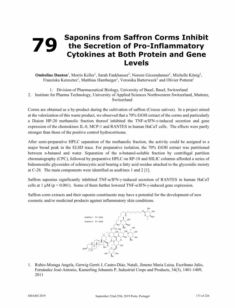
79 Saponins from Saffron Corms Inhibit the Secretion of Pro-Inflammatory Cytokines at Both Protein and Gene
Levels
Ombeline Danton1, Morris Keller1, Sarah Fankhauser2, Noreen Giezendanner2, Michelle König2, Franziska Keresztez1, Matthias Hamburger1, Veronika Butterweck2 and Olivier Potterat1
1. Division of Pharmaceutical Biology, University of Basel, Basel, Switzerland
2. Institute for Pharma Technology, University of Applied Sciences Northwestern Switzerland, Muttenz, Switzerland
Corms are obtained as a by-product during the cultivation of saffron (Crocus sativus). In a project aimed at the valorization of this waste product, we observed that a 70% EtOH extract of the corms and particularly a Diaion HP-20 methanolic fraction thereof inhibited the TNF-α/IFN-γ-induced secretion and gene expression of the chemokines IL-8, MCP-1 and RANTES in human HaCaT cells. The effects were partly stronger than those of the positive control hydrocortisone.
After semi-preparative HPLC separation of the methanolic fraction, the activity could be assigned to a major broad peak in the ELSD trace. For preparative isolation, the 70% EtOH extract was partitioned between n-butanol and water. Separation of the n-butanol-soluble fraction by centrifugal partition chromatography (CPC), followed by preparative HPLC on RP-18 and HILIC columns afforded a series of bidesmosidic glycosides of echinocystic acid bearing a fatty acid residue attached to the glycosidic moiety at C-28. The main components were identified as azafrines 1 and 2 [1].
Saffron saponins significantly inhibited TNF-α/IFN-γ-induced secretion of RANTES in human HaCaT cells at 1 μM (p < 0.001). Some of them further lowered TNF-α/IFN-γ-induced gene expression.
Saffron corm extracts and their saponin constituents may have a potential for the development of new cosmetic and/or medicinal products against inflammatory skin conditions.
ROOH
O
O
O
O O
O
OOH
OHOH
O
OOH
OHOH
OH
O
O
OO
HOOH
OH
OH
O
OO
O
OH
OH
OHOH
HOOH
H
H
H
Azafrine 1 R = GlcA
Azafrine 2 R = GalA
O
1. Rubio-Moraga Angela, Gerwig Gerrit J, Castro-Diȧz, Natali, Jimeno Maria Luisa, Escribano Julio, Fernȧndez José-Antonio, Kamerling Johannis P, Industrial Crops and Products, 34(3), 1401-1409, 2011
SMASH 2019 September 22nd-25th, 2019 Porto, Portugal 173 of 226
80 Method Optimization for Determination of Okadaic Acid in
Solution by Quantitative NMR Spectroscopy with External Standard
Method, and Validation by a Collaborative Study
Taichi Yamazaki1, Ryuichi Watanabe2, Tsuyoshi Kato3 and Masahiko Numata1
1. National Institute of Advanced Industrial Science and technology (AIST), National Metrology
Institute of Japan (NMIJ), Tsukuba, Ibaraki, Japan 2. National Research Institute of Fisheries Science (NRIFS), Yokohama, Kanagawa, Japan
3. Japan Food Research Laboratories (JFRL), Tama, Tokyo, Japan Okadaic acid is one of the diarrhea shellfish toxins. An instrumental analysis of diarrhea shellfish toxins has been introduced into many laboratories all over the world, where the regulatory level of the okadaic acid group was set as 0.16 mg/kg at CODEX in 2008. Certified reference materials (CRMs) of the diarrhea shellfish toxins are required for the instrumental analysis. The National Research Council (NRC) Canada provides CRMs of which concentrations have been determined by qNMR with an external standard method [1]. This approach is expected to provide determinations for high concentration precious samples such as diarrhea shellfish toxins in solutions.
We have also tried to establish accurate qNMR spectroscopy for development of the diarrhea shellfish toxin standard solutions as CRMs [2]. In this research, we optimized sample preparation processes and measurement conditions of qNMR spectroscopy with the external standard method for the determination of low concentration okadaic acid in solution. Although the internal standard method is more accurate than the external standard method in general [3], the external standard method has an advantage that the precious sample is secured from the contamination with standard materials. Approximately 0.4 mg/mL of okadaic acid solution was determined directly by 1H NMR spectroscopy with the external standard method. Its relative expanded uncertainty was 6 % and the result was validated by qNMR with the internal standard method. Moreover, a collaborative study among three laboratories was performed and the accuracy and variation factors of qNMR spectroscopy were discussed. The variation by the external standard method was larger compared with that by the internal standard method. The main variation factor in this method was caused by changing sample tubes into the NMR instrument. For this reason, reliable values could be obtained by relatively larger number of measurements. In the collaborative study, the concentration of an okadaic acid solution was determined with approximately 6 % of variation between three laboratories. The details of the studies will be discussed in our presentation.
1. Burton I. W., Quilliam M. A., Walter J. A, Anal. Chem., 77, 3123, 2005. 2. R. watanabe, C. sugai. T. Yamazaki, et al., Toxins , 8, 294, 2016. 3. Christopher H C, G Joseph R, Christine M S, Magn. Reson. Chem., 51, 705, 2013.
SMASH 2019 September 22nd-25th, 2019 Porto, Portugal 174 of 226
81 Polymorphism and Tautomerism in Crystal Structures of Azoles and Benzazoles: SSNMR and GIPAW
Calculations
Rosa M. Claramunt1, Carla I. Nieto1, Marta Marín‐Luna2, Felipe Reviriego3, Ibon Alkorta4 and José Elguero4
1. Departamento de Química Orgánica y Bio‐Orgánica, UNED, 28040 Madrid, Spain
2. Departamento de Química Orgánica, Universidad de Vigo, 36310 Vigo, Spain 3. Instituto de Ciencia y Tecnología de Polímeros, CSIC, 28006 Madrid, Spain
4. Instituto de Química Médica, CSIC, 28006 Madrid, Spain Polymorphism refers to the ability of a compound in the solid state to exist in two or more spatial arrangements, namely, polymorphs, keeping unaltered its molecular connectivity [1,2,3]. Tautomerism is a related term that involves the interconversion between two isomers trough an electrofuge group (being called prototropy if the group is a proton). These concepts are especially relevant in medicinal chemistry, reinforcing or blocking the activity of some drugs [4]. In this sense, the azole and benzazole families are known for being a representative example of tautomerism due to the easy prototropic process between the N atoms of the heterocycle five membered ring [5].
We have used solid-state NMR together with GIPAW calculations to investigate polymorphism and tautomerism in three pairs of compounds, two based on the pyrazole scaffold, 3(5)‐phenyl‐1H‐pyrazole 1 and 3(5)‐phenyl‐4‐bromo‐1H-pyrazole 2, and a indazole derivative, 3‐phenyl‐1H(2H)‐indazole 3. Phenylpyrazole 1 is one of the few examples known that presents desmotropy; that is, it crystallizes in two different tautomers 1a (HUMLUW) and 1b (UHENIE). On the other hand, compounds 2 and 3 present two different polymorphs each in the crystal lattice; in the case of 2 (PAMTAY and PAMTAY01) it is the 3‐phenyl tautomer, and in the case of 3 (UHENUQ and UHENUQ01) the 1H‐tautomer. Statistical analysis of the computed 13C‐NMR and 15N‐NMR chemical shifts indicates that the GIPAW approach reproduces with a high degree of accuracy the experimental data [6,7].
Acknowledgements. Thanks are given to the UNED research group "Sistemas Supramoleculares Bioorgánicos", the Universidad de Vigo and the CTI of CSIC for their continued support.
1. Bernstein, J, Cryst. Growth Des. 11, 632-650. 2011. 2. Gavezzotti, AJ, J. Pharm. Sci. 96, 2232-2241, 2007. 3. Elguero, J, Cryst. Growth Des. 11, 4731-4738, 2011. 4. Cruz‐Cabeza, AJ, Bernstein, J, Chem. Rev. 114, 2170-2191, 2014. 5. Elguero, J, Alkorta, I, R. M. Claramunt, RM, P. Cabildo, P, P. Cornago, P, Farrán, MA, García, MA,
López, C, Pérez‐Torralba, M, María, DS, Sanz, D, Chem. Heterocycl. Comp. 49, 177-202, 2013. 6. Marín-Luna, M, Alkorta, I, Elguero, J, Magn. Reson. Chem. 56, 164-171, 2018. 7. Marín-Luna, M, Claramunt RM, Nieto, CI, Alkorta, I, Elguero, J, Reviriego, F, Magn. Reson. Chem.
DOI: 10.1002/mrc.4824.
SMASH 2019 September 22nd-25th, 2019 Porto, Portugal 175 of 226
82 NOMAD – NMR Online Management and Datastore
Shyam M. Reyal1, Simone I. Conte1, Michalis Psalios1, Ignacy Debicki1, Dasha Savanovich1,
and Tomas Lebl2
1. School of Computer Science, University of St Andrews, Scotland, UK 2. EaStCHEM - School of Chemistry, University of St Andrews, Scotland, UK
In the last decade, amount of NMR data that large NMR facilities produce has dramatically increased due to high efficiency of fully automated instruments. However, this large quantity of data is not accordingly stored and shared despite of its considerable intrinsic value. NMR spectroscopy is lacking a global data depository that would be an equivalent of CCDC for X-ray crystallography data. The status quo has not been changing much despite of the open access data enforcement of research councils and initiatives like Go Fair [1] and NMReDATA [2]. We believe that the core of the issue is at the bottom of the pyramid. Data management in academic NMR laboratories is rather poor as the raw data are usually stored on a network drive without any metadata and search facilities. When the research is concluded, finding and uploading data into research data depository like Figshare or Zenodo becomes a troublesome commitment that researchers rather avoid at any cost.
NOMAD tries to solve the problem from the bottom up by providing smart and complete solution for NMR laboratory data management that enables to store NMR data securely and robustly together with provenance meta-data and facilitates sharing and audibility. It automatically captures and stores data generated by NMR instruments and provides seamless tools for annotating, sharing and editing the data through easily accessible web browser interface. Furthermore, NOMAD offers a centralised dashboard for the management of large automated NMR laboratories that operate 24 hours per day, 7 days per week. In long term, we envisage that NOMAD could form a peer-to-peer network of nodes that would facilitate exchange of raw NMR data between participants in accordance with FAIR Data Principles and potentially lead the way towards a decentralised NMR big data depository.
1. Wilkinson, M. D. et al., Scientific Data, 3, 160018, 2016. (doi:10.1038/sdata.2016.18) 2. Pupier, M. et al., Magn Reson Chem., 56, 703–715, 2018. (doi: 10.1002/mrc.4737)
SMASH 2019 September 22nd-25th, 2019 Porto, Portugal 176 of 226
83 ROESY Based Structural Studies of Anomalous High Order Complexes
Between Daclatasvir and Some Cyclodextrins
Antonio Salgado1, Sulaiman Krait2, Gerhard K. E. Scriba2 and Bezhan Chankvetadze3
1. NMR Spectroscopy Centre (CERMN), University of Alcalá, Alcalá de Henares, Madrid, Spain 2. Department of Pharmaceutical Chemistry, Friedrich-Schiller-University Jena, Jena, Germany
3. Institute of Physical and Analytical Chemistry, Tbilisi State University, Tbilisi, Georgia In routine separations of daclatasvir (DCV) enantiomers by capillary electrophoresis (CE), an unexpected and persistent plateau was observed in the electropherograms when native γ-CD was used as the chiral selector [1]. In order to rationalize this behaviour, NMR ROESY experiments were conducted to probe into the structure of the supramolecular aggregates (complexes) formed by daclatasvir enantiomers and the chiral selectors [2]. It was seen that the 1H NMR spectrum of pure enantiomer (S,S,S,S)-DCV, upon mixing with γ-CD under conditions that mimicked those in the CE runs, showed three different sets of signals assigned to DCV and other two due to γ-CD. One of the γ-CD set of signals was noticeably shifted upfields. Full signal assignment was done on the basis of selective TOCSY, COSY and HSQCED experiments and by comparison to data of DCV and γ-CD alone. It was concluded that the three sets of DCV signals corresponded to two different modes of complexation. In the first of them, a DCV molecule was inserted into the γ-CD cavity (1:1 complexes), whereas the second mode of complexation consisted of two stacked DCV molecules accommodated inside the CD macrocycle (2:1 complexes). Detailed structures of these complexes could be derived based on ROESY results.
1. Scriba, G. K. E., et al., manuscript in preparation. 2. Salgado, A., Chankvetadze, B., J. Chromatogr. A, 1467, 95-144, 2016.
SMASH 2019 September 22nd-25th, 2019 Porto, Portugal 177 of 226
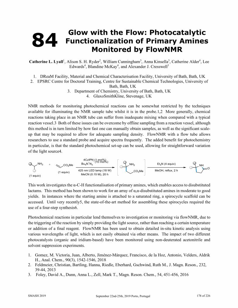
84 Glow with the Flow: Photocatalytic Functionalization of Primary Amines
Monitored by FlowNMR Catherine L. Lyall1, Alison S. H. Ryder2, William Cunningham3, Anna Kinsella3, Catherine Alder4, Lee
Edwards4, Blandine McKay4, and Alexander J. Cresswell3
1. DReaM Facility, Material and Chemical Characterisation Facility, University of Bath, Bath, UK 2. EPSRC Centre for Doctoral Training, Centre for Sustainable Chemical Technologies, University of
Bath, Bath, UK 3. Department of Chemistry, University of Bath, Bath, UK
4. GlaxoSmithKline, Stevenage, UK NMR methods for monitoring photochemical reactions can be somewhat restricted by the techniques available for illuminating the NMR sample tube whilst it is in the probe.1,2 More generally, chemical reactions taking place in an NMR tube can suffer from inadequate mixing when compared with a typical reaction vessel.3 Both of these issues can be overcome by offline sampling from a reaction vessel, although this method is in turn limited by how fast one can manually obtain samples, as well as the significant scale-up that may be required to allow for adequate sampling density. FlowNMR with a flow tube allows researchers to use a standard probe and acquire spectra frequently. The added benefit for photochemistry in particular, is that the standard photochemical set-up can be used, allowing for straightforward variation of the light source4.
NH2 NH2
(1 equiv)(1 equiv) 425 nm LED lamp (18 W)
4CzIPN (1 mol%)Bu4N+N3
− (10 mol%)
MeCN (0.15 M), 20 h
+ NHO
CO2Me
Et3N (4 equiv)CO2Me
MeOH, reflux, 2 h
This work investigates the α-C-H functionalisation of primary amines, which enables access to disubstituted lactams. This method has been shown to work for an array of α,α-disubstituted amines in moderate to good yields. In instances where the starting amine is attached to a saturated ring, a spirocycle scaffold can be accessed. Until very recently5, the state-of-the-art method for assembling these spirocycles required the use of a four-step synthesis6.
Photochemical reactions in particular lend themselves to investigation or monitoring via flowNMR, due to the triggering of the reaction by simply providing the light source, rather than reaching a certain temperature or addition of a final reagent. FlowNMR has been used to obtain detailed in-situ kinetic analysis using various wavelengths of light, which is not easily obtained via other means. The impact of two different photocatalysts (organic and iridium-based) have been monitored using non-deuterated acetonitrile and solvent suppression experiments.
1. Gomez, M. Victoria, Juan, Alberto, Jiménez-Márquez, Francisco, de la Hoz, Antonio, Velders, Aldrik H., Anal. Chem., 90(3), 1542-1546, 2018
2. Feldmeier, Christian, Bartling, Hanna, Riedle, Eberhard, Gschwind, Ruth M., J. Magn. Reson., 232, 39-44, 2013
3. Foley, David A., Dunn, Anna L., Zell, Mark T., Magn. Reson. Chem., 54, 451-456, 2016
SMASH 2019 September 22nd-25th, 2019 Porto, Portugal 178 of 226
4. Hall, Andrew M. R., Broomfield-Tagg, Rachael, Camilleri, Matthew, Carbery, David R., Codina, Anna, Wittaker, David T. E., Coombes, Steven, Lowe, John P., Hintermair, Ulrich, Chem. Commun., 54, 30-33, 2018
5. Mullen, Philip, Hugues, Miela M., McKerveya, Anthony, Tetrahedron Lett., 51, 3216-3217, 2010 6. Ye, Juntao, Kalvet, Indrek, Schoenebeck, Franziska, Rovis, Tomislav, Nat. Chem., 10, 1037-1041,
2018
SMASH 2019 September 22nd-25th, 2019 Porto, Portugal 179 of 226
85 NMR Spectroscopy and Magnetometry of the Cobalt(II)
Single Molecule Magnet with Unusual Coordianation Geometry
Dmitry Aleshin1,2, Alexander Pavlov1, Rosa Diego3,4, Mohanad Darawsheh3, Josha Nehrkorn5, Yulia
Nelyubina1, Olivier Roubeau6, Valentin Novikov1 and GuillemAromí3,4 1. A. N. Nesmeyanov Institute of Organoelement Compounds, Russian Academy of Sciences, Moscow,
Russia 2. Mendeleev University of Chemical Technology of Russia, Moscow, Russia
3. Departament de Química Inorgánica i Orgánica, Universitat de Barcelona, Barcelona, Spain 4. Institute of Nanoscience and Nanotechnology of the University of Barcelona, Barcelona, Spain
5. Max Planck Institute for Chemical Energy Conversion, Mülheim and der Ruhr, Germany 6. Instituto de Ciencia de Materiales de Aragón (ICMA),CSIC and Universidad de Zaragoza, Zaragoza,
Spain Single molecule magnets (SMMs) are compounds that keep magnetization on the molecular scale, unlike conventional bulk magnets that exhibits collective long-range magnetic ordering. In view of above, SMMs have several potentional applications in many areas, the most promising one is high density fata storage. SMMs keep magnetization due to the presence of the magnetization reversal energy barrier U, caused by zero field splitting energy D. Since D has negative values in SMMs, the direct transition from Ms = -S to Ms = +S is forbidden by the selection rules. The greatest value of U among 3d metals-based SMMs are shown by Co(II) complexes[1].
The obtained dinucleur cobalt(II) complex has unusual geometry of the coordination polyhedron. It is intermediate between trigonal prism (TP) and trigonal antiprism (TAP). According to single crystal X-ray diffraction data, the distorsion angle is equal to 38o (while distorsion angles for TP and TAP are equal to 0o and 60o, respectively). Such complexes are very interesting for understanding of magnetostructural correlations for cobalt(II)-based SMMs [2].
For determination of electron structure of this complex we applied NMR spectroscopy and magnetometry, allowing to determine axial anisotropy (Δχax) and isotropic value of the magnetic susceptibility tensor. For room temperature Δχax = 0.05 A3 is strongly lower than for the both TP (0.26 A3) [3] and TAP (0.26 A3) [4] geometries. By comparison of NMR and magnetometry data for Co2L3 and similar CoZnL3 complexes we excluded exchange coupling between two Co+2 ions as origin of such low magnetic anisotropy. In this poster we will discussapplicationof NMR spectroscopy and magnetometry for electron structure study of the binuclear cobalt(II) complex.
SMASH 2019 September 22nd-25th, 2019 Porto, Portugal 180 of 226
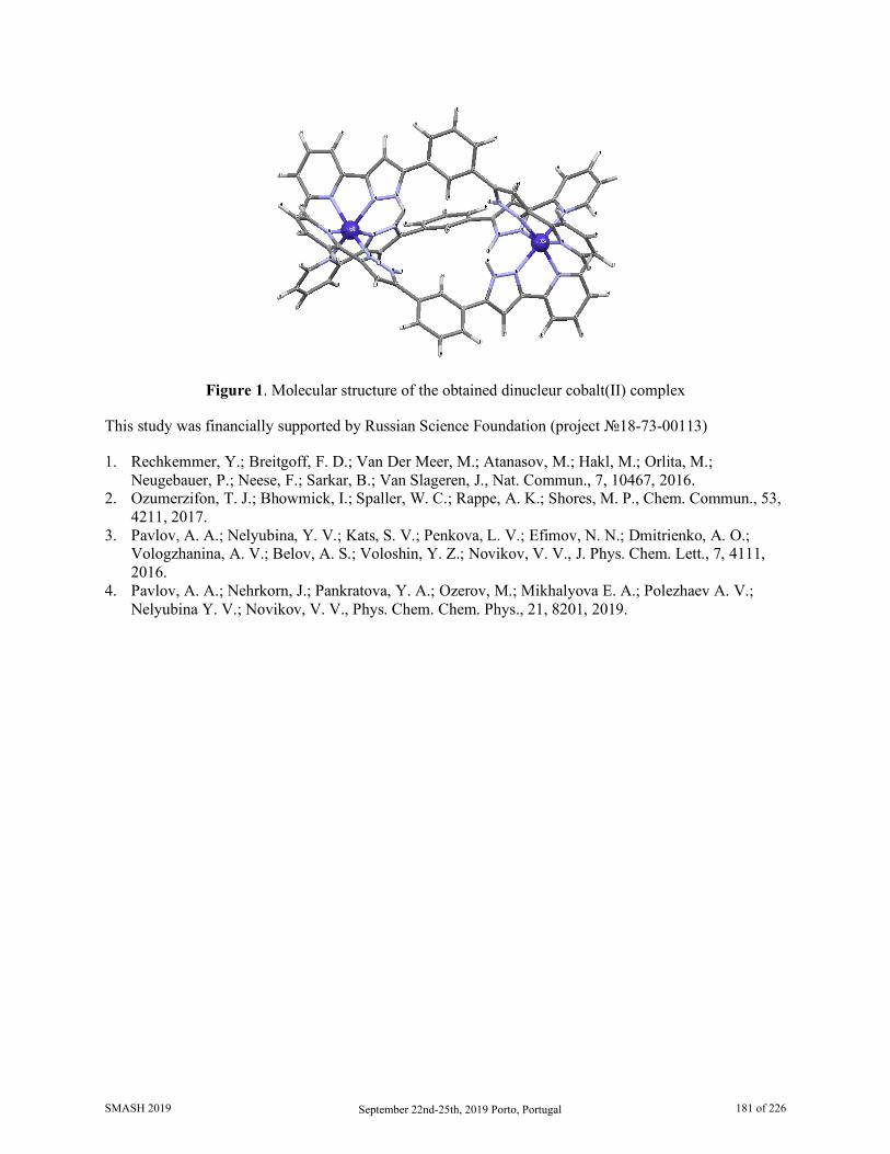
Figure 1. Molecular structure of the obtained dinucleur cobalt(II) complex
This study was financially supported by Russian Science Foundation (project №18-73-00113)
1. Rechkemmer, Y.; Breitgoff, F. D.; Van Der Meer, M.; Atanasov, M.; Hakl, M.; Orlita, M.; Neugebauer, P.; Neese, F.; Sarkar, B.; Van Slageren, J., Nat. Commun., 7, 10467, 2016.
2. Ozumerzifon, T. J.; Bhowmick, I.; Spaller, W. C.; Rappe, A. K.; Shores, M. P., Chem. Commun., 53, 4211, 2017.
3. Pavlov, A. A.; Nelyubina, Y. V.; Kats, S. V.; Penkova, L. V.; Efimov, N. N.; Dmitrienko, A. O.; Vologzhanina, A. V.; Belov, A. S.; Voloshin, Y. Z.; Novikov, V. V., J. Phys. Chem. Lett., 7, 4111, 2016.
4. Pavlov, A. A.; Nehrkorn, J.; Pankratova, Y. A.; Ozerov, M.; Mikhalyova E. A.; Polezhaev A. V.; Nelyubina Y. V.; Novikov, V. V., Phys. Chem. Chem. Phys., 21, 8201, 2019.
SMASH 2019 September 22nd-25th, 2019 Porto, Portugal 181 of 226
86 NMR Crystallography for the Acceleration of the Development of
New Medicines
Andrew Tatton, Luca Russo and Tran Pham
GSK R&D, Medicinal Science and Technology, Stevenage, Hertfordshire, UK Progression of new medicines to market often requires acceleration in the development of drug candidate molecules. In the solid form space, this can lead to some challenges since a lack of solid form knowledge can pose a high risk to the development of a medicine. Crystal structure analysis is therefore essential to obtaining an understanding of the solid form properties of a drug candidate molecule. While the gold standard is single-crystal X-ray diffraction, when a project is accelerated it may not always be possible or practical to obtain single crystals for all the relevant solid forms of that molecule of sufficient quality to enable a full understanding of the linkage between the crystal structure and the molecular properties which may give insight into its effectiveness as a medicine. Recent advancements in NMR crystallography coupled with density functional theory calculation have demonstrated the capability of two highly complementary techniques, namely solid-state NMR spectroscopy and X-ray powder diffraction, to provide an accurate crystal structure determination from a powder sample of a drug substance.[1,2]
In this poster a general crystal structure determination approach is proposed, including NMR crystallography, which can be incorporated into the development of new medicines. We demonstrate that the approach is capable of providing the crystal structures of drug candidates rapidly and accurately from powder samples without the need for potentially time-consuming or challenging crystal growth, therefore providing the characterisation data required to understand the solid forms for drug candidates and as a result enable acceleration of the development of medicines for patients.
1. Clark, Stewart J. ; Segall, Matthew D.; Pickard, Chris J.; Hasnip, Phil J. ; Probert, Matt I. J.; Refson, Keith ; Payne, Mike C., Z. Kristallogr. Cryst. Mater., 220, 567-570, 2005.
2. Tatton, Andrew S.; Blade, Helen; Brown, Steven P.; Hodgkinson, Paul; Hughes, Leslie P.; Nilsson Lill, Sten O.; Yates, Jonathan R., Cryst. Growth Des. 18, 3339-3351, 2018.
SMASH 2019 September 22nd-25th, 2019 Porto, Portugal 182 of 226

87 Helium Recovery and Recycling at GSK Stevenage
Andy Edwards and Tran Pham
GSK R&D, Medicinal Science and Technology, Stevenage, Hertfordshire, UK
NMR spectroscopy is used at all stages in the discovery and development of new medicines. At its heart is a superconducting magnet cooled by liquid helium to a temperature of 4 K. Helium continually boils off at a low rate and, once lost from the magnet, it will escape from the earth's atmosphere and be lost in space forever. NMR spectrometers at GSK Stevenage consume approximately 4000 L of liquid helium each year. During a complete refurbishment of the NMR laboratories at Stevenage, bringing all the spectrometers into a single laboratory for the first time, there was an opportunity to design and build infrastructure for helium recovery and recycling. In this new laboratory, all 9 magnets are connected via flexible metal hoses into a ring main which is routed into a helium collection "balloon". Once full, a high power compressor compresses the gas to 200 bar in cylinders which are then collected for recycling to liquid helium off-site. The new infrastructure is now fully operational, recovering and recycling helium to help sustain this finite resource.
SMASH 2019 September 22nd-25th, 2019 Porto, Portugal 183 of 226
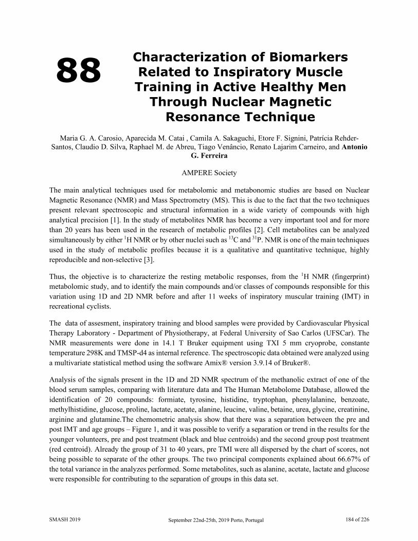
88 Characterization of Biomarkers Related to Inspiratory Muscle Training in Active Healthy Men
Through Nuclear Magnetic Resonance Technique
Maria G. A. Carosio, Aparecida M. Catai , Camila A. Sakaguchi, Etore F. Signini, Patrícia Rehder-
Santos, Claudio D. Silva, Raphael M. de Abreu, Tiago Venâncio, Renato Lajarim Carneiro, and Antonio G. Ferreira
AMPERE Society
The main analytical techniques used for metabolomic and metabonomic studies are based on Nuclear Magnetic Resonance (NMR) and Mass Spectrometry (MS). This is due to the fact that the two techniques present relevant spectroscopic and structural information in a wide variety of compounds with high analytical precision [1]. In the study of metabolites NMR has become a very important tool and for more than 20 years has been used in the research of metabolic profiles [2]. Cell metabolites can be analyzed simultaneously by either 1H NMR or by other nuclei such as 13C and 31P. NMR is one of the main techniques used in the study of metabolic profiles because it is a qualitative and quantitative technique, highly reproducible and non-selective [3].
Thus, the objective is to characterize the resting metabolic responses, from the 1H NMR (fingerprint) metabolomic study, and to identify the main compounds and/or classes of compounds responsible for this variation using 1D and 2D NMR before and after 11 weeks of inspiratory muscular training (IMT) in recreational cyclists.
The data of assesment, inspiratory training and blood samples were provided by Cardiovascular Physical Therapy Laboratory - Department of Physiotherapy, at Federal University of Sao Carlos (UFSCar). The NMR measurements were done in 14.1 T Bruker equipment using TXI 5 mm cryoprobe, constante temperature 298K and TMSP-d4 as internal reference. The spectroscopic data obtained were analyzed using a multivariate statistical method using the software Amix® version 3.9.14 of Bruker®.
Analysis of the signals present in the 1D and 2D NMR spectrum of the methanolic extract of one of the blood serum samples, comparing with literature data and The Human Metabolome Database, allowed the identification of 20 compounds: formiate, tyrosine, histidine, tryptophan, phenylalanine, benzoate, methylhistidine, glucose, proline, lactate, acetate, alanine, leucine, valine, betaine, urea, glycine, creatinine, arginine and glutamine.The chemometric analysis show that there was a separation between the pre and post IMT and age groups – Figure 1, and it was possible to verify a separation or trend in the results for the younger volunteers, pre and post treatment (black and blue centroids) and the second group post treatment (red centroid). Already the group of 31 to 40 years, pre TMI were all dispersed by the chart of scores, not being possible to separate of the other groups. The two principal components explained about 66.67% of the total variance in the analyzes performed. Some metabolites, such as alanine, acetate, lactate and glucose were responsible for contributing to the separation of groups in this data set.
SMASH 2019 September 22nd-25th, 2019 Porto, Portugal 184 of 226

Figure 1. Graphs of PCA scores (left) and loadings (right), PC1 vs PC2, for blood serum samples evaluating pre and post IMT groups, being volunteers aged 20 to 30 years pre IMT - black color, 20 to 30 years post IMT – blue color, 31 to 40 years old pre IMT - green color and 31 to 40 years post IMT - red
color.
In addition, with PCA there was a separation between two groups - the control group and those who received one of the IMT training - Figure 2. The metabolites that contributed the most to this separation were lactate, leucine, glycine and glucose.
Figure 2. Graphs of PCA scores (left) and loadings (right), PC1 vs PC2, for blood serum samples evaluating two IMT groups, control (black) and IMT trained (blue).
NMR combined with chemometrics was a powerful tool that allowed the identification of chemical markers related to the age of the volunteers in the proposed study and the pre and post IMT. Twenty metabolites have been identified so far, with glucose, acetate, glycine and betaine being the main chemical compounds responsible for the discrimination of the volunteer groups.
1. Zhang, Ai Hua & Sun, Hui & Wang, Ping & Han, Ying & Wang, Xi-Jun. The Analyst. 137. 293-300. 2011.
2. Markley, John L, Brüschweiler, Rafael, Edison, Arthur S, Eghbalnia, Hamid R, Powers, Robert, Raftery, Daniel , Wishart, David S. Biotechnology. 43. 34-40. 2017.
3. Emwas, Abdul-Hamid, Roy, Raja, McKay, Ryan T., Tenori, Leonardo, Saccenti, Edoardo, Gowda, Nagana G. A., Raftery, Daniel, Alahmari, Fatimah, Jaremko, Lukasz, Jaremko, Mariusz, Wishart, David S. Metabolites. 9, 123. 2019.
SMASH 2019 September 22nd-25th, 2019 Porto, Portugal 185 of 226
89 NMR and Docking Studies of Novel Macrolide Conjugates
Predrag Novak1, Ivana Mikulandra1, Iva Habinovec1, Tomislav Jednačak1, Branimir Bertoša1, Sonja
Škevin1, Mirjana Bukvić2 and Ivan Grgičević2
1. Department of Chemistry, Faculty of Science, University of Zagreb, HR-1000 Zagreb, Croatia 2. Fidelta Ltd., HR-10000 Zagreb, Croatia
Macrolide antibiotics are effective and well-tolerated therapeutic agents for treating infectious diseases owing to their high efficacy and safety [1]. They have been in widespread clinical use for over 60 years and are effective against Gram-positive and certain Gram-negative microorganisms. Macrolides exert their biological activity by binding to the 50S bacterial ribosome subunit at early stage of the translation process. They bind to the ribosomal 23S rRNA in domain V at or near the peptidyl transferase region and block the exit tunnel through which the nascent peptides leave the ribosome. However, in spite of a number of existing antibiotics, the emerging multi-drug resistant microbial pathogens present serious and challenging problems in medical treatment which demand novel and more effective antimicrobial agents to be discovered.
We have shown that NMR spectroscopy could reveal valuable data on free and bound state macrolide conformations [2-4] which can guide efforts directed to discovery of novel, more potent inhibitors. Here we present our recent NMR studies of new macrolide conjugates, the macrozones prepared by coupling of azithromycin and thiosemicarbazones. Macrozones have shown good activity against Gram positive strains, some Gram negative strains and also against efflux-mediated resistant Streptococcus pneumoniae. In order to characterize binding epitopes and asses bound conformations of macrozones we applied a combination of NMR experiments such as transferred nuclear Overhauser effect spectroscopy (trNOESY) and saturation transfer difference (STD) and molecular modeling.
We thank the Croatian Science Foundation for the financial support (project Macrozones, IP-2018-01-8098).
1. Arsić Biljana, Novak Predrag, Barber Jill, Rimoli Maria Grazia, Kragol Goran, Sodano Federica, Macrolides: properties, synthesis and applications, Walter de Gruyter, Berlin/Boston, 2018
2. Novak Predrag, Tatić Iva, Tepeš Predrag, Koštrun Sanja and Barber Jill, J. Phys. Chem. A, 110, 580-588, 2006
3. Kosol Simone, Schrank Eveline, Bukvić Krajačić Mirjana, Wagner E. Gabriel, Meyer N. Helge, Göbl Christoph, Rechberger N. Gerald, Zangger Klaus, Novak Predrag, J. Med. Chem. 55, 5632-5636, 2012
4. Novak Predrag, Barber Jill, Čikoš Ana, Arsić Biljana, Plavec Janez, Lazarevski Gorjana, Tepeš Predrag and Košutić-Hulita Nada, Bioorg. Med. Chem., 17, 5857-5867, 2009
SMASH 2019 September 22nd-25th, 2019 Porto, Portugal 186 of 226
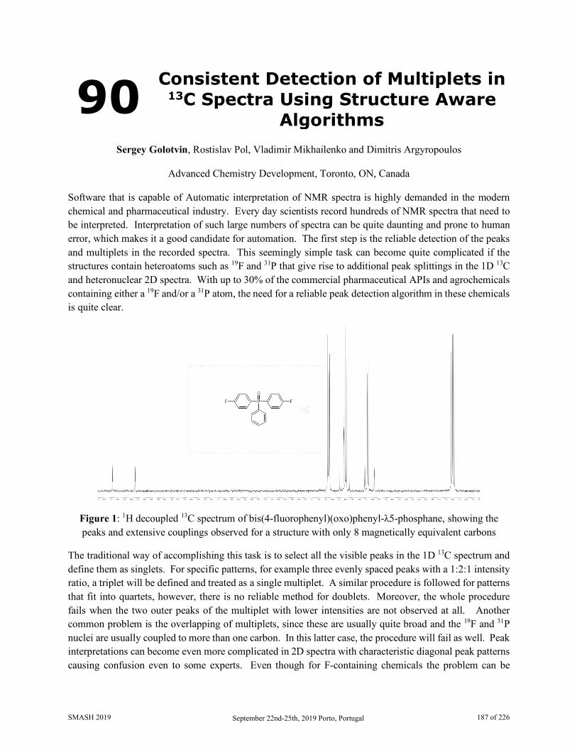
90 Consistent Detection of Multiplets in 13C Spectra Using Structure Aware
Algorithms
Sergey Golotvin, Rostislav Pol, Vladimir Mikhailenko and Dimitris Argyropoulos
Advanced Chemistry Development, Toronto, ON, Canada Software that is capable of Automatic interpretation of NMR spectra is highly demanded in the modern chemical and pharmaceutical industry. Every day scientists record hundreds of NMR spectra that need to be interpreted. Interpretation of such large numbers of spectra can be quite daunting and prone to human error, which makes it a good candidate for automation. The first step is the reliable detection of the peaks and multiplets in the recorded spectra. This seemingly simple task can become quite complicated if the structures contain heteroatoms such as 19F and 31P that give rise to additional peak splittings in the 1D 13C and heteronuclear 2D spectra. With up to 30% of the commercial pharmaceutical APIs and agrochemicals containing either a 19F and/or a 31P atom, the need for a reliable peak detection algorithm in these chemicals is quite clear.
Figure 1: 1H decoupled 13C spectrum of bis(4-fluorophenyl)(oxo)phenyl-λ5-phosphane, showing the peaks and extensive couplings observed for a structure with only 8 magnetically equivalent carbons
The traditional way of accomplishing this task is to select all the visible peaks in the 1D 13C spectrum and define them as singlets. For specific patterns, for example three evenly spaced peaks with a 1:2:1 intensity ratio, a triplet will be defined and treated as a single multiplet. A similar procedure is followed for patterns that fit into quartets, however, there is no reliable method for doublets. Moreover, the whole procedure fails when the two outer peaks of the multiplet with lower intensities are not observed at all. Another common problem is the overlapping of multiplets, since these are usually quite broad and the 19F and 31P nuclei are usually coupled to more than one carbon. In this latter case, the procedure will fail as well. Peak interpretations can become even more complicated in 2D spectra with characteristic diagonal peak patterns causing confusion even to some experts. Even though for F-containing chemicals the problem can be
SMASH 2019 September 22nd-25th, 2019 Porto, Portugal 187 of 226

alleviated by using costly required hardware and recording a spectrum with additional 19F decoupling, there is no such practical option for 31P.
Here we present a more generalized method for multiplet detection in such spectra, which resembles the way a human analyst would follow. Since a proposed structure usually exists with the positions of the fluorine and phosphorus atoms defined, the human expert would specifically know what to look for, instead of starting from zero. The way this can be implemented in software is by predicting the spectrum of the proposed structure and then looking for similar patterns in the experimental spectra. If such patterns are found then the multiplets are defined correctly and the analysis can proceed without getting a false negative result, and need for manual inspection. This study will demonstrate examples of overlapped and low-intensity multiplets arising from 19F and/or 31P couplings and how these are consistently detected using this approach, as well as characteristic 2D spectra with the associated problems completely resolved.
SMASH 2019 September 22nd-25th, 2019 Porto, Portugal 188 of 226

91 13C Solid State NMR Spectroscopy as a Method for Probing the Efficiency
of Biomass Pretreatments for Second Generation Ethanol
Production
Tatiana S Ribeiro1,3, Tatiane S Bianquini1, Camila Florencio2, Luis Alberto Colnago2, Cristiane S. Farinas2, Marcelo Carmo3
1. DCNME, Federal University of São Carlos, Araras-SP, Brazil.
2. Embrapa Instrumentation, São Carlos-SP, Brazil 3. IEK-3, Forschungszentrum Jülich, Jülich, Germany
The global energy system is heavily dependent on fossil fuels, which negatively impacts the environment. Therefore research into renewable energy sources is increasing to meet a variety of sustainability requirements [1]. 2G ethanol production is one of the main topics since cellulose is the most common polymer in nature [2]. The biomass pretreatment process can facilitate access for enzyme action [3] but it is necessary to identify the most adequate pretreatment for each biomass-enzyme system. The fast characterization of biomass structure can contribute to process definition and continuous monitoring by indicating the performance of each method to open the structure and increase enzyme action [3]. 13C ss-NMR was used to investigate the changes in cellulose, hemicellulose and lignin, as shown in Figure 1. The liquid hot water process (LWH), steam explosion process (SE) and steam exploded and delignified process (SED) were can be analyzed before and after the enzymatic treatment with different commercial enzymes (Cellic CTec2 and Cellic CTec3). Lignin carbon signals are extensive due to the more complex and disordered its structure. Peaks 3 (62.5 ppm) and 7 (84 ppm) predominantly correspond to the carbons of the amorphous region of cellulose, while the peaks 4 (65 ppm) and 8 (88.9 ppm) correspond to the carbons of the crystalline region of cellulose [4]. For LWH treatment, a strong decrease in signals 1 and 17, corresponding to hemicellulose after the pretreatment process. Peaks 6 and 9 have disappeared, following the increase in the spectral resolution in the region of 50-70 ppm, reinforcing the hemicellulose removal [5]. The lignin region did not show significant changes, suggesting that most lignin was retained in the bagasse and remains in the sample. It was also observed that after the enzymatic hydrolysis with Cellic CTec3 enzyme, the lignin region becomes more intense. The higher intesity on peaks 4 and 8 was observed, indicating an increase in the crystalline part of the cellulose. For the SE process, the lignin signals decrease as expected, indicating the better lignin removal, while hemicellulose was retained, as shown by the higher intensity on peaks 1 and 17. Compared to SED, this last treatment was more effective in removing lignin and hemicellulose, as expected and evinced by the lower intensity of peaks 1 and 2 (and related peaks). With hemicellulose removal in all pretreated samples, the signal s from 50 and 100 ppm have become more evident due to the increased effective cellulose content (peaks 4 and 8 regard to crystalline cellulose). Of all pretreatments, the best results were LHW followed by the enzymatic action of Cellic Ctec3 because it was observed that lignin and hemicellulose were retained in the bagasse. In addition, it was observed that crystalline part of bagasse increase and this suggests that amorphous part o f bagasse was removed.
SMASH 2019 September 22nd-25th, 2019 Porto, Portugal 189 of 226
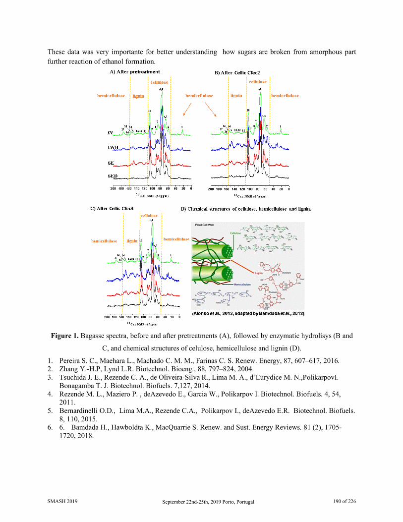
These data was very importante for better understanding how sugars are broken from amorphous part further reaction of ethanol formation.
Figure 1. Bagasse spectra, before and after pretreatments (A), followed by enzymatic hydrolisys (B and
C, and chemical structures of celulose, hemicellulose and lignin (D).
1. Pereira S. C., Maehara L., Machado C. M. M., Farinas C. S. Renew. Energy, 87, 607–617, 2016. 2. Zhang Y.-H.P, Lynd L.R. Biotechnol. Bioeng., 88, 797–824, 2004. 3. Tsuchida J. E., Rezende C. A., de Oliveira-Silva R., Lima M. A., d’Eurydice M. N.,PolikarpovI.
Bonagamba T. J. Biotechnol. Biofuels. 7,127, 2014. 4. Rezende M. L., Maziero P. , deAzevedo E., Garcia W., Polikarpov I. Biotechnol. Biofuels. 4, 54,
2011. 5. Bernardinelli O.D., Lima M.A., Rezende C.A., Polikarpov I., deAzevedo E.R. Biotechnol. Biofuels.
8, 110, 2015. 6. 6. Bamdada H., Hawboldta K., MacQuarrie S. Renew. and Sust. Energy Reviews. 81 (2), 1705-
1720, 2018.
SMASH 2019 September 22nd-25th, 2019 Porto, Portugal 190 of 226
92 31P NMR Study of
Trimethylphosphine Probe Adsorbed on Y Zeolite Modified with Boron
Jessyka Padilla1, Juan Poveda1, Daniel Molina1 and Alexander Guzmán2
1. Universidad Industrial de Santander, Carrera 27 calle 9 Bucaramanga, COL 2. ECOPETROL S.A. Instituto Colombiano del petróleo, Piedecuesta, COL
The Y zeolite is a microporous crystalline synthetic aluminosilicate with a pore opening of 7.4 Å, acidity adjustable due to the Si/Al ratio and high thermal and hydrothermal stability, which makes these materials suitable for their application in heterogeneous catalysis 1–3. Nowadays, great difficulties have been generated in the refinery processes being that heavy and extra heavy oils production have increased, which have an higher proportion of heavy fractions whose molecules have average molecular diameter between 1.2 to 15 nm and it limits its diffusion inside of the micropores of the zeolite Y as is synthesized 4–6. To improve diffusion properties in zeolites, treatments of dealumination and desilication post-synthesis by steaming or acid leaching to produce mesopores with heterogeneous distribution 7,8. Mi, S. et. al. sugested that boron insertion in Y zeolite structure promotes hydrolysis of the Si-O-Al and Si-O-B bonds during steaming resulting in an interconnected mesoporous system better than is obtained by conventional dealumination and genereating higher catalytic activity compared with zeolite without boron 9. In this study, Y zeolite modified with boron was obtained by direct hydrothermal synthesis using Water glass, Sodium aluminate solution and Boric acid (H3BO3) as reagent sources. It was found that the boron leaching of the structure is possible by washing to generate an increase in the volume of mesopores. 11B MAS NMR spectra confirmed tetrahedral sites of boron in B-Y remnants in the structure before and after steaming, and its effect on acid properties of zeolites was determined by 31P MAS NMR of trimethylphosphine adsorbed. The structural elucidation of samples was also resolute by X-ray diffraction and 29Si, 27Al and 27Al MQ MAS NMR. In addition, Argon adsorption/desorption isotherms were obtained at liquid Ar temperature of 83 K and used for calculation of BET specific surface area , pore size distribution and area and volume of pores (BJH and MP). 1. Baerlocher, C., McCusker, L. B. & Olson, D. Atlas of zeolite framework types. Structure
Commission of the International Zeolite Association 12, (Elsevier, 2007). 2. Karami, D. & Rohani, S. A Novel Approach for the Synthesis of Zeolite Y. Ind. Eng. Chem. Res. 48,
4837–4843 (2009). 3. Vermeiren, W. & Gilson, J.-P. Impact of Zeolites on the Petroleum and Petrochemical Industry. Top.
Catal. 52, 1131–1161 (2009). 4. Chen, W., Han, D., Sun, X. & Li, C. Studies on the preliminary cracking of heavy oils : Contributions
of various factors. Fuel 106, 498–504 (2013). 5. Pan, M. et al. Construction and practical application of a novel zeolite catalyst for hierarchically
cracking of heavy oil. J. Catal. 369, 72–85 (2019). 6. Primo, A. & Garcia, H. Zeolites as catalysts in oil refining. Chem. Soc. Rev. 43, 7548–7561 (2014). 7. Silaghi, M. C., Chizallet, C. & Raybaud, P. Challenges on molecular aspects of dealumination and
desilication of zeolites. Microporous Mesoporous Mater. 191, 82–96 (2014).
SMASH 2019 September 22nd-25th, 2019 Porto, Portugal 191 of 226

8. Silaghi, M. C., Chizallet, C., Sauer, J. & Raybaud, P. Dealumination mechanisms of zeolites and extra-framework aluminum confinement. J. Catal. 339, 242–255 (2016).
9. Mi, S. et al. Catalytic function of boron to creating interconnected mesoporosity in microporous Y zeolites and its high performance in hydrocarbon cracking. J. Catal. 347, 116–126 (2017).
SMASH 2019 September 22nd-25th, 2019 Porto, Portugal 192 of 226
93 How the Methyl Groups Affect the Spin-Orbit Component of 13C
Chemical Shift
Renan V. Viesser and Cláudio F. Tormena
Institute of Chemistry, University of Campinas, Campinas, SP, Brazil The spin-orbit (SO) coupling is known as the main factor responsible for heavy atoms strongly shield light nuclei. Carbon nuclei often show smaller chemical shifts (δ13C) when directly bound to heavy atoms, such as iodine, than when bound to light ones.[1] The SO coupling is a relativistic effect that causes the external magnetic field to induce spin polarization at the iodine atom, which is transmitted to carbon via covalent interactions.[2] Although the SO effect on the δ13C is known, there are some experimental behaviours that are not entirely understood, e.g., the effect of methyl groups on the carbon ipso bounded to iodine in methylated derivatives of iodobenzene (Figure 1). An interesting increase of 7 ppm for the δ13C (Table 1) is observed when iodine and methyl substituents of benzenes are in an ortho relationship between them. To understand how methyl groups could affect the heavy atom effect on the δ13C, the shielding mechanisms involved were investigated by DFT calculations, using ADF 2017, and localized molecular orbital analyses generated by the NBO 6.0 program.
Figure 1. Structure of studied compounds.
As shown in Table 1, methyl groups in ortho position decrease the SO component of the shielding tensor (σSO) in comparison to iodobenzene, while variations in diamagnetic (σdia) and paramagnetic (σpara) terms are negligible. This effect is addictive when two methyl are present (compound 5), but absent for methyl in meta (compound 3) and para (compound 4) positions. It indicates that methyl groups have a proximity effect on the shielding transmission from iodine atom to carbon nucleus.
Table 1. Experimental (δexp) and theoretical (δtheor) 13Cipso chemical shifts, and total shielding tensor (σtotal) and their diamagnetic (σdia), paramagnetic (σpara), and spin-orbit (σSO) components for studied compounds.
Compound σdia σpara σSO σtotal δtheor δexp 1 256.8 -193.7 29.4 92.5 100.9 94.4 2 255.6 -194.6 24.9 85.9 107.4 101.2 3 256.6 -192.0 28.9 93.5 99.9 94.5 4 256.8 -189.0 29.3 97.1 96.3 90.2 5 254.1 -195.5 20.7 79.3 113.9 108.4
1. Kaupp, M., Malkina, O. L., Malkin, V. G., Pyykkö, P., Chem. Eur. J., 4, 118-126, 1998. 2. Viesser, R. V., Ducati, L. C., Tormena, C. F., Autschbach, A., Phys. Chem. Chem. Phys., 20, 11247-
11259, 2018.
SMASH 2019 September 22nd-25th, 2019 Porto, Portugal 193 of 226
94 Deconvoluting Compound Mixtures by Ratio Analysis NMR Spectroscopy
(RANSY) of Low-Resolution Chromatographic Fractions
Ignacio Pérez-Victoria
Fundación MEDINA, Armilla, Granada, Spain
The concept of ratio analysis NMR spectroscopy (RANSY) has been developed in the context of metabolomics for identifying all the peaks of a specific metabolite on the basis of the ratios of peak heights or integrals [1]. Metabolomic studies involve the analysis of a set of NMR spectra (corresponding to a set of samples like human serum, urine, etc.). RANSY is based on the fact that peaks belonging to the same metabolite will show constant ratios with each other across all spectra in the set. The recently described development E-RANSY (extractive RANSY) is based on the extraction of metabolites from the same sample using a single organic solvent (such as ethyl acetate) at different pH values to exploit the simplified NMR spectra provided by the differential pH-dependent extraction of metabolites [2]. While RANSY was originally developed for deconvoluting compounds among various samples, each belonging to a different biological specimen, E-RANSY generates the required spectra set (for applying the ratio analysis algorithm) from just a single sample, obtained from a single biological specimen, through the variable pH extraction procedure in order to deconvolute the compounds present in that mother sample.
In this work, the RANSY concept is proposed as an approach to deconvolute mixtures of small organic molecules relying on rapid low-resolution chromatography for spectra simplification rather than on the differential extraction exploited in the E-RANSY development. Basically, the low-resolution chromatographic fractions (still mixtures of compounds at variable ratios) are analyzed by NMR and the RANSY algorithm is employed to identify the peaks belonging to each compound in the mother sample. The preliminary results obtained after applying this strategy for deconvoluting compounds in model mixtures will be presented and discussed.
Overall, the presented approach seems promising and shows potential applicability for example in the dereplication of natural products in complex extracts in a manner resembling the recently reported pattern recognition strategy based on 13C NMR and centrifugal partition extraction [3].
1. Wei S., Zhang J., Liu L., Ye T., Nagana Gowda G. A., Tayyari F., Raftery D., Anal. Chem., 83, 7616-7623, 2011
2. Paudel L., Nagana Gowda G. A., Raftery D., Anal. Chem., 91, 7373-7378, 2019 3. Hubert J., Nuzillard J-M., Purson S., Hamzaoui M., Borie N., Reynaud R., Renault J-H., et al., Anal.
Chem., 86, 2955-2962, 2014
SMASH 2019 September 22nd-25th, 2019 Porto, Portugal 194 of 226
95 Fast Acquisition of HMBC Spectra
Peter W. A. Howe
Syngenta Ltd
The 1H,13C HMBC experiment is central to structure elucidation in today’s NMR laboratory [1]. In contrast to other 2D NMR experiments, little work has been reported on optimising HMBC spectra for fast acquisition. This poster considers the sources of artefacts in HMBC spectra and shows how they can be minimized. Acceptable spectra can be obtained with no phase cycling, and a previously-reported modification to the sequence allows Ernst-Angle pulsing [2]. Changes to pulsed-field gradients (PFGs) to suppress radiation damping allows the acquisition of spectra in protonated solvents. These changes make it possible to acquire high-quality spectra in 4 minutes, without the use of non-uniform sampling (NUS). The ability to acquire HMBC spectra in 4 minutes opens up a wider range of possibilities for NMR laboratories, including higher throughput and the acquisition of spectra with several coupling evolution delays to enhance information content. In combination with NUS, it will allow the application of HMBC in reaction monitoring.
1. Furrer, J., Concepts Magn. Reson., 43: 177-206 (2014). DOI:10.1002/cmr.a.21317 2. Furrer, J., Chem. Commun., 46, 3396-3398 (2010). DOI: 10.1039/C000964D
SMASH 2019 September 22nd-25th, 2019 Porto, Portugal 195 of 226
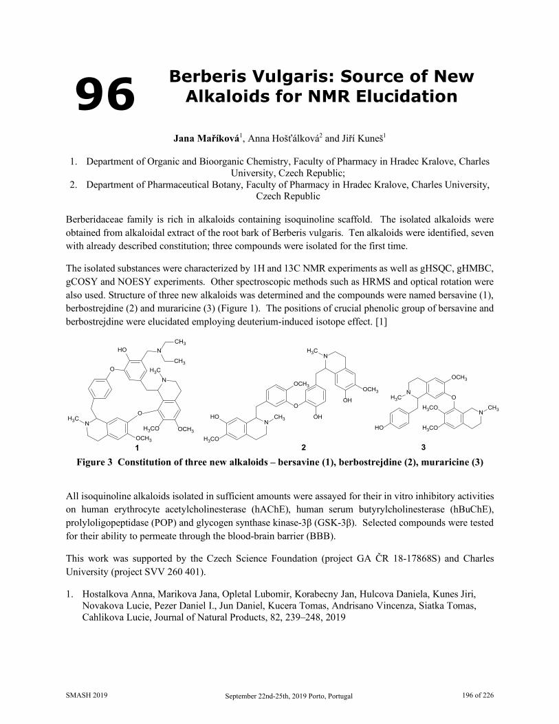
96 Berberis Vulgaris: Source of New Alkaloids for NMR Elucidation
Jana Maříková1, Anna Hošťálková2 and Jiří Kuneš1
1. Department of Organic and Bioorganic Chemistry, Faculty of Pharmacy in Hradec Kralove, Charles
University, Czech Republic; 2. Department of Pharmaceutical Botany, Faculty of Pharmacy in Hradec Kralove, Charles University,
Czech Republic Berberidaceae family is rich in alkaloids containing isoquinoline scaffold. The isolated alkaloids were obtained from alkaloidal extract of the root bark of Berberis vulgaris. Ten alkaloids were identified, seven with already described constitution; three compounds were isolated for the first time.
The isolated substances were characterized by 1H and 13C NMR experiments as well as gHSQC, gHMBC, gCOSY and NOESY experiments. Other spectroscopic methods such as HRMS and optical rotation were also used. Structure of three new alkaloids was determined and the compounds were named bersavine (1), berbostrejdine (2) and muraricine (3) (Figure 1). The positions of crucial phenolic group of bersavine and berbostrejdine were elucidated employing deuterium-induced isotope effect. [1]
NCH3
OCH3
H3CO
HO
N
OH
OCH3
OH
H3C
O
1 2
NH3C
OCH3
O
N
HO
H3CO
H3CO
CH3
3
NH3C
HO
OCH3H3CO
N
CH3
CH3
OCH3
NH3C
O
O
Figure 3 Constitution of three new alkaloids – bersavine (1), berbostrejdine (2), muraricine (3)
All isoquinoline alkaloids isolated in sufficient amounts were assayed for their in vitro inhibitory activities on human erythrocyte acetylcholinesterase (hAChE), human serum butyrylcholinesterase (hBuChE), prolyloligopeptidase (POP) and glycogen synthase kinase-3β (GSK-3β). Selected compounds were tested for their ability to permeate through the blood-brain barrier (BBB).
This work was supported by the Czech Science Foundation (project GA ČR 18-17868S) and Charles University (project SVV 260 401).
1. Hostalkova Anna, Marikova Jana, Opletal Lubomir, Korabecny Jan, Hulcova Daniela, Kunes Jiri, Novakova Lucie, Pezer Daniel I., Jun Daniel, Kucera Tomas, Andrisano Vincenza, Siatka Tomas, Cahlikova Lucie, Journal of Natural Products, 82, 239–248, 2019
SMASH 2019 September 22nd-25th, 2019 Porto, Portugal 196 of 226
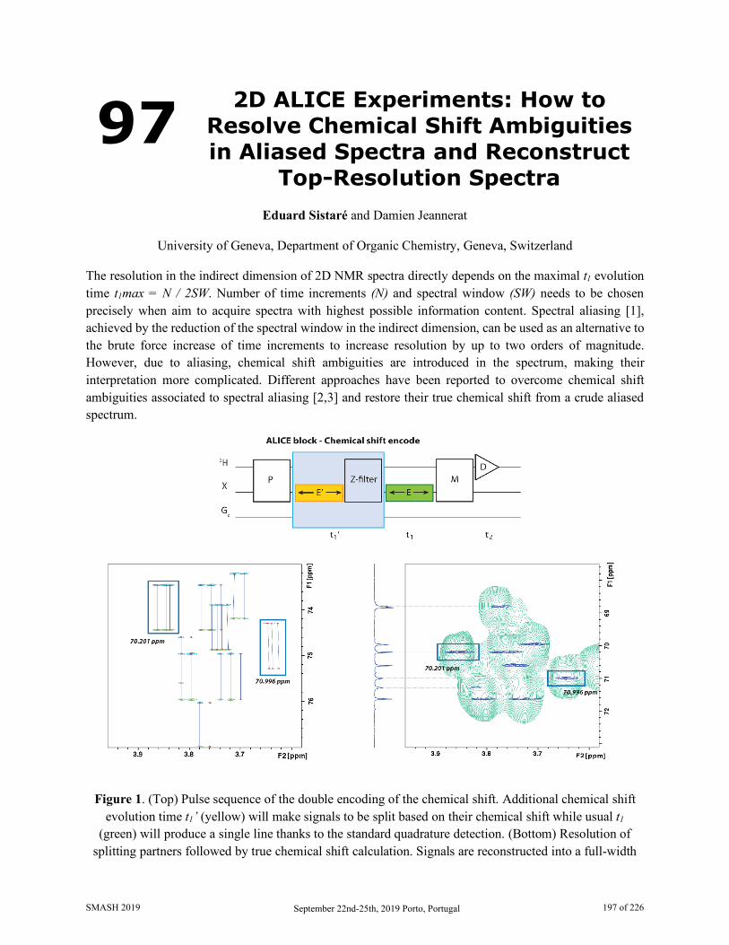
97 2D ALICE Experiments: How to Resolve Chemical Shift Ambiguities in Aliased Spectra and Reconstruct
Top-Resolution Spectra
Eduard Sistaré and Damien Jeannerat
University of Geneva, Department of Organic Chemistry, Geneva, Switzerland The resolution in the indirect dimension of 2D NMR spectra directly depends on the maximal t1 evolution time t1max = N / 2SW. Number of time increments (N) and spectral window (SW) needs to be chosen precisely when aim to acquire spectra with highest possible information content. Spectral aliasing [1], achieved by the reduction of the spectral window in the indirect dimension, can be used as an alternative to the brute force increase of time increments to increase resolution by up to two orders of magnitude. However, due to aliasing, chemical shift ambiguities are introduced in the spectrum, making their interpretation more complicated. Different approaches have been reported to overcome chemical shift ambiguities associated to spectral aliasing [2,3] and restore their true chemical shift from a crude aliased spectrum.
Figure 1. (Top) Pulse sequence of the double encoding of the chemical shift. Additional chemical shift evolution time t1’ (yellow) will make signals to be split based on their chemical shift while usual t1
(green) will produce a single line thanks to the standard quadrature detection. (Bottom) Resolution of splitting partners followed by true chemical shift calculation. Signals are reconstructed into a full-width
SMASH 2019 September 22nd-25th, 2019 Porto, Portugal 197 of 226
spectrum and placed based on its previously calculated chemical shift. The resolution enhancement using the ALICE sequence can be observed when comparing the reconstructed top-resolution spectrum (blue)
with its homologous standard low-resolution spectrum (green).
A general methodology to deal with chemical shift ambiguities generated by spectral aliasing is here presented. The ALICE (ALiasing order Information by Chemical shift Encoding) methodology [4] is used to encode chemical shift information along the F1 dimension. The inclusion of an additional evolution time block t1’ without quadrature discrimination, incremented simultaneously with t1, results in a signal split, which distance is a function of its chemical shift. The ALICE methodology is made compatible with the FOPA approach [5] in order to avoid ambiguities associated to signal split distance. An algorithm processing aliased spectra is used to identify the splitting pattern generated by the ALICE block and to recalculate the true chemical shift of each pair of peaks. A full-width reconstructed spectrum with top resolution is obtained by placing each reconstructed pair of peaks to its previously calculated true chemical shift value in the indirect dimension.
1. Jeannerat, D., Encyclopedia of Magnetic Resonance, John Wiley, Chichester, 2011 2. Ramirez-Gualito, K., Jeannerat, D., Magn. Reson, Chem., 2015, 53, 901 3. Foroozandeh, M., Jeannerat, D., Magn. Reson. Chem., 2015, 53, 894 4. Sistaré, E., PhD thesis, University of Geneva (Geneva), 2019 5. Sheberstov, K, Sistaré, E., Jeannerat, D., Magn. Reson. Chem., Revision submitted
SMASH 2019 September 22nd-25th, 2019 Porto, Portugal 198 of 226
98 Residual Dipolar Coupling Analysis for the Structure Elucidation of a Set
of Atropomeric Cyclic Peptides
Karl Z. Demmans1, Diego B. Diaz2, Solomon D. Appavoo2, Andrei K. Yudin2 and Darcy C. Burns1
1. CSICOMP NMR Facility, University of Toronto Chemistry Department, Toronto, ON, Canada 2. Andrei Yudin Research Group, University of Toronto Chemistry Department, Toronto, ON, Canada
Cyclic peptides have garnered attention in recent years for their application as viable antibodies[1-3]. To accurately simulate their bio-compatibility, first their structure must be determined. This is of the utmost importance as the various stereoisomers of cyclic peptides may affect biological processes in drastically different mechanisms of action. To achieve this goal, Nuclear Magnetic Resonance spectroscopy, through the determination of Residual Dipolar Couplings in combination with Density Functional Theory, has emerged as a practical method for diastereomeric structure elucidation[4,5]. Herein we present, using the aforementioned techniques, the structure determination of a set of atropomeric cyclic peptides employing poly-HEMA as the aligning media in DMSO-d6.
1. Belding, L.; Zaretsky, S.; Yudin, A. K.; Dudding, T., J. Org. Chem., 83, 9119-9124, 2018 2. Mendoza-Sanchez, R.; Corless, V. B.; Nguyen, Q. N. N.; Bergeron-Brlek, M.; Frost, J.; Adachi, S.;
Tantillo, D. J.; Yudin, A. K., Chem. Eur. J., 23, 13319-13322, 2017 3. Chung, B. K. W.; White, C. J.; Scully, C. C. G; Yudin, A. K., Chem. Sci., 7, 6662-6668, 2016 4. Burns, D. C.; Mazzola, E. P.; Reynolds, W. F., Nat. Prod. Rep., 36, 919-933, 2019 5. Liu, Y.; Navarro-Vázquez, A.; Gil, R. R.; Griesinger, C.; Martin, G. E.; Williamson, R. T., Nature
Protocols, 14, 217-247, 2019
SMASH 2019 September 22nd-25th, 2019 Porto, Portugal 199 of 226

99 Treatment Response to Fiber in Diabetes: NMR-Based Metabolomic
Approach
Helena Pelantová1, Martina Čermáková1, Blanka Šedivá2, Jan Gojda3, Monika Cahová4 and Marek Kuzma1
1. Institute of Microbiology of the CAS, Prague, Czech Republic
2. University of West Bohemia, Plzeň, Czech Republic 3. University Hospital Královské Vinohrady, Prague, Czech Republic
4. Institute for Clinical and Experimental Medicine, Prague, Czech Republic There is strong evidence that gut microbiota and microbiome-derived metabolites play a key role in many metabolic diseases, including type 2 diabetes [1]. Gut microbial ecology is largely modulated by diet in humans. Therefore, dietary interventions are a potential tool to impact host health [2]. The promising strategy aiming to optimize the gut environment is prebiotic treatment with dietary fiber (particularly inulin) that is transformed by different microbial species to final metabolite(s) with beneficial biological effect. Our project is based on the hypothesis that the therapeutic potential of the specific prebiotic (inulin) treatment is individual and depends on pre-existing microbiota composition which is also reflected in the metabolic profiles of host biofluids and fecal extracts. Therefore, determination of individual gut microbiome/metabolome patterns may serve as a predictive marker of the efficacy of this dietary intervention.
In the ongoing study, overweight/obese patients with new-onset diabetes were administered inulin (10 g/day) on every-day basis during three months without any other antidiabetic medication. Before and at the end of the intervention period, participants were subjected to metabolic characterization (euglycemic-hyperinsulinemic clamp) and stool, urine, and serum samples for NMR metabolomic analysis were collected. The fecal short-chain fatty acids (butyrate and propionate) response after acute inulin load was also assessed by NMR spectroscopy.
Inulin intervention was associated with the improvement of insulin sensitivity in one third of patients. NMR-based metabolomic analysis of samples collected before the intervention study aimed to describe specific responders/non-responders metabolomic profiles. Some common trends in metabolic levels in fecal extracts were revealed, e.g., differences in the concentrations of ethanol, glutamate, bile acids, ferulic acid, and nicotinic acid. In the future, metabolomic data will be correlated with the appropriate biochemical parameters and the results of the ongoing gut metagenome characterization to search for complex patterns which may help tailor dietary intervention with fiber to the individual patient.
Acknowledgements: This work is supported by the Ministry of Health of the Czech Republic, grant no. NV18-01-00040
1. Tilg H. and Moschen A.R., Gut, 63(9), 1513-1521, 2014 2. Kovatcheva-Datchary P., Nilsson A., Akrami R. et al., Cell Metab., 22(6), 971-982, 2015
SMASH 2019 September 22nd-25th, 2019 Porto, Portugal 200 of 226
100 Complexing Behavior of Diacetylcurcumin with Mg and Zn
Seen by Liquid and Solid NMR
William Meza1, Yair Alvarez1, Marco A. Obregón1, Julia Cassani2, W.F. Reynolds2 and Raúl G. Enríquez1
1. Instituto de Química, UNAM Ciudad Universitaria, Circuito Exterior, Mexico
2. Departamento de Sistemas Biológicos, UAM, Mexico City, Mexico. Curcumin and curcuminoids are chelating agents due to the eta-diketone functionality, capable of producing stable complexes with a series of metal ions and receive at present a great deal of attention due to their pharmacological importance. These complexes have low crystallinity and their characterization by SCDRX is often impeded. The characterization by techniques such as IR, UV-Vis, and MS often provide poor structural information, so establishing a metal-ligand ratio becomes is difficult task. [1,2].
In view of the high in vitro cytotoxicity and extremely low in vivo toxicity of diacetylcurcumin metal complexes with Zn and Mg towards cancer cell lines a detailed study of the complexing behavior using solid and liquid NMR was undertaken. [1, 2]
We studied the formation of metal complexes of diacetylcurcumin using high-resolution NMR in DMSO-d6 and CP-MAS (TOSS) with increasing metal-ligand ratios. The initial supramolecular interaction can be followed by spectral changes caused by the metal linkage at the site of complexation until a stable spectral change is reached. Solid phase tests of mechanical mixtures were studied by CPMAS and spectral changes were also observed using this approach.
1. W. Meza-Morales, J. C. Machado-Rodriguez, Y. Alvarez-Ricardo, M. A. Obregón-Mendoza, A. Nieto-Camacho, R. A. Toscano, M. Soriano-García, J. Cassani and R. G. Enríquez, Molecules, 24, 910-927, 2019.
2. W. Meza-Morales, M. Mirian Estévez-Carmona, Y. Alvarez-Ricardo, M. A. Obregón-Mendoza, J. Cassani, M. T. Ramírez-Apan, C. Escobedo-Martínez, M. Soriano-García, W. F. Reynolds and R. G. Enríquez, Molecules, 24, 1598-1616, 2019.
SMASH 2019 September 22nd-25th, 2019 Porto, Portugal 201 of 226
101 Effective Bayesian Approach for Automated Quantitative Analysis
of Benchtop NMR Data Ellen Steimers1, Yevgen Matviychuk2, Kerstin Münnemann1, Daniel J. Holland2 and Erik von Harbou1
1. Laboratory of Engineering Thermodynamics, Technische Universität Kaiserslautern, Kaiserslautern,
Germany, 2. Department of Chemical and Process Engineering, University of Canterbury, Christchurch, New
Zealand Low-cost, user-friendly benchtop NMR instruments have been developed to make NMR spectroscopy more accessible to the general laboratory chemist and are often touted as a ‘one-click’ solution for spectral acquisition. Unfortunately, the lower field strength of their permanent magnets entails lower spectral resolution and sensitivity compared to the traditional superconducting NMR equipment. Due to significant peak overlap in benchtop NMR spectra, the standard peak integration often becomes inadequate for accurate quantification. This challenging problem calls for the development of more sophisticated signal processing algorithms.
Here, we present an effective, fully-automated approach for the analysis of benchtop NMR data, which allows us to immediately determine the mixture composition without the need for any manual NMR data processing. Our method is based on the idea of model-based quantification coupled with an effective Bayesian problem formulation. The use of quantum mechanical models to describe ideal signatures of mixture components makes our method invariant to the spectrometer field strength – an important prerequisite for the analysis of benchtop NMR data. On the other hand, incorporating all available prior knowledge about the studied system facilitates the model adjustment to low-resolution spectra.
We demonstrate the performance of our approach using the practical example of quantifying the compositions of several mixtures of alcohols and acetates. First, we study the dataset acquired on a superconducting 400 MHz NMR spectrometer and reveal dependence between the chemical shifts of different functional groups, which vary with the changing concentrations of species. Then, we summarize this knowledge in terms of Bayesian priors and use them to analyze a dataset acquired on a 43 MHz benchtop NMR instrument. Rather surprisingly, the application of this method to benchtop NMR data yields a comparable or even slightly better accuracy than the combination of traditional processing approaches with well-resolved high field NMR spectra. An average error of 1.6 % in concentrations is achieved, with the maximum error for any species being less than 3.5 %. We expect our method to generalize well to other similar systems of small molecules, extending the range of applications of benchtop NMR to cases where peak overlap and higher-order coupling effects would otherwise prevent an accurate quantification with standard approaches.
SMASH 2019 September 22nd-25th, 2019 Porto, Portugal 202 of 226

102 Efficient Approaches for Addressing Spectral Ambiguities in Computer Assisted Structure
Elucidation (CASE) Systems
Rostislav Pol, Mikhail Elyashberg, Sergey Golotvin and Dimitris Argyropoulos
Advanced Chemistry Development, Toronto, Ontario, Canada Since their development over 50 years ago, Computer Assisted Structure Elucidation (CASE) systems (or Expert Systems, ES) have significantly facilitated the de novo structural elucidation of both natural and synthesized organic compounds, especially in cases where the traditional (manual) methods were very challenging or even impossible to perform [1,2]. Current ES are based on 1D and 2D NMR spectra, given that the molecular formula has already been determined by HR-MS. At present, there are several free and commercially available CASE systems which offer a few advantages [3]: i) They deliver all (without any exception) structures which can be deduced from a given set of NMR data; ii) The application of fast empirical methods for NMR chemical shift prediction allows the program to select the most probable structure; iii) If necessary, DFT based chemical shift calculations can be used to confirm the selected structure; iv) ES are now capable of suggesting a 3D model of the elucidated structure.
In spite of the recent advancements, ES are still susceptible to a series of limitations which also impede structure elucidation by a human expert. These limitations are mainly associated with the ambiguity of the experimental data, as well as the overlapping of characteristic chemical shift ranges in NMR spectra. Experimental ambiguity can result from low resolution 2D spectra, which prohibits the confident assignment of closely spaced signals. Further, ambiguity can often be due to the hybridization state of carbon nuclei that appear in the same regions of NMR spectra. For example, a 13C NMR signal observed at 90 ppm can belong either to a sp2 or a sp3-hybridized carbon connected to one or two oxygen atoms. If a molecule contains atoms that can have variable valences (e.g. N and/or P), all their possible valences should be explored during structure generation. Ambiguity can also be observed in some cases where carbon nuclei show no HMBC or COSY correlations at all; this is especially evident in hydrogen deficient molecules leading to "floating" atoms, i.e. atoms that could potentially be connected to any other atom. The presence of "floating" atoms significantly increases both the size of the output file and the time of structure generation.
In order to remove uncertainty from spectroscopic data, additional experiments are usually carried out; for example, high resolution 2D NMR spectra, such as highly inflated NUS or band selective spectra are collected to reliably assign the peaks. Additional spectroscopic data (IR, Raman, UV-Vis) could help to identify the characteristic groups and resolve the hybridization or valence status. However, such solutions are not useful in all cases.
In such cases, the only remaining solution is the exhaustive investigation of all alternatives resulting from the presence of any ambiguity. For example, if there are five carbon nuclei with signals in the range of 70-120 ppm, then 25=32 combinations of hybridization (sp3 or sp2) have to be generated and checked. This could significantly increase the structure generation time.
SMASH 2019 September 22nd-25th, 2019 Porto, Portugal 203 of 226
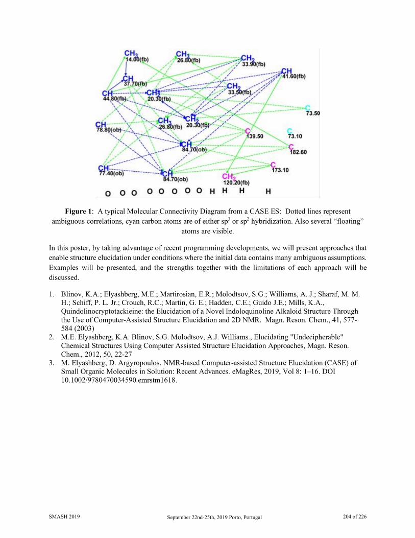
Figure 1: A typical Molecular Connectivity Diagram from a CASE ES: Dotted lines represent ambiguous correlations, cyan carbon atoms are of either sp3 or sp2 hybridization. Also several “floating”
atoms are visible.
In this poster, by taking advantage of recent programming developments, we will present approaches that enable structure elucidation under conditions where the initial data contains many ambiguous assumptions. Examples will be presented, and the strengths together with the limitations of each approach will be discussed.
1. Blinov, K.A.; Elyashberg, M.E.; Martirosian, E.R.; Molodtsov, S.G.; Williams, A. J.; Sharaf, M. M. H.; Schiff, P. L. Jr.; Crouch, R.C.; Martin, G. E.; Hadden, C.E.; Guido J.E.; Mills, K.A., Quindolinocryptotackieine: the Elucidation of a Novel Indoloquinoline Alkaloid Structure Through the Use of Computer-Assisted Structure Elucidation and 2D NMR. Magn. Reson. Chem., 41, 577-584 (2003)
2. M.E. Elyashberg, K.A. Blinov, S.G. Molodtsov, A.J. Williams., Elucidating "Undecipherable" Chemical Structures Using Computer Assisted Structure Elucidation Approaches, Magn. Reson. Chem., 2012, 50, 22-27
3. M. Elyashberg, D. Argyropoulos. NMR-based Computer-assisted Structure Elucidation (CASE) of Small Organic Molecules in Solution: Recent Advances. eMagRes, 2019, Vol 8: 1–16. DOI 10.1002/9780470034590.emrstm1618.
SMASH 2019 September 22nd-25th, 2019 Porto, Portugal 204 of 226
103 A Construction of a Crystal Lattice from Intermolecular Interactions
Jan Sykora and Vladimir Hejtmanek
Institute of Chemical Process Fundamentals, Czech Academy of Sciences, Rozvojová 135, 165 02
Prague, Czech Republic While intramolecular distances determined in the solid state can serve for determination of a correct conformation of a single molecule in a crystal packing, the intermolecular distances can be utilized for a building of crystal packing itself and subsequent derivation of standard crystallographic parameters. A crystal packing can be reconstructed using a limited number of intermolecular interactions as demonstrated here on the crystal structure of L-histidine·HCl·H2O [1].
The geometric restrictions based on intermolecular interactions are applied gradually within a supervised procedure, narrowing the scope of possible arrangement of two adjacent molecules.[2] Using a list of available intermolecular interactions[3,4] and implementing the additional condition that every molecule has the same surroundings in the crystal lattice, a basic repetitive motif of two histidine molecules can be derived. This motif can be subsequently used for the construction of molecular chains containing information on translation in one dimension. The other two dimensions can be then obtained by the construction of 2D-sheets and their mutual assembly into the final 3D structure. Complete molecular packing finally enables the derivation of standard crystallographic parameters that can be used for verification of the structural model obtained. The procedure based on intermolecular interactions derived from solid-state NMR data can be employed especially when standard crystallographic parameters are not available and traditional methods based on X-ray diffraction fail. Hydrogen atoms show high relevance for such structure solution, similarly to other fields of NMR spectroscopy.
Therefore, further development of methods for detection and quantification of intra- and intermolecular interactions is in high demand. More interactions mean lower degrees of freedom and a narrower scope of possible solutions. Regarding intermolecular interactions, only the shortest ones ranging from 2.5–4.0 Å are meaningful. The number of required distances can vary for different crystallographic systems. It seems that the construction of molecular frameworks of systems of low symmetry will be easier due to a low number of molecules in the unit cell, and therefore also in the basic repetitive motif. In some cases, a coarse crystal packing could be obtained using just a fragment of the molecule. Furthermore, the construction of crystal packing of achiral molecules crystallizing in centrosymmetric groups will probably be more difficult, as a mirror image has to be considered in every single step.
1. Fuess, H.; Hohlwein, D.; Mason, S.A. Acta Cryst. B33, 654, 1977. 2. Hejtmanek, V.; Dracinsky, M.; Sýkora, J. Crystals 9, 159, 2019. 3. Reddy, G.N.M.;Malon,M.;Marsh, A.; Nishiyama, Y.; Brown, S.P. Anal. Chem. 88, 11412, 2016. 4. Duong, N.T.; Raran-Kurussi, S.; Nishiyama, Y.; Agarwal, V. J. Phys. Chem. Lett. 9, 5948, 2018.
SMASH 2019 September 22nd-25th, 2019 Porto, Portugal 205 of 226
104 Solid State NMR Characterisation of Novel Fluorinated Cellulose
Materials
Juan C. Muñoz García1, Peterson de Andrade2, Giulia Pergolizzi2, Valeria Gabrielli1, Sergey Nepogodiev2, Robert Harniman3, Rinat Nigmatullin3, Dinu Iuga4, Stephen J. Eichhorn3, Jesús Angulo1,
Robert A. Field2 and Yaroslav Z. Khimyak1 1. School of Pharmacy, University of East Anglia, Norwich Research Park, Norwich NR4 7TJ, United
Kingdom 2. John Innes Centre, Norwich Research Park, Colney Ln, Norwich NR4 7UH, United Kingdom
3. Bristol Composites Institute (ACCIS), University of Bristol, Bristol BS8 1TR, United Kingdom 4. The UK 850 MHz Solid State NMR Facility, Department of Physics, University of Warwick,
Coventry CV4 7AL, United Kingdom The application of 19F NMR to probe chemical and biological interactions is constantly increasing due to the 100% isotopic abundance of 19F,1 the extremely high sensitivity of 19F to changes in local environment avoiding overlapping signals, and its almost total absence in biological receptors in nature. The introduction of 19F in molecules represent a very powerful strategy to monitor conformational changes,2 ligand-receptor interactions3 and assembly processes4 in complex and heterogeneous systems such as cellulose. We have accomplished the cellodextrin phosphorylase synthesis of per-6-monofluorinated cellulose and a series of cellulose oligomers containing a single deoxy-fluoro glucose per cellulose chain (at the reducing end), all yielding a degree of polymerization of about 7-9 glucose units. We have characterised their ordered and disordered domains by 1H{19F}-13C and 19F{1H}-13C cross polarization (CP) solid state NMR and microscopy. Our results show that 2- and 6-fluorination at the reducing end does affect the mesoscale order of enzymatic cellulose, thus representing a powerful chemical strategy to monitor self-assembly during cellulose synthesis. On the other hand, the 3-monofluorinated derivative assembled into a more disordered material by breaking the intra-chain hydrogen bond to the oxygen ring. Interestingly, fluorination at position 6 in all glucose units gave rise to a more disordered cellulose material containing some ordered domains showing a novel type of cellulose allomorphism. This work demonstrates that phosphorylases can facilitate as a “bottom up” assembly of biomaterials with novel properties, which are used across industries from diverse areas (food, health, cosmetics, pharmaceuticals, etc).
1. Chen, H.; Viel, S.; Ziarelli, F.; Peng, L., Chem. Soc. Rev., 42 (20), 7971–7982, 2013. 2. Danielson, M. A.; Falke, J. J., Annu. Rev. Biophys. Biomol. Struct., 25, 163–195, 1996. 3. Yanamala, N.; Dutta, A.; Beck, B.; Van Fleet, B.; Hay, K.; Yazbak, A.; Ishima, R.; Doemling, A.;
Klein Seetharaman, J., Chem. Biol. Drug Des., 75 (3), 237–256, 2010. 4. Leung, R. L. C.; Robinson, M. D. M.; Ajabali, A. A. A.; Karunanithy, G.; Lyons, B.; Raj, R.;
Raoufmoghaddam, S.; Mohammed, S.; Claridge, T. D. W.; Baldwin, A. J.; et al., J. Am. Chem. Soc., 139 (15), 5277–5280, 2017.
SMASH 2019 September 22nd-25th, 2019 Porto, Portugal 206 of 226

105 Enhancement and Evaluation of CAST/CNMR Database Focused on
Diterpenoids
Hiroyuki Koshino1, Ken-ichi Kimura2, Shungo Koichi3 and Hiroko Satoh4,5
1. RIKEN Center for Sustainable Resource Science, Wako, Japan 2. Graduate School of Arts and Science, Iwate University, Morioka, Japan
3. Department of Systems and Mathematical Science, Nanzan University, Nagoya, Japan 4. Research Organization of Information and Systems, Tokyo, Japan
5. Department of Chemistry, University of Zurich, Zurich, Switzerland The CAST/CNMR system and its database have been developed. This system is composed of two functions for prediction of 13C NMR chemical shift values from a query chemical structure (CAST/CNMR Shift Predictor1) and elucidation of chemical structures from a query of 13C NMR chemical shift values (CAST/CNMR Structure Elucidator2). These functions use a database of reported/observed 13C NMR chemical shift values associated with their 3D chemical structures described using CAST coding method1. The datbase is composed mostly of the data from peer-reviewed journals, and the data registration has been done after a careful check, partly by using two functions of CAST/CNMR system. Indeed, we have found incorrect data when developing the database and revised some natural products by total synthesis and by using CAST/CNMR sytems.3-7 Recently, we have been studying the biologically active compounds in amber as yeast Ca2+ signal tranduction inhibitors. After first characterization of a new compound named kujigamberol (15,20-dinor-5,7,9-labdatrien-18-ol),8,9 we have isolated several new diterpenoids with diverse carbon skeletons from Japanese Kuji amber,10-12 Dominican amber,13 and Burmese amber.14 To use the CAST/CNMR system effectively for structure characterzation of those natural products, enhancement and evaluation of CAST/CNMR database is important. So we added new data of a few hundred diterpenoids, especially focused on carbon skeletons of labdane, clerodane, halimane, abietane, pimarane, isopimarane, and related norditerpenoids such as podocarpane, which have been found in amber. In the evaluation process of each data by using the CAST/CNMR system, we could find some mistake of assignments of NMR signals and also structures in some case.
1. H. Satoh et al, Tetrahedron, 2005, 61, 7431-7437 and references cited therein. 2. S. Koichi et al, J. Chem. Inf. Model., 2014, 54, 1027-1035. 3. H. Koshino et al, Tetrahedron Lett., 2006, 47, 4623-4626. 4. S. Takahashi et al, J. Org. Chem., 2007, 72, 4578-4581. 5. S. Takahashi et al, J. Org. Chem., 2014, 79, 9373-9380. 6. S. Takahashi et al, J. Org. Chem., 2016, 81, 11222-11234. 7. S. Takahashi et al, Bioorg. Med. Chem. Lett., 2018, 28, 930-933. 8. K. Kimura et al, Fitoterapia, 2012, 83, 907-912. 9. Y. Q. Ye at al, Bioorg. Med. Chem. Lett., 2012, 22, 4259-4262. 10. T. Kawamura et al, Org. Geochem., 2018, 120, 12-18. 11. T. Uchida et al, J. Nat. Prod., 2018, 81, 1070-1074. 12. E. Shimizu et al, Fitoterapia, 2019, 134, 290-296. 13. T. Abe et al, Fitoterapia, 2016, 113, 188-194. 14. T. Uchida et al, Fitoterapia, 2019, 134, 422-428.
SMASH 2019 September 22nd-25th, 2019 Porto, Portugal 207 of 226

106 Effect of Nonpolar External Solvents on the Interfaces of
Reverse Micelles Formulated with the Protic Ionic Liquid-like
Surfactant Imim-DEHP
Cristian Lépori1, Juana J. Silber2,3, N. Mariano Correa2,3 and R. Darío Falcone2,3
1. Instituto de Física Enrique Gaviola, CONICET, Universidad Nacional de Córdoba, Córdoba, Argentina
2. Instituto para el Desarrollo Agroindustrial y de la Salud, CONICET, Universidad Nacional de Río Cuarto, Río Cuarto, Argentina
3. Departamento de Química, Universidad Nacional de Río Cuarto, Río Cuarto, Argentina Reverse micelles (RMs) are self-assembled supramolecular systems that are obtained by dissolving surfactant molecules in an organic solvent of low polarity. These systems are capable of dissolving an appreciable amount of water inside and therefore have numerous applications [1]. One of the surfactants that forms RMs is sodium bis-(2-ethylhexyl) phosphate (Na-DEHP). On the other hand, ionic liquids (ILs) are liquid salts with a melting point lower that 100ºC, which have interesting properties. In general, they can be classified into aprotic ionic liquids (AILs) and protic ionic liquids (PILs), the latter being different because they always present a proton available generally in the cation to establish hydrogen bonding. An area still less explored is the use of ILs with amphiphilic properties to generate RMs that allow to encapsulate water or another polar solvent. For this reason, a new PIL-like surfactant has been synthesized: 1-methylimidazolium bis-(2-ethyl-hexyl)phosphate (imim-DEHP), which can form RMs in toluene and n-heptane [2]. When comparing imim-DEHP RMs with Na-DEHP RMs in both solvents, it is observed that in n-heptane the aggregates formed by imim-DEHP are larger, while in toluene they are of similar sizes.
In the present work, results obtained using 1H and 31P NMR technique in the micellar system formed by toluene/imim-DEHP/water and n-heptane/imim-DEHP/water are shown. All the experiments were carried out varying the water content of the RMs, defined as W0 (W0 = [water]/[surfactant]) and at a constant surfactant concentration. The results using 31P and 1H NMR suggest a larger penetration of toluene towards the micellar interface (comparing with n-heptane), producing that imim+ counterion to be located more towards the micellar interior, moving away from the interface. This fact strongly affects the water-surfactant interaction in both RMs. Thus, in toluene/imim-DEHP the encapsulated water interacts with both the phosphate anion and the imim+ cation. On the other hand, in n-heptane/imim-DEHP water interacts strongly with the phosphate group, while it interacts weakly with its counterion.
In conclusion, the imim-DEHP surfactant, which is a PIL, generates RMs in toluene and n-heptane with different interfaces, which is very interesting for a future application as nano-reactors.
1. Correa N.M., Silber J.J., Riter R.E., Levinger N.E., Chem. Rev., 2012, 112, 4569–4602 2. Lépori C.M.O., Silber J.J., Falcone R.D., Correa N.M., RSC Advances, 2017, 7, 44743-44750
SMASH 2019 September 22nd-25th, 2019 Porto, Portugal 208 of 226

107 ³JCH Coupling in Unsaturated Compounds: The Effect of
Neighboring Groups
Uenifer R. Couto¹, Armando Navarro-Vázquez², and Cláudio F. Tormena¹
1. Institute of Chemistry, University of Campinas, Campinas, Brazil 2. Departamento de Química Fundamental, CCEN, Universidade Federal de Pernambuco, Recife, Brazil Configuration assignment of disubstituted double bonds are usually straight forward through nOe cross-relaxation and 3JHH. The assignment through 3JHH is accomplished through the relationship of the coupling with the dihedral angle between the coupled nuclei by a cosine series, as proposed by Martin Karplus [1], which is also applicable for unsaturated compounds, in terms of the well-known relationship 3JHH(cis)<3JHH(trans). On the other hand, trisubstituted double bonds lack vicinal 3JHH coupling and nOe’s absence may be ambiguous. Fortunately, several authors [2] have found that the 3JCH coupling constant follow the same trend predicted by Karplus for 3JHH, which may be used for configurational assignment. In this study we use the Density Functional Theory (DFT) to investigate the behaviour of a series of substituted olefins to verify the dependence of the coupling not with the variation of the dihedral between the coupled nuclei, but rather with the nature and orientation of the substituents with respect to the double bond. We evaluated a series of disubstituted and trisubstitued α,β-unsaturated carbonyl compounds varying the nature and orientation of the carbonyl group. For comercially available compounds the experimental 3JCH couplings were measured using the dsel-HSQMBC-IPAP pulse sequence [3].
For some of the studied compounds a strong modulation of the 3JCH coupling with the orientation of the carbonyl group was observed. This modulation increases the magnitude of 3JCH for s-trans conformations and was greater for aldehydes and ketones. The presence of a α-cyano substituent does not affect the observed modulation. In the case of aldehyde, the difference in the 3JCH coupling can reach around 3 Hz from s-cis to s-trans conformations, which can result in an inversion of the well-known relationship 3J(cis)<3J(trans) for some compounds. In fact, for 1-carboxaldehydecyclohexene the experimental values for the 3JCH(cis) and 3JCH(trans) are 9.2 and 6.7 Hz, respectively. According to DFT computations, this inversion is observed for the s-trans conformer, but not for s-cis. The modulation of 3JCH according to the carbonyl dihedral for the studied compounds follows a Karplus-like cosine dependence which has been implemented in the MSpin-JCoupling software package [3].
In summary, it was demonstrated that the 3JCH coupling for H-C=C-C(=O) fragment is highly dependent on the orientation of the carbonyl group and the magnitude of the modulation is dependent on the carbonyl nature. For the special case of aldehydes, the modulation may lead to the inversion of the well-known relationship 3J(cis)<3J(trans) which might result to misassignment of the configuration of double bonds.
1. (a) Karplus, M., J. Chem. Phys., 30, 11-15, 1959; (b) Karplus, M., J. Am. Chem. Soc., 85, 2870, 1963.
2. (a) Bifulco, G., et al., Chem. Rev. 107, 3744-3779, 2007; (b) Aydin, R., Günther, H., Magn. Reson. Chem., 28, 448-457, 1990. (c) Palermo, G., Riccio, R., Bifulco, G., J. Org. Chem., 75, 1982, 2010.
3. Couto, U.R., Navarro-Vázquez, A., Tormena, C.F., Magn Reson Chem., 2019 (DOI: 10.1002/mrc.4885)
SMASH 2019 September 22nd-25th, 2019 Porto, Portugal 209 of 226
108 Slice Selective NMR Analysis of Lanthanum Picrate Distribution in Extraction Studies by Calixarene
Ketone Derivatives Paula M. Marcos1,2, José R. Ascenso3,Véronique Hubscher-Bruder4, Rachel Schurhammer5, Ana S.D.
Ferreira6, and Eurico J. Cabrita6
1. CQE, Faculdade de Ciências da Universidade de Lisboa, Lisboa, Portugal. 2. Faculdade de Farmácia da Universidade de Lisboa, Lisboa, Portugal
3. CQE, Instituto Superior Técnico, Lisboa, Portugal 4. Université de Strasbourg, CNRS, IPHC UMR 7178, Strasbourg, France
5. Université de Strasbourg, Laboratoire de Modélisation et Simulations Moléculaires, UMR 7140, Strasbourg, France
6. UCIBIO, Faculdade de Ciências e Tecnologia Universidade Nova de Lisboa, Caparica, Portugal. In the field of host-guest chemistry, many studies have focused on the binding ability of calixarene derivatives with carbonyl groups at their lower rims towards metal ions [1]. We have been synthesising dihomooxacalix[4]arene derivatives containing the ketone group at the lower rim and studying their binding abilities towards alkali, alkaline earth, transition and heavy metal cations[2,3]. In the course of these studies, we have extended our research into trivalent lanthanide ions [4]. We report here a comparative study on the binding properties of the tetraphenyl ketone of p-tert-butyldihomooxacalix[4]arene (1) and the tetraphenyl ketone of p-tert-butylcalix[4]arene (2), both in the cone conformation, towards trivalent lanthanum cation. This has been assessed by extraction studies with the correspondent metal picrate from aqueous solutions into dichloromethane. The distribution of picrate anions between the two phases was monitored using slice selective NMR and two internal standards for concentration determination [5,6], Molecular dynamics (MD) simulations and Potential of Mean Force (PMF) free energy calculations were done for ligands 1 and 2 in pure solvents and at the CH2Cl2/water interface to add further information on the complexation and extraction processes.
1. Calixarenes and Beyond; Neri P., J.L. Sessler J.L., M-X. Wang, Eds.; Springer International
Publishing Switzerland, 2016 2. Marcos P.M., Félix S., Ascenso J.R, Segurado M.A.P, Mellah B., Abidi R., Hubscher-Bruder V.,
Arnaud-Neu F., Supramol. Chem., 18, 285-297, 2006 3. Marcos C:PM, Félix S., Ascenso J.R., Segurado M.A.P., Thuéry P., Mellah B., Michel S., Hubscher-
Bruder V., Arnaud-Neu F., New J. Chem., 31, 2111-2119, 2007
SMASH 2019 September 22nd-25th, 2019 Porto, Portugal 210 of 226

4. Marcos P.M., Ascenso J.R., Segurado M.A.P, Cragg P.J., Michel S., Hubscher-Bruder V., Arnaud-Neu F., Supramol. Chem. 23, 93-101,2011.
5. Mantel C., Bayle P.A., Hediger S., Berthon C., Bardet M., Magn. Reson. Chem. , 48 (8), 600−606, 2010.
6. Mitrev Y., Simova S., Jeannerat D., Chem. Commun., , 52, 5418-5420, 2016.
SMASH 2019 September 22nd-25th, 2019 Porto, Portugal 211 of 226
109 NMR Measurements on Lignin and Black Liquor: Towards Industrial
Applications for the Pulp and Paper Industry
Jerk Rönnols1, Lars Wallbäcks1, Hélène Freichels2, Ernesto Danieli2, and Fredrik Aldaeus1
1. RISE, Stockholm, Sweden
2. Magritek GmbH, Aachen, Germany A major challenge and oppurtunity for the pulp and paper industry is valorization of lignin, which today is considered waste and is to a large extent burnt for heat. Various types of chemical treatments subsequent to precipitation out of the process liquid (the black liquor) have been suggested, aiming at products such as fuels, adhesives and carbon fibers. Both precipitated lignins and black liquors are challenging sample matrixes, containg a large variety of compounds of varying molar masses of which far from all is known. NMR spectroscopy is commonly used in academic environments for investigations of certain bulk properties of lignin, e.g. content and nature of hydroxyl functionalities and intermolecular bond types. In the presented work developments of NMR methods for analysis of lignin and black liquor aiming towards industrial applications are described. These methods consist of two parts: 1) diffusion measurements on precipitated lignins at both high (9.4 T) and low (1.0 – 1.5 T) field strength and 2) 1H NMR measurements on black liquors gathered from pulp mills and subsequent principal component analysis.
The diffusion measurments performed at high field strength were calibrated for molar mass by orthogonal measurements by MALDI-TOF [1]. Good agreements where found between the weight average molar mass of the polymers and the diffusion constants. This has proven to be a reliable tool also in comparison with other typical methods, such as size exclusion chromatography. These experiments were followed by measurements on a benchtop system based on a permanent magnet, the Magritek Spinsolve, in which the diffusion constants were readily obtained with good repeatbility and short experiment time [2]. The application of these measurements on benchtop spectrometers potentially enables NMR spectroscopy as an in-house tool for molar mass determinations in the industrial environment.
The chemical composition of the black liquor – the highly alkaline liquid remaining after pulping – depends on the pulping conditions and variations in the feedstock. Increased online knowledge on this composition could be useful for downstream processing, such as evaporaiton and precipitation, and also for modifications of the upstream processes. We have performed 1H NMR measurements on black liquor samples gathered over two months time at two different pulp mills, at high field strength. The resulting spectra were subjected to principal component analysis and matched towards analysis data obtained from orthogonal methods of measurement, in search for correlations. These meausrements reperesent the first steps taken towards online NMR-measurements in the pulp and paper industry.
1. Rönnols, Jerk; Jacobs, Anna; Aldaeus, Fredrik, Holzforschung, 71(7-8), 563-570, 2017. 2. Rönnols, Jerk; Danieli, Ernesto; Freichels, Hélène; Aldaeus, Fredrik, Holzforschung, in press, DOI:
10.1515/hf-2018-0282, 2019.
SMASH 2019 September 22nd-25th, 2019 Porto, Portugal 212 of 226
110 New NOAH Modules for Structure Elucidation and Analysis of Small
Molecules
Ēriks Kupče1 and Tim D. W. Claridge2
1. Bruker (UK) Ltd., Banner Lane, Coventry, CV4 9GH, UK 2. Department of Chemistry, University of Oxford, Chemistry Research Laboratory, Mansfield Road,
Oxford, OX1 3TA, UK As many as five conventional NMR pulse sequences based on 1H direct detection have been combined into NMR supersequences named NOAH (NMR by Ordered Acquisition using 1H detection) [1-4]. This approach offers significant time savings and increases the efficiency of NMR experiments as compared to conventional data recording since only a single recovery (relaxation) delay (d1) is employed in the combined pulse sequences. Here we introduce several new NOAH modules designed for NMR supersequences for the time. This allows more efficient structure characterisation of small molecules in the solution state (see Fig. 1). The triplet NOAH-3 BSX supersequences provide HMBC (B), HSQC (S), and one homonuclear correlation experiment of choice, X = N (NOESY), R (ROESY), T (TOCSY), Cc (CLIP-COSY) or similar according to the NOAH principle.
We show that double isotope filters (ZZ-filters) increase the flexibility of module permutation within the NMR supersequences, optimising combinations exploiting 15N and 13C nuclides. The time-shared 2BOB module apended to the ZZ-HMBC module yields NOAH-2 BO supersequence that provides sufficient information for structure elucidation of small molecules, such as ajmalicine and giberellic acid from a single measurement. The NOAH-2 BO demonstrates the possibilities of constructing NMR supersequences that include time-shared experiments (here 2BOB). Finally, we provide examples of incorporating multiple receiver experiments into NMR supersequences thus opening new avenues for designing information rich NMR experiments. All pulse programs are available from the Bruker Online User Library [5].
SMASH 2019 September 22nd-25th, 2019 Porto, Portugal 213 of 226
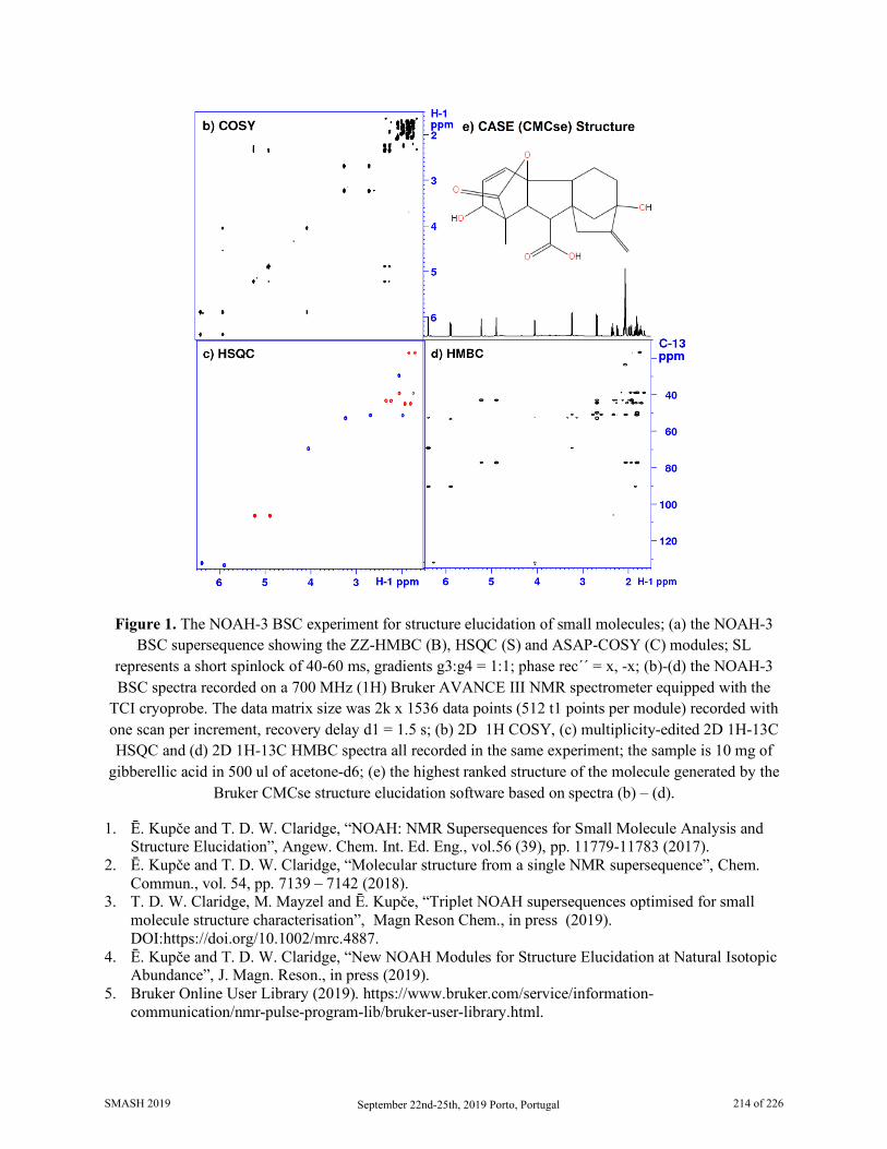
Figure 1. The NOAH-3 BSC experiment for structure elucidation of small molecules; (a) the NOAH-3 BSC supersequence showing the ZZ-HMBC (B), HSQC (S) and ASAP-COSY (C) modules; SL
represents a short spinlock of 40-60 ms, gradients g3:g4 = 1:1; phase rec´´ = x, -x; (b)-(d) the NOAH-3 BSC spectra recorded on a 700 MHz (1H) Bruker AVANCE III NMR spectrometer equipped with the
TCI cryoprobe. The data matrix size was 2k x 1536 data points (512 t1 points per module) recorded with one scan per increment, recovery delay d1 = 1.5 s; (b) 2D 1H COSY, (c) multiplicity-edited 2D 1H-13C HSQC and (d) 2D 1H-13C HMBC spectra all recorded in the same experiment; the sample is 10 mg of
gibberellic acid in 500 ul of acetone-d6; (e) the highest ranked structure of the molecule generated by the Bruker CMCse structure elucidation software based on spectra (b) – (d).
1. Ē. Kupče and T. D. W. Claridge, “NOAH: NMR Supersequences for Small Molecule Analysis and Structure Elucidation”, Angew. Chem. Int. Ed. Eng., vol.56 (39), pp. 11779-11783 (2017).
2. Ē. Kupče and T. D. W. Claridge, “Molecular structure from a single NMR supersequence”, Chem. Commun., vol. 54, pp. 7139 – 7142 (2018).
3. T. D. W. Claridge, M. Mayzel and Ē. Kupče, “Triplet NOAH supersequences optimised for small molecule structure characterisation”, Magn Reson Chem., in press (2019). DOI:https://doi.org/10.1002/mrc.4887.
4. Ē. Kupče and T. D. W. Claridge, “New NOAH Modules for Structure Elucidation at Natural Isotopic Abundance”, J. Magn. Reson., in press (2019).
5. Bruker Online User Library (2019). https://www.bruker.com/service/information-communication/nmr-pulse-program-lib/bruker-user-library.html.
SMASH 2019 September 22nd-25th, 2019 Porto, Portugal 214 of 226
111 Towards the Discovery of Influenza NS1 Inhibitors: A Combined in Silico and NMR Fragment-based Approach
Andreia E. S. Cunha1, Pedro Cruz1, Zaida Almeida1, Pedro Castanheira2, Carlos J. V. Simões1,3, Rui M.
M. Brito1,3
1. Chemistry Department and Coimbra Chemistry Centre, Faculty of Science and Technology, University of Coimbra, 3004-535 Coimbra, Portugal;
2. Biocant - Biotechnology Innovation Center, Parque Tecnológico de Cantanhede, Núcleo 4, Lote 8, 3060-197 Cantanhede, Portugal;
3. BSIM Therapeutics, Instituto Pedro Nunes, 3030-199 Coimbra, Portugal Influenza viruses are major human pathogens responsible for respiratory diseases affecting millions of people worldwide and characterized by high morbidity and significant mortality. The rapid emergence of influenza virus strains resistant to current antivirals highlights the need for the development of new classes of antivirals. With this in mind, several structural and functional studies have indicated influenza’s nonstructural protein 1 (NS1) as a potential therapeutic target [1]. NS1 has two distinctive structural domains, the dsRNA-binding domain (RBD) and the C-terminal effector domain (ED). To identify new potential NS1 inhibitors, we have devised a computational workflow supported in fragment-based approaches followed by experimental validation using Nuclear Magnetic Resonance (NMR).
Our experimental data shows that, in solution, the RBD-NS1 is mostly a dimer. [1H-15N]-HSQC spectra of RBD-NS1 reveal large chemical shift dispersion, thus providing a useful tool for compound screening based on chemical shift perturbation (CSP). Following the computational screening, we have experimentally tested eight fragments. The NMR results highlight the success of our computational workflow and the advantage of NMR based experimental validation in drug discovery.
1. Engel, D. (2013) The influenza virus NS1 protein as a therapeutic target, Antivir Res 99, 409–416.
SMASH 2019 September 22nd-25th, 2019 Porto, Portugal 215 of 226
112 Quantitation of Multiple Crystal Forms of Magnesium Stearate in
Tablet Formulations Using Variable Contact Time
Experiments in CP/MAS SSNMR
Julie L. Calahan1, Daniel DeNeve2 and Eric J. Munson2
1. University of Kentucky, Department of Pharmaceutical Sciences, Lexington, KY, USA 2. Purdue University, Dept of Industrial and Physical Pharmacy, West Lafayette, IN, USA
Magnesium stearate (MgSt) is a natural product commonly used as a lubricant in pharmaceutical manufacturing and is the most common excipient in pharmaceutical formulations (108 of the top 200 formulations). It is a complex mixture of fatty acid salts, composed primarily of stearate and palmitate, and can exist in multiple hydration states. It is typically used at ~0.5 - 2% levels (w/w), as too low a concentration leads to tableting problems, and too high a concentration leads to dissolution issues. Its low concentration of ~1% and complicated structure makes detection in formulated products extremely difficult. We are using 13C SSNMR spectroscopy to study both bulk MgSt and MgSt in formulations. Traditional analytical techniques cannot detect MgSt in the formulations due to its low (~0.5 - 2%) concentration. In order to enhance sensitivity, we use 13C labeled MgSt to characterize MgSt in formulations with 13C SSNMR spectroscopy.
13C spectra were acquired on a 9.4T spectrometer using home-built 7.5mm MAS modules at 4 kHz at room temperature and processed using the Tecmag software package. Quantitative data for the different crystal forms of MgSt were obtained for tablet formulations using CP/MAS experiments. To mitigate the effect of CP, variable contact time experiments were performed. Areas were calculated for the peaks corresponding to different crystal forms and extrapolated back to the intercept to obtain the peak areas. Synthesized lots of 13C labeled MgSt were prepared by: 1) melting 13C labeled stearic and palmitic acids together at 70 ºC and precipitating MgSt with Mg(OH)2 or 2) dissolving the acids in water and making ammonia soap with NH4OH at pH 9 and then precipitating out MgSt using MgCl2. The 13C SSNMR spectra of the carbonyl region has been used to identify the five unique forms of MgSt. These correspond with a disordered form, an anhydrous form and three hydrate forms. The aliphatic region in 13C SSNMR spectra can also provide an indication of composition differences, i.e. stearate:palmitate ratio. Additionally, it was found that in tablets, the MgSt dihydrate form can undergo form conversion to the monohydrate form under compaction pressure. In tablets containing a mixture of MgSt monohydrate, dihydrate and trihydrate crystal forms, the percentage of dihydrate decreases with increasing compaction pressure, with a corresponding increase in the percentage of monohydrate in the tablets. This study shows how SSNMR can be used to quantify the amount of each form in a mixture of MgSt crystal forms. Using variable contact times with CP/MAS experiments, it is possible to track and quantitate crystal form changes of MgSt, both in bulk and in tablet formulations.
SMASH 2019 September 22nd-25th, 2019 Porto, Portugal 216 of 226

Supported by NSF I/UCRC Center for Pharmaceutical Development and the PhRMA Foundation. Eric Munson is a partial owner of Kansas Analytical Services, a company that provides solid-state NMR services to the pharmaceutical industry. The results presented here are from his academic work at the University of Kentucky, and no data from Kansas Analytical Services are presented here. Funding was provided by the Center for Pharmaceutical Development, an NSF Industry-University Cooperative Research Center, and Iowa State University.
SMASH 2019 September 22nd-25th, 2019 Porto, Portugal 217 of 226

113 A Single-Sample for Performance Monitoring of NMR Systems
Amy S. Freund, Michael Fey
Bruker BioSpin, Billerica, MA, USA
As seen in a recent publication[1], there is an increased awareness by both spectromscopists and regulating bodies that NMR instrument tests should be done regularly in order to validate the peformance of NMR spectrometers. This is especially important when running analyses requiring a high degree of system reproducibility. One example of such an analysis is qNMR using an external reference sample. However, much of the routine work we run involves comparison of current data with older data, or data from another instrument. These inter-instrument comparisons are difficult when systems vary in their inherent stability over time.
There are many tests available for monitoring individual aspects of performace (shimming, sensitivity, quality)each requiringhtheir own dedicated sample. Here we highlight the utility of running multiple NMR performance tests on a single sample (2mM sucrose in H2O:D2O (9:1) (99.9aton % D), DSS 0.5 mM). With this sample we are able to test multiple aspects of performance; shimming, pulse calibration, sensitivity, and quantitativeness. Results can be viewed indivdually, but also in conjunction with other tests. Seeing highly consistent sensitivty values might lead one to believe the system is running fine, yet the proton p90 test results show some wildly varying numbers (see green and purple systems.)
We will show that a single sample, multiple test setup allows access to test results that improves our ability to troubleshoot these dicrepencies, and resolve the underlying issues. In this initial testing phase, we collected data from 8 separate NMR systems of various fields and probe-types over a period of 3-4 months. Our sample was controlled through barcode recognition, enabling us to assume the same sample was used throughout the test. We also know the position of the sample in the probe was maintained throughout the testing period because the sample was fixedin into a spinner. All steps, from instrument preparation steps (lock, tune, shim. etc…), data acquisition, data processing, and data analysis were run with automation software under fully automated conditions. As a result, human decisions and abilities were taken out of the picture. 1. Monokhova, Yulia B; Diehl, Bernd W. K., Magnetic Resonance in Chemistry, 57, 110-117, 2019.
SMASH 2019 September 22nd-25th, 2019 Porto, Portugal 218 of 226
114 The Approximation of a Single Alignment Tensor and the MDOC
Approach
Pavleta Tzvetkova,1 Maria Enrica Di Pietro,1 Emine Sager,1 Thomas Gloge,1 Armando Navarro-Vázquez,2 Alvar Gossert,4 Ina Dix,3 Trixie Wagner,3 Axel Meissner,3 Ulrich Sternberg,1,5 Burkhard Luy1
1. Institute of Organic Chemistry and Institute for Biological Interfaces 4 – Magnetic Resonance, Karlsruhe Institute of Technology (KIT), Fritz-Haber-Weg 6, 76131 Karlsruhe, Germany;
2. Departamento de Química Fundamental, Universidade Federal de Pernambuco, Cidade Universitária, Recife, PE 50740-560, Brazil;
3. Novartis Pharma AG, Novartis Institutes for Biomedical Research, 4002 Basel, Switzerland; 4. 4Institut für Molekularbiologie und Biophysik, ETH Zürich, 8093 Zürich, Switzerland;
5. COSMOS-Software, Jena, Germany. Residual dipolar couplings (RDCs) and other residual anisotropic NMR parameters provide valuable structural information of high quality and quantity, bringing detailed structural models of flexible molecules in solution in reach. Corresponding data interpretation so far is based on the concept of a so-called alignment tensor, which, however, is ill-defined for flexible molecules. The concept is also only applied to a single or a small set of lowest-energy structures, ignoring the effect of vibrational averaging.
Here we provide a simple mathematical model introducing an alignment tensor for every anisotropic interaction measured for a molecule of interest. From the model easily the limitations of current single alignment tensor calculations can be derived as well as the more complicated multiple alignment tensor multiple conformer models. Example calculations are performed based on a orientational and structural ensemble derived from so-called MDOC simulations, for which resulting interactions are also represented in the laboratory frame.
In the MDOC calculations leading to the orientational and structural ensemble, RDC restraints are represented by their full 3D tensor in the laboratory frame, which can be derived considering spatial averaging of the anisotropic interactions [1]. Enforced rotational averaging of each individual tensorial restraint leads to structural ensembles that best fulfil experimental data. Using one-bond RDCs, the approach has been implemented in the MD program COSMOS and extensively tested. A detailed description of the underlying theory, including the special treatment of force fields for stable and fast MD runs, and applications regarding configurational and conformational analyses of small-to-medium-sized organic molecules with different degrees of flexibility are also given.
1. P. Tzvetkova, U. Sternberg, T. Gloge, A. Navarro-Vázquez, B. Luy, Chemical Science, 2019, available online.
SMASH 2019 September 22nd-25th, 2019 Porto, Portugal 219 of 226
115 A Unique Glyceryl Diglycoside Identified in the Thermophilic, Radiation-Resistant Bacterium
Rubrobacter Xylanophilus
Pedro Lamosa1, Eva C. Lourenço1, Filipa d’Avó2, Ana Nobre2, Tiago M. Bandeiras1,3, Milton S. da Costa4, M. Rita Ventura1 and Helena Santos1
1. Instituto de Tecnologia Química e Biológica, Universidade Nova de Lisboa, Av. da República-EAN,
2780-157 Oeiras, Portugal 2. CNC - Center for Neuroscience and Cell Biology, University of Coimbra, 3004-517 Coimbra,
Portugal 3. IBET – Instituto de Biologia Experimental e Tecnológica, Apartado 12, 2780-901 Oeiras, Portugal
4. Department of Life Sciences, University of Coimbra, 3001-401 Coimbra, Portugal The solute pool of the actinobacterium Rubrobacter xylanophilus has been investigated as a function of the growth temperature and concentration of NaCl in the medium [1]. Changing the carbon source from glucose to maltose in a minimal growth medium led to the accumulation of an unknown organic compound whose structure was established in the present study as: (2R)-2-(1-O-α-D-mannopyranosyl)-3-(1-O-α-D-glucopyranosyl)-glycerate (MGlyG). In addition to this newly identified diglycoside, the solute pool of R. xylanophilus included trehalose, mannosylglycerate, di-myo-inositol phosphate and di-N-acetyl-glucosamine phosphate. The structure of MGlyG was established by NMR and confirmed by chemical synthesis. The availability of g-amounts of the synthetic material allowed us to perform stabilization tests on three model enzymes (malate dehydrogenase, staphylococcal nuclease, and lysozyme), and compare the efficacy of MGlyG with other natural glyceryl glycosides, such as α-D-mannosyl-D-glycerate, α-D-glucosyl-D-glycerate and α-D-glucosyl-(1-6)-α-D-glucosyl-(1-2)-D-glycerate.
1. Empadinhas, N.; Mendes, V.; Simões, C.; Santos, M. S.; Mingote, A.; Lamosa, P.; Santos, H.; da Costa, M. S., Extremophiles, 11, 667-673, 2007.
SMASH 2019 September 22nd-25th, 2019 Porto, Portugal 220 of 226
116 Recent Applications of Online NMR Reaction Monitoring in the
Pharmaceutical Industry
Mark J. Milton, Takashi Ouchi, William Lee, Liam Gordon, Mark T. Zell
Takeda Pharmaceuticals International Co., 40 Landsdowne Street, Cambridge, MA 02139 The use of NMR spectroscopy to monitor chemical reactions has become increasingly popular, as it provides a quantitative picture of all NMR active species in a reaction mixture. NMR is well suited for interrogating transient intermediates, providing kinetic information via multiple NMR active nuclei, and providing quantitative information for monitoring mass balance. We make use of a wide range of NMR systems in our reaction monitoring work, ranging from 60 to 500 MHz depending on the application to be investigated. We have recently begun utilizing smaller, more flexible NMR systems for reaction monitoring to take advantage of their increased portability. We will provide an overview of our portable benchtop NMR system that is used for reaction monitoring in collaboration with our process chemistry group. Examples of our work utilizing a benchtop NMR system for monitoring a Grignard reaction and a distillation process at line will be presented to demonstrate some of the advantages low-field NMR systems can provide. We will also briefly discuss our adventures incorporating other analytical techniques for reaction monitoring (IR, MS), along with our attempts at the incorporation of lab automation to aid the operator with oversight of the wide array of equipment required to monitor a given reaction.
SMASH 2019 September 22nd-25th, 2019 Porto, Portugal 221 of 226
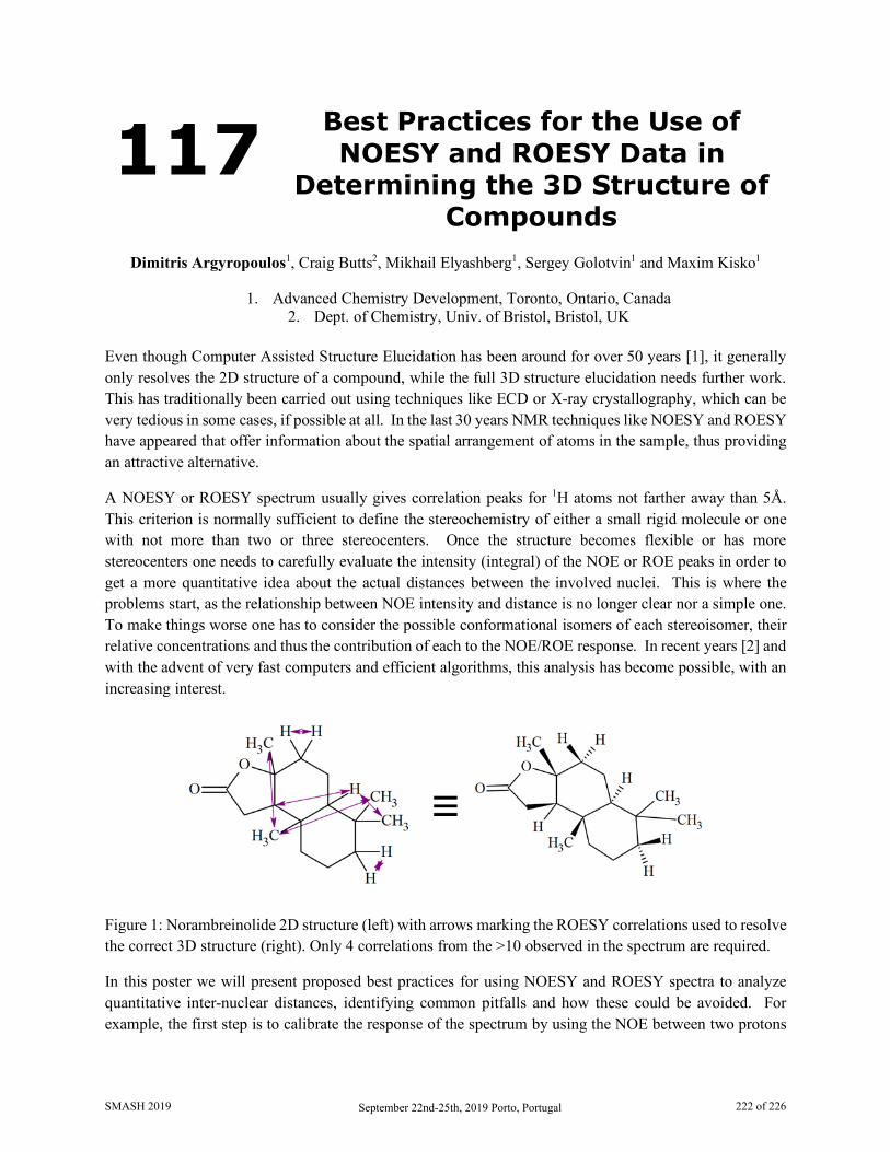
117 Best Practices for the Use of NOESY and ROESY Data in
Determining the 3D Structure of Compounds
Dimitris Argyropoulos1, Craig Butts2, Mikhail Elyashberg1, Sergey Golotvin1 and Maxim Kisko1
1. Advanced Chemistry Development, Toronto, Ontario, Canada 2. Dept. of Chemistry, Univ. of Bristol, Bristol, UK
Even though Computer Assisted Structure Elucidation has been around for over 50 years [1], it generally only resolves the 2D structure of a compound, while the full 3D structure elucidation needs further work. This has traditionally been carried out using techniques like ECD or X-ray crystallography, which can be very tedious in some cases, if possible at all. In the last 30 years NMR techniques like NOESY and ROESY have appeared that offer information about the spatial arrangement of atoms in the sample, thus providing an attractive alternative.
A NOESY or ROESY spectrum usually gives correlation peaks for 1H atoms not farther away than 5Å. This criterion is normally sufficient to define the stereochemistry of either a small rigid molecule or one with not more than two or three stereocenters. Once the structure becomes flexible or has more stereocenters one needs to carefully evaluate the intensity (integral) of the NOE or ROE peaks in order to get a more quantitative idea about the actual distances between the involved nuclei. This is where the problems start, as the relationship between NOE intensity and distance is no longer clear nor a simple one. To make things worse one has to consider the possible conformational isomers of each stereoisomer, their relative concentrations and thus the contribution of each to the NOE/ROE response. In recent years [2] and with the advent of very fast computers and efficient algorithms, this analysis has become possible, with an increasing interest.
Figure 1: Norambreinolide 2D structure (left) with arrows marking the ROESY correlations used to resolve the correct 3D structure (right). Only 4 correlations from the >10 observed in the spectrum are required.
In this poster we will present proposed best practices for using NOESY and ROESY spectra to analyze quantitative inter-nuclear distances, identifying common pitfalls and how these could be avoided. For example, the first step is to calibrate the response of the spectrum by using the NOE between two protons
SMASH 2019 September 22nd-25th, 2019 Porto, Portugal 222 of 226
that are held at a fixed, known distance in the structure (e.g. the distance within a CH2 group with two diastereotopic geminal protons), however, the distance between two ortho aromatic protons is not a good choice – and we’ll explain why. Another pitfall is the treatment of CH3 groups, with 3 chemically equivalent hydrogens which present 3 different distances to their coupling partners in any static conformer, but give rise to only one NOESY/ROESY peak (the same is true for CH2 groups with equivalent protons). Here, we will explain the reasons behind such observations and present the correct method for averaging of the response using some examples.
1. Elyashberg, M., Argyropoulos, D., NMR-based Computer-assisted Structure Elucidation (CASE) of Small Organic Molecules in Solution: Recent Advances, eMagRes, 2019, Vol 8: 1–16. DOI 10.1002/9780470034590.emrstm1618
2. Butts, C. P., Jones, C. R., Towers, E. C., Flynn, J. L., Appleby, L., Barron, N. J., Interproton distance determinations by NOE – surprising accuracy and precision in a rigid organic molecule, Org. Biomol. Chem., 2011, 9, 177-184
SMASH 2019 September 22nd-25th, 2019 Porto, Portugal 223 of 226
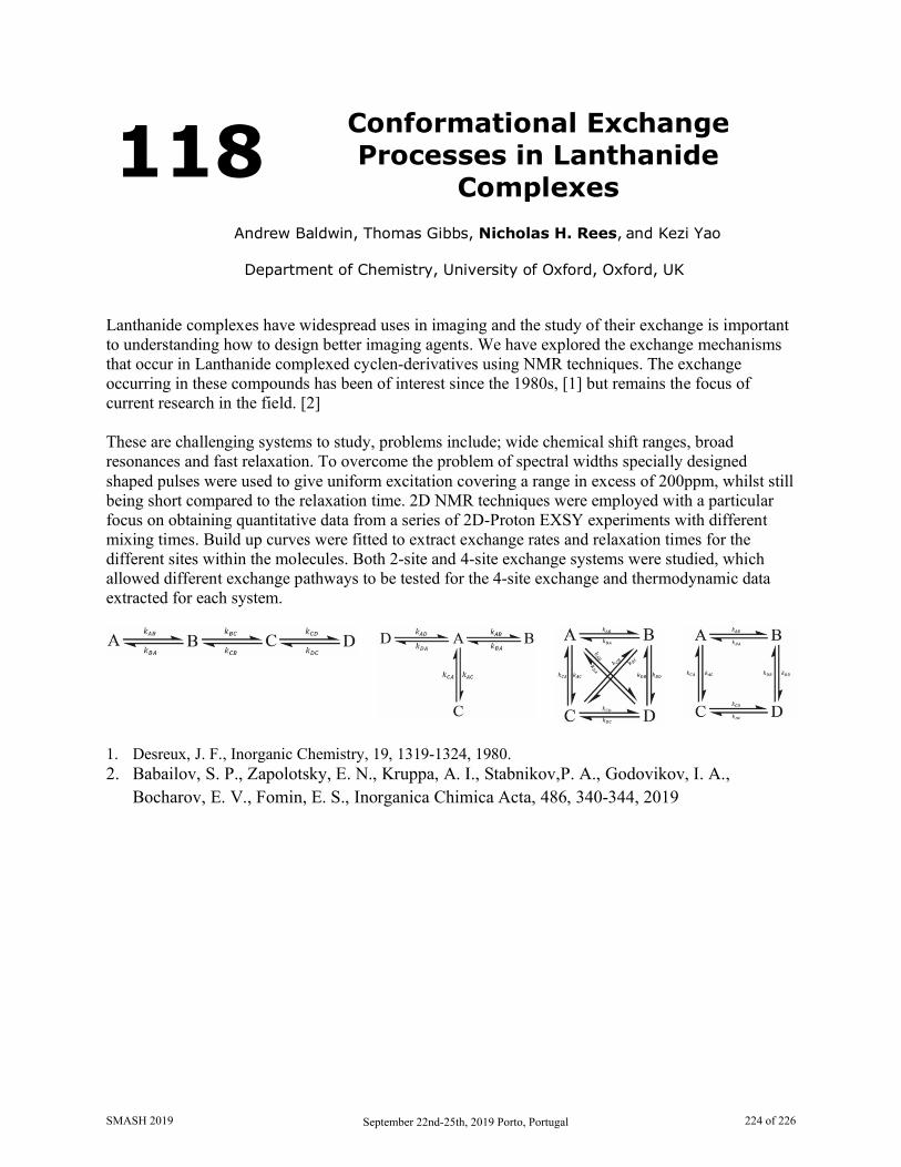
118 Conformational Exchange Processes in Lanthanide
Complexes
Andrew Baldwin, Thomas Gibbs, Nicholas H. Rees, and Kezi Yao
Department of Chemistry, University of Oxford, Oxford, UK
Lanthanide complexes have widespread uses in imaging and the study of their exchange is important to understanding how to design better imaging agents. We have explored the exchange mechanisms that occur in Lanthanide complexed cyclen-derivatives using NMR techniques. The exchange occurring in these compounds has been of interest since the 1980s, [1] but remains the focus of current research in the field. [2] These are challenging systems to study, problems include; wide chemical shift ranges, broad resonances and fast relaxation. To overcome the problem of spectral widths specially designed shaped pulses were used to give uniform excitation covering a range in excess of 200ppm, whilst still being short compared to the relaxation time. 2D NMR techniques were employed with a particular focus on obtaining quantitative data from a series of 2D-Proton EXSY experiments with different mixing times. Build up curves were fitted to extract exchange rates and relaxation times for the different sites within the molecules. Both 2-site and 4-site exchange systems were studied, which allowed different exchange pathways to be tested for the 4-site exchange and thermodynamic data extracted for each system.
1. Desreux, J. F., Inorganic Chemistry, 19, 1319-1324, 1980. 2. Babailov, S. P., Zapolotsky, E. N., Kruppa, A. I., Stabnikov,P. A., Godovikov, I. A.,
Bocharov, E. V., Fomin, E. S., Inorganica Chimica Acta, 486, 340-344, 2019
SMASH 2019 September 22nd-25th, 2019 Porto, Portugal 224 of 226
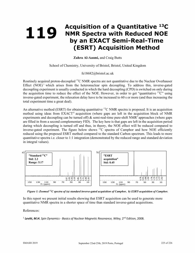
119 Acquisition of a Quantitative 13C NMR Spectra with Reduced NOE
by an EXACT Semi-Real-Time (ESRT) Acquisition Method
Zahra Al-Aasmi, and Craig Butts
School of Chemistry, University of Bristol, Bristol, United Kingdom
Routinely acquired proton-decoupled 13C NMR spectra are not quantitative due to the Nuclear Overhauser Effect (NOE) i which arises from the heteronuclear spin decoupling. To address this, inverse-gated decoupling experiment is usually conducted in which the hard decoupling (CPD) is switched on only during the acquisition time to reduce the effect of the NOE. However, in order to get “quantitative 13C” using inverse-gated experiment, the relaxation delay have to be increased to 60 s or more (and thus increasing the total experiment time a great deal). An alternative method (ESRT) for obtaining quantitative 13C NMR spectra is proposed. It is an acquisition method using ideas from EXACTii acquisition (where gaps are left in the acquisition block of NMR experiments and decoupling can be turned off) & semi-real-time pure-shift NMR3 approaches (where gaps are filled in from a second complementary FID). The key here is that gaps are left in the acquisition period during which decoupling is turned off and thus, in theory, the NOE effect will be reduced compared to inverse-gated experiment. The figure below shows 13C spectra of Camphor and how NOE efficiently reduced using the proposed ESRT method compared to the standard Carbon spectrum. This leads to more quantitative spectra i.e. closer to 1:1 integration (demonstrated by the reduced range and standard deviation in integral values).
Figure 1: Zoomed 13C spectra of (a) standard inverse-gated acquisition of Camphor, b) ESRT-acquisition of Camphor.
In this report we present initial results showing that ESRT acquisition can be used to generate more quantitative NMR spectra in a shorter space of time than standard inverse-gated acquisitions. References:
1 Levitt, M.H. Spin Dynamics - Basics of Nuclear Magnetic Resonance, Wiley, 2nd Edition, 2008.
a) b)
1030507090110130150f1 (ppm)
5.30
5.48
5.72
6.14
5.86
4.18
5.30
1.04
0.97
“Standard 13C” Std: 2.2 Range: 5.17
1030507090110130150f1 (ppm)
1.92
1.77
1.72
1.74
1.78
1.90
1.88
0.93
0.78
“ESRT acquisition” Std: 0.45
SMASH 2019 September 22nd-25th, 2019 Porto, Portugal 225 of 226

2EXtended ACquisition Time (EXACT) NMR—A Case for′ Burst′ Non‐Uniform Sampling. Ndukwe, I.E., Shchukina, A., Kazimierczuk, K., Cobas, C. and Butts, C.P. ChemPhysChem, 2016, 17, 2799-2803. 3 Semi-real-time acquisition for fast pure shift NMR at maximum resolution. Kiraly, P., M., Nilsson, Morris, G.A. Journal of Magnetic Resonance, 2018, 293, 19-27.
SMASH 2019 September 22nd-25th, 2019 Porto, Portugal 226 of 226