skin
TRANSCRIPT
SKIN
https://www.beautyflash.co.uk/skin-facts.html

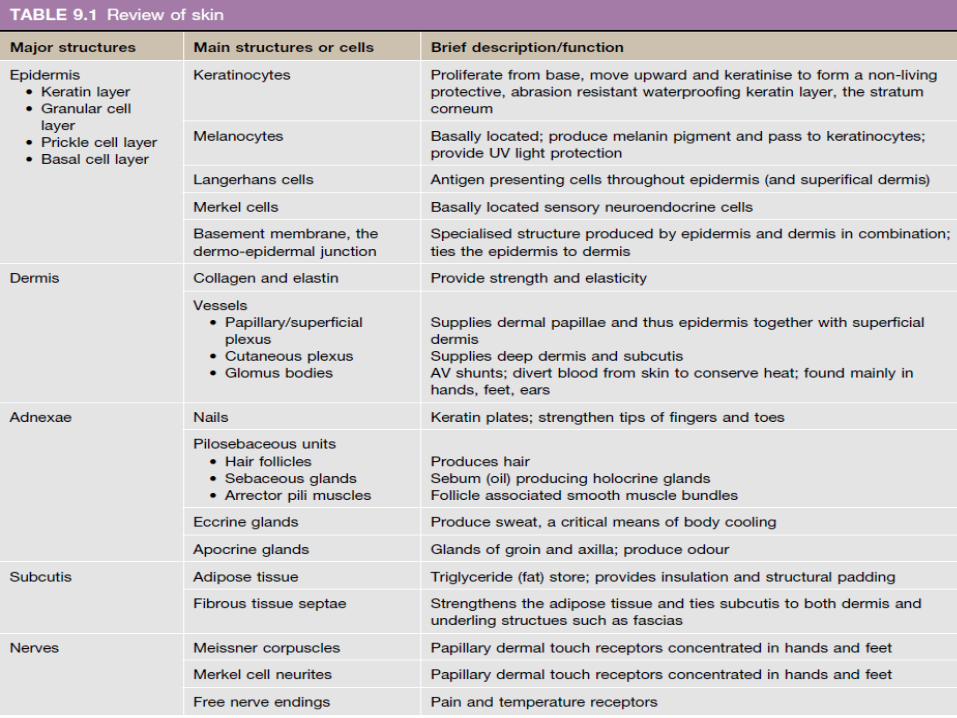
Integumentary System• Refers to the skin plus its appendages.(hair
follicles, sweat glands, sebaceous glands, nails, mammary
glands)
Skin• Covers the surface of the body and is
composed of two layers: epidermis and dermis.
• The deeper fascial layer, the hypodermis/subcutaneous fascia(anatomy), is not considered part of skin.

Serves Several Important Functions: • Protection against injury-Barrier
• Desiccation, and infection – Immunologic
• The regulation of body temperature – Homeostasis
• The absorption of UV radiation for synthesis of vitamin D – Endocrine
• Secretes sweat, sebaceous and apocrine glands- Exocrine
• And the reception of sensory stimuli (tactile, thermal, and pain) from the external environment. - Sensory

• Epidermis and dermis interdigitate with each other to form an irregular contour.
• Dermal papillae (ridges) project into the epidermis to produce epidermal ridges (which can be seen on the finger tips with the naked eye).
• May also be classified as thick or thin, depending on the thickness of epidermis.
Skin architecture
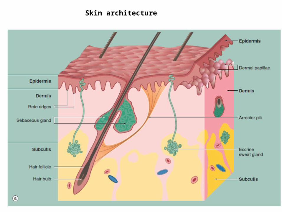
Skin architecture

• Photomicrograph of a section of skin.
• epidermis E,• keratin K,• papillary dermis PD• reticular dermis RD• subcutis SC,• eccrine (sweat)
glands EG• ducts ED.
rete ridges
dermal papillae
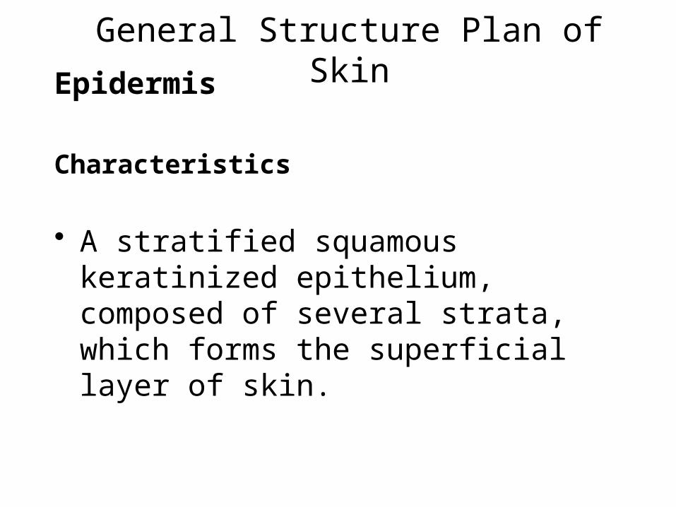
General Structure Plan of SkinEpidermis
Characteristics
• A stratified squamous keratinized epithelium, composed of several strata, which forms the superficial layer of skin.

• Is constantly being regenerated by its keratinocytes (every 2 to 4 weeks) via mitotic activity that occurs mostly at night.
• Epithelial layers of the epidermis include the following strata: basale, spinosum, granulosum, lucidum, and corneum:
1. Stratum Basale (germinativum)
• Is the deepest layer of cells, attached directly to the basal lamina by hemidesmosomes.
• Cuboidal to columnar in shape and frequently seen undergoing division.
• Melanocytes (pigment cells) and Merkel cells are also present in this layer.
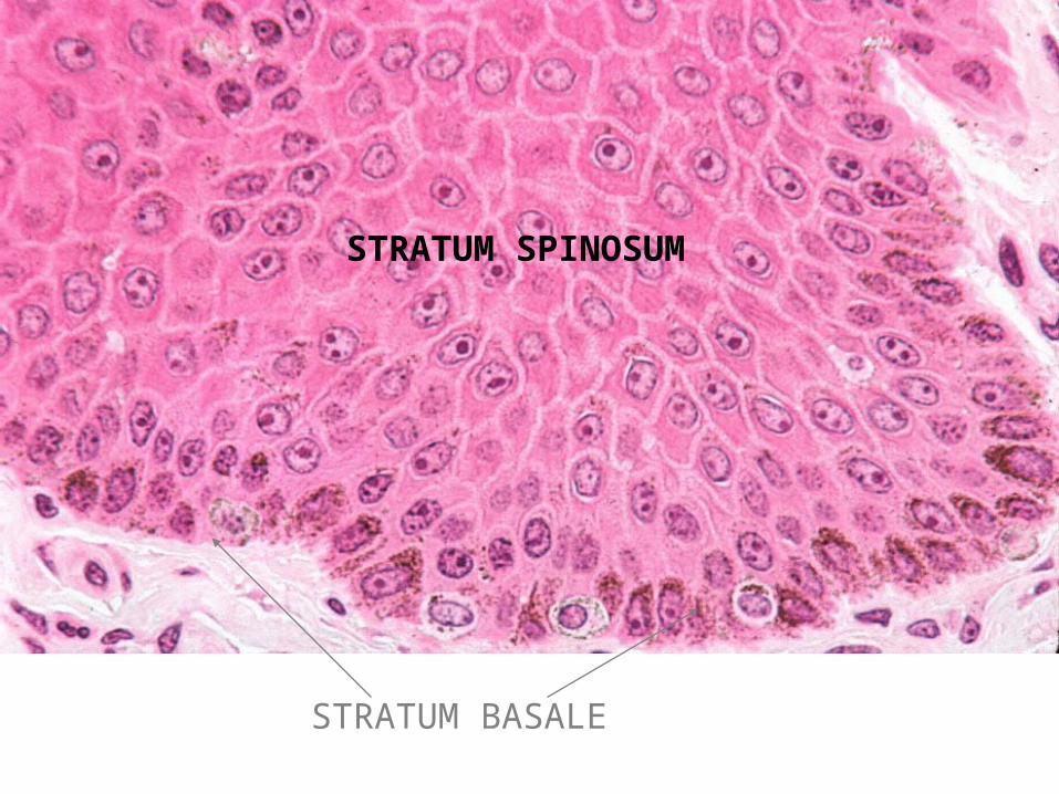
STRATUM BASALE
STRATUM SPINOSUM
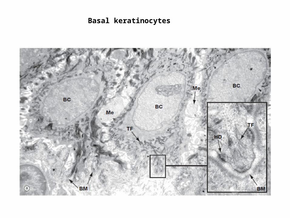
Basal keratinocytes
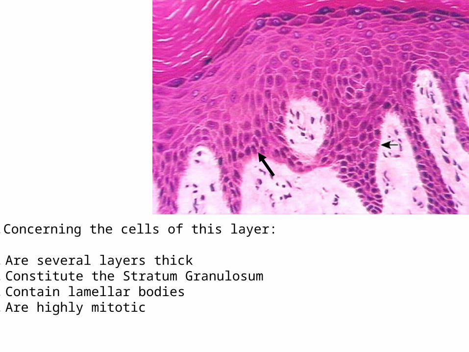
2.Concerning the cells of this layer:
A. Are several layers thickB. Constitute the Stratum GranulosumC. Contain lamellar bodiesD. Are highly mitotic

2. Stratum Spinosum
• consists of a few layers of polyhedral (prickle) keratinocytes.
• these cells have extensions, or so-called “intercellular bridges”, where desmosomes attach the cells to each other- Prickle cells.
• Superficial layer cells- elongate
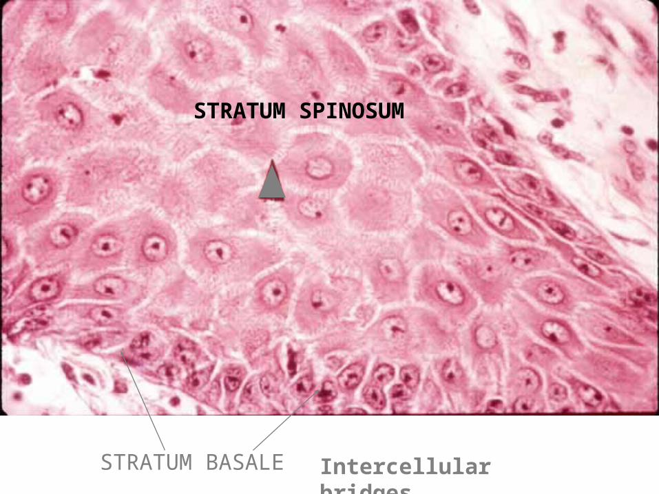
STRATUM SPINOSUM
STRATUM BASALE Intercellular bridges

Keratinocyte from prickle cell layer
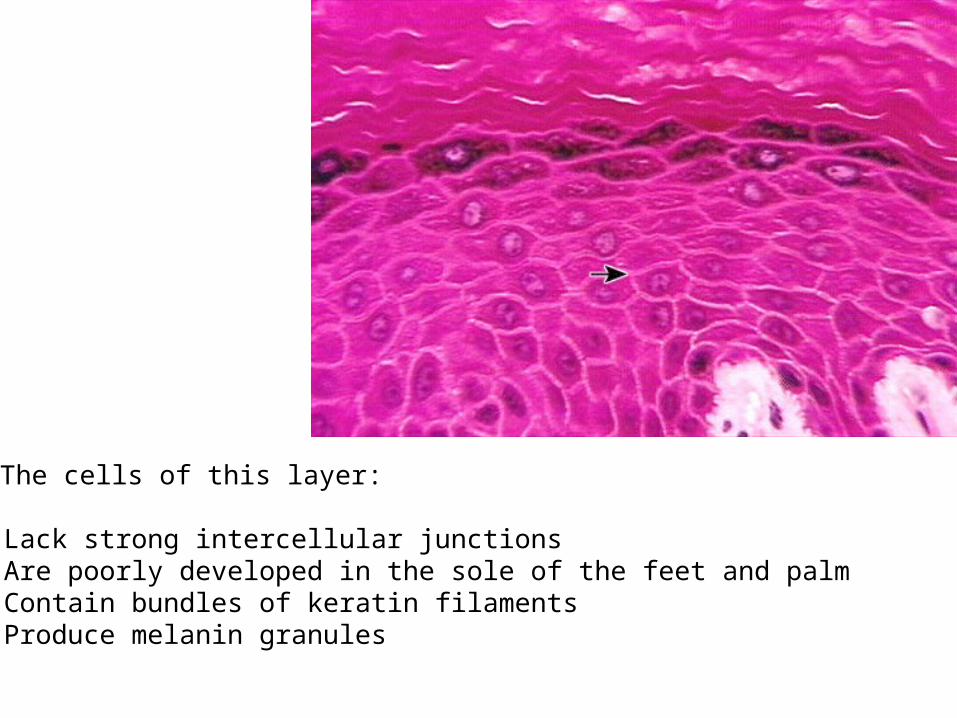
5.The cells of this layer:
A. Lack strong intercellular junctionsB. Are poorly developed in the sole of the feet and palmC. Contain bundles of keratin filamentsD. Produce melanin granules
3. Stratum Malpighii
• refers to the stratum basale and the stratum spinosum grouped together.
4. Stratum Granulosum
• the layer of the epidermis where cells accumulate keratohyalin granules and bundles of intermediate keratin filaments (tonofilaments) and become flattened.
• Granules have precursors for production of filaggrin which aggregates cells in stratum corneum.
• Also produce membrane bound lamellar bodies - epidermal water barrier
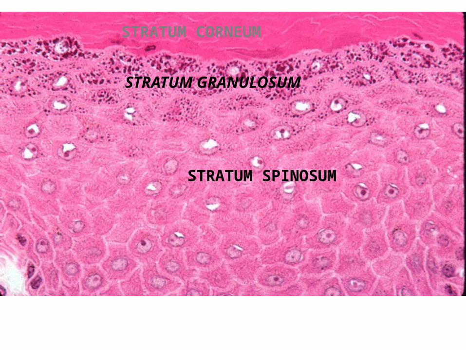
STRATUM SPINOSUM
STRATUM GRANULOSUM
STRATUM CORNEUM
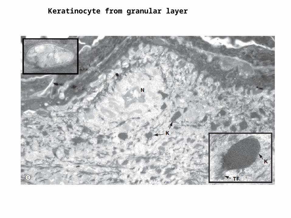
Keratinocyte from granular layer
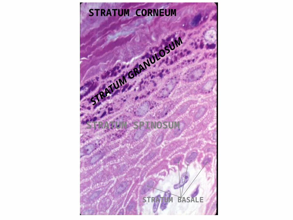
STRATUM SPINOSUM
STRATUM GRANULOSUM
STRATUM CORNEUM
STRATUM BASALE
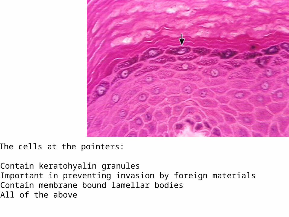
6.The cells at the pointers:
A. Contain keratohyalin granulesB. Important in preventing invasion by foreign materialsC. Contain membrane bound lamellar bodiesD. All of the above

5. Stratum Lucidum
• a clear homogeneous layer, which is often difficult to distinguish in histological sections.
• nuclei and organelles are not present in this layer, and the cells contain a substance known as eleidin, which is believed to be a transformation product of keratohyalin.
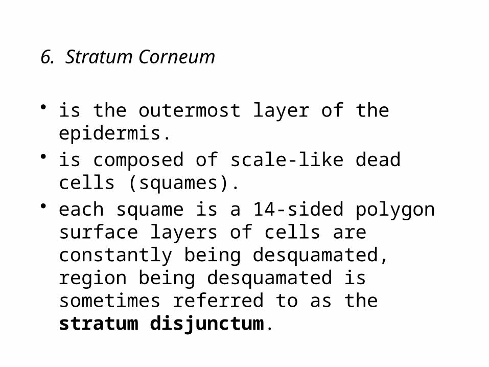
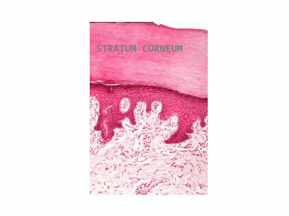
6. Stratum Corneum
• is the outermost layer of the epidermis.• is composed of scale‑like dead cells
(squames).• each squame is a 14‑sided polygon surface
layers of cells are constantly being desquamated, region being desquamated is sometimes referred to as the stratum disjunctum.
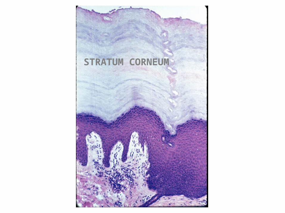
STRATUM CORNEUM

11.Intercellular bridges are characteristic of this layer:
A.B.C.D.
A
B
C
D
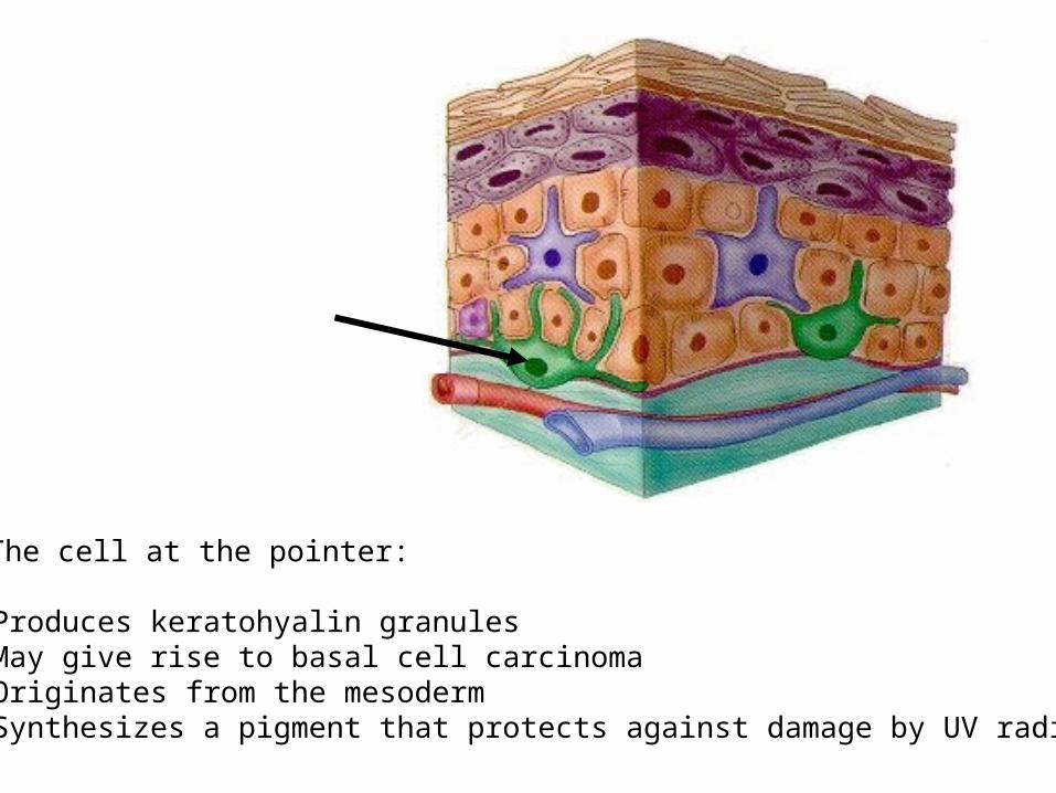
8.The cell at the pointer:
A. Produces keratohyalin granulesB. May give rise to basal cell carcinomaC. Originates from the mesodermD. Synthesizes a pigment that protects against damage by UV radiation

7. Other Cell Types in the Epidermis
Melanocytes
• are present in the stratum basale and derived from the neural crest.
• synthesize brown melanin pigment in oval organelles called melanosomes.
melanocyte
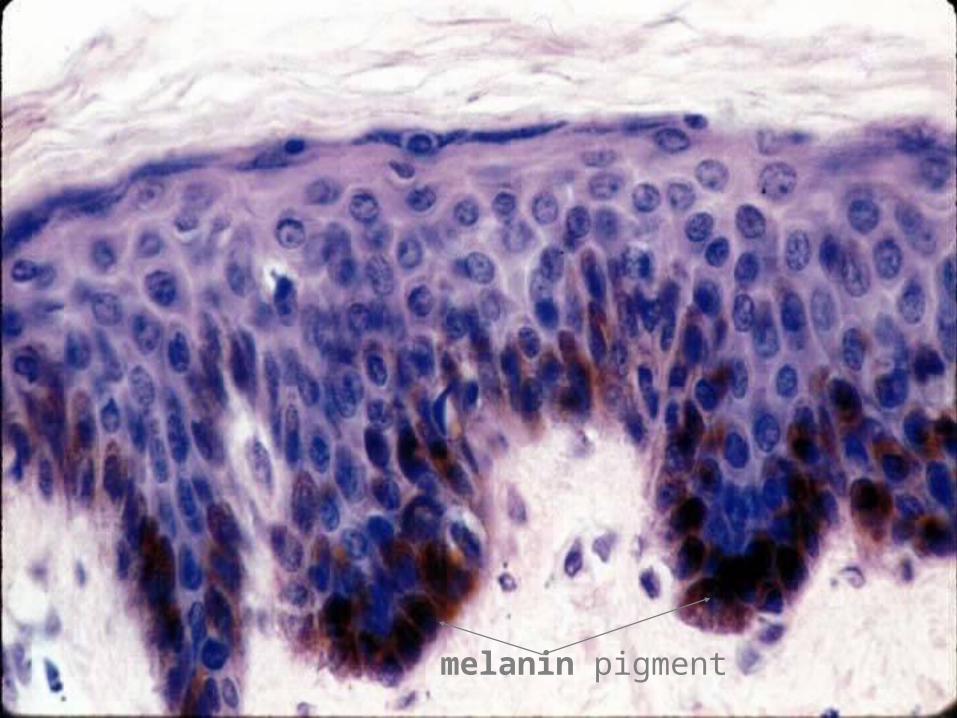
melanin pigment

Langerhans Cells
• are dendritic‑shaped cells derived from the bone marrow.
• are present mainly in the stratum spinosum.• contain distinct paddle‑shaped
membrane‑bounded granules (Birbeck granules).
• function in presenting antigen to lymphocytes and thereby play a role in contact allergic responses.

Langerhans cells- LCytoplasmic Process -CP
Birbeck granule –tennis racket shape
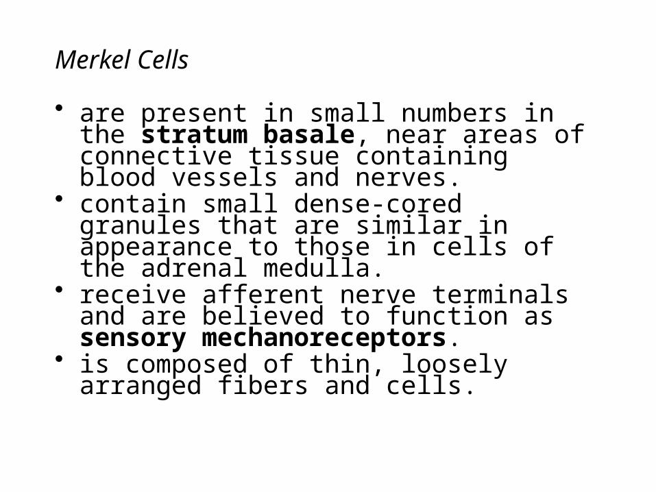
Merkel Cells
• are present in small numbers in the stratum basale, near areas of connective tissue containing blood vessels and nerves.
• contain small dense‑cored granules that are similar in appearance to those in cells of the adrenal medulla.
• receive afferent nerve terminals and are believed to function as sensory mechano receptors.
• is composed of thin, loosely arranged fibers and cells.
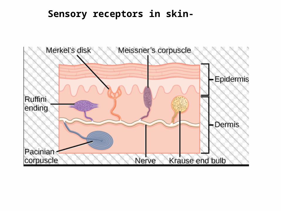
Sensory receptors in skin-
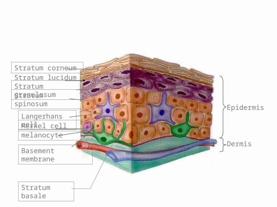
Epidermis
Dermis
Stratum corneumStratum lucidumStratum granulosumStratum spinosum
Langerhans cellMerkel cellmelanocyte
Basement membrane
Stratum basale

Dermis
1. Characteristics
• layer of skin underlying the epidermis that consists of dense, irregular connective tissue.
• contains collagen (Type I) fibers in abundance and networks of thick elastic fibers.
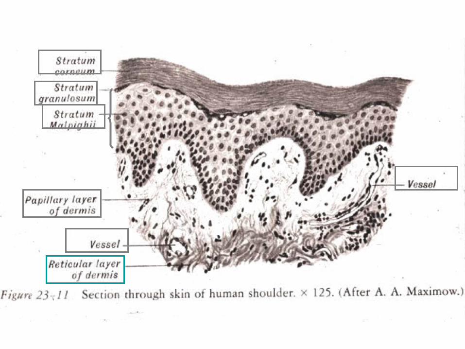
• is divided into a superficial papillary layer and a deeper more extensive reticular layer, but there is no distinct boundary between them.
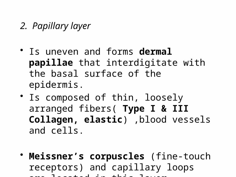
2. Papillary layer
• Is uneven and forms dermal papillae that interdigitate with the basal surface of the epidermis.
• Is composed of thin, loosely arranged fibers( Type I & III Collagen, elastic) ,blood vessels and cells.
• Meissner’s corpuscles (fine-touch receptors) and capillary loops are located in this layer.

“Papillary layer”
Meissner’s corpuscles
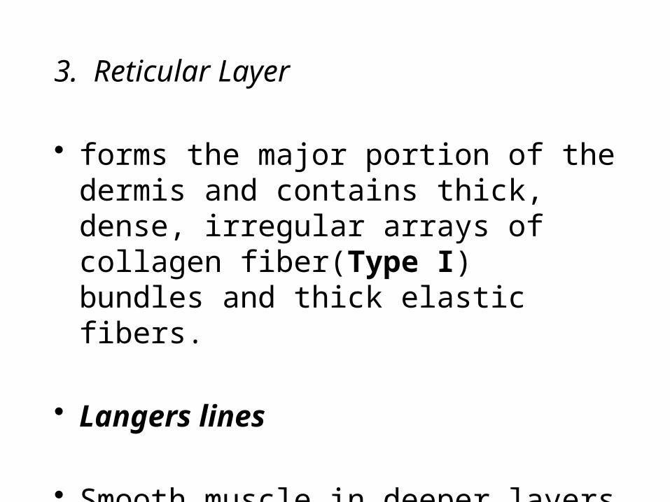
3. Reticular Layer
• forms the major portion of the dermis and contains thick, dense, irregular arrays of collagen fiber(Type I) bundles and thick elastic fibers.
• Langers lines
• Smooth muscle in deeper layers certain areas of skin- scrotum, perineum.

Encapsulated Nerve Endings
• consisting of Pacinian corpuscles (pressure receptors) and Krause’s end bulbs (cold and pressure receptors) may be present in the deeper regions of the dermis.
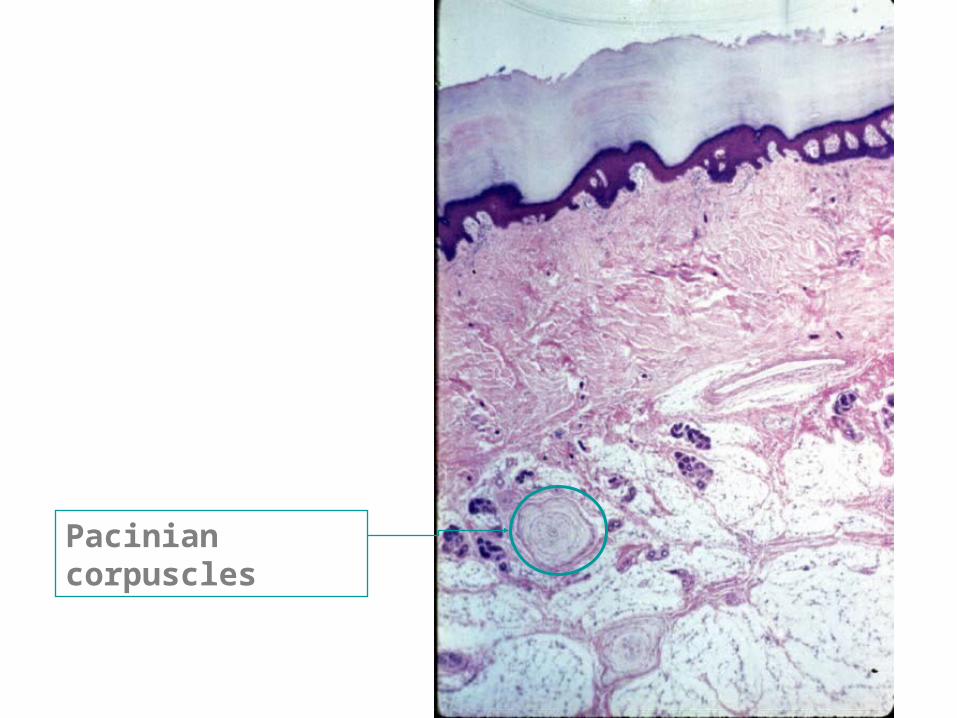
Pacinian corpuscles
Two Types of Skin
1. Thick Skin• has a thick epidermis that is characterized by
a prominent stratum corneum.• lines the palms of the hands and the soles of
the feet.• lacks hair follicles, sebaceous glands, and
arrector pili muscle bundles.
STRATUM CORNEUM
2. Thin Skin
• has a thin epidermis with a less prominent stratum corneum.
• is present over most of the body surface and contains hair follicles, sebaceous glands, and arrector pili muscle bundles.
• stratum lucidum and stratum granulosum are seldom seen in thin skin, although individual cells are present that show characteristics of these layers.
• the dermis is usually thicker in this area than in thick skin.


7.Identify the circumscribed structure as:
A. ThermoregulatorB. OsmoreceptorC. ChemoreceptorD. Pressure receptor

GlandsEccrine Sweat Glands
• are distributed in skin throughout the body.
• are simple tubular coiled glands that have a secretory unit composed of three cell types: dark cells, clear cells, and myoepithelial cells.
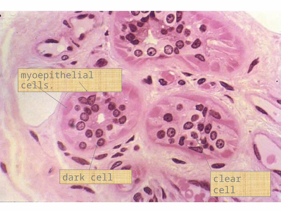
clear celldark cell
myoepithelial cells.

Eccrine sweat glands and ducts-
Ducts Lined by stratified cuboidal Epithelieum.

9. Concerning this gland:
A. Is a holocrine glandB. Is absent in thick skinC. Secretes an oily material called sebumD. Has ducts lined by stratified cuboidal epith.

Apocrine Glands
• Are large specialized sweat glands located in axilla, the areola of the nipple, and the circumanal region.
• Do not begin to function until puberty and are responsive to hormonal influences.
• Have a large coiled secretory portion enveloped by scattered myoepithelial cells.

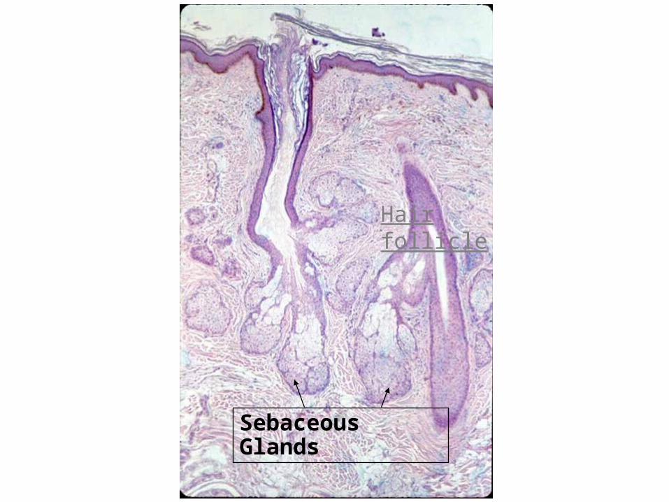
Sebaceous Glands
• Consist of several sacs (alveoli) that empty into a short duct, which in turn empties into the neck of a hair follicle.
• Cells at the periphery of the alveoli are flattened and inactive, but near the ducts mitosis are common.

Sebaceous Glands
Hair follicle
Sebaceous Glands
Hair follicle