skin and soft tissue infections 3 -...
TRANSCRIPT
Skin and soft tissue infections 3
M Al Madadha
Sources :
Harrisons infectious diseases 2nd edition, Oxford Handbook of Infectious Diseases and Microbiology 2nd edition

Hand foot and mouth disease
• Occurs in outbreaks, typically in schools (we had one a few months ago in nov)
• The typical disease:
• Children under 10 are mostly affected, more-so under 5
• The causative agents are• Coxsackie virus A16 (an enterovirus)
• Enterovirus 71 ( HAS BEEN ASSOCIATED WITH ENCEPHALITIS AND MYOCARDITIS)

• Atypical disease:
• Higher age groups ( adults and teenagers)
• Caused by coxsackie A6
• Can have severe presentation with fever, arthralgia and flu like symptoms with the rash as vesicles that affect (the nose, cheeks, extensor arms, elbows, thighs, buttocks, groin)
• Both typical and atypical disease are transmitted by fecooral route, or direct close contact to rash
Signs and symptoms of HFMD
Symptoms and signs:
• URT symptoms before the skin lesions occur
• With fever, malaise and pharyngitis!
• The rash is typically on the soles and palms and buttocks
• The rash:
• ORAL : football shaped (eye shaped) painful vesicles, involves the buccal mucosa and tongue. (spares posterior pharynx as opposed to herpangina that spares anterior pharynx)
• SKIN: red papules that progress to gray vesicles on the soles and palms and buttocks

https://cdn1.dailyhealthpost.com/wp-content/uploads/2016/11/hand-foot-and-mouth-disease-918x420.jpg

Rx
• Only symptomatic treatment ( treat fever, pain)
• Must maintain hydration
Consider hospital admission in severe illness (enterovirus A71) which has high morbidity and mortality

Smallpox
• • Smallpox is caused by variola virus, anي orthopoxvirus.
• There are two strains: variola major (mortality 20–50%) and variola minor
• • The last reported case was in Somalia in 1977, and the virus was declared eradicated by the WHO in 1980.
• Virus stocks exist in two laboratories, and there are concerns about its potential use as a bioterrorism agent
• • The incubation period is 10–12 days and is followed by a prodromal period of 1–2 days.
• The centrifugal rash is initially maculopapular and progresses to vesicles, pustules, and scabs over 1–2 weeks.
• Death may occur with fulminant disease.
• • Diagnosis may be confirmed by EM or PCR (to differentiate it from other poxviruses).
• • There is no specific treatment (supportive)

• Orf is caused by a DNA virus related to smallpox virus (parapoxvirus).
• Also called soremouth infection and infects the fingers of individuals who work around goats and sheep.
• Usually coming into contact with infected animals (petting, feedings, harnesses,bite) , but can also transmit between people.
• There is no treatment , however, the papules may become infected by bacteria, or immune compromised
• Can be infected multiple times through out life (each time is less severe).
• Six stages: each about a week
• Small papule nodules ulcerate and crust

http://www.farmhealthonline.com/wp-content/uploads/2015/09/Orf-MSD-animal-health.co_.uk_.jpghttp://c.ymcdn.com/sites/www.aocd.org/resource/resmgr/ddb_high/orf_1_high.jpg

Molluscum contagiosum
• virus induces flaccid vesicles on the skin of healthy and immunocompromised individuals.
• Mcc 2-11 year olds, in adults as part of STD- transmission by direct contact
• Single or small clusters of vesicles (<30) raised papules, may look like vesicles, not erythematous!, with central umbilication
• On face and trunk, pubis and rarely mucosa
• Resolve on their own (cryotherapy or other dermatologic treatments can be done) may be present for YEARS
• If associated with HIV SEVERE

https://www.healthline.com/hlcmsresource/images/galleries/Molluscum-Contagiosum/molluscum_contagiosum_adult_stomach.jpg

Infections Associated with Crusted Lesions

Impetigo •
• Caused by S. aureus and/or GASusual skin flora, with local trauma that allows colonizing bacteria to break through.
• Commonly affects children (2-5 years) in tropical/subtropical regions; also prevalent in temperate regions in summer months (so warm weather >).
• It is the most common bacterial skin infection in children
• 70% are non bullous and appear as crusted legions
• It is highly contagious (scratching, towels, clothing, autoinfection and spread in daycares)

• • Clinical features—occurs on the face and extremities.
• Lesions start as small macule or papule small vesicles (with erythema) develop into flaccid bullae rupture, releasing a yellow discharge which forms thick crusts.
• Usually seen with regional LAP
• Can cause cellulitis ( deeper infection, see later)
• Or PSGN ( post strep GN)
• • Treatment—mupirocin is the best topical agent.
Patients who have numerous lesions or who do not respond to topical treatment should receive oral antibiotics (flucloxacillin or cefalexin).
If MRSA is suspected/isolated, then treatment with doxycycline, clindamycin, or co-trimoxazole.


http://diseasespictures.com/wp-content/uploads/2013/12/Impetigo-2.jpg

Ecthyma
• • Punched-out ulcers surrounded by raised deep red/violet margins invades into the dermis and leaves highly inflamed regions on the sides
• • Caused by S. aureus or GAS. Other similar lesions (ecthyma gangrenosum) may occur with P. aeruginosa in neutropenic (reduced neutrophils in blood) patients.
• • Empiric treatment is with flucloxacillin or cephalexin (unless cultures yield streptococci alone, in which case penicillin is appropriate).
• Antipseudomonal agents, e.g. piperacillin-tazobactam, should be given for P. aeruginosa infections.

http://www.pcds.org.uk/ee/images/made/ee/images/uploads/clinical/Ecthyma_gangrenosum_pseud_bact_800_600_70_http:www.pcds.org.ukeeassetsimgwatermark.gif_0_0_80_r_b_-5_-5_.jpg
Dermatophytes
• A group of fungi capable of invading the dead keratin of skin, hair, and nails (require keratin for their growth).
• They are spread by direct contact with patients or animals or soil
• Clinical classification is by age group:
Children :
-tinea capitis (scalp hair and the commonest in children),
-tinea corporis (trunk and limbs)
-tinea faciale (face)

cont.
Adolescents:
-tinea manuum and pedis (palms and soles – athletes foot-, and the commonest overall worldwide)
-tinea unguium (nail—also known as onychomycosis)
Adults:
-tinea cruris (groin) AKA jock itch
-tinea barbae (beard area and neck)
-Tinea corporis Gladiatorum ( wrestlers)

Ringworm, corporis, circinata
https://www.news-medical.net/image.axd?picture=Ringworm%20shutterstock_484652500%5B12%5D_1.jpg
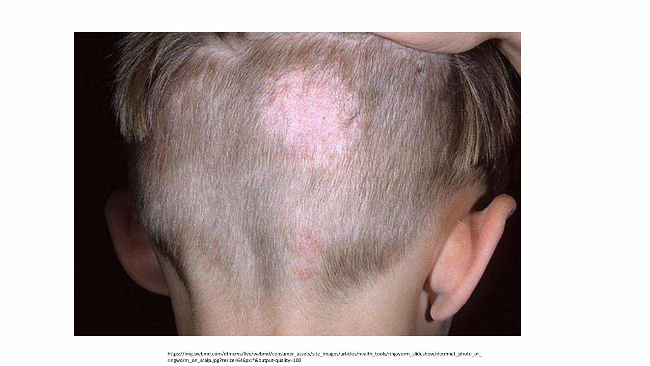
https://img.webmd.com/dtmcms/live/webmd/consumer_assets/site_images/articles/health_tools/ringworm_slideshow/dermnet_photo_of_ringworm_on_scalp.jpg?resize=646px:*&output-quality=100

http://ringwormtinea.com/wp-content/uploads/2015/02/11.jpg
Demarcation lines hint at A possible dermatophyteInfection, rather than acnevulgaris

Tinea corporis psoriasis

https://www.sporthuesca.com/piscinas-antihigienicas/
http://mulicia.pixnet.net/blog/post/26749579-%E7%94%B2%E7%99%AC-onychomycosis%EF%BC%8Ctinea-unguium

DIAGNOSIS
• The goal is to distinguish dermatophytoses from other causes of skin inflammation (such as psoriasis).
• Infections caused by bacteria, other fungi, and noninfectious disorders (psoriasis, contact dermatitis) may have similar features.
• KOH mounts of skin scrapings and infected hairs demonstrate hyphae.
• Some species fluoresce by a U.V. lamp.
• Culture is used when KOH preparations are negative.

TREATMENT AND PREVENTION
• Many local skin infections resolve spontaneously without chemotherapy.
• Topical use of tolnaftate, allylamines, or azoles is usually sufficient .
• Nail bed and more extensive skin infections require systemic therapy with griseofulvin or itraconazole and terbinafine + combined with topical therapy.
• Therapy must be continued over weeks to months, and relapses may occur.
• No specific preventive measures such as vaccines exist.
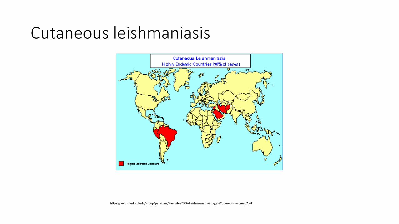
Cutaneous leishmaniasis
https://web.stanford.edu/group/parasites/ParaSites2006/Leishmaniasis/images/Cutaneous%20map2.gif

Vector and parasite
• Transmitted by sandflies• Trypanosome (single flagellae)• Has two forms: disease causing (resistant) form and infectious motile form• On CBC shows reduced cell count (red, white or all), Dx by culture• Deemed as a neglected tropical illness (causes mortality with small effort
to battle it)• Has many types (other than visceral),• 1) old world in Asia, Africa and Europe long incubation time (MONTHS
2-24)• Skin lesion(s) on the face or leg: papules become necrotic and then
pigmented scars ( papule at site of bite small nodules painLESSulcer crust (pigmented) leave ugly scars after healing (social issues)
https://www.cdc.gov/parasites/images/leishmaniasis/home_page_image_leishmaniasis.jpg


https://www.ncbi.nlm.nih.gov/pmc/articles/PMC4183486/figure/pntd-0003208-g001/

• Becoming an increasing problem in surrounding countries (syria and iraq) with poor sanitation and reduced living standards.
• L. major and L. tropics is most common
• Resolve over months, round depressed scars remain
• May resemble other skin lesions (nodular lymphangitis) caused by waterborne pathogens (such as Sporothrix schenckii, Nocardiabrasiliensis, Mycobacterium marinum, Leishmania (Viannia).
• Nodular lymphangitis- granulamatous reaction to these pathogens on the path of lymphatics

https://online.epocrates.com/data_dx/reg/920/img/920-1-hlight.jpg
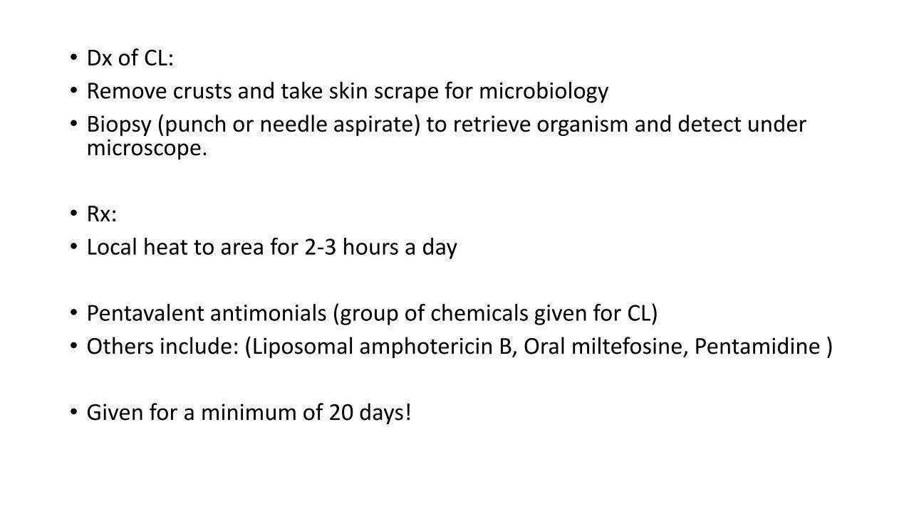
• Dx of CL:
• Remove crusts and take skin scrape for microbiology
• Biopsy (punch or needle aspirate) to retrieve organism and detect under microscope.
• Rx:
• Local heat to area for 2-3 hours a day
• Pentavalent antimonials (group of chemicals given for CL)
• Others include: (Liposomal amphotericin B, Oral miltefosine, Pentamidine )
• Given for a minimum of 20 days!

Infections Associated with Bullae

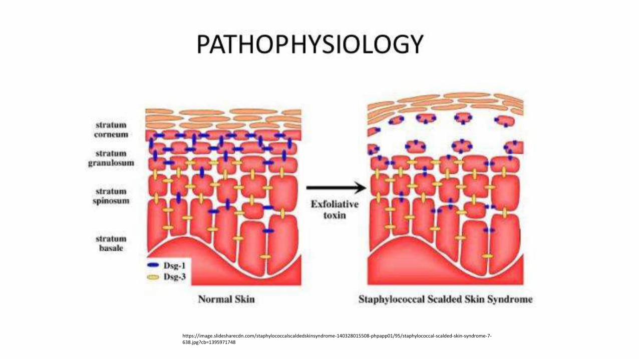
• Staphylococcal scalded-skin syndrome (SSSS) in neonates is caused by a toxin (exfoliatin) from phage group II (bacteria is infected by this phage, acquires the gene to produce the toxin)
• S. aureus. SSSS must be distinguished from toxic epidermal necrolysis (TEN), which occurs primarily in adults, is drug-induced, and is associated with a higher mortality rate.
• Punch biopsy with frozen section is useful in making this distinction since the cleavage plane is the stratum corneum in SSSS and the stratum germinatum in TEN
Staphylococcal scalded-skin syndrome (SSSS)
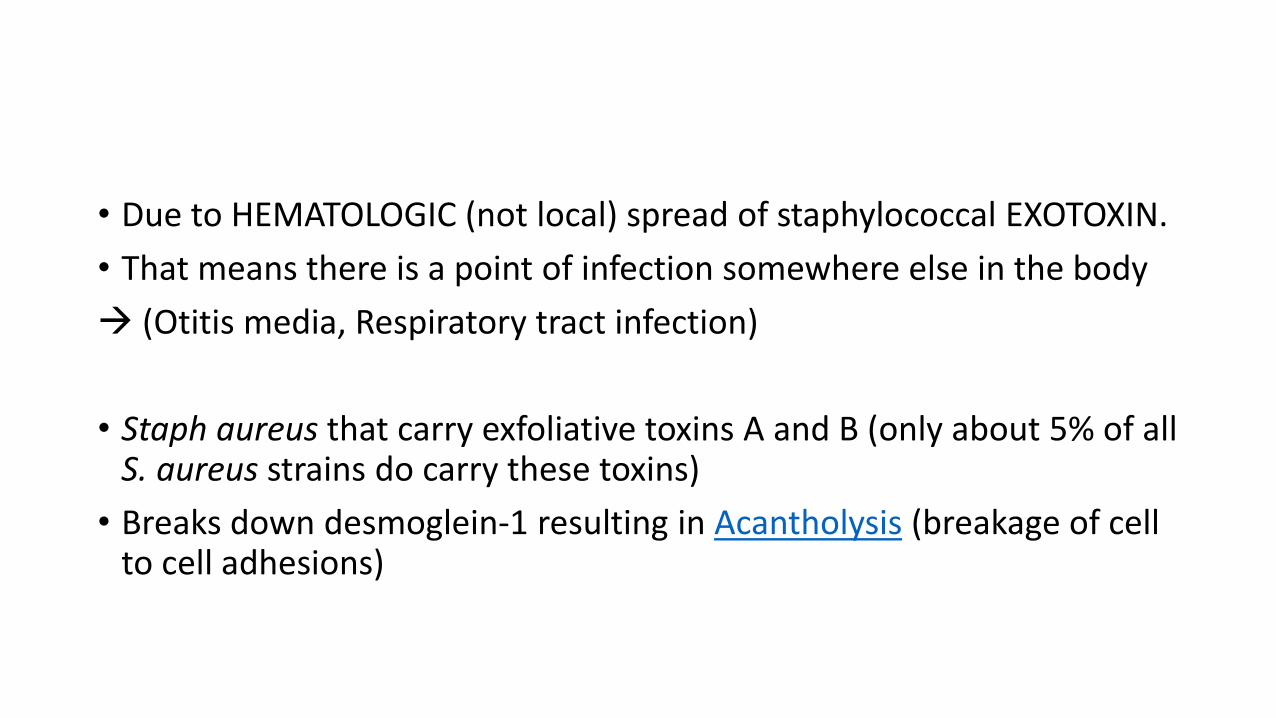
• Due to HEMATOLOGIC (not local) spread of staphylococcal EXOTOXIN.
• That means there is a point of infection somewhere else in the body
(Otitis media, Respiratory tract infection)
• Staph aureus that carry exfoliative toxins A and B (only about 5% of all S. aureus strains do carry these toxins)
• Breaks down desmoglein-1 resulting in Acantholysis (breakage of cell to cell adhesions)
https://image.slidesharecdn.com/staphylococcalscaldedskinsyndrome-140328015508-phpapp01/95/staphylococcal-scalded-skin-syndrome-7-638.jpg?cb=1395971748

https://upload.wikimedia.org/wikipedia/commons/thumb/3/3e/OSC_Microbio_21_02_SSSS.jpg/300px-OSC_Microbio_21_02_SSSS.jpg

http://www.dermnet.com/dn2/allJPG3/Staphylococcal-Scalded-Skin-Syndrome-3.jpg

SSSS
• Symptoms:• Preceded by a prodromal illness ( URTI ) or• Otitis Media, Pharyngitis, Conjunctivitis• Then the actue phase hits: fever! + malaise (loss of fluids) and red
painful skin with bullae formation
• Signs:• Paper thin (peeling) skin• Large flaccid BLISTERS, more in the flexor creases (see image above)• Mucous membranes are SPARED!• Positive Nikolsky’s sign (detects acantholysis)

You make the bullae spread further by rubbing the side from affected skin to non affected skin

SSSS Dx and Rx
• Dx: blood cultures are often positive (remember hematologic spread of toxin –bacteremia)
• Skin biopsy will show typical acantholysis
• In lab we can do exotoxin assay
• Rx:
• MUST ADMIT THE CHILD ( burn unit or ICU!)
• Systemic IV antibiotics are given (anti MRSA or anti S. aureus) +
• Systemic steroids ( only if patient doesn’t look toxic/otherwise it is not used)
• In severe cases (IV Immunoglobulins and plasmapheresis) removal of plasma

As for TEN
• TEN, primarily seen in adults is potentially fatal.
• Intravenous γ-globulin is a promising treatment for TEN.
http://i.dailymail.co.uk/i/pix/2016/12/07/11/3B2162BB00000578-4009166-image-a-41_1481111581525.jpg

Necrotizing Fasciitis
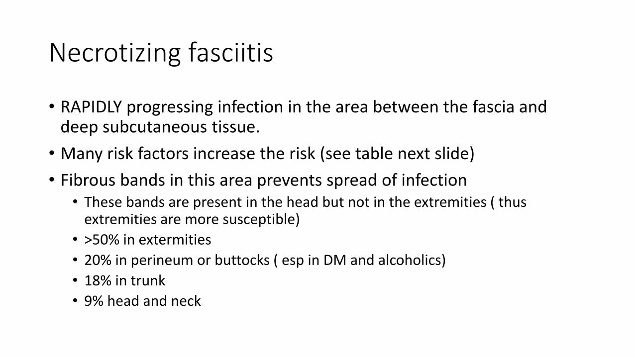
Necrotizing fasciitis
• RAPIDLY progressing infection in the area between the fascia and deep subcutaneous tissue.
• Many risk factors increase the risk (see table next slide)
• Fibrous bands in this area prevents spread of infection• These bands are present in the head but not in the extremities ( thus
extremities are more susceptible)
• >50% in extermities
• 20% in perineum or buttocks ( esp in DM and alcoholics)
• 18% in trunk
• 9% head and neck

• Necrotizing fasciitis (GAS) and gas gangrene (anaerobic clostridialinfection) also induce bulla formation.
• In the USA, the estimated incidence of invasive GAS infection is 3.5 cases per 100000 persons—necrotizing infections account for 6% of these.
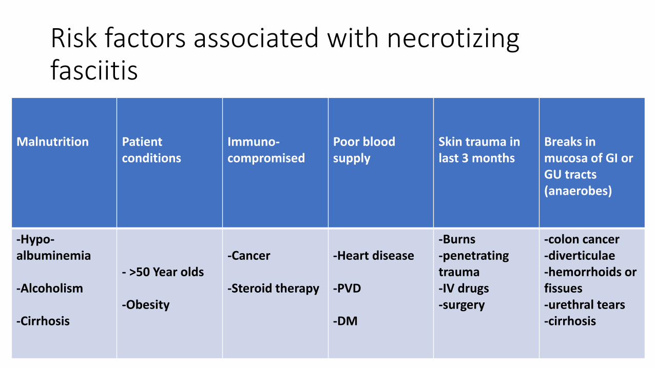
Risk factors associated with necrotizing fasciitis
Malnutrition Patient conditions
Immuno-compromised
Poor blood supply
Skin trauma in last 3 months
Breaks in mucosa of GI or GU tracts (anaerobes)
-Hypo-albuminemia
-Alcoholism
-Cirrhosis
- >50 Year olds
-Obesity
-Cancer
-Steroid therapy
-Heart disease
-PVD
-DM
-Burns-penetrating trauma-IV drugs-surgery
-colon cancer-diverticulae-hemorrhoids or fissues-urethral tears-cirrhosis

Signs and Symptoms – occur in order
• Pain/tenderness• Unexplained fever (Early diagnosis may be difficult when pain or
unexplained fever is the only presenting manifestation, remember infection is deep, might not present with pain yet )
• Swelling• Dark red induration• BULLAE, filled with blueish or purple fluid• Thrombosis of dermal blood vessels (The affected area becomes
anaesthetic as a result of small vessel thrombosis and destruction of superficial nerves)
• Extension to deep fascia with rapid spread• Most progressed symptoms : toxicity , shock and multi organ failures

Pain / redness
http://www.antimicrobe.org/new/printout/e11printout/e11cause/e11cau9.jpg

Dark red induration /swelling

Progressing to bullae /thrombosis
https://osuemed.files.wordpress.com/2012/09/diff-patient-with-blebs.jpg
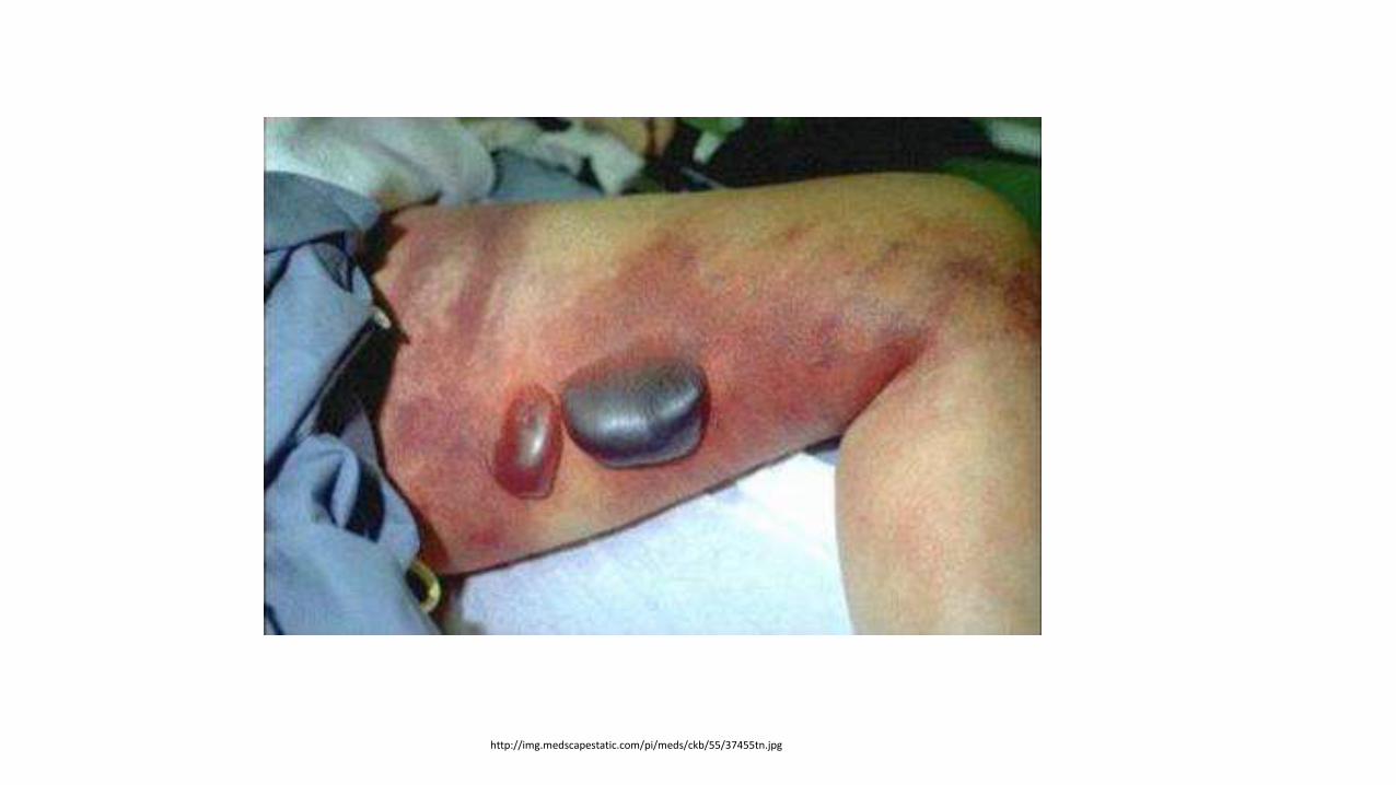
http://img.medscapestatic.com/pi/meds/ckb/55/37455tn.jpg

Microbiology causes:
• A) Polymicrobial (• Type I necrotizing fasciitis involves at least one anaerobic species ( Bacteroides or Peptostreptococcus spp.), as well as one or more facultative anaerobic species (e.g. non-GAS, E. coli, Enterobacter, Klebsiella, Proteus spp.).
• usually a mix of aerobes and anaerobic bacteria ( clostridium perfringens)
• 1 - Break in Gastrointestinal or Genitourinary mucosa, typically on trunk and extremities
• 2- Fournier's Gangrene (in genitalia/perineal area)
• 3- mixed infection usually have comorbid states ( DM, PVD, immunecompromised)
Microbiology causes…cont
• B) Type II necrotizing fasciitis is usually caused by GAS alone or in combination with other species (e.g. S. aureus). Group A , Beta hemolytic strep (GAS), S. pyogenes +- S. aureus
• Strains of MRSA that produce the PantonValentine leukocidin (PVL) toxin have been reported to cause necrotizing fasciitis.
• 1) usually following trauma in otherwise healthy individual
or IV drug abusers (skin popping)
2) Fasciitis progresses to skin contusions due to seeding by transient bactermia
3) Gas production if mixed infections occurs
4) Severe toxicity and renal impairments shock
5) Myositis ( destruction of muscle tissue markedly increases CPK)
6) Mortality is high (upto 50%!)

Skin popping/drug abuse = basically SubQinjections in same area over and over

• Necrotizing fasciitis caused by mixed aerobic-anaerobic bacteria begins with a breach in the integrity of a mucous membrane barrier, such as the mucosa of the gastrointestinal or genitourinary tract.
• The portal can be a malignancy, a diverticulum, a hemorrhoid, an anal fissure, or a urethral tear.
• Other predisposing factors include peripheral vascular disease, diabetes mellitus, surgery, and penetrating injury to the abdomen.
• Leakage into the perineal area results in a syndrome called Fournier’s gangrene, characterized by massive swelling of the scrotum and penis with extension into the perineum or the abdominal wall and legs.

• In the newborn, necrotizing fasciitis may complicate omphalitis and spread to involve the abdominal wall, flanks, and chest wall.
• Fournier’s gangrene is a form of necrotizing fasciitis that affects the male genitals and is usually polymicrobial.
• Craniofacial necrotizing fasciitis is usually associated with trauma and caused by GAS.
• Cervical necrotizing fasciitis is usually associated with dental or pharyngeal infections and is polymicrobial.
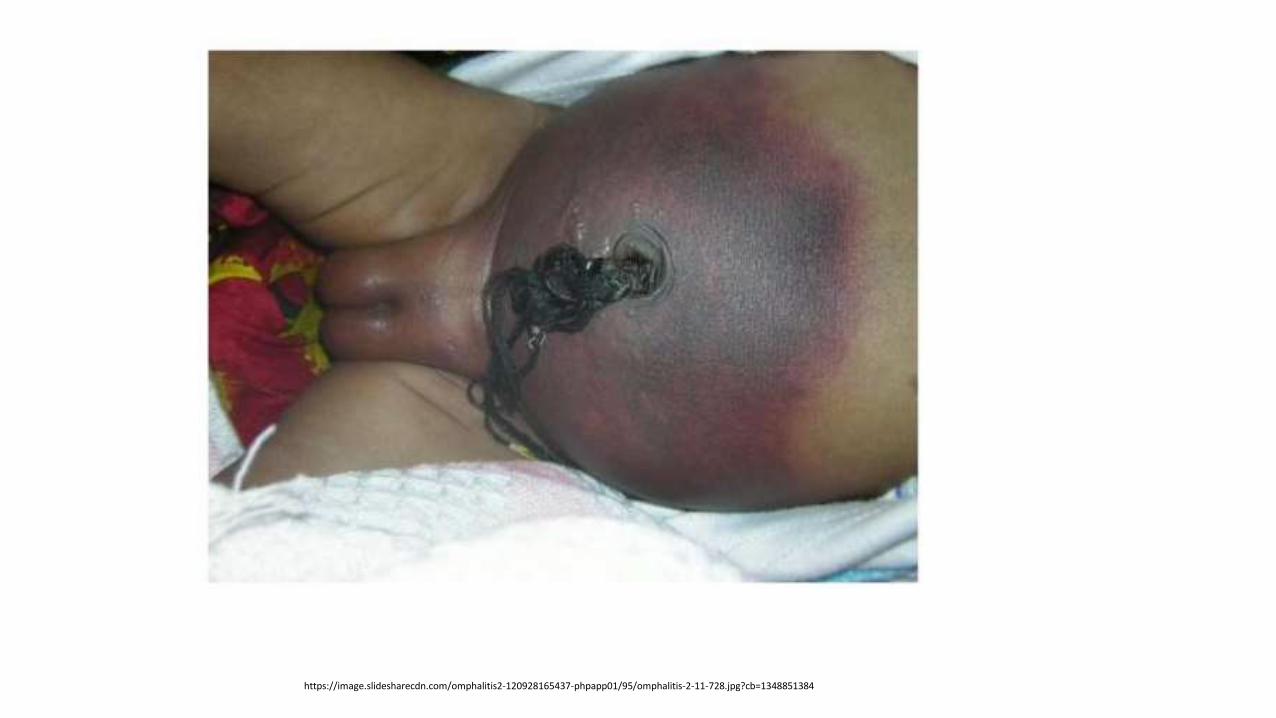
https://image.slidesharecdn.com/omphalitis2-120928165437-phpapp01/95/omphalitis-2-11-728.jpg?cb=1348851384

Dx of necrotizing fasciitis
Clinical findings are suggestive + surgical exploration/sample:
a) Altered mental status
b) Soft tissue infection signs (redness/swelling/pain) 70-80% of casesBullae Pain is typically exaggerated out of examTenderness is outside the red erythematous borders (indicates further progress)
c) are only seen in ¼ of cases
d) Fever in less than 50% of the cases!
e) Low BP in 21%
f) crepitation (feeling of air pockets under skin upon examination) in 20%
Rx - empiric
• 3 drug combo/ 2 drug combo/ 1 drug ( each +MRSA coverage)
3 drug combo :
• 1- anaerobic coverage (and inhibits ribosomal production of toxins)= Clindamycin
• 2- G +ve coverage (Ampicillin-sulbactam) or ( Piperacillin-tazobactam)
• 3- G-ve coverage (Ciprofloxacin)

• 2 drug combo (Cefotaxime covers G+ and G- bacteria) + (anaerobic coverage by metronidazole or clindamycin)
• 1 drug combo ( Carbapenem 0 Imipenem, meropenem, ertapenem)
• The MRSA coverage to be added to any chosen empiric regimen includes = Vancomycin or Linezolid
• Hemorrhagic bullae may indicate presence of vibrio vulnificus, in which case doxycycline is used

Rx.
• Surgical debridement, and treatment in hospital Emergency surgical exploration and debridement confirm the diagnosis and are the mainstay of therapy.
• Reducing compartment pressure in extremities
• Prophylaxis for exposed house hold members (penicillin, rifampin, clindamycin or azirthromycin)