skills in minor surgical procedures for general …...skills in minor surgical procedures for...
TRANSCRIPT

7
Skills in Minor Surgical Procedures for General Practitioners
Jose María Arribas Blanco and María Hernández Tejero Faculty of Medicine, Autonomous University of Madrid, Madrid,
Spain
1. Introduction
Minor surgery is defined as a set of procedures in which short surgical techniques are applied on superficial tissues. Local anaesthesia is often required for these procedures and their complication-rate as well as the risk involved is low.
Lesions and problems requiring these procedures for diagnostic or therapeutical reasons are frequently seen by general practitioners both in the outpatient setting (excision of skin lesions, for instance) as well as in the emergency care setting (wound suturing, for example). Therefore, for family doctors proficiency in minor surgical procedures is an additional tool for good medical practice and acquiring skills in minor surgical procedures has become a critical part of medical training, and has adjusted their professional profile to the problems and needs arising in te daily practice of family medicine.
Achieving clinical excellence in these procedures depends on the involved practitioner’s
level of technical training, since surgical training is the best way to learn the correct surgical
gestures and proper use of surgical instruments. Trainees must also understand that a
sound diagnostic approach plus the observance of basic principles of good medical practice
are prerequisites for clinical excellence in their practice of minor surgical procedures
It is shown that the performance of minor surgery in primary care is good for patients, for health care system and for the practitioner; In addition to being fun and rewarding. However, those procedures are not without risks, both during surgery and afterwards. Therefore, any surgical procedure performed by the family physician, is necessary (in addition to proper surgical technique and appropriate therapeutic indications) provide a comprehensive, clear and complete information to the patient, which will be reflected with the Informed Consent signed by the patient before carrying out any minor surgical intervention.The realization of Informed Consent is not a guarantee of exculpate in the event of a judicial proceeding by a complaint from a medical act of minor surgery. The judge will assess good medical practice and clinical experience based on the lex artis (proper patient selection and indications for its implementation, use of good surgical technique ..). Therefore, and although the academic programs of the Family Medicine Specialty explicitly enable the family doctor to perform minor surgical procedures, it is recommended that primary care physicians have medical insurance with specific coverage for surgical techniques.
www.intechopen.com

Primary Care at a Glance – Hot Topics and New Insights
102
This chapter will try and help general practitioners master minor surgical procedures by
answering the following questions:
1. Where should general practitioners perform minor surgical procedures? Optimal infrastructure and medical furniture for a Minor Surgery operating room and doctor’s preparation
2. Which are the instruments and materials involved? Surgical instruments (plus their handling) and suture materials for basic and advanced surgery.
3. How to perform minor surgical procedures in the primary care setting? Anaesthesia techniques: local anaesthetic infiltration and regional blocks Basic and Advanced Surgical Techniques in Minor Surgery
4. Preoperative considerations
Body areas of risk in minor surgery. Basic knowledge of the topographic anatomy of the
skin. The most common pathologies in minor surgery: diagnostic criteria
5. Performance of the surgical procedure
Elliptical excision
6. Good clinical practice in minor surgery and complications in minor surgery.
2. The minor surgery room and physician’s preparation
2.1 The minor surgery room
Minor surgical procedures do not involve very sophisticated devices. However, some basic
requirements in terms of infrastructure and equipment must be met1,2.
Although some minor surgical procedures could be performed in a consulting room or
office in the primary care setting, it is recommended that each facility has a specific room for
these procedures. This room (Fig 1) must include:
Surgical room: a well-ventilated, square or rectangular, 15-20 square- meter room is
necessary, with a suitable temperature and a good source of artificial light. It is imperative
that it is clean, but it does not require sterile isolation. The surgical room should be cleaned
properly at the end of the surgical session, particularly after contaminated procedures (e.g.
abscesses). Having a room exclusively dedicated to surgical procedures is ideal, but a well-
equipped treatment room is acceptable. It is advisable to have a sink with a mixer tap
control plus an automatic soap applicator for hand washing.
Operating table: it should be located in the centre of the room to allow easy access from
both sides. Height-adjustable, articulated tables are preferred. It should be of washable
material. In any case, it is essential to have a table that allows the doctor to work in comfort,
both standing and sitting. Low beds used for physical examination are not acceptable, since
doctors will be forced to work in awkward positions.
Doctor’s stool: long procedures are best performed in a sitting position. A height-adjustable
stool on wheels is, therefore, necessary.
Side Table: it is used to place the surgical instruments and material used during the
surgery. It must have wheels and be height- adjustable, and it should be placed near the
www.intechopen.com

Skills in Minor Surgical Procedures for General Practitioners
103
surgical field, facilitating the procedure. Placing surgical material on the patient must be
avoided, to prevent it from falling in case the patient moves during the procedure.
Fig. 1. Well-equipped Room of Minor Surgery.
Lamp: it must provide adequate lighting with, at least, 45,000 lux of iluminance. Lamps may
be fixed to the wall or ceiling of the room but portable lamps with wheels are also
acceptable. These lamps can be moved in several directions, their light intensity cam be
modulated and their spotlight can be focused. It is advisable to have another auxiliary lamp
with a magnifying glass, which will be useful for removing foreign bodies or working under
magnification.
Showcase and containers: some space should also be left for storing consumables and surgical instruments. There should also be properly marked containers for bio contaminated material, and a disposal system in accordance with current health legislation.
Resuscitation equipment: although life-threatening events are extremely rare in minor surgery, it is nonetheless essential to have a crash trolley with cardiopulmonary resuscitation equipment, including material for vascular access, airway intubation, saline, drugs for resuscitation (e.g. epinephrine, atropine, bicarbonate) and a defibrillator.
Sterilization system: any medical facility performing surgical procedures must have an autoclave to sterilize surgical equipment or set up an external circuit to sterilize the material.
www.intechopen.com

Primary Care at a Glance – Hot Topics and New Insights
104
2.2 Physician’s preparation for minor surgery
Performing minor surgical procedures carries some risk of transmission of infectious diseases (such as HCV and HIV), both from patient to doctor and vice versa. To minimize this risk, universal precautions should be adopted and applied by all physicians performing invasive procedures to any patient regardless of their serological status. These measures include the use of appropriate clothing and accessories and proper hand washing as well as sterile technique for surgical glove placement.
Surgical attire: In minor surgical procedures we consider essential the use of surgical shirts and trousers (“scrubs”) or gowns and sterile gloves. The use of surgical masks and eye goggles is considered highly desirable. Disposable gowns are very useful.
Hand washing: There are different methods of surgical scrubbing. Hygienic scrubbing involves using a normal soap solution (no brush) and washing thoroughly all skin folds for at least 20 seconds and is appropriate for all minor surgical procedures. Anatomic scrubbing is left for major surgery.
Time span from scrubbing to glove placement should never exceed 10 minutes.
Sterile glove placement: surgical gloves are sterile and single use and are available in various sizes. Some manufacturers use a numerical sizing (from 6 ½ to 8 ½) while others use an alphanumeric sizing (XS, S, M, L, XL). There are models with and without latex as well as powder-free models.
Glove placement should be completed without contaminating the outer surface of the glove,
i.e. the inner or powdered part of the glove can be touched with the hands, while the outer
or non- powdered surface should only be touched with the other glove.
Unquestionably, gloves act as a barrier against body fluids from accidental cuts or
punctures. In some surgeries with increased risk of glove perforation the use of double
gloves is recommended, because it decreases the risk of perforation and, therefore, the risk
of patient to doctor contamination.
3. Surgical instruments (handling) and suture material
3.1 surgical instruments for minor surgery
General practitioners should have a thorough knowledge of surgical instruments, including their handling and maintenance. The quality, condition and type of instruments used in any procedure can affect its outcome. Choosing the right instruments for each surgical intervention is, therefore, an important issue1.
In the following paragraphs we will briefly describe the main features of the instruments recommended for minor surgical procedures.
Scalpel: it allows the surgeon to cut with precision through the skin and other tissues and is also used for non-blunt dissection.
A number 3 handle with number 11 and 15 blades must be available. The scalpel blade is installed on the handlle in a unique position, matching the blade guide with the handle guide. The scalpel is handled with the dominant hand like a pencil (fig. 2), allowing small
www.intechopen.com

Skills in Minor Surgical Procedures for General Practitioners
105
and precise incisions. The hand should be partially supported on the working surface to increase precision. Using the contralateral hand the skin should be tightened perpendicularly to the direction of the incision. The blade should cut the skin perpendicularly (bevelled incisions should be avoided), except in hairy areas (scalp or eyebrows) where the incision should be parallel to the hairshafts, to avoid damaging the follicles.
Fig. 2. Correct way of managing of the scalpel
Scissors: they are used both for cutting or sectioning tissues and different materials such as sutures, drains, and bandages, and dissecting through different tissues.
A 14 cm. long curved blunt May scissors (cutting scissors) and an 11.5 cm curved blunt
Metzenbaum scissors (dissecting scissors) should be available. The use of dissecting scissors
for cutting materials is not recommended.
Scissors are handled by inserting part of the distal phalanges of the thumb and fourth finger
into the rings, and then supporting the second finger on the branches of the instrument. For
blunt dissection (using Metzenbaum scissors), scissors are inserted with the tip closed and
are then opened, separating the tissues in more or less anatomical layers. For sharp
dissection scissors are inserted with the tip open and the blades are then closed, cutting the
tissue.
www.intechopen.com

Primary Care at a Glance – Hot Topics and New Insights
106
Dissection manoeuvres should be performed gently and with a good exposure of the surgical field, never in a blind fashion, to avoid irreversible damage to anatomical structures. To this aim, it is essential to know the topographic anatomy of the operative site.
Needle-holder: needle-holders are meant to hold curved needles while stitching.Their jaws
are especially designed to hold needles safely and atraumatically. The needle is held 2/3 of
the way back from its point. A small or medium (12 to 15 cm.) standard needle holder with a
tip suitable for needles up to 4/0 is recommended. Long needle holders are not
recommended for minor surgeries.
Like other instruments with rings, the needle holder is handled by inserting a portion of the distal phalanges of the thumb and fourth finger of the dominant hand into the rings, while the index finger is directed towards the tip of the instrument (Fig 3). When performing the suture, the needle holder should describe a prono-supination movement to facilitate the passage of the needle through the tissues. The angle of entry of the needle into the skin should be 90 ° for a proper edge eversion of the wound. The non-dominant hand holds the skin with a dissecting forceps or a retractor, to oppose the pressure of the needle
Fig. 3. Correct way of managing of needle-holders
Dissecting Forceps: Use of a 12 cm- long Adson forceps with teeth to handle the skin, plus a toothless Adson forceps for suture removal is recommended. If Adson forceps are not
www.intechopen.com

Skills in Minor Surgical Procedures for General Practitioners
107
available, they may be replaced by two standard forceps, one with and one without teeth. It is important not to manipulate the skin using non-toothed forceps.
Used with the nondominant hand, forceps are the most important auxiliary instrument.
They allow the surgeon to expose the tissues that are to be incised, dissected and sutured,
while the other hand uses the main surgical instrument. Forceps are handled similar to
holding a pencil, between the first, second and third fingers.
Haemostats: 2 or 3 12 cm curved non-toothed Mosquito forceps must be available.
Haemostats are used to pull tissue, for haemostasis and, in some cases, for blunt dissection
in absence of small scissors. Haemostats are handled by inserting the thumb and fourth
finger through the rings, while the second finger is directed towards the tip of the
instrument.
Although a basic set of surgical instruments (including the previously described
instruments [Fig 4]) is enough for most minor surgical interventions, certain surgical
procedures require familiarity with the use of especial instruments or equipment such as
curettes, punches, the bovie, or surgical retractors.
Fig. 4. Basic Set of instruments of minor surgery: scalpel (handle of the number 3 for scalpel number 15), scissors of May, Adson forceps with teeth, needle-holders and Mosquito forceps
www.intechopen.com

Primary Care at a Glance – Hot Topics and New Insights
108
Surgical retractors: these instruments are used to expose the surgical field through separation or retraction of the edges of the wound. If an assistant is available, he or she will hold the retractors. Otherwise, the surgeon will hold the retractor in his/her non dominant hand. In minor surgery, it is advisable to have a Senn-Mueller retractor (which is also called double-end retractor, due to its having a wide plate on one side and three sharp hooks on the other). Another useful retractor for delicate surgery is the simple skin hook.
Biopsy punch: it is an instrument consisting of a handle and a cylindrical cutting edge (trephine) for obtaining tissue biopsies. They are usually disposable and are manufactured in different diameters (2 to 8 mm), the most useful in minor surgery being the 4 mm punch. These instruments allow the surgeon to obtain full- thickness samples of the skin. They are handled with the dominant hand, performing rotational movements of the instrument to cut the skin and obtain the sample3.
Curette: it is an instrument consisting of a handle and a spoon-shaped or cutting ring end that allows scraping of lesions on the skin surface. They can be disposable or not, and they are manufactured in different diameters. The curette is handled with the dominant hand using a simple surgical technique that involves "scraping" or enucleating different types of superficial, hyperkeratotic or raised partial-thickness skin lesions.
Cryosurgical equipment: these are devices that spray a cryogen, which is usually liquid nitrogen to treat skin lesions. The cryogen may also be applied by using a swab4. The cryogen is stored in tanks or containers to prevent its evaporation (Fig 5). There are mobile units equipped with a nitrogen- spraying mechanism, which are endowed with a range of nozzles and probes that allow the surgeon to control the intensity of the spray, depending on the dimensions and location of the lesion that needs to be treated.
Electrocautery: the electrocautery or bovie is an electrical device consisting of a central unit that applies an electric current through a sterile terminal with capacity to coagulate and cut through different tissues. It also consists of a ground to close the electrical circuit (Fig 6). There are different terminals depending on the type of procedure that is to be performed5.
A set of surgical instruments for minor surgical procedures should include:
- One 14-16 cm-long standard needle holder (Webster, Crile-Wood, Hegar) - Two curved non-toothed Mosquito hemostats - One 14 cm-long standard dissecting forceps or 1 Adson dissecting forceps with teeth - One 14 cm-long standard non-toothed dissecting forceps - One scalpel handle # 3, with No. 15 disposable blades - One 14 cm-long curved or straight blunt May scissors - One 14 cm-long curved blunt Metzenbaum scissors
Optional: 1 or 2 double-end retractors (Senn-Muller), and a sterile marker. An optimal allocation should include electrosurgical and cryosurgical equipment
Care of surgical instruments
Surgical instruments are expensive. With proper care, an instrument should last ten years or more. Eventually instruments deteriorate through normal use, but most of the damage is
www.intechopen.com

Skills in Minor Surgical Procedures for General Practitioners
109
due to improper cleaning and handling. On the other hand, instruments should always be used in sterile conditions since minor surgical procedures require sterilization of all surgical material. In accordance, the following guidelines should be respected:
Fig. 5. Equipo de criocirugía. Portable unit of cryosurgery for liquid nitrogen. It consists of a thermos of stainless steel, covered with a structure of bronze and stainless steel, with a special system of valves and a great mouth of filling, the pulverization being controlled by means of a trigger. They are light and of easy transport.
- Separate (using gloves) single-use sharps and throw them in the container for biocontaminated material.
- Do not place the instruments in saline, which can deteriorate the instruments and do not allow organic matter to dry on the instruments after use. Instruments should be placed into a container with some disinfecting solution (glutaraldehyde phenolate, disinfecting solution of phenol, sodium tetraborate, glutaraldehyde or 0.05% chlorhexidine solution).
- Sterilize. The most appropriate method is using an autoclave with quality control of the sterilization process
www.intechopen.com

Primary Care at a Glance – Hot Topics and New Insights
110
Fig. 6. Unit of electrobisturí, terminal and capture of land.
3.2 Suture materials
Different types of suture materials are available: threads, staples, adhesive sutures and tissue adhesives. The use of a particular suture material or a particular type of needle can make a difference in surgical outcome. Suture choice should be based on scientific criteria, and tempered by good practical experience.
Thread sutures provide a secure wound closure and ensure the strongest wound- support and minimal wound- dehiscence rate compared to other types of closure6,7, but require the use of anesthesia, operating time is increased, tissue is traumatized, foreign bodies are inserted in the wound and the risk of disease transmission by accidental inoculation is increased.
Conventional sutures may be replaced by mechanical sutures, which reduce surgical time or by adhesive tapes, which provide lower reactivity and a lower infection rate, as previously mentioned. Tissue glues or adhesives arise in this context as an alternative to the usual procedures8.
Sutures
They are classified according to their origin (natural, such as silk, or synthetic polymers that produce less tissue reaction), their configuration (monofilament or multifilament), their size
www.intechopen.com

Skills in Minor Surgical Procedures for General Practitioners
111
(the thickness of the suture is measured using a zero-scale [USP system] with more zeros meaning finer sutures) (fig 7). The most commonly used in minor surgery range from 2 / 0 to 4 / 0 or 5 / 0, the finest sutures are usually attached to smaller needles and require the use of more precise needle holders.
Fig. 7. Information of on of suture: (1) calibre of the thread (system USP and metric), (2) trade name of the suture, (3) composition and physical structure of the thread, (4) length of the thread, (5) color of the thread, (6) model of needle (every manufacturer uses different references), (7) I draw from the needle to scale 1:1, (8) circumference of the needle (expressed in parts of circle), (9) section of the needle, (10) length of the needle, (11) expiry date, (12) indexes of the manufacturer, (13) indicator of sterile packing.
The size and type of suture will be selected depending on the anatomical site, the type of wound and on the patient’s features (Table 1).
Features of main sutures
Nonabsorbable sutures: They are not degraded by the body (or their degradation process is very slowly). They are used for skin wounds in which stitches that are to be removed or for internal structures that must maintain a constant tension (like tendons and ligaments):
1. Silk: Suitable for skin suture and for removable sutures in general, although it may cause significant tissue reaction.
www.intechopen.com

Primary Care at a Glance – Hot Topics and New Insights
112
2. Nylon: Indicated for precise skin sutures and internal structures that must maintain constant tension (e.g. tendons). Nylon is more difficult to handle but it causes minimal tissue reaction
3. Polypropylene: Indicated in continuous intradermal skin closure. It is a very soft suture with high package memory and, therefore, it requires more knots for secure tying. Polypropylene causes minimal tissue reaction
Anatomical region Skin suturing Subcutaneous suturing
(whenever necessary)
Stitch removal
Adults children
scalp staples
2/0 silk
Vicryl® or Dexon® 3/0 7-9 6-8
eyelids 6/0 monofilament
6/0 silk
- 3-5 3-5
ears 4/0-5/0 monofilament
4/0-5/0 silk
- 4-5 3-5
nose 4/0 monofilament
4/0 silk
Vicryl® or Dexon® 4/0 4-6 3-5
lips 4/0 monofilament
4/0 silk
Vicryl® or Dexon® 4/0 4-6 4-5
Forehead and face
neck
4/0-5/0 monofilament
4/0-5/0 silk
Vicryl® or Dexon® 4/0 4-6 3-5
Trunk / abdomen 3/0-4/0 monofilament
Vicryil® or Dexon® 3/0 7-12 7-9
back 3/0-4/0 monofilament
Vicryil® or Dexon® 3/0 12-14 14
Upper limb / hand 4/0 monofilament
Vicryil® or Dexon® 3/0 8-10 7-9
Pulp of fingers 4/0 monofilament
- 10-12 8-10
Lower extremity 3/0 monofilament
staples
Vicryil® or Dexon® 3/0 8-12 7-10
foot 4/0 monofilament
Vicryil® or Dexon® 3/0 10-12 8-10
penis 4/0 monofilament
Vicryil® or Dexon® 3/0 7-10 6-8
Mouth and tongue 3/0 Vicryil® - - -
Table 1. Indications of types of sutures and time for stitch removal
Absorbable Sutures: A suture is considered absorbable if, when placed under the skin
surface, it loses most of its tensile strength in 60 days. Complete resorption is, thus, not
required for a suture to be considered absorbable. These sutures gradually disappear from
the body by biological absorption or hydrolysis, causing an inflammatory reaction in the
body. Absorbable sutures are use for deep or non-removable suturing.
www.intechopen.com

Skills in Minor Surgical Procedures for General Practitioners
113
1. Polyglactin 910: Indicated in dermal suturing, subcutaneous tissue, deep suturing and ligatures of small vessels.
2. Polyglycolic acid: its indications are similar to the previous
Stitch removal
The lapse of time (in days) recommended for the removal of stitches, together with an indication of the type of suture thread is described in the table 1.
On the face, where the scar will be visible, it is important to remove stitches as soon as possible, while Steri-Strip ® is placed for 7 extra days, since during this period there is risk of wound dehiscence after minor trauma. In other anatomical regions, where the cosmetic result is not as important and the healing process is not as fast as in the face, the sutures should be left longer. In particular, in periarticular areas –due to continuous movement, and in the lower extremities, where the healing process is slower, the stitches will be removed later than usual.
Suturing needles
Needles are designed to carry the suture through tissues with minimal damage. Needle selection depends on the type of tissue to be sutured, its accessibility and suture thickness.
There are straight needles which are handled with the fingers, and are not used in minor surgery, and curved needles, which are handled with the needle holder, allowing greater accuracy and accessibility. Curved needles have different arcs, those of 3 / 8 circle or ½ circle being the most useful in minor surgery.
According to their section, needles are classified as triangular, conical or spatulate. Triangular needles have sharp edges that allow suturing through highly-resistent tissues such as skin, subcutaneous tissue and fascia and, therefore, are considered as first choice in minor surgery.
Curved needles are used with the needle holder, since its jaws are especially designed to hold needles safely and atraumatically. Needle holders should be selected in accordance with the size of the needle and the surgical area (shorter needle holders are preferred in minor surgery).
Staples
staples are available in different widths (W: Wide staples, R: normal staples) and are applied by disposable staplers preloaded with a variable number of staples (35 staples for large staplers, 10 for small ones). The use of staples versus conventional sutures has certain advantages such as the speed with which the suture is performed, low resistance and no tissue reaction
Indications: In linear wounds on the scalp, trunk and limbs, and for temporary closure of wounds in patients to be transferred or with other serious injuries.
Contraindications: Wounds on face and hands. Staple use is contraindicated in regions that are going to be studied through CT or MRI.
Staple application and removal: Staples are applied with the dominant hand, while the nondominant hand everts the skin edges using dissecting forceps with teeth. Time for staple
www.intechopen.com

Primary Care at a Glance – Hot Topics and New Insights
114
removal parallels time for suture removal in each anatomical region. Staple removal is performed using a staple extractor which is also provided by the stapler distributor.
Adhesive sutures
Adhesive sutures consist of adhesive tapes made of porous paper and capable of approximating the edges of a wound or incision. Sterile presentations are available in various widths and lengths, but can be cut to the proper size as required. Their advantages over conventional sutures are speed and ease of application. Besides, local anesthesia is not necessary and no “suture cross-hatching” is produced.
Indications: linear and superficial wounds with little tension. The regions where they are used most are: the forehead, chin, malar eminence, chest, non-articular surfaces of the limbs and fingertips. They are also a good choice for elderly patients and patients under treatment with corticosteroids, whose skin is thin and fragile and as wound-reinforcement after stitch removal.
Contraindications: irregular wounds, wounds closed under tension, wounds producing continous oozing or discharge, wounds on the scalp and hairy areas, skin folds and joint surfaces
Application and removal of adhesive sutures: The wound should be dry, free of blood or
secretions; substances may be added to increase skin adhesiveness. The suture tape is cut,
before removing its protective paper, to the adequate size and is then applied to the wound
using dissecting forceps without teeth or fingers, first on one edge of the wound and then
the other and along the wound.
Time for adhesive suture removal parallels time for conventional suture removal in each
anatomical region. Unlike conventional sutures, any wound closed with adhesive suture
should not be wet for the first few days, due to the risk of tape detachment.
Tissue adhesives (glues)
One of the latest advances in the treatment of wounds has been the development of tissue adhesives. These products (cyanoacrylates) act as an adhesive, producing an epidermal plane closure, so they are used as topical agents that bind to the most superficial epithelial layer (stratum corneum) and hold together the wound edges. The product forms a bridge over the edges of wounds, lacerations and incisions, holding them together for 7 to 14 days. During this lapse of time, normal wound reparation takes place under the adhesive. After 7 to 14 days, most of the adhesive is shed along with the stratum corneum before degradation occurs.
In the areas of greatest wound tension or in deeper wounds, cyanoacrylates can be used in
conjunction with sutures in the subcutaneous plane.
Application technique
After placing the patient in supine position and once cleaning and hemostasis of the wound
have been completed, tissue adhesive will be applied as follows:
- Accurately approximate the wound edges using fingers or dissecting forceps. - Apply the adhesive on the outer surface of the skin, preventing it from entering the
interior of the wound.
www.intechopen.com

Skills in Minor Surgical Procedures for General Practitioners
115
- Keep the edges in contact for 30 to 60 seconds. After this time a proper degree of polymerization will have been reached. Final adhesive tension is reached within two minutes of application, and can be checked by gently pulling appart the wound edges. The application process is repeated an average of three times. After polymerization the wound can be inspected through the transparent adhesive film.
- After adhesive application, the wound does not require dressings. The wound should be kept dry 5 days and then it can get wet with caution, avoiding prolonged contact with water (bath). The glue will disappear after 7-10 days
Warnings for correct use
Should the adhesive penetrate within a wound, it will be considered a foreing body and will be eliminated through debridement. Should it contact the eyes, use of an ophthalmic ointment is recommended, since its emollients will facilitate the removal of the adhesive, along with ocular occlusion for twenty-four hours. The adhesive is usually easily detached from the eyelashes, without any need for cutting them. Should the adhesive reach the cornea, it can be extracted as a foreign body or a conservative attitude may be adopted, waiting for spontaneous detachment.
Current indications
Tissue adhesives are a good alternative for closing lacerations that meet the following criteria1,8:
- Require 4 / 0 or finer sutures - Not associated with multiple trauma - In patients without peripheral vascular disease, diabetes mellitus, bleeding diathesis,
history of keloid formation - The cause of the wound is not an animal bite, puncture, presure sore, or crush injury
causing stellate lacerations - Wound should present no visual sign of local or systemic active infection,
contamination or visible or devitalized tissue within an active rash - Not located in lip vermillion, mucous membranes or very hairy areas
4. Surgical procedures and techniques of anesthesia in minor surgery
4.1 Basic surgical maneuvers
Practice of minor surgical procedures requires knowledge of proper technique for handling surgical instruments (described above). Surgical knowledge and technique are also essential for carrying out minor surgical procedures.
The practice of any surgical procedure, however minimal, is not without risks. The possibility of complications during and after surgery must always be kept in mind. A satisfactory outcome of surgery should never be guaranteed, since the results of surgical treatment are not always predictable, and depend on many factors, involving not only the physician’s skills, but also the patient.
Surgical incision and dissection
A correct design of the incision is important in any cutting technique, so that enough exposure of the lesion is obtained without damaging any important anatomical structures
www.intechopen.com

Primary Care at a Glance – Hot Topics and New Insights
116
and, at the same time, a cosmetically acceptable scar is produced. It is therefore essential to know the anatomy of the area being treated, as well as the basic technique for obtaining optimal cosmetic and functional results.
Dissection is a maneuver that involves detaching layers of tissue similar to others to which
they are attached. There are two ways to dissect tissue: a so-called blunt dissection (in which
tissue is not sectioned but spread apart and is usually performed using Metzembaun
scissors or mosquito forceps) and a cutting dissection, which is done with a scalpel or
scissors. In minor surgery, the most common level of dissection should be: for the face and
neck, the junction between the dermis and subcutaneous tissue, for the scalp, the subgaleal
plane, and for the trunk and extremities, the junction between the superficial and deep
fascia
Incisions in minor surgery
To plan a surgical incision certain elements, such as the anatomy of the surgical area, the relaxed skin tension lines and the biology of the lesion to be treated must be taken into consideration.
Surgical incisions or excisions should be oriented so that they result in an acceptable scar, both cosmetically and functionally. To do this, incisions must parallel the minimal tension lines, which match facial expression lines and skin relaxation lines (Fig 8). Diagrams of the relaxed skin tension lines are available for correct incision planning.
Design of the incisions or excisions also must take into account the type of lesion to be treated. For excisional biopsies, it is necessary to leave an adequate margin (1-2 mm) of healthy skin both around the lesion and in depth, depending on each lesion. For partial or incisional biopsies, the incision should be designed so that it can be included in a future excision. In many cases, marking the planned incision helps the surgeon not to lose reference after draping of the surgical field. The incision can be marked prior to skin antiseptic preparation or a previously sterilized marking pen can be used in the surgical field after skin preparation and draping.
Types of incisions for minor surgery
Incision: Used for surgical exposure of deeper tissues (e.g., lipomas, epidermal cysts, lymph
node biopsies) or for drainage of abscesses. Incisions can be straight, angled or curved
depending on the anatomic area involved and the type of surgery.
Elliptical excision: Used to remove skin lesions with a margin of healthy skin around the
lesion and in depth. As a general rule, the length of the ellipse should be 3 times its width
and the ends must form a 30- degree angle (Fig 9). It should be oriented along the lines of
minimal tension and not along the axis of the lesion to be excised.
Tangential Excision: Also called "skin shave", it involves the removal by scalpel or scissors
of very superficial lesions, eliminating only the most superficial layers of the skin. The defect
created is allowed to heal by secondary intention. Shave can only be used to remove certain
lesions affecting only the most superficial layers of the skin and for which diagnosis is
certain.
www.intechopen.com

Skills in Minor Surgical Procedures for General Practitioners
117
Fig. 8. Graphs of the lines of minimal tension ( the lines of Langers)
www.intechopen.com

Primary Care at a Glance – Hot Topics and New Insights
118
Fig. 9. Characteristics of the Elliptical excision.
Haemostasis
It is a surgical maneuver that not only controls bleeding, but allows clear vision of surgical anatomy. Most episodes of bleeding in minor surgery (where incisions or injuries very rarely involve major blood vessels) are controllable with pressure with a gauze or surgical towel. Moreover, applying a compressive bandage on the wound in the immediate postoperative period reduces the possibility of collecting a hematoma or seroma.
Types of hemostasis
- A tourniquet is not a method of hemostasis "per se", but provides temporary control of bleeding, allowing wound exploration and reducing surgical time. Its use in minor surgery is limited to the fingers (nail surgery, etc) and should not exceed 15 minutes.
- Hemostats. After identifying a bleeding vessel, the surgeon clampes it with the tip of a toothless-hemostat and checks the interruption of bleeding. Attempts to clamp blindly a bleeding vessel in the depth of a bleeding wound must be avoided at all costs because of the risk of damaging important structures (eg nerves or tendons).
- Ligatures are threads that, when tied around a blood vessel, occlude its lumen and prevent bleeding. After identification of the bleeding vessel, it should be fixed using a hemostat. The ligature (a single 3/0 thread) should be passed below the clamp and several knots tied. The ends are left short.
www.intechopen.com

Skills in Minor Surgical Procedures for General Practitioners
119
- In hemostasis by electrocoagulation the bovie is used in coagulation mode.
Suture techniques
Their goal is to approximate similar tissues so that proper healing of the wound ensues. For an optimal surgical closure the following principles should be remembered:
1. Tension must be avoided: suturing a wound under tension decreases the blood supply to its edges, increasing healing problems and the risk of infection.
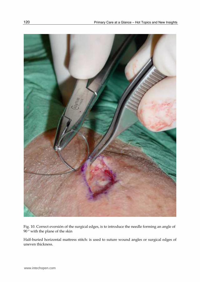
2. Eversion of the wound edges: due to the tendency of scars to contract over time, if surgical edges are left slightly elevated above the plane of the skin, they will flatten over time, producing a more cosmetically acceptable result. One of the keys to proper surgical skin edge eversion is to introduce the needle at a 90-degree angle with the plane of the skin so that the suture, once tied, lifts the skin (fig 10).
3. Closure by layers: for most minor surgical interventions a single (cutaneous) layer
closure is enough. However, if there is any tension, if the wound is very deep and
involves several surgical layers or if there is much dead space, a multi-layer closure
may become necessary. A multi-layer wound closure requires thick fascia or dermis for
the placement of internal sutures, because fatty tissue lacks consistency to support
sutures.
4. Type of suture material: it is a less important factor than the previous principles. If a
suture is removed too late it will cause scarring in the areas of entry and exit of the
suture ("cross-hatching"). To avoid it, stitches shall be removed as soon points as
possible. The choice of suture material and its thickness are also important.
Interrupted sutures
Interrupted sutures are those in which each stitch is independent of the next one. Interrupted suturing is the most appropriate suturing technique for minor surgery, as it helps to distribute stress, promotes the drainage of the wound and stitch removal is easier.
Simple stitch (percutaneous): it is the suture technique of choice for skin suturing in minor
procedures and is used alone or in combination with buried stitches in deeper wounds.
Simple stitch with inverted knot (buried): Used to approximate the deep planes, reducing
tension, and to obliterate dead spaces before skin suturing. It is not necessary in superficial
wounds. Absorbable material is used, leaving the knot in the depth of the wound, thus
reducing the chances of suture exposure through the incision. The knot is cut flush to
decrease the amount of foreign material within the wound.
Mattress stitch or "U" stitch: vertical mattress stitch: a stitch useful in areas of loose skin (back
of the hand, elbow), where the wound edges tend to invaginate. In addition to providing
good eversion of wound edges, this suture provides good obliteration of dead space,
avoiding the need for buried sutures in shallow wounds.
Horizontal mattress stitch: This stitch also provides a good eversion of wound edges,
especially in areas where the dermis is thick (e.g. Palm of the hand and sole of the foot).
www.intechopen.com
Primary Care at a Glance – Hot Topics and New Insights
120
Fig. 10. Correct eversión of the surgical edges, is to introduce the needle forming an angle of 90 º with the plane of the skin
Half-buried horizontal mattress stitch: is used to suture wound angles or surgical edges of uneven thickness.
www.intechopen.com

Skills in Minor Surgical Procedures for General Practitioners
121
Running sutures
They impede the drainage of the wound, so they are contraindicated if infection is suspected and in heavily contaminated wounds. Suture removal is somewhat more difficult, and there is no chance to remove stitches in several sessions.
Simple running suture: It is a sequence of stitches with an initial and a final knot. It takes short time to perform but it makes difficult to adjust skin tension and does not always provide adequate wound edge eversion. It is seldom used in minor surgery.
Intradermal (subcuticular) running suture: This type of suture allows wound suturing without breaking the skin, prevents “cross-hatching” and provides an optimal cosmetic result. It is performed by passing the suture through the dermis horizontally along the entire wound. At both ends suture can go out through the skin (removable intradermal suture), for which nonabsorbable monofilament suture material is selected or a knot may be tied inside the wound (non- removable intradermal suture) for which absorbable material is used. Use of multifilament suture material as silk for intradermal suturing should be avoided, as it would be very difficult to remove the suture material. Intradermal sutures are used on wounds where it will be necessary to maintain the suture a long time (more than 15 days). There should be no tension on the wound. In minor surgery their usefulness is limited.
Knot-tying
Instrumental knot-tying is performed with a needle holder and a curved needle. In minor surgery, where the surgical area is superficial and accessible to surgical instruments, instrumental knot-tying is the preferred technique because it provides more precise suturing and significant savings in suture material, except for suture-ligation of small blood vessels, where manual tying is preferred. The recommended technique is the surgeon’s knot which consists of a double loop followed by several simple loops. The advantage of this knot is the resistance of its first double loop, which prevents knot untying as the surgeon proceeds with the following loops.
When knot-tying a multifilament thread (e.g. Silk) three loops are usually enough (first a double loop plus two single loops). When knot-tying a monofilament thread (eg. Nylon, polypropylene) an extra loop should be added, to increase knot security.
The knots should be placed to one side of the wound, instead of placing them on top of the incision. This will allow better visualization of the wound, interfere less with the healing and facilitate stitch removal.
4.2 Techniques of local anesthesia in minor surgery
Local anesthetics are drugs that block the transmission of nerve impulses causing, at least, the absence of pain sensation in the area of injection.
According to a small chemical difference, local anesthetics can be classified into two groups:
Esters (procaine, tetracaine, chloroprocaine, benzocaine.... which is obsolete due to its high incidence of sensitization) and amides (lidocaine, mepivacaine, bupivacaine, prilocaine, etidocaine and ropivacaine). For their remarkable safety and efficacy we will .only use amides, namely lidocaine and mepivacaine
www.intechopen.com

Primary Care at a Glance – Hot Topics and New Insights
122
Available presentations
The concentration of the anesthetic is expressed in%. We must know that a concentration of
1% means that 100 ml of the solution contain 1 g of anesthetic and 10 ml of the solution
contain 100 mg of the anesthetic. To calculate the concentration in mg / ml, the
concentration percent should be multiplied by 10; thus, a 2 ml ampoule of 2% mepivacaine
contains 20 mg of mepivacaine for each ml of the solution, i.e. this 2 ml ampoule contain 40
mg of mepivacaine.
Maximum doses and features
LIDOCAINE.- 10 ml Amp. 1% (100 mg), 2% (200 mg), 5% (500 mg)
Dose without vasoconstrictor: 3-4 mg / kg maximum 300 mg (3 amp 10 ml 1%)
Dose with vasoconstrictor: 7 mg / kg maximum 500 mg (5 amp 10 ml 1%) Onset of action: 2-4 min. Induces vasodilation. Duration: 1-2 hours, depending on the dose and breadth of the area.
In adults, concentration should range between 0.5 and 1% and total dose should not exceed 30 ml.
In children, concentration should range between 0.25 and 0.50% and total dose should not exceed 4 mg / kg.
MEPIVACAINE.- 10 ml Amp. 1% (100 mg) and 2 ml Amp. 2% (40 mg) Dose without vasoconstrictor: 400 mg (4-5 mg / kg) Dose with vasoconstrictor. 500mg (7mg/Kg) Onset of action: 2-5 min. Induces less vasodilation. Duration: 1-1.5 hours, depending on the dose and breadth of the area.
In adults, mepivacaine should be used at a 1% concentration and total dose should not exceed 40 ml.
For children 0.25 to 0.50% mepivacaine should be used and total dose should not exceed 7 mg / kg.
Adverse effects of local anesthetics
- Local effects: Pain, hematoma, injury to nerve trunks. - Systemic effects: 1. Due to overdose toxicity. These effects appear when the maximum
recommended dose is exceeded or the correct dosage is used but applied intravascularly. From a clinical standpoint, toxicity is associated to the central nervous system (CNS: tinnitus, metallic taste, numbness, dizziness, twitching, etc.) and cardiovascular system (CV: hypotension, arrhythmias, cardiac arrest). 2. Due from an allergic reaction. 3. Due to psychogenic reaction (vasovagal syncope), this is the most common adeverse effect.
Use of vasoconstrictors
The asociation of vasoconstrictors with local anesthetics improves the safety profile of the anesthetic and also allows for better visualization of the surgical field. The most widely used is adrenaline and the maximum dose (as a vasoconstrictor) must not exceed 250 micrograms in adults or 10 micrograms / kg in children.
www.intechopen.com

Skills in Minor Surgical Procedures for General Practitioners
123
The recommended concentration is a dilution of 1:100,000 or 1:200,000 (best) which is prepared by mixing 0.1 mg of adrenaline (0.1 ml of 1:1000 adrenaline) in 10 ml of anesthetic to obtain a 1:100,000 dilution, or in 20 ml to obtain a 1:200,000 dilution.
Due to the risk of necrosis and delayed healing, adrenaline should not be used in acral areas (fingers and toes,), or in devitalized or traumatized skin. Except in such circumstances, using the anesthetic with vasoconstrictor is highly recommended.
Basic techniques of local anesthesia
Topical anesthesia
In recent years, topical anesthetics have been developed as an alternative to infiltration both for intact skin and for lacerations and mucosae, especially in children. Topical anesthetics used in minor surgical procedures and their characteristics are shown in the table 2:
Anesthetic Mode of use
characteristics Indications Complications Not indicated
LET® (4 % lidocaine, 0,1 % epinephine 1:2000, 0,5 % tetracaíne)
1-3 ml applied directly on wound for 15-30 min
Onset 20-30 min after aplication. Duration of effect has not been clearly established
Can be effective in children for face and scalp lacerations and less effective in limbs
No important adverse effects reported
For mucosae and acral areas
EMLA® lidocaine 25 mg/ml plus prilocaine 25 mg/ml,
1-2 gr of cream should be applied for each 10 cm2 of intact skin and occluded Or apply in patch Maximum dose is 10 grams
Onset 60-120 min after aplication. Duration of effect is 30-120 min. not useful on palms of hands and soles of feet
Admitted for procedures on intact skin: scraping and shaving (seborrheic keratosis, Molluscum contagiosum, dermal nevus), cryosurgery (warts, condylomas), electrosurgery (small fibroids and spiders), laser hair removal, pre-anesthesia for infiltration
Local mild irritation, contact dermatitis. There have been reports of Metahemoglobinemia in children aged <6 months
For wounds or deep tissues
Table 2. Topical anesthetics used in minor surgical procedures and their characteristics
Infiltration anesthesia: angular and peri-lesional9
1. Angular infiltration: From the point of entry, the anesthetic is infiltrated in three or more different directions, like a fan (Fig 11).
2. Perilesional infiltration: Starting from each point of entry the anesthetic is infiltrated in a single direction, so that after several injections, the lesion will have been surrounded by anesthetic, and the diferent points of entry will be forming a polyhedral figure (Fig 12).
3. Linear Infiltration: If the lesion to be operated on is a skin laceration, the anesthetic should be directly infiltrated into the wound edges in a linear fashion. If the wound is
www.intechopen.com

Primary Care at a Glance – Hot Topics and New Insights
124
bruised and has irregular edges, it is preferable to use a perilesional technique from the uninjured area, and follow along the margins of the wound to avoid introducing microbial contamination.
Fig. 11. Anesthesic angular infiltration: it infiltrates following three or more different directions, like a fan
www.intechopen.com

Skills in Minor Surgical Procedures for General Practitioners
125
Fig. 12. Infiltration perilesional: the injury surrounds itself by means of different infiltrations
Loco-regional block
There are multiple blocks, digital blocks being the most common minor surgery. The digital
block will allow the surgeon to act on all types of fingers injuries (lacerations, wound
debridement, soft tissue injuries, foreign body removal, pathology of the nail).
www.intechopen.com

Primary Care at a Glance – Hot Topics and New Insights
126
A thin needle is inserted at the base of the proximal phalanx in a dorsal and lateral location. The needle is inserted to the point where the collateral palmar digital nerve should lie and, after aspiration, 0.5-1 mL of local anesthetic is injected. The needle is removed until its tip is just below the skin and, after aspiration, a dose of anesthetic is again infiltrated in a subcutaneous plane along the side of the base of the phalanx.
Then, the needle will be inserted on the opposite side and infiltration will proceed in a similar way.
Before the procedure, the surgeon should wait 10-15 minutes for a complete effect of the block. The total injected volume should not exceed 4 ml, because higher volumes can compress digital vessels and cause ischemia. For the same reason, anesthetics used in digital blocks should contain no vasoconstrictor.
5. Preoperative considerations
5.1 Diagnostic criteria for the most common lesions in minor surgery
The indications for minor surgery are related to the diagnosis of the patient’s lesion. A misdiagnosis can cause incorrect treatment and lead to loss of clinical information relevant to the patient's prognosis. It is therefore imperative that general practitioners have an extensive knowledge of the lesions most frequently treated by minor surgery. When in doubt about the nature of any given lesion, preoperative diagnosis should always be confirmed by other specialists10. For each lesion there is a surgical procedure of choice. Other techniques may also be valid or they may even be contraindicated.
The following paragraphs contain an overview of the most important diagnostic consideration in lesions usually treated with minor surgery.
Seborrheic keratoses
Seborrheic keratoses are benign light brown lesions initially measuring 1-3 mm which after years of growth can turn into more or less pigmented 1-6 cm plaques with a rough or waxy surface. These lesions are easily treated with curettage, electrosurgery or cryosurgery. In case of doubt, an incisional biopsy should be sent for histopathological analysis.
Epidermal cysts
Also known as epithelial cysts, epidermoid cysts or sebaceous cysts, they are firm nodules, measuring from 0.2 to 5 cm in diameter, formed by a layer of stratified squamous epithelium or epithelium of a hair follicle, which are located in the dermis or subcutaneous tissue. They are not adherent to deep tissues and in many instances a central keratin- rich pore can be seen. Queratin is the main component inside the cyst. The most common location is on the face, neck, chest, upper third of back and scrotum. Their treatment is surgical removal for cosmetic reasons or due to recurrent infections
Warts
They are a form of benign epithelial hyperplasia induced by human papilloma virus (HPV). The clinical presentations of the cutaneous infection by HPV include:
Verruca Vulgaris: The treatment of choice is 15-20% salicylic acid with petroleum jelly, applied topically for 2-3 weeks or liquid nitrogen.
www.intechopen.com

Skills in Minor Surgical Procedures for General Practitioners
127
Plantar wart: The treatment of choice is 40% salicylic acid with petroleum jelly, applied topically for 3 weeks followed first by curettage and then by liquid nitrogen. Surgery and electrocoagulation are not indicated
Molluscum
Pearly white papules of 1-5 mm (sometimes even bigger) with central dimpling. They may appear isolated or in groups in the neck, trunk, anogenital area or eyelids. They are very common in children and in patients with HIV / AIDS with extensive lesions which difficult to eradicate.
Their first choice treatment is cryosurgery, curettage or electrodesiccation.
Lipoma
Lipomas are slow-growing benign tumors of mature adipose tissue. They are a frequent cause of consultation due to mechanical or aesthetic reasons. Lipomas appear as soft, elastic, smooth or multilobulated tumors of variable size (from 1 or 2 cm to 15 cm or more) with ill-defined borders, and not adherent to deep planes.
Lipomas are generally asymptomatic and patients usually refer the emergence of a "lump" whose location, size and visibility will determine how urgent such consultation will be. In most cases, a simple physical examination will establish a correct diagnosis of lipoma.
Lipomas are treated by surgical removal. Their removal by either a general practitioner or a surgeon usually depends on the size, location and positionof the lipoma in the subcutaneous plane.
Fibroma pendulum, skin tags
These are pedunculated fleshy-looking benign tumours, whose colour is either similar to that of the surrounding skin or darker. Their size varies from a few mm (which usually appear in groups and are located in the neck, armpits, and skin folds) up to 2-5 mm (which usually appear as pedunculated and solitary lesions).
Their treatment is justified for cosmetic reasons, and good surgical results usually ensue. These lesions can be treated by skin shave, electrodesiccation and cryosurgery.
Melanocytic nevi
They are acquired lesions in the form of macules or papules or small nodules (<1 cm) and are constituted by groups of melanocytes located in the epidermis, dermis or both areas and rarely in the subcutaneous tissue. They are very common in Caucasians and family incidence has been clearly established. Sun exposure contributes to the induction of these lesions.
Most appear in early childhood and peak in young adults to regress and disappear gradually and are asymptomatic. By contrast, dysplastic nevi continue to appear as new lesions over a lifetime and do not show signs of involution. The indication for removal will be established when any of the following characteristics is seen:
- Presentation site: scalp, mucous membranes, anogenital area - Time of evolution: de novo appearance in adulthood - Color: variegated or modified - Borders: always have been irregular or have just turned irregular
www.intechopen.com
Primary Care at a Glance – Hot Topics and New Insights
128
- Symptoms: If the nevus starts to itch, bleed or hurt - Epiluminescence microscopy: criteria of dysplasia
Actinic keratoses
These are yellowish brown dry- looking maculopapular lesions, with a rough and scaly texture located in sun-exposed areas of middle-aged patients. Actinic keratoses are more prevalent in males. Most of these lesions are present on the skin for years and 13-25% of them will lead to a squamous cell carcinoma.
The treatment of choice is the topical application of 5% 5-fluorouracil or Imiquimod. If lesions are scarce and localized, they may be treated with liquid nitrogen.
Basal cell carcinoma
Basal cell carcinoma is the most common skin malignancy. Although it can occur at any age, its incidence raises abruptly after the age of 40 with a history of heavy sun exposure. These lesions are more prevalent in the face and scalp followed by the ears and upper chest and back.
Excision should be considered for any slow-growing pale pink, brown or flesh-colored, flat or slightly elevated new skin lesion located in the face, ear, neck, back or scalp with a pearly or waxy appearance, that also meets any of the following criteria:
- Blood vessels visible in the lesion or in the adjacent skin - Keratin pearls sometimes visible with a loupe - Appearance of a scar with no history of previous wound in the area - Non-healing ulcer
Squamous cell Carcinoma
This is a malignant tumor that usually appears on a previous premalignant lesion.
It takes different clinical forms, in this case as a crust-covered superficial ulcer that grows insidiously over months. In other cases it may appear as a hard papule or nodule.
Squamous-cell carcinomas require a multidisciplinary therapeutical approach involving
dermatologists, surgeons, radiotherapists, and chemotherapists.
Melanoma
Of all skin malignancies, melanoma has the worst prognosis. There are different clinical
presentations and, for their detection, the "ABCDE-rule of melanoma" , which is described in
the following table, should never be overlooked (Table 3).
A-B-C-D-E rule for any lesion clinically suspicious of melanoma
A Asimetry
B Border: irregular and scalloped
C Colour: mottled, irregular shades of brown, black, gray and pink
D Diameter over 6 mm
E Subtle or obvious skin elevation (on illuminated sideview) and serial assessment of growth
Table 3. A-B-C-D-E rule for any lesion clinically suspicious of melanoma
www.intechopen.com

Skills in Minor Surgical Procedures for General Practitioners
129
Any lesion suspicious of melanoma should be referred to a dermatologist. These lesions should never undergo biopsy by skin shave or curettage, nor should any destructive procedure (cryosurgery or electrosurgery) be used in their diagnosis or therapy.
5.2 Body areas of risk in minor surgery
Certain body areas are considered high-risk areas for minor surgical procedures, due to the superficial location of some anatomical structures, which are likely to be injured during surgery. To avoid injury to these structures, it is necessary to know their theoretical path and keep the surgery, whenever possible, on a superficial plane (superficial subcutaneous tissue). Other areas are also considered high-risk areas due to the potential esthetic impact of poor surgical technique.
High-risk areas for minor surgery include the facial and cervical regions, axillary and supraclavicular regions, wrists, hands and fingers, the groin, the popliteal fossa and the feet (Fig 13 A, B).
Fig. 13, A, B: The zones of risk in Minor Surgery include the facial and cervical region, the region axilar and supraclavicular, the wrists, hands and fingers, the inguinal region, the hollow poplíteo and the feet
www.intechopen.com

Primary Care at a Glance – Hot Topics and New Insights
130
Along with the previously-mentioned areas, we must also consider those regions with a greater tendency to develop pathological scars. The deltoid and shoulder region, the sternal and interscapular region, and the skin of black patients and children are especially prone to hypertrophic scarring and keloids. Thus, before any skin lesion is removed in any of these areas it is important to discuss this possibility with the patient, especially in cases where the excision pursues aesthetic improvent, and is not indicated for diagnostic or therapeutic reasons.
6. How to perform a surgical procedure: Elliptical excision step by step
Elliptical excision is a skin excision technique with a spindle-shaped design. This ellipse
should include all skin layers plus some subcutaneous fat, in order to remove skin lesions
with a safety margin both around and under them. This technique not only allows for
simultaneous diagnosis and treatment, but also facilitates closure producing good cosmetic
results. It is, therefore, the ideal technique to remove the majority of skin lesions11-14.
Before surgery the patient should be informed about the procedure and its technical details
before asking them to sign the informed consent form.
The procedure involves the following steps:
1. Design of the incision which is drawn with a marker using the following parameters: the longitudinal axis of the ellipse will be three times longer than its transverse axis and will be parallel to skin tension lines. Its ends will form a <30 °angle to avoid "dog ears". There must be a 1-2 mm margin between the lesion and the incision (some lesions may require more generous margins).
2. Preparation of the surgical field: cleaning and antisepsis 3. Local anesthetic injection, covering the entire edge of the incision and the tissue to be
sectioned and sutured 4. Superficial skin incision along the marked ellipse, going through the entire dermis to
prevent jagged edges. The incision is made with a clean cut of the scalpel, which is held like a pencil, and any sawing movement should be avoided. Using the nondominant hand, the skin should be stretched or pinched along the previously marked incision line (Fig 14).
5. En bloc excision of the lesion using the nondominant hand the skin is stretched with toothed-forceps from one end of the ellipse and, using the edge of the blade, the deep wedge-shaped incision is made (always under direct vision, Fig 15), until fat is reached and the lesion is, thus, removed en bloc.
6. Haemostasis of the surgical area: if anesthesia with vasoconstrictor is used bleeding will be scarce and hemostasis will be easily achieved by applying digital pressure with a gauze.
7. Wound closure by layers: most minor surgical procedures only require suturing of the skin surface, but if there is any tension, the incision is deep and involves several tissue layers or there is dead space, suturing several surgical planes may be required. The deep-layer suture should be performed with absorbable material using an inverted-knot technique. Then, the superficial closure with nonabsorbable suture will be performed. The number of stitches will depend on the tension of the wound, suture thickness and chosen suture technique (Fig 16).
8. Sterile dressing placement after cleaning the surgical área
www.intechopen.com

Skills in Minor Surgical Procedures for General Practitioners
131
9. Submission of the resected specimen to pathology, in a container with 10% formalin. 10. Follow-up: After 48 hours the wound can be washed gently, and the patient should be
warned of postoperative risks and taught how to take care of the surgical wound. No surgical procedute is complete until the pathology report has been received and the patient informed of the results and prognosis.
Fig. 14. Incision is done giving a clean cut of the scalpel, not sawing, taking the handle of the scalpel as a pencil, traccionando (or pinching the zone) with the fingers
www.intechopen.com

Primary Care at a Glance – Hot Topics and New Insights
132
Fig. 15. En bloc excision: traction is realized (with forceps with teeth) from an end of the spindle and with the edge of the scalpel deep incision is realized
www.intechopen.com

Skills in Minor Surgical Procedures for General Practitioners
133
Fig. 16. Suturing of the skin surface
7. Good clinical practice in minor surgery
Preoperative
For most basic minor surgical procedures, no preoperative work-up is needed. However, since we, as general practitioners, operate on our own patients, we have inmediate access to their medical history which can completed with a series of simple questions that will help us detect those patients in whom surgery poses especial risks or is contraindicated. The table 4 summarizes the precautions of minor surgery in primary care:
www.intechopen.com
Primary Care at a Glance – Hot Topics and New Insights
134
- Diabetes Mellitus and peripheral vascular diseasse when planning surgery in the lower extremities - In patients with chronic use of immunosuppressors (corticosteroids) - Do not add vasoconstrictor to local anesthetic in patients with: arrhythmia, severe hypertension, hyperthyroidism, pheochromocytoma, pregnancy, and in anesthesia of the fingers - Anatomic areas of risk - Use of oral anticoagulants and antiplatelet agents should be sought for. The risk-benefit of their suspension should be studied prior to surgery. In most patients at low risk, minor surgical procedures can be performed without altering the anticoagulation regimen if their INR is within therapeutic range (Antithrombotic and Thrombolytic Therapy: American College of Chest Physicians Evidence-Based Clincial Practice Guidelines (8th Edition)2008) Specific precautions with the use of electrocautery - Keep the patient’s skin clear from any metallic object. Metal dentures, implants, prosthesis, IUD are not contraindicated - Do not use alcohol or use the bovie near a source of oxygen. - Use latex gloves for insulation Special precautions for cryosurgery - Patients with areas of potential circulatory compromise due to the risk of necrosis. - Hairy areas in which hair loss could ensue. - Hyperpigmented areas (black or dark skin), where the use of cryosurgery can leave areas of hypopigmentation. - Patients with high levels of cryoglobulins.
Table 4. Precautions minor surgery
Premedication
In patients with increased anxiety, use of preoperative sedation can be considered. 5-10 mg
oral or sublingual diazepam, or 1-5 mg sublingual lorazepam can be administered 30
minutes before surgery.
Contraindications for minor surgery
- With few justified exceptions, no malignant skin lesion should be surgically treated by the general practitioner. Similarly, all lesions labeled as malignant after pathological analysis, should promt consultation with other specialists.
- Allergy to local anesthetics: If there is doubt about a history of allergic reactions, the patient should see the allergist prior to surgery.
- Pregnancy: If the patient is pregnant, surgery should be deferred until the end of pregnancy, to avoid unnecessary risks. If malignancy is suspected, the patient should be referred to a specialist.
www.intechopen.com

Skills in Minor Surgical Procedures for General Practitioners
135
- Acute intercurrent disease: if the patient suffers an acute illness at the time of surgery
(heart failure, liver failure, arrhythmia, flu or pneumonia), surgery should be postponed
until recovery.
- Doubt about patient’s motivations: surgery is contraindicated in patients whose
motivation is questionable or in whom there is excessive preoccupation about the
aesthetic result
- Patients with psychiatric disorders or uncooperative patients are not eligible for surgery
in primary care. In general it is not advisable to involve uncooperative patients.
- Refusal to sign the informed consent form is a contraindication for any minor surgery
procedure or technique.
Intraoperative complications
Vasovagal syncope is the most fequent complication and is more common in young men.
Warning signs are flushing, pallor, sweating, weakness, nausea (occasionally, vomiting), etc.
After these simptoms, some patients may lose consciousness.
Treatment consists of placing the patient in Trendelenburg’s position, administering oxygen
and i.v. fluids if needed and, in severe cases (long-lasting or associated with bradycardia)
use of 0.5-1 mg sc or iv atropine can be considered (maximum dose, 2 mg). Generally, most
of patients recover spontaneously over a period of seconds to a few minutes.
All medical premises where minor surgical procedures are performed should have CPR
equipment and medication.
Postoperative complications
- Hematoma-seroma: fluid collection under the sutures that occurs whenever cavities or
dead space have been left in the surgical site. To prevent their formation, a correct
intraoperative hemostasis is paramount plus suturing the wound in layers with no gaps
and, finally, applying a compressive bandage.
- Infection can occur in up to 1% of minor surgical patients and it appears as swelling,
redness of the wound edges and, sometimes, purulent discharge. Symptoms such as
fever and / or chills are only rarely seen. Infections are treated by removing some of the
stitches, plus daily cleaning and disinfection of the wound and allowing the wound to
close by secondary intention. If necessary, a drain may be inserted into the wound and
an oral antibiotic regimen may be initiated.
- Wound dehiscence: The separation of the edges of the wound before proper healing.
There are predisposing factors secondary to surgery (excessive tension on the edges of
the wound, use of inappropriate suture material or early suture withdrawal) or it may
be the final phase of other complications such as hematoma or infection. After wound
dehiscence, wound repair will take place by secondary intention, resulting in a poor
cosmetic result.
- Hypertrophic scar and keloid scarring are deviations from normal wound-repair process. Hypertrophic scars do not grow beyond the initial limits of the primary wound whereas in a keloid the scar tissue extends beyond the initial boundaries of the scar, forming a permanent bulk of scar tissue. Keloids are very difficult to prevent and their most frequent location is in the chest, shoulders and upper back, in young people and
www.intechopen.com

Primary Care at a Glance – Hot Topics and New Insights
136
blacks. Treatment of these scars is difficult and results are often discouraging. Occlusive treatment or steroid injections may be tried.
8. References
[1] Arribas JM (2006). Cirugía menor y procedimientos en medicina de familia (2ª edición). Madrid: Jarpyo Editores,
[2] Czarnowski C, Ponka D, Rughani R, Geoffrion P. See one. Do one. Teach one.: Office-based minor surgical procedures. Can Fam Physician. 2008 Jun;54(6):893.
[3] Zuber TJ. Punch biopsy of the skin. Am Fam Physician. 2002 Mar 15;65(6):1155-8, 1161-2, 1164.
[4] Freiman A, Bouganim N. History of cryotherapy. Dermatol Online J. Aug 1 2005;11(2):9 [5] Hainer BL. Electrosurgery for the skin. Am Fam Physician. 2002 Oct 1;66(7):1259-66. [6] Kudur MH, Pai SB, Sripathi H, Prabhu S. Sutures and suturing techniques in skin
closure. Indian J Dermatol Venereol Leprol. 2009 Jul-Aug;75(4):425-34. [7] Moy RL, Lee A, Zalka A. Commonly used suturing techniques in skin surgery. Am Fam
Physician. 1991 Nov;44(5):1625-34. [8] Singer AJ, Quinn JV, Hollander JE. The cyanoacrylate topical skin adhesives. Am J
Emerg Med. 2008 May;26(4):490-6. [9] Achar S, Kundu S Principles of office anesthesia: part I. Infiltrative anesthesia. Am Fam
Physician. 2002 Jul 1;66(1):91-4. [10] Klaus Wolff y Richard Allen Johnson (2009). Fitzpatrick's Color Atlas and Synopsis of
Clinical Dermatology 6th edition. El McGraw-Hill Companies, Inc. New York [11] Hussain W, Mortimer NJ, Salmon PJ. Optimizing technique in elliptical excisional
surgery: some pearls for practice. Br J Dermatol. 2009 Sep;161(3):697-8. Epub 2009 Jun 25.
[12] Czarnowski C, Ponka D, Rughani R, Geoffrion P. Elliptical excision: minor surgery video series. Can Fam Physician. 2008 Aug;54(8):1144
[13] Zuber TJ. Fusiform excision. Am Fam Physician. 2003 Apr 1;67(7):1539-44, 1547-8, 1550. [14] Wu T. Plastic surgery made easy - simple techniques for closing skin defects and
improving cosmetic results. Aust Fam Physician. 2006 Jul;35(7):492-6.
www.intechopen.com

Primary Care at a Glance - Hot Topics and New InsightsEdited by Dr. Oreste Capelli
ISBN 978-953-51-0539-8Hard cover, 446 pagesPublisher InTechPublished online 27, April, 2012Published in print edition April, 2012
InTech EuropeUniversity Campus STeP Ri Slavka Krautzeka 83/A 51000 Rijeka, Croatia Phone: +385 (51) 770 447 Fax: +385 (51) 686 166www.intechopen.com
InTech ChinaUnit 405, Office Block, Hotel Equatorial Shanghai No.65, Yan An Road (West), Shanghai, 200040, China
Phone: +86-21-62489820 Fax: +86-21-62489821
"Both among scientists and clinical practitioners, some find it easier to rely upon trivial explanations, whileothers never stop looking for answers". With these surprising words, Augusto Murri, an Italian master in clinicalmedicine, reminds us that medical practice should be a continuous journey towards knowledge and the qualityof care. The book brings together contributions by over 50 authors from many countries, all around the world,from Europe to Africa, from Asia to Australia, from North to South America. Different cultures are presentedtogether, from those with advanced technologies to those of intangible spirituality, but they are all connectedby five professional attributes, that in the 1978 the Institute of Medicine (IOM)1 stated as essentials ofpracticing good Primary Care: accessibility, comprehensiveness, coordination, continuity and accountability.The content of the book is organized according to these 5 attributes, to give the reader an internationaloverview of hot topics and new insights in Primary Care, all around the world.
How to referenceIn order to correctly reference this scholarly work, feel free to copy and paste the following:
Jose Maria Arribas Blanco and Maria Hernandez Tejero (2012). Skills in Minor Surgical Procedures forGeneral Practitioners, Primary Care at a Glance - Hot Topics and New Insights, Dr. Oreste Capelli (Ed.), ISBN:978-953-51-0539-8, InTech, Available from: http://www.intechopen.com/books/primary-care-at-a-glance-hot-topics-and-new-insights/skills-in-minor-surgical-procedures-in-family-medicine

© 2012 The Author(s). Licensee IntechOpen. This is an open access articledistributed under the terms of the Creative Commons Attribution 3.0License, which permits unrestricted use, distribution, and reproduction inany medium, provided the original work is properly cited.