site.iugaza.edu.pssite.iugaza.edu.ps/maziz/files/2014/02/understanding-the... · web view• the...
TRANSCRIPT

Chapter 4Understanding the cardiovascular system
The cardiovascular system (CVS) begins its activity when the fetus is barely 1 month old (21st day), and it’s the last system to cease activity at the end of life. The heart, arteries, veins, and lymphatic vessels make up the CVS. These structures:
transport life-supporting oxygen and nutrients to cells, remove metabolic waste products, and carry hormones from one part of the body to another.
Circulation requires normal heart function, which propels blood through the system by continuous rhythmic contractions.Despite advances in disease detection and treatment, cardiovascular disease remains the leading cause of
death. Heart attack, or myocardial infarction (MI), is the primary cause of cardiovascular-related deaths. MI typically occurs with little or no warning.
Oxygen balancing actA critical balance exists between myocardial oxygen supply and demand. A decrease in oxygen supply
or an increase in oxygen demand can disturb this balance and threaten myocardial function. The four major determinants of myocardial oxygen demand
are: heart rate, contractile force, muscle mass, and ventricular wall tension. Cardiac workload and oxygen demand increase if the heart rate speeds up or if the force of contractions becomes stronger. This can occur in hypertension, ventricular dilation, or heart muscle hypertrophy.
The heart’s law of supply and demandIf myocardial oxygen demand increases, so must oxygen supply. To effectively increase oxygen supply,
coronary perfusionmust also increase. Tissue hypoxia causes coronary arteries to dilate and increases coronary blood flow.
Normal coronary vessels can dilate and increase blood flow five to six times above resting levels. However, stenotic, diseased vessels can’t dilate, so oxygen deficit may result.
One-way ticketNormally, blood flows unimpeded across the valves in one direction. The valves open and close in
response to a pressure gradient. When the pressure in the chamber proximal to the valve exceeds the pressure in the chamber beyond the valve, the valves open. When the opposite occurs, the valves close. The valve leaflets, or cusps, are so responsive that even a pressure difference of less than 1 mm Hg between chambers will open and close them.
How low can you flow?Valvular disease is the major cause of low blood flow. A diseased valve allows blood to flow backward
across leaflets that haven’t closed securely. This phenomenon is called regurgitation. The backflow of blood through the valves forces the heart to pump more blood, increasing cardiac workload. The valve opening may also become restricted and impede the forward flow of blood. This is referred to as stenosis.
The heart may fail to meet the tissues’ metabolic requirements for blood and fail to function as a pump. Eventually, the circulatory system may fail to perfuse body tissues, and blood volume and vascular tone may be altered.
Inner awareness and how the heart respondsThe body closely monitors both blood volume and vascular tone. Blood flow to each tissue is monitored
by micro-vessels, whichmeasure how much blood each tissue needs and control the local blood flow. The nerves that control
circulation also help directblood flow to tissues. The heart pays attention to the tissues’ demands. It responds to the return of blood
through the veins and to nerve signals that make it pump the required amounts of blood.Under pressureArterial pressure is carefully regulated by the body: If it falls below or rises above its normal mean
level, immediate circulatorychanges occur. If arterial pressure falls below normal, then an increase occurs in:
1الصفحة

• heart rate • force of contraction • constriction of arterioles.If arterial pressure rises above normal, these changes occur:• reflex slowing of heart rate • decreased force of contraction • vasodilation.Risk factorsRisk factors for CVD fall into two categories: modifiable and non-modifiable.Modifiable risk factors These factors include:• elevated serum lipid levels • hypertension(HTN) • cigarette smoking• diabetes mellitus (DM) • sedentary lifestyle • stress• obesity—especially abdominal • excessive intake of saturated fats, carbohydrates, and salt.Non-modifiable risk factors age male gender family history race.Better to be young at heart; Susceptibility to CVD increases with age; disease before age 40 is
unusual. However, the age-disease correlation may simply reflect the longer duration of exposure to other risk factors.
An estrogen effect? Women are less susceptible than men to heart disease until after menopause; then they become as susceptible as men. One theory proposes that estrogen has a protective effect.
Nature vs. nurture A positive family history also increases a person’s chances of developing premature CVD. For example, genetic factors can cause some pronounced, accelerated forms of atherosclerosis such as lipid disease. However, family history of CVD may reflect a strong environmental component. Risk factors—such as obesity or a lifestyle that causes tension—
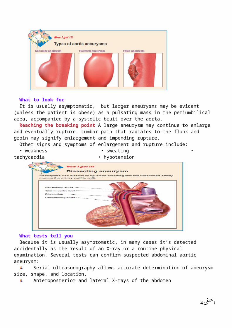
may recur in families.Color blind ; Although it affects all races, blacks are most susceptible to cardiovascular disease.Cardiovascular disordersThe disorders discussed in this section include:• abdominal aortic aneurysm • cardiac tamponade • cardiogenic shock• coronary artery disease (CAD) • dilated cardiomyopathy • heart failure • hypertension hypertrophic cardiomyopathy • MI • pericarditis • rheumatic fever and rheumatic heart disease.Abdominal aortic aneurysmIt is, an abnormal dilation in the arterial wall occurs in the aorta between the renal arteries and the iliac
branches. In a false aneurysm, the out pouching occurs when the entire vessel wall is injured and leads to a sac formation affecting the artery or heart. It is more common in men and most prevalent in ages 50 to 80. Arteriosclerosis is responsible for 95% of cases.
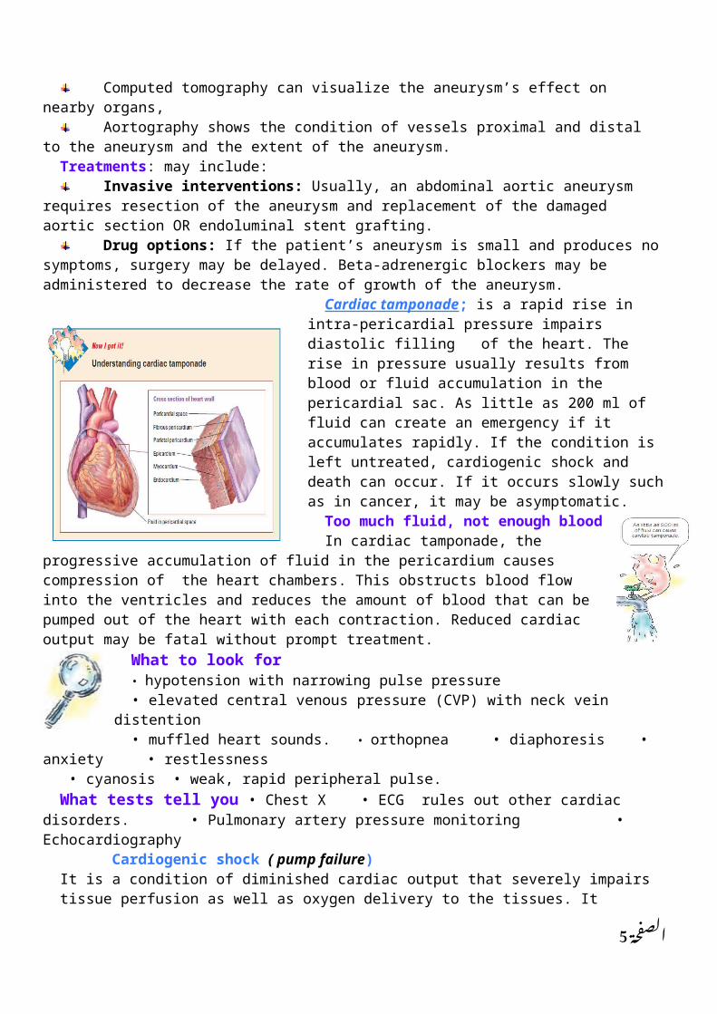
It begins locallyFirst, a local weakness in the muscular layer of the aorta (tunica media), due to degenerative changes,
allows the inner layer (tunica intima) and outer layer (tunica adventitia) to stretch outward. Blood pressure within the aorta progressively weakens the vessel walls and enlarges the aneurysm. Aneurysms can dissect or rip when bleeding into the weakened artery causes the artery wall to split.
2الصفحة

What to look forIt is usually asymptomatic, but larger aneurysms may be evident (unless the patient is obese) as a
pulsating mass in the periumbilical area, accompanied by a systolic bruit over the aorta.Reaching the breaking point A large aneurysm may continue to enlarge and eventually rupture.
Lumbar pain that radiates to the flank and groin may signify enlargement and impending rupture. Other signs and symptoms of enlargement and rupture include:• weakness • sweating • tachycardia • hypotension
What tests tell youBecause it is usually asymptomatic, in many cases it’s detected accidentally as the result of an X-ray or
a routine physical examination. Several tests can confirm suspected abdominal aortic aneurysm:Serial ultrasonography allows accurate determination of aneurysm size, shape, and location.Anteroposterior and lateral X-rays of the abdomen Computed tomography can visualize the aneurysm’s effect on nearby organs, Aortography shows the condition of vessels proximal and distal to the aneurysm and the extent
of the aneurysm.Treatments: may include:
Invasive interventions: Usually, an abdominal aortic aneurysm requires resection of the aneurysm and replacement of the damaged aortic section OR endoluminal stent grafting.
Drug options: If the patient’s aneurysm is small and produces no symptoms, surgery may be delayed. Beta-adrenergic blockers may be administered to decrease the rate of growth of the aneurysm.
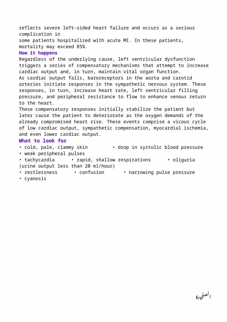
Cardiac tamponade; is a rapid rise in intra-pericardial pressure impairs diastolic filling of the heart. The rise in pressure usually results from blood or fluid accumulation in the pericardial sac. As little as 200 ml of fluid can create an emergency if it accumulates rapidly. If the condition is left untreated, cardiogenic shock and death can occur. If it occurs slowly such as in cancer, it may be asymptomatic.
Too much fluid, not enough bloodIn cardiac tamponade, the progressive
accumulation of fluid in the pericardium causes compression of the heart chambers. This obstructs blood flow into the ventricles and reduces the amount of blood that can be pumped out of the heart with each contraction. Reduced cardiac output may be fatal without prompt treatment.
What to look for• hypotension with narrowing pulse pressure• elevated central venous pressure (CVP) with neck vein distention
3الصفحة
• muffled heart sounds. • orthopnea • diaphoresis • anxiety • restlessness • cyanosis • weak, rapid peripheral pulse.What tests tell you • Chest X • ECG rules out other cardiac disorders. • Pulmonary artery
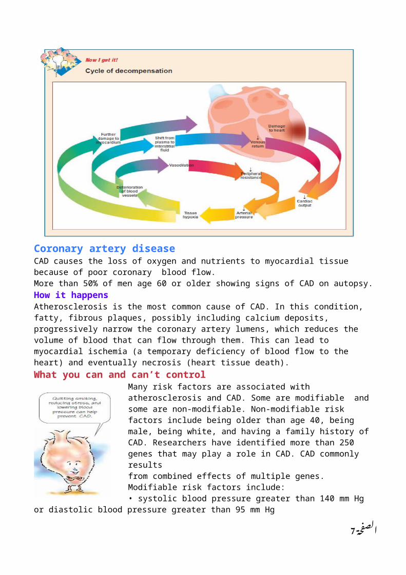
pressure monitoring • Echocardiography Cardiogenic shock ( pump failure)It is a condition of diminished cardiac output that severely impairs tissue perfusion as well as oxygen delivery to the tissues. It reflects severe left-sided heart failure and occurs as a serious complication insome patients hospitalized with acute MI. In these patients, mortality may exceed 85%. How it happensRegardless of the underlying cause, left ventricular dysfunction triggers a series of compensatory mechanisms that attempt to increase cardiac output and, in turn, maintain vital organ function.As cardiac output falls, baroreceptors in the aorta and carotid arteries initiate responses in the sympathetic nervous system. These responses, in turn, increase heart rate, left ventricular fillingpressure, and peripheral resistance to flow to enhance venous return to the heart.These compensatory responses initially stabilize the patient but later cause the patient to deteriorate as the oxygen demands of the already compromised heart rise. These events comprise a vicous cycle of low cardiac output, sympathetic compensation, myocardial ischemia, and even lower cardiac output.What to look for• cold, pale, clammy skin • drop in systolic blood pressure • weak peripheral pulses• tachycardia • rapid, shallow respirations • oliguria (urine output less than 20 ml/hour)• restlessness • confusion • narrowing pulse pressure • cyanosis
Coronary artery diseaseCAD causes the loss of oxygen and nutrients to myocardial tissue because of poor coronary blood flow. More than 50% of men age 60 or older showing signs of CAD on autopsy. How it happensAtherosclerosis is the most common cause of CAD. In this condition, fatty, fibrous plaques, possibly including calcium deposits, progressively narrow the coronary artery lumens, which reduces the volume of blood that can flow through them.
4الصفحة
This can lead to myocardial ischemia (a temporary deficiency of blood flow to the heart) and eventually necrosis (heart tissue death).What you can and can’t control
Many risk factors are associated with atherosclerosis and CAD. Some are modifiable and some are non-modifiable. Non-modifiable risk factors include being older than age 40, being male, being white, and having a family history of CAD. Researchers have identified more than 250 genes that may play a role in CAD. CAD commonly resultsfrom combined effects of multiple genes. Modifiable risk factors include:• systolic blood pressure greater than 140 mm Hg or diastolic blood pressure greater than 95 mm Hg• increased low-density and decreased high-density lipoprotein levels• smoking (risk dramatically drops within 1 year of quitting)
• stress •obesity, which increases the risk of diabetes mellitus, hypertension, and high cholesterol• inactivity • diabetes mellitus, especially in women.From aerobic to anaerobicTransient ischemia causes reversible changes at the cellular and tissue levels, depressing myocardial function. Untreated, it can lead to tissue injury or necrosis. Oxygen deprivation forces the myocardium to shift from aerobic to anaerobic metabolism. As a result, lactic acid (the end product of anaerobic metabolism) accumulates. This reduces cellular pH.With each contraction, less bloodThe combination of hypoxia, reduced energy availability, and acidosis rapidly impairs left ventricular function. The strength of contractions in the affected myocardial region is reduced as the fibers shorten inadequately with less force and velocity. In addition, the ischemic section’s wall motion is abnormal. This generally results in less blood being ejected from the heart with each contraction..Compliance countsThese increases in left-sided heart pressures is magnified by changes in wall compliance induced by ischemia. Compliance is reduced, magnifying the elevation in pressure. During ischemia, sympathetic nervous system response leads to slight elevations in blood pressure and heart rate before the onsetof pain. With the onset of pain, further sympathetic activation occurs.What to look forAngina is the classic sign of CAD. The patient may describe a burning, squeezing, or crushing tightness in the sub-sternal or precordial area that radiates to the left arm, neck, jaw, or shoulder blade. He may clench his fist over his chest or rub his left arm when describing it. Pain is commonly accompanied by nausea, vomiting, fainting, sweating, and cool extremities. Angina commonly occurs after physical exertion but may also follow emotional excitement, exposure to cold, or the consumption of a large meal.When to label it stable or unstable If the pain is predictable and relieved by rest or nitrates, it’s called stable angina. If it increases in frequency and duration and is more easily induced, it’s called unstable or unpredictable angina.
5الصفحة
What tests tell you • ECG during an episode of angina shows ischemia, as demonstrated by T-wave inversion, ST-segment depression and, possibly, arrhythmias • Treadmill or bicycle exercise stress test may provoke chest pain• Coronary angiography reveals the location and extent of coronary artery stenosis or obstruction.
• Myocardial perfusion imagingTreating CAD:Controlling riskNoninvasive measures : Drug therapy also consists of nitrates, such as nitroglycerin, isosorbide dinitrateInvasive measures; coronary artery bypass graft (CABG) surgery, percutaneous transluminal coronaryangioplasty (PTCA), and laser angioplasty.Heart failureWhen the myocardium can’t pump effectively enough to meet the body’s metabolic needs, heart failure occurs. Pump failure usually occurs in a damaged left ventricle, but it may also happen in the right ventricle. Usually, left-sided heart failure develops first. Heart failure is classified as:• high-output or low-output• acute or chronic • left-sided or right• forward or backward. Symptoms of heart failure may restrict a person’s ability to perform activities of daily living and severely affect quality of life.But the good news is…Advances in diagnostic and therapeutic techniques have greatly improved the outlook for these patients. However, the prognosis still depends on the underlying cause and its response to treatment.How it happensHeart failure may result from a primary abnormality of the heart muscle—for example, an infarction—that impairs ventricular function and prevents the heart from pumping enough blood.Heart failure may also be caused by problems unrelated to MI:• Mechanical disturbances in ventricular filling during diastole, occur in mitral stenosis secondary to rheumatic heart disease or constrictive pericarditis and in atrial fibrillation.• Systolic hemodynamic disturbances—such as excessive cardiac workload caused by volume overload or pressure overload, as in mitral or aortic insufficiency, which leads to volume overload.Factors favorable to failureCertain conditions can predispose a patient to heart failure include:• arrhythmias, such as tachyarrhythmias, which can reduce ventricular filling time; and bradycardia, which can reduce cardiac output• pregnancy and thyrotoxicosis, which increase cardiac output• pulmonary embolism, which elevates PAP, causing right-sided heart failure• infections, which increase metabolic demands and further burden the heart• anemia, which leads to increased cardiac output to meet the oxygen needs of the tissues
6الصفحة
• increased physical activity, increased salt or water intake, emotional stress, or failure to comply with the prescribed treatment regimen for the underlying heart disease. Getting complicatedEventually, sodium and water may enter the lungs, causing pulmonary edema, a life-threatening condition. Decreased perfusion to the brain, kidneys, and other major organs can cause them tofail. MI can occur because the oxygen demands of the overworked heart can’t be met.Acute or insidiousThe patient’s underlying condition determines whether heart failure is acute or insidious. Heart failure is commonly associated with systolic or diastolic overloading and myocardial weakness. As stress on the heart muscle reaches a critical level, the muscle’s contractility is reduced and cardiac output declines. Venous input to the ventricle remains the same, however.The long and short of itWhen blood in the ventricles increases, the heart compensates, or adapts. Adaptations may be short term or long term:• Short-term adaptations—As the end-diastolic fiber length increases, the ventricular muscle responds by dilating and increasing the force of contraction. • Long-term adaptations—Ventricular hypertrophy increases the heart muscle’s ability to contract and push its volume of blood into the circulation.Compensation may occur for long periods before signs and symptoms develop.
7الصفحة

8الصفحة

What to look for; The early signs and symptoms of heart failure include: • fatigue • exertional, paroxysmal, and nocturnal dyspnea• neck vein engorgement • hepatomegaly.Later signs and symptoms include:• tachypnea • palpitations • dependent edema • unexplained, steady weight gain• nausea • chest tightness • slowed mental response • anorexia • hypotension • diaphoresis • narrow pulse pressure • pallor • oliguria• hemoptysis • cyanosis • marked hepatomegaly • pitting ankle enema• sacral edema in bedridden What tests tell youThese tests help diagnose heart failure:• ECG reveals ischemia, tachycardia, and extrasystole.
• Echocardiogram identifies the underlying cause as well as the type and severity of the heart failure.• Laboratory studies, such as B-type natriuretic peptide, confirm the presence of heart failure.• Chest X-ray shows increased pulmonary vascular markings, interstitial edema, or pleural effusion and cardiomegaly.HypertensionHypertension is an intermittent or sustained elevation of diastolic or systolic blood pressure. Generally, a sustained systolic blood pressure of 139 mm Hg or higher or a diastolic blood pressure of 89 mm Hg or higher indicates hypertension. Listen up—this is essentialThe two major types of hypertension are essential (also called primary or idiopathic) and secondary. The etiology of essential hypertension, the most common type, is complex. It involves several interacting homeostatic mechanisms. Hypertension is classified as secondary if it’s related to a systemic disease that raises peripheral vascular resistance or cardiac output. Malignant hypertension is a severe, fulminant form of the disorder that may arise from either type.How it happensHypertension may be caused by increases in cardiac output, total peripheral resistance, or both. Cardiac output is increased by conditions that increase heart rate or stroke volume. Peripheral resistance is increased by factors that increase blood viscosity or reduce the lumen size of vessels, especially the arterioles. Family history, race, stress, obesity, a diet high in fat or sodium, use of tobacco or hormonal contraceptives, a sedentary lifestyle, and aging may all play a role. Their effects continue to be studied.Sly as a foxEssential hypertension usually begins insidiously as a benign disease. If left untreated, even mild cases can cause major complications and death. Carefully managed treatment, which may include lifestyle modifications and drug therapy, improves prognosis.Why? Why? Why?Several theories help to explain the development of hypertension. Such as:• changes in the arteriolar bed, causing increased resistance• abnormally increased tone in the sensory nervous system , causing increased peripheral vascular resistance• increased blood volume resulting from renal or hormonal dysfunction• an increase in arteriolar thickening caused by genetic factors, leading to increased peripheral vascular resistance
9الصفحة
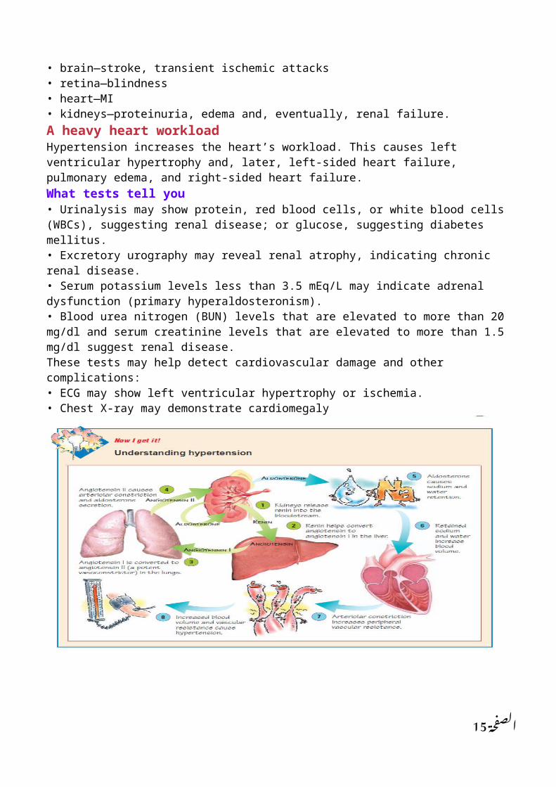
• abnormal renin release resulting in the formation of angiotensin II, which constricts the arterioles and increases blood volumeSecondary hypertension; Secondary hypertension may be caused by:• renovascular disease • renal parenchymal disease• pheochromocytoma • primary hyperaldosteronism• Cushing’s syndrome • diabetes mellitus• dysfunction of the thyroid, pituitary, or parathyroid gland• coarctation of the aorta • pregnancyUnderneath it allThe pathophysiology of secondary hypertension is related to the underlying disease. For example, consider these points:• The most common cause of secondary hypertension is chronic renal disease. Insult to the kidney from chronic glomerulonephritis or renal artery stenosis interferes with sodium excretion, the renin-angiotensin-aldosterone system, or renal perfusion. This causes blood pressure to rise.• In Cushing’s syndrome, increased cortisol levels raise blood pressure by increasing renal sodium retention, angiotensin II levels, and vascular response to norepinephrine.• In primary aldosteronism, increased intravascular volume, altered sodium concentrations in vessel walls, or very high aldosterone levels cause vasoconstriction (increased resistance).• Pheochromocytom is a secreting tumor of chromaffin cells, usually of the adrenal medulla. It causes hypertension due to increased secretion of epinephrine and norepinephrine. Epinephrine functions mainly to increase cardiac contractility and rate. Norepinephrine functions mainly to increase peripheral vascular resistance.Late complicationsComplications occur late in the disease and can attack any organ system. Cardiac complications include CAD, angina, MI, heart failure, arrhythmias, and sudden death. Neurologic complications includestroke and hypertensive encephalopathy. Hypertensive retinopathy can cause blindness. Renovascular hypertension can lead to renal failure. What to look forHypertension usually doesn’t produce signs and symptoms until vascular changes in the heart, brain, or kidneys occur. Severely elevated blood pressure damages the intima of small vessels, resultingin fibrin accumulation in the vessels, local edema and, possibly, intravascular clotting.Location, location, locationSymptoms depend on the location of the damaged vessels, for example: • brain—stroke, transient ischemic attacks• retina—blindness• heart—MI• kidneys—proteinuria, edema and, eventually, renal failure.A heavy heart workloadHypertension increases the heart’s workload. This causes left ventricular hypertrophy and, later, left-sided heart failure, pulmonary edema, and right-sided heart failure.What tests tell you• Urinalysis may show protein, red blood cells, or white blood cells (WBCs), suggesting renal disease; or glucose, suggesting diabetes mellitus.• Excretory urography may reveal renal atrophy, indicating chronic renal disease. • Serum potassium levels less than 3.5 mEq/L may indicate adrenal dysfunction (primary hyperaldosteronism).• Blood urea nitrogen (BUN) levels that are elevated to more than 20 mg/dl and serum creatinine levels that are elevated to more than 1.5 mg/dl suggest renal disease.These tests may help detect cardiovascular damage and other complications:
10الصفحة

• ECG may show left ventricular hypertrophy or ischemia.• Chest X-ray may demonstrate cardiomegaly
Myocardial infarctionMI, an acute coronary syndrome, results from reduced blood flow through one of the coronary arteries. This causes myocardial ischemia, injury, and necrosis. Leading the wayMI is one of the leading causes of death. Death usually results from cardiac damage or complications. sudden deaths occur within 1 hour after the onset of symptoms. Men are more susceptible to MI than premenopausal women, although the incidence is increasing in women who smoke and take hormonal contraceptives. The incidence in postmenopausal women is similar to that in men.How it happensMI results from occlusion of one or more of the coronary arteries. Occlusion can stem from atherosclerosis, thrombosis, platelet aggregation, or coronary artery stenosis or spasm.Predisposing factors include:• aging • diabetes mellitus• elevated serum triglyceride, low-density lipoprotein and cholesterol, and decreased serum
11الصفحة

high-density lipoprotein levels• excessive intake of saturated fats, carbohydrates, or salt • hypertension • obesity• positive family history of CAD • sedentary lifestyle • smoking• stress • use of amphetamines or cocaine.Susceptibility increases with ageElderly patients are more prone to complications and death. The most common complications after an acute MI include:• arrhythmias • cardiogenic shock • heart failure causing pulmonary edema • pericarditis.Other complications include:• rupture of the atrial or ventricular septum, ventricular wall, or valves• ventricular aneurysms• mural thrombi causing cerebral or pulmonary emboli• extensions of the original infarction • psychological problems caused by fear of another MI or organic brain disorder from tissue hypoxia• personality changes.Heavy reductionsMI results from prolonged ischemia to the myocardium with irreversible cell damage and muscle death. Functionally, MI causes:• reduced contractility with abnormal wall motion• altered left ventricular compliance• reduced stroke volume• reduced ejection fraction• elevated left ventricular end-diastolic pressureInsult…that is…ischemia added to injuryAll MIs have a central area of necrosis or infarction surrounded by an area of injury. The area of injury is surrounded by a ring of ischemia. Tissue regeneration doesn’t occur after an MI because the affected myocardial muscle is dead.A compensatory kickScar tissue that forms on the necrotic area may inhibit contractility. When this occurs, the compensatory mechanisms (vascular constriction, increased heart rate, and renal retention of sodium and water) kick in to try to maintain cardiac output. Ventricular dilation also may occur. If a lot of scar tissue forms, contractility may be greatly reduced. The patient may develop heart failure orcardiogenic shock.What to look for; The cardinal symptom of MI is persistent, crushing substernal pain that may radiate to the left arm, jaw, neck, or shoulder blades. The pain is commonly described as heavy, squeezing, or crushing and may persist for 12 hours or more. However, in some patients—particularly elderly or diabetic patients—pain may not occur at all. In others, it may be mild and confused with indigestion.An infarction on the horizon?In patients with CAD, angina of increasing frequency, severity, or duration (especially if not provoked by exertion, a heavy meal, or cold and wind) may signal an impending infarction.Other clinical effects include:• a feeling of impending doom • fatigue • nausea • vomiting
12الصفحة

• shortness of breath • cool extremities • diaphoresis • anxiety • restlessness.Fever is unusual at the onset of an MI, but a low-grade temperature may develop during the next few days. Blood pressure varies. Hypotension or hypertension may occur.What tests tell youThese tests help diagnose MI:• Serial 12-lead ECG may be normal or inconclusive during the first few hours after an MI. Abnormalities include serial STsegment depression and ST-segment elevation and Q waves, • Serum creatine kinase (CK) levels are elevated, especially the CK-MB isoenzyme, the cardiac muscle fraction of CK.• Troponin I, a structural protein found in cardiac muscle, is elevated. Troponin levels increase within 4 to 6 hours of myocardial injury.• Myoglobin is released with cardiac muscle damage and elevated levels may be detected as soon as 2 hours after an MI.• Echocardiography shows ventricular wall dyskinesia • Nuclear ventriculography can show acutely damaged muscle by picking up accumulations of radionuclide, which appear as a “hot spot’’ on the film.
Rheumatic fever and rheumatic heart diseaseA systemic inflammatory disease of childhood, acute rheumatic fever develops after infection of the upper respiratory tract with group A beta-hemolytic streptococci. It mainly involves the heart, joints,
13الصفحة
central nervous system, skin, and subcutaneous tissues and commonly recurs. If rheumatic fever isn’t treated, scarring deformity of the cardiac structures results in rheumatic heart disease.The disease strikes most often during cool, damp weather in the winter and early spring. All in the family?Rheumatic fever tends to run in families, lending support to the existence of genetic predisposition. Environmental factors also seem to be significant in development of the disorder. For example,in lower socioeconomic groups, the incidence is highest in children between ages 5 and 15, probably due to malnutrition and crowded living conditions.How it happensRheumatic fever appears to be a hypersensitivity reaction. For some reason, antibodies produced to combat streptococci react and produce characteristic lesions at specific tissue sites. Because only about 0.3% of people infected with Streptococcus bacteria contract rheumatic fever, altered immune response probably is involved in its development or recurrence.Getting complicatedThe mitral and aortic valves are commonly destroyed by rheumatic fever’s long-term effects. Their malfunction leads to severe heart inflammation (called carditis) and, occasionally, produces pericardial effusion and fatal heart failure. Of the patients who survive this complication, about 20% die within 10 years. Carditis develops in up to 50% of patients with rheumatic fever and may affect the endocardium, myocardium, or peri- cardium during the early acute phase. Later, the heart valves may be damaged, causing chronic valvular disease. Follow the infectionThe extent of heart damage depends on where the infection strikes:• Myocarditis produces characteristic lesions in the interstitial tissue of the heart as well as cellularswelling and fragmentation of interstitial collagen. These lesions lead to formation of progressively fibrotic nodules and interstitial scars.• Endocarditis causes valve leaflet swelling, erosion along the lines of leaflet closure, and blood, platelet, and fibrin deposits, which form beadlike vegetation. Endocarditis strikes the mitralvalve most commonly in females and the aortic valve in males. It affects the tricuspid valves in both sexes and, rarely, affects the pulmonic valve.What to look forIn 95% of patients, rheumatic fever follows a streptococcal infection that appeared a few days to 6 weeks earlier. A temperature of at least 100.4º F (38° C) occurs. Most patients complain of migratory joint pain or polyarthritis. Swelling, redness, and signs of effusion usually accompany such pain, which most commonly affects the knees, ankles, elbows, and hips.Rash talkAbout 5% of patients (usually those with carditis) develop a nonpruritic, macular, transient rash called erythema marginatum. This rash gives rise to red lesions with blanched centers. These same patients may also develop firm, movable, nontender subcutaneous nodules about 3 mm to 2 cm in diameter, usually near tendons or bony prominences of joints. These nodules persist for a few days to several weeks.What tests tell youNo specific laboratory tests can determine the presence of rheumatic fever, but these test results support the diagnosis:• WBC count and ESR may be elevated during the acute phase; blood studies show slight anemia caused by suppressed erythropoiesis during inflammation.• C-reactive protein is positive, especially during the acute phase.• Cardiac enzyme levels may be increased in severe myocarditis.• Throat cultures may continue to show group A beta-hemolytic streptococci• ECG reveals no diagnostic changes, but 20% of patients show a prolonged PR interval.• Chest X-ray shows normal heart size, except with myocarditis, heart failure, and pericardial effusion.
14الصفحة
• Echocardiography helps evaluate valvular damage, chamber size, ventricular function, and the presence of a pericardial effusion.• Cardiac catheterization evaluates valvular damage and left ventricular function in severe cardiac dysfunction• Antistreptolysin-O titer is elevated in 95% of patients within 2 months of onset.
15الصفحة

Quick Quiz:
1. Which factor is a major modifiable risk factor for CAD?A. High cholesterol B. Genetic predisposition C. Age D. Family history
2. Which of the following is the major pathophysiologic effect of cardiac tamponade?A. Atelectasis B. Hypertension C. Compressed heart D. Distended pericardium
3. Which liver enzyme stimulates the adrenal cortex to secretealdosterone?A. Angiotensin IB. Angiotensin IIC. ReninD. Antidiuretic hormone
16الصفحة
17الصفحة