single rooted maxillary first molar with type i can al ... · gajjela et al; single rooted...
TRANSCRIPT

DOI: 10.21276/aimdr.2017.3.3.DE8
Case Report ISSN (O):2395-2822; ISSN (P):2395-2814
Annals of International Medical and Dental Research, Vol (3), Issue (3) Page 38
Section: Dentistry
Single Rooted Maxillary First Molar With Type I Canal Configuration: Case Report. Rani Samyukta Gajjela1, Girija. S. Sajjan2, Kalyan Satish R3, Lavanya Sireesha Putchala4,
Prasanthi Gonapa5, Uday Podugu5, Chaithanya Reddy6 1Assistant Professor, Department of Conservative Dentistry and Endodontics, Vishnu Dental College, Bhimavaram, India.
2Professor and HOD, Department of Conservative Dentistry and Endodontics, Vishnu Dental College, Bhimavaram, India.
3Professor, Department of Conservative Dentistry and Endodontics, Vishnu Dental College, Bhimavaram, India.
4BDS, Department of Conservative Dentistry and Endodontics, Vishnu Dental College, Bhimavaram, India.
5Postgraduate student, Department of Conservative Dentistry and Endodontics, Vishnu Dental College, Bhimavaram, India.
6Assistant Professor, Department of Prosthodontics, Vishnu Dental College, Bhimavaram, India.
Received: April 2017 Accepted: April 2017
Copyright: © the author(s), publisher. Annals of International Medical and Dental Research (AIMDR) is an Official Publication of “Society for Health Care & Research Development”. It is an open-access article distributed under the terms of the Creative Commons Attribution Non-Commercial License, which permits unrestricted non-commercial use, distribution, and reproduction in any medium, provided the original work is properly cited.
ABSTRACT Atypical root canal morphology in multirooted teeth poses a perplexing situation during diagnosis and endodontic treatment. Ample knowledge of the anatomic variations of teeth is essential for successful endodontic treatment. Various studies have reported maxillary first molars with additional roots, canals, fused roots and c shaped canals. Rarest variation is to have a single root with type 1 canal configuration which is 0.02% according to Ingle. The present case report highlights the root canal treatment of a rare case of single rooted maxillary first molar with single canal. Keywords: Maxillary first molar; single canal; single root
INTRODUCTION Anatomic variations in maxillary molars are frequent. Although additional roots and extra canals are commonly encountered, the possibility of less number of roots and canals also exists. These deviations are also one of the major causes for endodontic treatment failure owing to inadequate cleaning, shaping and sealing of root canal system. In dental literature, the presence of single rooted maxillary first molar is rare. This case report describes the successful nonsurgical endodontic management of a maxillary first molar with a single root and root canal. Name & Address of Corresponding Author Dr. G. Rani Samyukta, Assistant professor, Department of Conservative Dentistry and Endodontics, Vishnu Dental College, Vishnupur, Bhimavaram-534202, West Godavari district, Andhra Pradesh.
CASE REPORT A 33 year old male patient presented with a chief complaint of pain in left upper back teeth region for the past 2 months. The medical history was non-contributory. Patient experienced pain which was moderate, intermittent in nature, dull, throbbing type, aggravated with intake of hot and cold beverages and while there is change in posture.
Clinical examination revealed deep dentinal caries approximating pulp distally with respect to tooth # 26. The tooth was tender on percussion. Thermal and electrical pulp testing with cold test (Endo-Frost, Roeko, Langenau, Germany), heated gutta-percha and electric pulp tester (Parkell, Edgewood NY, USA), respectively, elicited a negative response. Intraoral periapical radiograph revealed coronal radiolucency approximating pulp distally with ill-defined radiolucency at the apex [Figure 1]. Bilaterally, radiographs revealed atypical anatomy of single root and canal in maxillary first and second molars [Figure 2]. Based on the clinical and radiographic findings, the diagnosis of nonvital tooth with symptomatic apical periodontitis was made and root canal treatment followed by full veneer crown was advised in relation to tooth # 26. After obtaining informed consent, access cavity preparation was done under rubber dam isolation. Under an operating microscope (Carl Zeiss Inc, Oberkochen, Germany), single wide root canal orifice was found in the center of the pulpal floor. Pulpal floor was further examined for other orifices but were not present. To confirm this morphology, stainless steel K files were placed in the root canal and multiple X-rays were taken in variable horizontal angulations which confirmed single root and canal [Figure 4a & b]. On instrumentation, all scouting files converged into a single broad canal. Working length was determined and biomechanical preparation was done using circumferential filing

Gajjela et al; Single Rooted Maxillary First Molar With Type I Canal Configuration
Annals of International Medical and Dental Research, Vol (3), Issue (3) Page 39
Section: Dentistry
technique until 80 k file size under 3% sodium hypochlorite irrigation. The canal was dried with paper points and calcium hydroxide was mixed with 2% chlorhexidine gluconate (Neelkanth Healthcare
Pvt. Ltd, Safe Plus, Rajasthan, India) to form a paste and placed in the canal using a lentulo spiral. The access cavity was filled with a temporary restorative material, IRM (Dentsply, Caulk, USA).
Figure 1-8: (1) Preoperative radiograph; (2) Preoperative radiograph showing single rooted contralateral tooth; (3) Access cavity preparation showing a central canal "Cn."; (4 a and b) Working length determination radiograph; (5) Master cone radiograph; (6) Sectional obturation; (7) Postobturation radiograph; (8) Postoperative radiograph after crown cementation. In the next visit, calcium hydroxide dressing was flushed out using alternating irrigation with 5.25% NaOCl and 17% EDTA. The canal was irrigated with final rinse of 2% chlorhexidine gluconate and was dried using paper points. The apical 5mm of root canal was obturated using a resin-sealer (AH Plus; Dentsply) and guttapercha [Figure 5] followed by backfilling using injectable thermoplasticized gutta-percha (Obtura III Spartan, Fenton, Missouri, USA) [Figure 6]. Postendodontic restoration was done with composite Filtek Z 250XT [Figure 7] and later PFM crown was fabricated and cemented using resin cement [Figure 8].
DISCUSSION In this clinical case report, unusual root canal morphology involving single roots and canals in all existing maxillary molars bilaterally should be taken into consideration. Kim Y et al., reported that asian
population shows higher prevalence for single rooted maxillary molars, which is inherited as an autosomal dominant trait.[1] In dentistry, from the past four decades, there is a quest to perceive, understand and comprehend the anatomical variations of teeth. Although extra canals are more of a rule rather than an exception, there is also possibility of fewer canals than the normally presumed canal morphology. Inadequate knowledge about frequency of single canal and attempting to search for another canal may result in perforation and failure of endodontic treatment.[2] In single rooted maxillary molars, as the root surface area for periodontal attachment is reduced they are at higher risk for periodontal disease progression. Therefore, periodontal health should be given due consideration. Various factors should be considered during endodontic treatment of single rooted maxillary molar with single canal. Use of multiple angulation
Gajjela et al; Single Rooted Maxillary First Molar With Type I Canal Configuration
Annals of International Medical and Dental Research, Vol (3), Issue (3) Page 40
Section: Dentistry
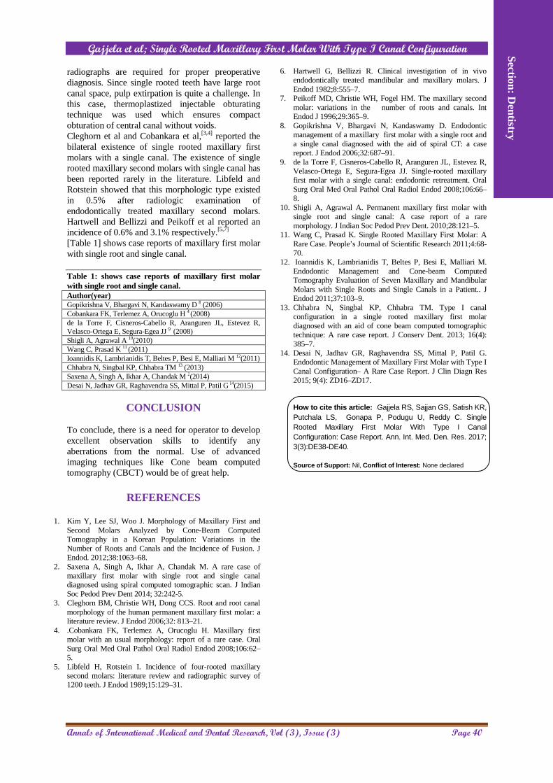
radiographs are required for proper preoperative diagnosis. Since single rooted teeth have large root canal space, pulp extirpation is quite a challenge. In this case, thermoplastized injectable obturating technique was used which ensures compact obturation of central canal without voids. Cleghorn et al and Cobankara et al,[3,4] reported the bilateral existence of single rooted maxillary first molars with a single canal. The existence of single rooted maxillary second molars with single canal has been reported rarely in the literature. Libfeld and Rotstein showed that this morphologic type existed in 0.5% after radiologic examination of endodontically treated maxillary second molars. Hartwell and Bellizzi and Peikoff et al reported an incidence of 0.6% and 3.1% respectively.[5,7] [Table 1] shows case reports of maxillary first molar with single root and single canal.
Table 1: shows case reports of maxillary first molar with single root and single canal. Author(year) Gopikrishna V, Bhargavi N, Kandaswamy D 8 (2006) Cobankara FK, Terlemez A, Orucoglu H 4 (2008) de la Torre F, Cisneros-Cabello R, Aranguren JL, Estevez R, Velasco-Ortega E, Segura-Egea JJ 9 (2008) Shigli A, Agrawal A 10(2010) Wang C, Prasad K 11 (2011) Ioannidis K, Lambrianidis T, Beltes P, Besi E, Malliari M 12(2011) Chhabra N, Singbal KP, Chhabra TM 13 (2013) Saxena A, Singh A, Ikhar A, Chandak M 2(2014) Desai N, Jadhav GR, Raghavendra SS, Mittal P, Patil G 14(2015)
CONCLUSION
To conclude, there is a need for operator to develop excellent observation skills to identify any aberrations from the normal. Use of advanced imaging techniques like Cone beam computed tomography (CBCT) would be of great help.
REFERENCES
1. Kim Y, Lee SJ, Woo J. Morphology of Maxillary First and
Second Molars Analyzed by Cone-Beam Computed Tomography in a Korean Population: Variations in the Number of Roots and Canals and the Incidence of Fusion. J Endod. 2012;38:1063–68.
2. Saxena A, Singh A, Ikhar A, Chandak M. A rare case of maxillary first molar with single root and single canal diagnosed using spiral computed tomographic scan. J Indian Soc Pedod Prev Dent 2014; 32:242-5.
3. Cleghorn BM, Christie WH, Dong CCS. Root and root canal morphology of the human permanent maxillary first molar: a literature review. J Endod 2006;32: 813–21.
4. .Cobankara FK, Terlemez A, Orucoglu H. Maxillary first molar with an usual morphology: report of a rare case. Oral Surg Oral Med Oral Pathol Oral Radiol Endod 2008;106:62–5.
5. Libfeld H, Rotstein I. Incidence of four-rooted maxillary second molars: literature review and radiographic survey of 1200 teeth. J Endod 1989;15:129–31.
6. Hartwell G, Bellizzi R. Clinical investigation of in vivo endodontically treated mandibular and maxillary molars. J Endod 1982;8:555–7.
7. Peikoff MD, Christie WH, Fogel HM. The maxillary second molar: variations in the number of roots and canals. Int Endod J 1996;29:365–9.
8. Gopikrishna V, Bhargavi N, Kandaswamy D. Endodontic management of a maxillary first molar with a single root and a single canal diagnosed with the aid of spiral CT: a case report. J Endod 2006;32:687–91.
9. de la Torre F, Cisneros-Cabello R, Aranguren JL, Estevez R, Velasco-Ortega E, Segura-Egea JJ. Single-rooted maxillary first molar with a single canal: endodontic retreatment. Oral Surg Oral Med Oral Pathol Oral Radiol Endod 2008;106:66–8.
10. Shigli A, Agrawal A. Permanent maxillary first molar with single root and single canal: A case report of a rare morphology. J Indian Soc Pedod Prev Dent. 2010;28:121–5.
11. Wang C, Prasad K. Single Rooted Maxillary First Molar: A Rare Case. People’s Journal of Scientific Research 2011;4:68-70.
12. Ioannidis K, Lambrianidis T, Beltes P, Besi E, Malliari M. Endodontic Management and Cone-beam Computed Tomography Evaluation of Seven Maxillary and Mandibular Molars with Single Roots and Single Canals in a Patient.. J Endod 2011;37:103–9.
13. Chhabra N, Singbal KP, Chhabra TM. Type I canal configuration in a single rooted maxillary first molar diagnosed with an aid of cone beam computed tomographic technique: A rare case report. J Conserv Dent. 2013; 16(4): 385–7.
14. Desai N, Jadhav GR, Raghavendra SS, Mittal P, Patil G. Endodontic Management of Maxillary First Molar with Type I Canal Configuration– A Rare Case Report. J Clin Diagn Res 2015; 9(4): ZD16–ZD17. How to cite this article: Gajjela RS, Sajjan GS, Satish KR, Putchala LS, Gonapa P, Podugu U, Reddy C. Single Rooted Maxillary First Molar With Type I Canal Configuration: Case Report. Ann. Int. Med. Den. Res. 2017; 3(3):DE38-DE40.
Source of Support: Nil, Conflict of Interest: None declared