silencer sirna starter kit - thermo fisher scientific - us
TRANSCRIPT

Silencer® siRNA Starter KitComplete Kit for Inducing and Monitoring RNAi
Part Number AM1640
cov_siRNA Starter Kit.fm Page 1 Thursday, August 19, 2010 11:42 AM

Silencer® siRNA Starter Kit
(Part Number AM1640)
Protocol
I. Introduction. . . . . . . . . . . . . . . . . . . . . . . . . . . . . . . . . . . . . . . . . . . . . . . . . . . . . . . 1
A. BackgroundB. Product DescriptionC. Silencer siRNA Starter Kit Components and Storage ConditionsD. Required and Optional Materials Not Provided with the KitE. Related Products Available from Applied Biosystems
II. siRNA Transfection . . . . . . . . . . . . . . . . . . . . . . . . . . . . . . . . . . . . . . . . . . . . . . . . . 7
A. Traditional and Reverse Transfection MethodsB. Basic Transfection Procedure
III. Monitoring Cytotoxicity & GAPDH Knockdown. . . . . . . . . . . . . . . . . . . . . . . . . 11
A. Quantitative and Qualitative Options for Evaluating RNAiB. KDalert GAPDH Assay Standard Fluorescence ProcedureC. qRT-PCR InstructionsD. KDalert and qRT-PCR Data InterpretationE. Demonstrating GAPDH Knockdown Using the Anti-GAPDH AntibodyE.I. Western Blot ProcedureE.II. Immunofluorescence ProcedureF. Optimizing siRNA Transfection Conditions
IV. Troubleshooting . . . . . . . . . . . . . . . . . . . . . . . . . . . . . . . . . . . . . . . . . . . . . . . . . . 26
A. Transfection Causes Extensive Cell DeathB. Troubleshooting the KDalert GAPDH AssayC. No Detectable Gene SilencingD. Gene Silencing Experiments Lack Reproducibility
V. Appendix . . . . . . . . . . . . . . . . . . . . . . . . . . . . . . . . . . . . . . . . . . . . . . . . . . . . . . . . 31
A. Traditional “Pre-plating” Transfection ProcedureB. Alternate KDalert Colorimetric Assay ProcedureC. Quality ControlD. Safety Information
siRNA Starter Kit.book Page 1 Thursday, August 19, 2010 11:54 AM

P/N 1640M Revision C Revision Date: August 19, 2010
For research use only. Not for use in diagnostic procedures. By use of this product, you accept the termsand conditions of all applicable Limited Label Licenses. For statement(s) and/or disclaimer(s) applicable to thisproduct, see below.
Information in this document is subject to change without notice. Applied Biosystems assumes no responsibil-ity for any errors that may appear in this document.
Applied Biosystems disclaims all warranties with respect to this document, expressed or implied, including butnot limited to those of merchantability or fitness for a particular purpose. In no event shall Applied Biosystemsbe liable, whether in contract, tort, warranty, or under any statute or on any other basis for special, incidental,indirect, punitive, multiple or consequential damages in connection with or arising from this document,including but not limited to the use thereof.
Literature Citation: When describing a procedure for publication using this product, please refer to it as theSilencer® siRNA Starter Kit.
Warranty and Liability: Applied Biosystems is committed to delivering superior product quality and perfor-mance, supported by industry-leading global service and technical support teams. Warranty information forthe accompanying consumable product is available at www.ambion.com/info/warranty in “Limited Warrantyfor Consumables,” which is subject to the exclusions, conditions, exceptions, and limitations set forth underthe caption “EXCLUSIONS, CONDITIONS, EXCEPTIONS, AND LIMITATIONS” in the full warrantystatement. Please contact Applied Biosystems if you have any questions about our warranties or would likeinformation about post-warranty support.
Patents and Licensing Notifications: The Silencer® siRNA Starter Kit is compatible with the 5' nucleasedetection and dsDNA-binding dye processes covered by patents owned or licensable by Applied Biosystems.No license under these patents is conveyed expressly, by implication, or by estoppel to the purchaser by thepurchase of this product. Further information on purchasing licenses may be obtained by contacting the Direc-tor of Licensing, Applied Biosystems, 850 Lincoln Centre Drive, Foster City, California 94404, USA. Thisproduct is licensed under European Patents 1144623, 1214945 and foreign equivalents from Alnylam Phar-maceuticals, Inc., Cambridge, USA and is provided only for use in academic and commercial research includ-ing in vitro and in vivo identification and validation of drug targets (but excluding the evaluation orcharacterization of this product as the potential basis for a siRNA-based drug) and not for any other commer-cial purposes. Information about licenses for commercial use (including discovery and development of siRNA-based drugs) is available from Alnylam Pharmaceuticals, Inc., 300 Third Street, Cambridge, MA 02142, USA.This product is manufactured under license from the Massachusetts Institute of Technology to U.S. PatentNos. 7,056,704 and 7,078,196 and pending counterparts.
Trademarks: Applied Biosystems, AB (Design), Ambion, RNAqueous, and Silencer are registered trade-marks, and KDAlert, MagMAX, MessageSensor, siPORT and NeoFX are trademarks of Applied BiosystemsInc. or its subsidiaries in the US and/or certain other countries. All other trademarks are the sole property oftheir respective owners.
© 2008, 2010 Ambion, Inc. All Rights Reserved.
siRNA Starter Kit.book Page 2 Thursday, August 19, 2010 11:54 AM

I.A. Background
Introduction
1
I. Introduction
A. Background
RNA interference (RNAi), the biological mechanism by which dou-ble-stranded RNA (dsRNA) induces gene silencing by targeting com-plementary mRNA for degradation, is revolutionizing the wayresearchers study gene function. Scientists can quickly and easily reducethe expression of a particular gene in nearly all metazoan systems to ana-lyze the effect that gene has on cellular function.
The RNAi mechanism In non-mammalian systems, introducing or expressing long dsRNAtriggers the RNAi pathway. The cytoplasmic nuclease Dicer first cleavesthe long dsRNA into 21–23 bp small interfering RNAs (siRNAs), thatthen unwind and assemble into RNA-induced silencing complexes(RISCs). The antisense siRNA strand then guides the RISC to comple-mentary RNA molecules, and the RISC cleaves the messenger RNA(mRNA), leading to specific gene silencing. Since most mammaliancells mount a potent antiviral response upon introduction of dsRNAlonger than 30 bp, researchers transfect cells with 21–23 bp siRNAs toinduce RNAi in these systems without eliciting the antiviral response(see Figure 1).
Figure 1. RNAi Triggered by siRNAs
RISC
siRNA
siRNAs associatewith RISC
Cleavage of mRNA by RISC
siRNAs
siRNA Starter Kit.book Page 1 Thursday, August 19, 2010 11:54 AM

Silencer® siRNA Starter Kit
I.B. Product Description2
Chemically synthesized
siRNAs
Currently, the most widespread application of RNAi is to transientlytransfect chemically synthesized siRNA into cultured mammalian cells,and assay the cells to monitor the RNAi effect. Ambion providesexpert-designed, guaranteed-to-silence siRNAs to >100,000 human,mouse, and rat targets (>98% of all human, mouse, and rat genes in theRefSeq database). The quality of siRNA can significantly influence RNAi experiments.siRNA should be free of reagents carried over from synthesis, such assalts and proteins. Also, dsRNA contaminants longer than 30 bp areknown to cause cytotoxicity. We recommend using column, HPLC, orgel purified, chemically synthesized siRNAs to ensure quality andpurity. Ambion offers an extensive line of siRNAs available individually and infunctional class-focused sets as Silencer® siRNA Libraries and SilencerCellReady™ Libraries. Individual siRNAs allow detailed analysis of anindividual gene’s role in one or more pathways, whereas sets of siRNAs(libraries) enable large scale screening experiments to tie genes to cellularfunction; for details, see our website at: www.ambion.com/siRNA.
B. Product Description
The Silencer® siRNA Starter Kit is ideal for researchers new to RNAiexperiments. It contains the reagents and procedures to demonstrategene silencing using RNAi, and it can also be used for siRNA deliveryoptimization experiments. Briefly, the kit includes control siRNAs,siPORT™ NeoFX™ Transfection Agent, and three different reagent setsto detect silencing of the GAPDH (glyceraldehyde 3-phosphate dehy-drogenase) gene, including the rapid KDalert™ GAPDH Assay Kit. Thereagents in the kit are discussed in more detail below.
Delivery of siRNAs into
cultured cells
For many immortalized cell lines, chemical transfection is the preferredmeans to deliver siRNA into your cells. The Ambion siPORT NeoFXTransfection Agent was designed expressly for this purpose.siPORT NeoFX is a proprietary mixture of lipids that functions by com-plexing with siRNAs to facilitate siRNA transfection in a broad range ofcell types with high efficiency and reproducibility. It is easy to use andhas minimal cytotoxic effects. However, the efficiency of siRNA trans-fection is strongly influenced by several parameters including the choiceof transfection agent, the amount of transfection agent, and the numberof cells plated.
GAPDH and Negative
Control #1 siRNA
The GAPDH siRNA targets the abundant, ubiquitously-expressedhousekeeping gene, GAPDH. It efficiently induces silencing in human,mouse, and rat cell lines, reducing both the mRNA and protein levels ofGAPDH. The Negative Control #1 siRNA is a nontargeting sequencethat has no significant homology to the sequences of human, mouse, or
siRNA Starter Kit.book Page 2 Thursday, August 19, 2010 11:54 AM

I.B. Product Description
Introduction
3
rat transcripts. The Negative Control #1 siRNA should have no effecton the mRNA and protein levels of GAPDH; it serves as a baseline formeasuring the effects of the GAPDH siRNA. The negative control canbe used to identify nonspecific effects such as nonsequence-specificsiRNA effects, cytotoxicity of the transfection agent and/or the siRNA,or suboptimal transfection conditions.
Reagents to detect gene
silencing
In RNAi experiments, gene silencing can be measured either at themRNA or protein level. The Silencer siRNA Starter Kit provides conve-nient methods for users to detect siRNA-mediated silencing of GAPDHgene expression at both the mRNA and protein levels. Quantitativemeasurements at the mRNA level can be obtained via qRT-PCR* usingthe GAPDH RT-PCR Primer Set. For quantitative evaluation ofGAPDH silencing at the protein level, we provide the KDalertGAPDH Assay. For labs that routinely perform Western blot and/orimmunofluorescence techniques, an Anti-GAPDH Antibody isincluded to demonstrate qualitative GAPDH knockdown. All the sup-plied detection reagents work equally well to detect silencing in human,mouse, and rat cells.
Reverse transfection This Procedure provides guidelines for transfection optimization usingreverse transfection. In reverse transfection, cells are transfected as theyadhere to a plate after trypsinization (Figure 2 on page 7). This methodbypasses several steps of the traditional “pre-plating” transfectionmethod, making it faster, easier, and often more effective. We recom-mend reverse transfection for convenience and ease. Furthermore, it canbe used with most chemical transfection agents and is effective in mostcultured cell types. (A procedure for traditional transfection is providedin section V.A on page 31).
Kit applications The Silencer siRNA Starter Kit is designed to provide an introduction toRNAi experiments in most types of adherent cultured cells. Note thatfor some cell lines, the siPORT NeoFX transfection agent provided withthe kit may not be the most appropriate. Our website has a list of rec-ommended siPORT Transfection Agents for different cell lines as wellas information on transfection conditions compiled from recent scien-tific literature and reported to Ambion by customers who are using ourproducts:
www.ambion.com/techlib/resources/delivery
* This product is compatible with the 5' nuclease detection and dsDNA-binding dye processes covered by patents owned or licens-able by Applied Biosystems. No license under these patents is conveyed expressly, by implication, or by estoppel to the purchaser by the purchase of this product. Further information on purchasing licenses may be obtained by contacting the Director of Licensing, Applied Biosystems, 850 Lincoln Centre Drive, Foster City, California 94404, USA.
siRNA Starter Kit.book Page 3 Thursday, August 19, 2010 11:54 AM

Silencer® siRNA Starter Kit
I.C. Silencer siRNA Starter Kit Components and Storage Conditions4
Reagents are provided for delivery of an extremely potent GAPDHsiRNA and the nontargeting Negative Control #1 siRNA. Further,these reagents can be used for transfection optimization experiments.RNAi-induced gene silencing can be detected using any of three meth-ods for detection of GAPDH.
C. Silencer siRNA Starter Kit Components and Storage Conditions
Properly stored kits are guaranteed for 6 months from the date received.
D. Required and Optional Materials Not Provided with the Kit
General lab equipment and
supplies
• Nuclease-free microcentrifuge tubes and barrier pipet tips (see www.ambion.com/prod/tubes for a complete listing)
• Constant temperature incubators
Cell culture material and
equipment
• Opti-MEM® I Reduced-Serum Medium (Invitrogen Cat #31985)• Routine tissue culture supplies and equipment
Material and equipment for
KDalert GAPDH Assay
(optional)
• 96 well plates: We recommend black U-bottom polypropylene 96well plates (e.g., ABgene® Microplate 0796/k). Alternatively clearpolystyrene plates can be used (recommended: BD FalconCat #353072 or Greiner Bio-One CellStar Cat #655180).
• Fluorescence plate reader (recommended) or UV-Vis plate reader• General lab equipment and supplies such as 15 mL or microcentri-
fuge tubes, multichannel pipettors (recommended), vortex mixer,ice, etc.
Amount Component Storage
1.75 mL Nuclease-free Water any temp*
* Store Nuclease-free Water at –20°C, 4°C, or room temp.
400 μL siPORT NeoFX Transfection Agent†
† Keep the tube of siPORT NeoFX tightly closed to prevent evaporation.
4°C
100 μg Anti-GAPDH Monoclonal Antibody (MAb) –20°C
250 μL GAPDH PCR Primers (3 μM forward primer, 3 μM reverse primer)
–20°C
100 mL KDalert Lysis Buffer –20°C
12.5 mL KDalert Solution A –20°C
100 μL KDalert Solution B –20°C
75 μL KDalert Solution C –20°C
100 μL GAPDH enzyme below –70°C
50 μL GAPDH siRNA, Human, Mouse, & Rat (50 μM)
below –70°C
50 μL Negative Control #1 siRNA (50 μM) below –70°C
siRNA Starter Kit.book Page 4 Thursday, August 19, 2010 11:54 AM

I.E. Related Products Available from Applied Biosystems
Introduction
5
Material and equipment for
qRT-PCR (optional)
• Reagents for isolation of total RNA from transfected cells: Anymethod for isolating high quality total RNA suitable for qRT-PCRcan be used. We recommend the following Ambion products:– MagMAX™-96 Total RNA Isolation Kit (P/N AM1830)– MagMAX-96 for Microarrays Total RNA Isolation Kit (P/N
AM1839)– RNAqueous®-4PCR Kit (P/N AM1914)
• Reagents and supplies for two-step qRT-PCR using SYBR® Green Idetection of amplified products: We recommend the following kitsand reagents for qRT-PCR detection of siRNA-induced GAPDHknockdown:– MessageSensor™ RT Kit (P/N AM1745)– SuperTaq™† Thermostable DNA Polymerase (P/N AM2050,
AM2052)– PCR primers for an endogenous control RNA such as 18S rRNA
or cyclophilin– SYBR Green I or comparable nucleic acid stain– ROX normalization dye – Thermal cycler for real-time PCR– Optical PCR plates or tubes
E. Related Products Available from Applied Biosystems
† Use of this product is covered by US patent claims and patent claims outside the US. The purchase of this product includes a limited, non-transferable immunity from suit under the foregoing patent claims for using only this amount of product for the purchaser’s own internal research. No right under any other patent claim (such as the patented 5’ Nuclease Process claims), no right to perform any patented method, and no right to perform commercial services of any kind, including without limitation reporting the results of purchaser's activities for a fee or other commercial consideration, is conveyed expressly, by implication, or by estoppel. This product is for research use only. Diagnostic uses under Roche patents require a separate license from Roche. Further information on purchasing licenses may be obtained by contacting the Director of Licensing, Applied Biosystems, 850 Lincoln Centre Drive, Foster City, California 94404, USA.
siPORT™ Amine Transfection
AgentP/N AM4502, AM4503
siPORT Amine is an easy-to-use proprietary blend of polyamines that deliverssiRNA into mammalian cells with minimal cytotoxicity.
MessageSensor™ RT KitP/N AM1745
The MessageSensor RT Kit for one-step qRT-PCR includes an optimized setof reagents for exceptionally sensitive reverse transcription. The kit isdesigned to be used for single-tube amplification of mRNA using eitherreal-time or end-point amplification strategies.
Silencer® siRNA
Transfection II KitP/N AM1631
The Silencer siRNA Transfection II Kit contains both siPORT™ NeoFX™ andsiPORT Lipid Transfection Agents in addition to a well-characterized siRNAtargeting human, mouse, and rat GAPDH. This kit is ideal for developing anoptimal transfection protocol for your cells. Also included are a highly vali-dated non-targeting negative control siRNA and a detailed Protocol.

Silencer® siRNA Starter Kit
I.E. Related Products Available from Applied Biosystems6
Silencer® CellReady™ siRNA
Transfection Optimization KitP/N AM86050
The Silencer® CellReady™ siRNA Transfection Optimization Kit facilitatesidentification of optimal siRNA delivery conditions in a high-throughput for-mat. Developed as a companion kit to the Silencer CellReady siRNA Librar-ies, the kit includes three 96-well Silencer CellReady Optimization Plates,each with 48 wells containing Silencer GAPDH siRNA and 48 wells platedwith Silencer Negative Control #1 siRNA. It also includes Ambion’slipid-based transfection agent, siPORT™ NeoFX™, as well as step-by-stepinstructions for rapid and efficient optimization of transfection conditions.The conditions identified using the kit are generally applicable for siRNAtransfections in 96-well plates.
Silencer® siRNA Labeling KitsP/N AM1632, AM1634
The Silencer siRNA Labeling Kits are used for labeling siRNA synthesizedwith the Silencer siRNA Construction Kit or synthesized chemically. LabeledsiRNA can be used to analyze the subcellular distribution of siRNA, in vivostability, transfection efficiency, or the capability of the siRNA to attenuatetarget gene expression.
Silencer® siRNA ControlsP/N AM4250–AM4639
see our web or print catalog
www.ambion.com/siRNA
Silencer siRNA Controls are chemically synthesized siRNAs for genes com-monly used as controls. Validated control siRNAs are available for genes suchas GAPDH, β-actin, cyclophilin, KIF11 (Eg5), GFP, and luciferase. ThesesiRNAs are ideal for developing and optimizing siRNA experiments and havebeen validated for use in human cells; many are also validated in mouse andrat cells.
Silencer® siRNAssee our web or print catalog
www.ambion.com/siRNA
Ambion Silencer Pre-designed siRNAs, Validated siRNAs, and siRNA Librar-ies are designed with the most rigorously tested siRNA design algorithm inthe industry. Silencer siRNAs are available for >100,000 human, mouse, andrat targets from our searchable online database. Because of their carefully opti-mized design, Silencer siRNAs are very effective, and they are guaranteed toreduce target mRNA levels by 70% or more. Furthermore, their exceptionalpotency means that Silencer siRNAs effectively induce RNAi at very low con-centrations, minimizing off-target effects.
siRNA Starter Kit.book Page 6 Thursday, August 19, 2010 11:54 AM

II.A. Traditional and Reverse Transfection Methods
siRNA Transfection
7
II. siRNA Transfection
A. Traditional and Reverse Transfection Methods
Delivery of synthetic siRNA molecules into adherent cells requires theuse of chemical transfection agents. Lipid-based transfection agents suchas siPORT NeoFX facilitate transfection by complexing into aggregateswith the negatively charged siRNA molecules. These siRNA-transfec-tion agent complexes are efficiently taken up by cells, presumably byendocytosis.Historically, the first step in transfection of adherent mammalian cells isto pre-plate the cells 24 hr before transfection. During this time, thecells recover from trypsinization, grow, and adhere to the culture plate.Reverse transfection is a time-saving, effective alternative in which cellsare transfected as they are passaged. Compared to the traditionalpre-plating method, equivalent or improved transfection efficiency isseen for many of the cell types tested at Ambion. In addition, the reversetransfection process is an entire day shorter than traditional transfection(Figure 2). Because cells are in suspension, a larger amount of cell sur-face is exposed to transfection agent/siRNA complexes, and this isthought to contribute to the improved transfection efficiency. This pro-cedure is written for reverse transfection, because it is the preferredmethod at Ambion; however, we also include a procedure for traditionalpre-plated transfection in section V.A starting on page 31.
Figure 2.Traditional and Reverse Transfection Methods
Mix transfectionagent with nucleic acid
Add transfectioncomplex to plated cells
Dilute transfectionagent
Harvest cellsPlate cellsand grow for 24 hr
Aliquot nucleic acidto transfect
Incubate and assay cells
Overlay with cells
Pre-platedTransfection
Day 1plate cells
Day 2transfect
Day 4assay
ReverseTransfection
Day 1transfect
Day 3assay
siRNA Starter Kit.book Page 7 Thursday, August 19, 2010 11:54 AM

Silencer® siRNA Starter Kit
II.B. Basic Transfection Procedure8
IMPORTANT
B. Basic Transfection Procedure
Preparation and planning Dilute the siRNAs to 2 μM
Follow the guidelines in the table below to prepare 2 μM solutions ofthe supplied siRNAs in nuclease-free microcentrifuge tubes:
Mix thoroughly and store the 2 μM siRNA solutions at –20°C for up to3 months.
Planning for transfection
For each set of transfection conditions, plan to include 3 replicate trans-fections (i.e., a total of 9 transfections for each set of transfection condi-tions) with each of the following:• GAPDH siRNA• Negative Control #1 siRNA• Nontransfected control: cells that are mock-transfected with
Opti-MEM I medium, but no transfection agent and no siRNA
NOTE
In this section, suggested initial transfection conditions are listed first and
reagent quantities for transfection optimization experiments follow. More
detailed suggestions for how to optimize transfection conditions are pro-
vided in section III.F. Optimizing siRNA Transfection Conditions starting on
page 22.
1. Prepare cells a. Trypsinize adherent cells.
Trypsinize healthy, growing, adherent cells using your routine pro-cedure. In general, healthy cells transfect better than poorly main-tained cells. Routinely subculturing cells before they becomeovercrowded or unhealthy will minimize instability in continuouscell lines from experiment to experiment. Information on basic cellculture technique can be found in Culture of Animal Cells: A Manualof Basic Technique (2000) Freshney, NY:Wiley-Liss.
b. Resuspend cells in normal growth medium.
For initial experiments, resuspend cells in normal growth mediumto 1 x 10⁵ cells/mL.
In order for cells to be transfected
before they re-adhere; it is important
to proceed immediately with the
following steps.
For subsequent transfection optimization experiments, we recom-mend testing from 5 x 10⁴ to 1.5 x 10⁵ cells/mL. To do this, firstsuspend cells at 1.5 x 10⁵ cells/mL and then dilute a portion of thesuspension further to 1 x 10⁵ cells/mL and 5 x 10⁴ cells/mL.
Table 1. Preparation of 2 μM siRNA solutions
Amount Component
16 μL GAPDH or Negative Control #1 siRNA
384 μL Nuclease-free water
siRNA Starter Kit.book Page 8 Thursday, August 19, 2010 11:54 AM

II.B. Basic Transfection Procedure
siRNA Transfection
9
Keep the cells at 37°C until they are needed in step 3. The table below shows the volume of cells needed per well of differ-ent-sized culture plates. Count the number of wells in your experi-ment, and calculate the total volume of cells you will need. Plan toprepare ~5% overage to account for pipetting error.
2. Prepare siRNA/siPORT
NeoFX complexes and
distribute into culture
plate wells
Bring siPORT NeoFX or other transfection agent and the Opti-MEM Imedium to room temp before use.a. Dilute siPORT NeoFX in Opti-MEM I medium in a sterile
conical tube.
b. Incubate diluted siPORT NeoFX for 10 min at room temp.
c. Dilute the 2 μM GAPDH or Negative Control #1 siRNA in
Opti-MEM I medium.
Be sure to dilute the siRNA supplied with the kit to 2 μM before useas described in Preparation and planning on page 8.
Amounts per transfection
Culture Plate Type
96-well 24-well 6-well
Volume of cells 80 μL 400 μL 2.4 mL
Number of cellsrecommended for first expt(range for optimization)
8 x 10³
(4–12 x 103)4 x 10⁴
(2–6 x 10⁴)2.4 x 10⁵
(1.2–3.6 x 10⁵)
Dilute siPORT NeoFX in Opti-MEM I
Dilute siRNAin Opti-MEM I
Incubate 10 minat room temp
Mix
Incubate 10 minat room temp
Amounts per transfection 96-well 24-well 6-well
siPORT NeoFX*recommended for first expt(range for optimization)
* Note that these volumes are appropriate for siPORT NeoFX, but may not be appropriate for other transfection agents. Use the manufacturer’s volume rec-ommendations for other transfection agents.
0.5(0.2–0.8
μLμL)
1(0.6–2
μLμL)
5(2–6
μLμL)
Opti-MEM I to final volume:
10 μL 50 μL 300 μL
Amounts per transfection 96-well 24-well 6-well
2 μM siRNA*recommended for first expt(range for optimization)
* We recommend initially using 30 nM final siRNA concentration in the trans-fection mixture at the end of step 3 on page 10, and to test 5–30 nM final siRNA concentration in optimization experiments.
1.5(0.25–1.5
μLμL)
7.5(1.5–7.5
μLμL)
45(7.5–45
μLμL)
Opti-MEM I to final volume:
10 μL 50 μL 300 μL
siRNA Starter Kit.book Page 9 Thursday, August 19, 2010 11:54 AM

Silencer® siRNA Starter Kit
II.B. Basic Transfection Procedure10
NOTE
d. Mix diluted siRNA with diluted siPORT NeoFX. Incubate at
room temp for 10 min.
Combine the diluted siPORT NeoFX (or other transfection agent)from step b with the diluted siRNA from step c. Mix gently bypipetting up and down or flicking the tube several times. Incubate 10 min at room temp. siRNA/siPORT NeoFX complexesform during this incubation.
e. Dispense the siRNA/siPORT NeoFX complexes into the
empty wells of a culture plate, and set up the nontransfected
controls.
If you plan to evaluate GAPDH
knockdown by Western blot or
immunofluorescence, we
recommend conducting
transfections in 24-well plates, for
immunofluorescence, plate cells
onto 12mm round glass coverslips.
Aliquot siRNA/siPORT NeoFX transfection complexes from step dinto the wells of the culture plate following the volume guidelinesshown below. Include 3 nontransfected control wells, containingOpti-MEM I medium, but no siRNA and no transfection agent.
3. Add cells to the
siRNA/siPORT NeoFX
complexes (and control
wells)
a. Transfer cells to the culture plate.
Gently mix the cells prepared in step 1 to resuspend, and pipet theminto wells of the culture plate containing siRNA/siPORT NeoFXcomplexes or wells set up as nontransfected controls.
b. Gently mix the cells and siRNA/siPORT NeoFX complexes.
Rock the plate gently back and forth to evenly distribute the com-plexes; avoid swirling, as this can cause contents to aggregate in thecenter of the well.
4. Incubate at 37°C for 24 hr,
then replace the culture
medium
Incubate the transfection mixture at 37°C in normal cell culture condi-tions for 24 hr. Then, replace the culture medium with fresh normalgrowth medium.
5. Assay for transfection
efficiency and
cytotoxicity
As an initial screen for severe cytotoxic effects, check the visual appear-ance of transfected cells for evidence of cell necrosis and/or apoptosisbefore investing the time to evaluate GAPDH knockdown and cytotox-icity in more detail as described in the next section.
Assay for GAPDH knockdown ~48 hr after transfection.
Amount per transfection 96-well 24-well 6-well
siRNA/siPORT NeoFX complex or Opti-MEM I for controls
20 μL 100 μL 600 μL
Amount per transfection 96-well 24-well 6-well
Volume of cells per well 80 μL 400 μL 2.4 mL
siRNA Starter Kit.book Page 10 Thursday, August 19, 2010 11:54 AM
III.A. Quantitative and Qualitative Options for Evaluating RNAi
Monitoring Cytotoxicity & GAPDH Knockdown
11
III. Monitoring Cytotoxicity & GAPDH Knockdown
A. Quantitative and Qualitative Options for Evaluating RNAi
GAPDH knockdown or silencing can be measured at either the mRNAor protein level. For convenience, economy, and speed, we recommendmonitoring GAPDH knockdown at the protein level using the KDalertGAPDH Assay. We also provide GAPDH PCR Primers designed formeasuring GAPDH knockdown at the mRNA level by qRT-PCR. Werecommend qRT-PCR if it is a routine procedure in your lab. Either theKDalert GAPDH Assay or qRT-PCR will yield data appropriate forquantitative analysis of your experimental results. If your goal is to sim-ply evaluate RNAi qualitatively, we provide brief instructions for usingthe Anti-GAPDH Antibody to demonstrate GAPDH knockdown usingWestern blotting or immunofluorescence in section III.E starting onpage 18.
KDalert GAPDH Assay The KDalert GAPDH Assay Kit provides a rapid, convenient, fluores-cence-based or colorimetric method for measuring the enzymatic activ-ity of GAPDH in cultured cells. The GAPDH knockdown for a giventransfection condition is determined from the ratio of GAPDH activityin cells transfected with GAPDH siRNA vs. cells transfected with Neg-ative Control #1 siRNA. Comparison of the GAPDH activity per cell in nontransfected cells andin cells transfected with Negative Control #1 siRNA is an indicator ofcytotoxicity caused by transfection.With the KDalert assay, we have found that evaluating cells forGAPDH knockdown 48 hr after transfection provides a good balancebetween reliable detection of protein knockdown and the elapsed timerequired to see such knockdown due to the half life of the protein.Slightly higher levels of GAPDH knockdown are typically seen 3 daysafter transfection.
qRT-PCR qRT-PCR using the supplied GAPDH RT-PCR Primer Set can alterna-tively be used to analyze GAPDH knockdown and cytotoxicity. Forthese experiments, isolate total RNA from the transfected cells approxi-mately 48 hr after transfection, then use the RNA in two-stepqRT-PCR using SYBR Green-based amplicon detection.
qRT-PCR data are analyzed similarly to data from the using theKDalert GAPDH Assay. GAPDH knockdown is determined by com-paring GAPDH mRNA levels in cells transfected with either theGAPDH siRNA or with Negative Control #1 siRNA, and cytotoxicityinformation is inferred from the GAPDH mRNA level in cells trans-fected with Negative Control #1 siRNA vs. nontransfected controls.
siRNA Starter Kit.book Page 11 Thursday, August 19, 2010 11:54 AM

Silencer® siRNA Starter Kit
III.B. KDalert GAPDH Assay Standard Fluorescence Procedure12
Comparison of KDalert and
qRT-PCR methods
In general, successful knockdown will cause a slightly greater reductionin GAPDH mRNA levels than in GAPDH protein levels. This is prob-ably because protein knockdown is influenced by the rates of proteinsynthesis and turnover, in addition to the rates of mRNA synthesis andturnover. For housekeeping genes such as GAPDH, the rate of cell divi-sion and concomitant protein synthesis during the transfection experi-ment also have an impact on knockdown levels.
Direct methods for analyzing
transfection-induced
cytotoxicity
KDalert and qRT-PCR provide indirect measurement of transfec-tion-induced cytotoxicity, which is sufficient in most cases for demon-strations of RNAi and initial transfection optimization experiments.There are also many ways to directly assess cell viability, and any estab-lished method that is appropriate for the cells in the experiment can beused. If you choose to directly measure cell viability (or total cell num-ber), some of the available methods are listed below:
• Trypan blue exclusion assay• alamarBlue® assay• Acid phosphatase or alkaline phosphatase assay• Flow cytometry • Fluorescence microscopy
B. KDalert GAPDH Assay Standard Fluorescence Procedure
The KDalert GAPDH Assay measures the conversion of NAD+ toNADH by GAPDH in the presence of phosphate and glyceralde-hyde-3-phosphate (G-3-P). The production of NADH under these con-ditions results in a fluorescence increase and a color change in thesamples. If you have access to a fluorescence plate reader, we recom-mend that you use it to analyze results, but the reaction rate can alterna-tively be measured colorimetrically using a visible plate reader orspectrophotometer. Under the recommended assay conditions, the rateof NADH production is proportional to the amount of GAPDHenzyme present. Thus the assay can be used to accurately determine theamount of GAPDH protein in a sample.
KDalert GAPDH AssayqRT-PCR for GAPDH mRNA
Measures: GAPDH activity (protein) GAPDH mRNA
Assay timing 48 hr after transfection 48 hr after transfection
Expected result ≥50% knockdown ≥70% knockdown
siRNA Starter Kit.book Page 12 Thursday, August 19, 2010 11:54 AM

III.B. KDalert GAPDH Assay Standard Fluorescence Procedure
Monitoring Cytotoxicity & GAPDH Knockdown
13
1. Prepare KDalert Master
Mix
a. On the day of the assay, 2 days after transfection, assemble a KDalertMaster Mix as described in Table 2. Prepare Master Mix only for thesamples to be assayed that day plus 5–10% overage (90 μL KDalertMaster Mix per reaction).
b. Mix thoroughly by inversion or gentle vortexing.The KDalert Master Mix can be stored at room temp for <60 min oron ice for several hours; allow it to warm up to room temp immedi-ately before use.
2. Adjust the fluorescence
plate reader settings
Turn on the fluorescence plate reader and set the data acquisitionparameters as follows:• Set the excitation wavelength at 560 nm and the emission wave-
length at 590 nm.If the fluorometer uses preset filters, use the filters closest to thesewavelengths. We have found that excitation and emission filters setto 545 and 575 nm, respectively, work well for the assay.
• Set the plate reader to kinetic mode, if available. • Set the gain to autoscale, if available. Alternatively, set the gain to
medium initially. • Set the temperature to room temperature.
3. Remove culture medium
from transfected cells
48 hr after siRNA transfection, aspirate the culture medium from trans-fected cells.
STOPPING POINT
The culture plate can be stored at –80°C after removing the culture medium.
Thaw frozen cells on ice before proceeding.
Table 2. KDalert Master Mix
Component Per sampleFor one 96 well plate
plus a standard curve*
* Instructions for including a GAPDH Enzyme standard curve as a positive control is described in section IV.B starting on page 26.
KDalert Solution A 88.8 μL 12.34 mL
KDalert Solution B 0.68 μL 95 μL
KDalert Solution C 0.47 μL 65 μL
siRNA Starter Kit.book Page 13 Thursday, August 19, 2010 11:54 AM

Silencer® siRNA Starter Kit
III.B. KDalert GAPDH Assay Standard Fluorescence Procedure14
4. Add KDalert Lysis Buffer Add KDalert Lysis Buffer to each well containing cells following the vol-ume guidelines in the table below.
(For the positive control reactions, add KDalert Lysis Buffer to emptywells of the assay plate.)
5. Incubate at 4°C for
20 min, then pipet the cell
lysate up and down
4–5 times
a. Incubate at 4°C for 20 min to lyse the cells.Alternatively, the lysis can be incubated on ice instead of at 4°C.
b. Pipet the cell lysate up and down 4–5 times (or shake the plate for20 sec at room temp) to homogenize the lysate.
6. Transfer 10 μL of lysate to
a 96 well plate
Transfer 10 μL of each lysate (for the positive control reactionsdescribed in section IV.B on page 26 transfer 10 μL of GAPDHEnzyme dilution–including the GAPDH Working Stock) to the wellsof a clean 96 well plate. 96 well plate recommendations are provided insection I.D. Required and Optional Materials Not Provided with the Kiton page 4.
7. Add 90 μL of KDalert
Master Mix to each
sample
Working quickly, add 90 μL of KDalert Master Mix to each sample. Itis important that all samples receive the KDalert Master Mix at approx-imately the same time. We recommend using a multichannel pipettor todispense the KDalert Master Mix quickly.
8. Measure the increase in
fluorescence at room
temp
Measure the increase in fluorescence of the samples at room temp.
The fluorescence plate reader settings are provided in section B.3 onpage 13.
If the plate reader is capable of real-time kinetic measurements, imme-diately measure the increase in fluorescence over a 4 minute interval,collecting data every 1–2 min.
For endpoint fluorescence measurements, measure the fluorescenceimmediately after adding the KDalert Master Mix, then allow the reac-tion to proceed for 4 minutes at room temperature and measure the flu-orescence again. Subtract the initial fluorescence reading from thesecond reading to determine the fluorescence increase for each sample.
Samples with a relatively high levels of GAPDH activity will acquire amagenta color approximately 15 min after the KDalert Master Mix isadded to the samples. This color does not affect the fluorescence readingand can be considered a visible indicator of GAPDH activity.
Plate Type
96-well 24-well 6-well
Lysis Buffer Volume (per well) 200 μL 1 mL 4 mL
siRNA Starter Kit.book Page 14 Thursday, August 19, 2010 11:54 AM

III.C. qRT-PCR Instructions
Monitoring Cytotoxicity & GAPDH Knockdown
15
C. qRT-PCR Instructions
We recommend the Ambion MessageSensor RT Kit (P/N AM1745)and SuperTaq® Polymerase (P/N AM2050) for qRT-PCR following therecommendations provided here and the instructions for two-stepRT-PCR provided in the appendix of the MessageSensor RT Kit Proto-col. Below we list the important protocol specifications for qRT-PCR ofGAPDH mRNA using the GAPDH PCR Primers:
Isolate total RNA from
transfected cells 48 hr after
transfection
Any method for obtaining high quality RNA suitable for RT-PCR canbe used to isolate total RNA from the transfected cells. Any of the fol-lowing Ambion products are well-suited for this application as they pro-vide high quality and purity RNA, and include procedures/reagents forremoval of genomic DNA:• MagMAX-96 Total RNA Isolation Kit (P/N AM1830)• MagMAX-96 for Microarrays Total RNA Isolation Kit
(P/N AM1839)• RNAqueous-4PCR Kit (P/N AM1914)
Recommended controls and
replicates
• Include reactions to amplify an endogenous control RNA such as18S ribosomal RNA or cyclophilin for normalization of results.
• Include a no-template (no RNA) RT reaction that is carried throughto become a no-template PCR negative control reaction.
• Include duplicate PCRs from each RT reaction.
Procedure
recommendations
• Use a two-step qRT-PCR procedure.• Use random primers for the reverse transcription.• Detect amplification reaction products using SYBR Green I or a
comparable nucleic acid stain.• We recommend the following real-time PCR cycling conditions:
At the end of the PCR,
perform dissociation
analysis and use the thermal
cycler software for Ct
analysis
• Perform dissociation analysis (melt-curve) on the reactions to iden-tify the characteristic peak associated with primer-dimers. SuccessfulPCR amplification should exhibit a single prominent peak that isreadily distinguishable from the primer-dimer observed in theno-template control reactions.
Stage Reps Temp Time
Initial denaturation 1 1 95°C 5 min
Amplification 2 40 95°C 15 sec
60°C 30 sec
72°C 30 sec
siRNA Starter Kit.book Page 15 Thursday, August 19, 2010 11:54 AM

Silencer® siRNA Starter Kit
III.D. KDalert and qRT-PCR Data Interpretation16
• Determine an appropriate cycle threshold (Ct) using the softwaresupplied by the thermal cycler manufacturer. We recommend usingthe automatic baseline determination feature.
D. KDalert and qRT-PCR Data Interpretation
Data interpretation
overview
Either the KDalert GAPDH Assay or qRT-PCR can be used to generatequantitative data about GAPDH knockdown. Successful transfection ofGAPDH into cells causes a reduction in the levels of GAPDH mRNAcompared to the GAPDH mRNA levels in cell transfected with Nega-tive Control #1 siRNA; this can be quantitated by qRT-PCR. Of coursea reduction in the amount of GAPDH mRNA will also result in knock-down of GAPDH protein in transfected cells. This reduction or knock-down of GAPDH at the protein level can be quantitatively evaluatedusing the KDalert assay. In this section we describe how to interpretdata from both of these assays.
Analysis of KDalert GAPDH
Assay data
For the KDalert GAPDH Assay, the GAPDH activity in each sample isdefined as the fluorescence increase over 4 min, (Δfluorescence) in arbi-trary units. This value is calculated automatically with a fluorescenceplate reader set to kinetic mode, or it can be calculated from static fluo-rescence measurements by subtracting the fluorescence reading just afterthe reaction is started (t0) from that at 4 minutes (t4 min).
Calculating % remaining gene expression and % knockdown
The percent remaining gene expression for a given transfection condi-tion can be determined from the ratio of the fluorescence increase forsamples transfected with GAPDH siRNA to the fluorescence increasefor samples transfected with Negative Control #1 siRNA:
where ΔfluorescenceGAPDH and ΔfluorescenceNeg #1are the mean fluores-cence increases for a given transfection condition for samples transfectedwith GAPDH siRNA and Negative Control #1 siRNA, respectively.
The level of gene knockdown for a given transfection condition is calcu-lated from the percent remaining gene expression:
% remaining expression 100 x ΔfluorescenceGAPDHΔfluorescenceNeg #1--------------------------------------------------=
% knockdown 100 % remaining expression–=
% knockdown 100 100ΔfluorescenceGAPDHΔfluorescenceNeg #1--------------------------------------------------×⎝ ⎠
⎛ ⎞–=
siRNA Starter Kit.book Page 16 Thursday, August 19, 2010 11:54 AM

III.D. KDalert and qRT-PCR Data Interpretation
Monitoring Cytotoxicity & GAPDH Knockdown
17
Transfection-associated toxicity
Transfection agents are somewhat toxic to cells. Since transfection withthe Negative Control #1 siRNA will not induce RNAi, any decrease inthe GAPDH levels in those cultures can be attributed to cytotoxicity.Specifically, compare the GAPDH activity in cells transfected withNegative Control #1 siRNA (ΔfluorescenceNeg #1) to that in nontrans-fected cells (ΔfluorescenceNontransfected) to examine the cytotoxic effects ofthe transfection conditions. Transfection conditions which do not causeany cytotoxicity would result in a ratio of 1, therefore, the closer thisratio is to 1, the less cytotoxic the transfection conditions.
Calculating the best transfection condition tested
Typically, there is an inverse relationship between transfection effi-ciency (and associated gene knockdown) and cell viability. A useful wayto describe the balance of transfection efficiency and cytotoxicity is theterm Optimal Balance Factor, or OBF. The OBF is calculated for eachtransfection condition as follows:
Typically, optimal transfection condition(s) are those which exhibit thehighest OBF value.
Analysis of qRT-PCR data Calculating GAPDH knockdown from Ct values
For these calculations, use the mean CT value from the duplicate PCRs. In qRT-PCR, the Ct of the experimental amplicon, GAPDH in theseexperiments, is proportional to the quantity of the target mRNA afternormalization to the Ct value for an endogenous control RNA such as18S ribosomal RNA or cyclophilin. This normalized Ct value is knownas ΔCT, and for the GAPDH amplicon, it is defined as follows:
ΔCT = CTfor GAPDH PCR– CT for endogenous control PCR
GAPDH knockdown is related to the difference in the ΔCTvalue forsamples transfected with GAPDH siRNA (ΔCTGAPDH) compared tothe ΔCT value for samples transfected with Negative Control #1 siRNA(ΔCTNeg#1). This is known as the ΔΔCT:
ΔΔCT = ΔCTGAPDH – ΔCTNeg#1
The percent knockdown can then be determined using the relation:% knockdown = 100 – 100 X 2–ΔΔCT
ΔfluorescenceNeg #1
ΔfluorescenceNontransfected-------------------------------------------------------------- cytotoxicity ratio=
OBF cytotoxicity ratio x % knockdown=
OBFΔfluorescenceNeg #1
ΔfluorescenceNontransfected-------------------------------------------------------------- x 100 100 x
ΔfluorescenceGAPDHΔfluorescenceNeg #1--------------------------------------------------⎝ ⎠
⎛ ⎞–=
siRNA Starter Kit.book Page 17 Thursday, August 19, 2010 11:54 AM

Silencer® siRNA Starter Kit
III.E. Demonstrating GAPDH Knockdown Using the Anti-GAPDH Antibody18
Evaluating transfection-associated cytotoxicity from CT values
Transfection agents are somewhat toxic to cells. Since transfection withthe Negative Control #1 siRNA will not induce RNAi, any decrease inGAPDH expression levels in those cultures can be attributed to cytotox-icity. Specifically, compare the ΔCT for GAPDH amplification in cellstransfected with Negative Control #1 siRNA (ΔCTNeg #1) to that innontransfected cells (ΔCTnontransfected) to examine the cytotoxiceffects of the transfection conditions. Transfection conditions which donot cause any cytotoxicity would result in a ratio of 1, therefore, thecloser this ratio is to 1, the less cytotoxic the transfection conditions.
ΔCTNeg #1 ÷ ΔCTnontransfected = cytotoxicity factor
Calculating the best transfection condition tested
Optimal conditions for siRNA transfection for a given cell type arethose which simultaneously maximize the percent knockdown and min-imize transfection-associated cytotoxicity. A useful way to describe thebalance of transfection efficiency and cytotoxicity is the term OptimalBalance Factor, or OBF. The OBF is calculated for each transfectioncondition as follows:
OBF = cytotoxicity factor X % knockdownOBF = (ΔCT Neg #1 ÷ ΔCT nontransfected) X (100 – 100 X 2–ΔΔCT)
Typically, optimal transfection condition(s) are those which exhibit thehighest OBF value.
E. Demonstrating GAPDH Knockdown Using the Anti-GAPDH Antibody
The Anti-GAPDH Monoclonal Antibody (MAb) provided with the kitis a specific, sensitive reagent that can be used to show GAPDH knock-down at the protein level. We include it for users who are familiar withWestern blotting and/or immunofluorescence and want to see qualita-tive evidence of GAPDH knockdown. As with the quantitative meth-ods, compare the GAPDH signal in samples transfected with theGAPDH siRNA to that in samples transfected with NegativeControl #1 siRNA and nontransfected control samples.
Anti-GAPDH Monoclonal
Antibody description
The Anti-GAPDH MAb is a mouse monoclonal antibody specific forGAPDH expressed by human, mouse, and rat cells. We have success-fully used it in Western blotting and immunofluorescence experiments;however, it also should be compatible with other routine proceduressuch as ELISA and immunoprecipitation.• Use anti-mouse secondary antibodies for detection of Anti-GAPDH
MAb.• For Western blotting, use the Anti-GAPDH MAb at 1 μg/mL.• For immunofluorescence, use the Anti-GAPDH MAb at 5 μg/mL.
siRNA Starter Kit.book Page 18 Thursday, August 19, 2010 11:54 AM
III.E.I. Western Blot Procedure
Monitoring Cytotoxicity & GAPDH Knockdown
19
Use the Anti-GAPDH MAb
with your routine protocols
Many laboratories routinely use Western blot and immunofluorescence,and if your lab has standard protocols for these techniques, we encour-age you to follow them. The procedures included here are used by somescientists at Ambion, and we provide them as a convenience, but theymay not be detailed enough for someone who is has never used thesetechniques. Current Protocols in Molecular Biology Ausubel FM, Brent R,Kingston RE, Moore DD, Seidman JG, Smith JA, Struhl K (eds.) JohnWiley & Sons, Inc. is an excellent source of detailed protocol informa-tion on these techniques.
E.I. Western Blot Procedure
Required reagents and
materials
• Routine cell culture supplies and reagents such as 1X PBS,trypsin-EDTA, pipettes, plates, tubes, etc.
• Lysis buffer: 50 mM HEPES pH 8.3, 420 mM KCl, 0.1% NP-40,1 mM EDTA
• Reagent to measure total protein concentration such as Lowryreagent
• Protein gel electrophoresis equipment and supplies: we recommendusing 12% acrylamide/bisacrylamide (29:1) SDS gels with a stackinggel for good separation of the 36 kDa GAPDH protein
• Western blotting reagents and equipment such as transfer membraneand apparatus, rocker platform agitator, blocking buffer: e.g.,1% dry milk in 1X PBS, PBST: 0.1% Tween 20 in 1X PBS,anti-mouse secondary antibody conjugated to the enzyme or ligandof choice, and appropriate detection reagents/equipment
1. Collect cells and wash
with 1X PBS
a. Collect the cells from a 24-well tissue culture plate usingtrypsin-EDTA and transfer to a 1.5 mL microcentrifuge tube.
b. Centrifuge the cells at 600 x g for 5 min, and discard the supernatant.
c. Wash with PBS by adding 500 μL 1X PBS and centrifuging as in theprevious step. Discard the PBS wash.
2. Lyse the cells on ice for
15 min
a. Add 100 μL of lysis buffer to each sample and vortex for 15 sec.
b. Incubate for 15 min on ice.
3. Collect the lysate and
determine the total
protein concentration
a. Centrifuge at 16,000 x g at 4°C for 10 min to pellet cellular debrisand transfer the supernatant to a fresh tube. Store the clarified lysateon ice for immediate use; alternatively store at –20°C.
b. Determine the total protein concentration.
siRNA Starter Kit.book Page 19 Thursday, August 19, 2010 11:54 AM

Silencer® siRNA Starter Kit
III.E.I. Western Blot Procedure20
NOTE
4. Perform PAGE, and
transfer protein to a
blotting membrane
a. Mix clarified lysates with gel loading buffer. The protein amount andsample volume will depend on the size of the gel, for minigels, werecommend running samples containing ~5 μg total protein per well.Analyze equal protein amounts of each sample.
b. Heat samples to 95°C for 3 min to denature proteins, then store onice while you set up the gel apparatus.
c. Load and run the gel.
d. Transfer the protein to a support membrane.
5. Detect GAPDH on the
membrane using
Anti-GAPDH MAb
a. Block nonspecific binding by immersing the membrane in blockingreagent for 1 hr.
b. Wash the membrane with PBST (0.1% Tween 20, 1X PBS) threetimes for 5 min each.
Unless otherwise noted, all of these
incubations are at room temp with
rocking.
c. Add 1 μg/mL Anti-GAPDH MAb diluted in fresh blocking solutionto the membrane and incubate for 1 hr.
d. Wash the membrane with PBST three times for 5 min each.
e. Add the secondary antibody diluted according to the supplier’srecommendations in fresh blocking solution and incubate for 1 hr.
f. Wash the membrane with PBST three times for 5 min each.
g. Detect the Anti-GAPDH MAb using an appropriate detectionmethod for the conjugated secondary antibody.
6. Expected result First compare the GAPDH signal from cells transfected with the Nega-tive Control #1 siRNA to that from nontransfected cells. The GAPDHsignal should be equivalent indicating that the transfection process doesnot disrupt GAPDH expression. Then compare the GAPDH signalfrom cells transfected with the GAPDH siRNA to that from cells trans-fected with Negative Control #1 siRNA. Effective gene silencing willresult in a lower signal (or no signal) from cells transfected with theGAPDH siRNA.
siRNA Starter Kit.book Page 20 Thursday, August 19, 2010 11:54 AM

III.E.II. Immunofluorescence Procedure
Monitoring Cytotoxicity & GAPDH Knockdown
21
E.II. Immunofluorescence Procedure
IMPORTANT
This procedure is compatible with cells transfected and grown on 12 mm
round glass cover slips in a 24 well tissue culture plate.
Required reagents and
materials
• Routine cell culture supplies and reagents such as 1X PBS, pipettes,plates, tubes, etc.
• 4% paraformaldehyde/PBS: Freshly prepare this solution by mixing0.4 g paraformaldehyde (powder) in 10 mL of 1X PBS, then add25 μL of 5M NaOH. Heat to 65°C until dissolved, approximately10 min, and cool to room temp before use
• 0.1% Triton X-100/PBS for cell permeabilization• 3% BSA/PBS for blocking• Anti-mouse secondary antibody conjugated with a fluorescent
marker [e.g., donkey anti-mouse IgG labeled with fluorescein(FITC)]
• Fluorescence microscope
1. Wash and fix the cellsNOTE
Use gentle agitation for all the incubation steps throughout the procedure
a. 48 hr after transfection, aspirate the media from the dish and washthe cells with 1 mL 1X PBS.
b. Remove the 1X PBS and add 400 μL of fresh4% paraformaldehyde/PBS to each well.
c. Incubate for 7 min at room temp with gentle agitation.
2. Wash and permeabilize
the cells
a. Discard the 4% paraformaldehyde/PBS and wash the cells with 1 mLof 1X PBS.
b. Remove the 1X PBS and add 500 μL of 0.1% Triton X-100/PBS.
c. Incubate for 7 min at room temp with gentle agitation topermeabilize cell membranes.
d. Remove 0.1% Triton X-100/PBS and wash the cells with 1 mL of1X PBS.
3. Block and wash the cells a. Remove the 1X PBS and block cells by adding 500 μL of3% BSA/PBS (blocking solution).
b. Incubate for 1 hr at room temp with gentle agitation.
c. Remove the blocking solution and wash cells with 1 mL of 1X PBS.
siRNA Starter Kit.book Page 21 Thursday, August 19, 2010 11:54 AM

Silencer® siRNA Starter Kit
III.F. Optimizing siRNA Transfection Conditions22
4. Incubate the cells with
5 μg/mL Anti-GAPDH
MAb in 1X PBS for 1 hr,
and wash the cells
a. Remove the 1X PBS, and add 500 μL of Anti-GAPDH MAb dilutedin 1X PBS at a final concentration of 5 μg/mL.
b. Incubate for 1 hr at room temp with gentle agitation.
c. Remove the diluted Anti-GAPDH MAb solution and wash the cellswith 1 mL of 1X PBS.
5. Detect the Anti-GAPDH
MAb
Use an anti-mouse secondary antibody with the detection technology ofyour choice to detect the Anti-GAPDH MAb. Follow the manufac-turer’s instructions for incubating with secondary antibody and detec-tion. You may also want to stain nuclei (e.g., with DAPI) to help withinterpretation of the immunofluorescence data.
6. Expected result First compare the GAPDH signal from cells transfected with the NegativeControl #1 siRNA to that from nontransfected cells. The signal should beequivalent indicating that the transfection process does not disruptGAPDH expression. Then compare the GAPDH signal from cells trans-fected with the GAPDH siRNA to that from cells transfected with Nega-tive Control #1 siRNA. Effective gene silencing will result in a lowersignal (or no signal) from cells transfected with the GAPDH siRNA.
F. Optimizing siRNA Transfection Conditions
When is transfection
optimization needed?
As a general guideline, we recommend optimizing transfection condi-tions so that ≥50% GAPDH knockdown as measured by KDalert or≥70% GAPDH knockdown as measured by qRT-PCR with ≤25% cyto-toxicity or cell death results. Figure 3 on page 23 shows an overview ofthe transfection optimization strategy described in this section.
Optimized transfection is a
balance of target
knockdown and cytotoxicity
The goal of transfection optimization is to identify the conditions thatwill provide good gene knockdown while minimizing transfec-tion-induced cytotoxicity for the particular cell type. Typically condi-tions which improve gene knockdown (e.g., increase in amount oftransfection agent) also result in increased cytotoxicity. Therefore, bothgene knockdown and cytotoxicity must be considered when interpret-ing optimization experiments—with a balance between the two repre-senting the ideal conditions for transfection. Once optimal conditionsare established, they should be kept constant among experiments for agiven cell type.
1. Choice of transfection
agent
Ambion siPORT NeoFX reagent is a highly effective transfection agentwith minimal cytotoxic effects. It is useful for a wide variety of adherentcultured mammalian cells. However, different cell types may vary intheir response to a given transfection agent. Therefore, in some
siRNA Starter Kit.book Page 22 Thursday, August 19, 2010 11:54 AM

III.F. Optimizing siRNA Transfection Conditions
Monitoring Cytotoxicity & GAPDH Knockdown
23
instances siPORT NeoFX may not efficiently transfect a particular celltype. In this case, test other transfection agents such as Ambion siPORTAmine (P/N AM4502, AM4503).
a. Follow the general procedure in section II.B (using 30 nM finalconcentration of siRNA) to test different transfection agents. Followthe transfection agent manufacturer’s recommendations for thevolume of transfection agent to use.
b. Assay for target knockdown and cytotoxicity. • If one of the transfection agents provides ≥50% GAPDH knock-
down as measured by KDalert or ≥70% GAPDH knockdown asmeasured by qRT-PCR with ≤25% cytotoxicty, no further opti-mization is necessary.
• If the observed results do not meet these criteria choose the trans-fection agent that gave better overall results, and proceed tostep 2 below.
2. Amount of transfection
agent
The volume of transfection agent used is a critical parameter to opti-mize; too little can limit transfection, but too much can be toxic. Theoverall transfection efficiency is influenced by the amount of transfec-tion agent complexed to the siRNA.
a. Follow the procedure in section II.B (using 30 nM finalconcentration of siRNA) to test 4 different volumes of transfectionagent in step II.B.2.
b. Assay for GAPDH knockdown and cytotoxicity. • If good GAPDH knockdown and minimal cytotoxicity (as
defined above) are obtained, no further optimization is necessary. • If >25% cytotoxicity is observed, proceed to step 3.
Figure 3. Transfection Optimization Strategy.
First, follow the procedure in section B. Basic Transfection Procedure on page 8 to evaluate the effectiveness of siPORTNeoFX. The goal is to achieve ≥50% GAPDH knockdown as measured by KDalert or ≥70% GAPDH knockdown asmeasured by qRT-PCR with ≤25% cytotoxicity. If these values are achieved, no further optimization is needed. Otherwise,optimize transfection using the strategy described here.
Optimizetransfection
agent(s)e.g., try siPORT™ Amine
siPORT™ NeoFX™(included in the kit)
If cytotoxicity encountered
Optimizecell exposure to
transfection agent
To enhance overall transfection efficiency, optimize cell density
If no cytotoxicity
Optimize transfection agent volume
To optimize knockdown
test different amounts of siRNA
siRNA Starter Kit.book Page 23 Thursday, August 19, 2010 11:54 AM

Silencer® siRNA Starter Kit
III.F. Optimizing siRNA Transfection Conditions24
• If acceptable levels of cytotoxicity are obtained, but GAPDHknockdown is insufficient, proceed to step 4.
3. Exposure time to
transfection agent
(if needed)
Although siPORT NeoFX was designed to minimize cytotoxicity,exposing cells to excessive amounts of transfection agent or for extendedtime periods can be detrimental to the overall health of the cell culture.
After determining the optimal volume of transfection agent forGAPDH knockdown, minimize cytotoxicity by adjusting the time thatcells are exposed to transfection complexes.
a. Replace the medium at 6 hr and 12 hr after transfection by carefullyaspirating the old medium from the well and adding fresh medium. Itis usually not necessary to wash cells.
b. Re-evaluate GAPDH knockdown and cytotoxicity. • If good GAPDH knockdown and minimal cytotoxicity (as
defined above) are obtained, no further optimization is necessary. • If GAPDH knockdown is insufficient, proceed to step 4.
4. Amount of siRNA The optimal amount of siRNA and its capacity for gene silencing areinfluenced in part by properties of the target gene, including the follow-ing: mRNA localization, stability, and abundance, and target proteinstability and abundance. If too much siRNA is used for transfection, itmay lead to off-target effects. Conversely, if too little siRNA is trans-fected, reduction of target-gene expression may be undetectable.Because there are so many variables involved, it is important to optimizethe siRNA amount for every cell line used, and in some cases, it mayeven be necessary to re-optimize for different targets.
To optimize the activity of transfected siRNAs, test 1, 3, 10, and 30 nM(final concentration) siRNA, using the transfection agent quantity andexposure time optimized in the experiments described above.
If GAPDH knockdown is still insufficient, even using 30 nM siRNA,try increasing the GAPDH siRNA concentration to 100 nM. IfGAPDH knockdown is still insufficient, proceed to step 5.
5. Cell density For most adherent cells, the optimal confluency for transfection is30–80%. The cell amounts listed as the range for optimization instep II.B.1 on page 8 provide guidelines for seeding different sized cul-ture plates to obtain 30–80% confluency after 24 hr of growth; thesenumbers are approximate because the exact number of cells required forseeding and transfection depends on cell type, size, and growth rate. • Follow the transfection procedure in section II.B, using the condi-
tions optimized in the steps above, while varying the cell plating den-sity across the wells of the culture dish so that cells will reach between30–80% confluency.
siRNA Starter Kit.book Page 24 Thursday, August 19, 2010 11:54 AM

III.F. Optimizing siRNA Transfection Conditions
Monitoring Cytotoxicity & GAPDH Knockdown
25
• Be sure to monitor cell viability during these experiments, as cell cul-tures can become unstable at low densities.
• Use the siRNA concentration optimized in step 4.
The optimal cell plating density results in the greatest reduction inGAPDH expression without creating instability in the cell line.
siRNA Starter Kit.book Page 25 Thursday, August 19, 2010 11:54 AM

Silencer® siRNA Starter Kit
IV.A. Transfection Causes Extensive Cell Death26
IV. Troubleshooting
A. Transfection Causes Extensive Cell Death
Too much transfection agent
was used
Titrate transfection agent over a broad dilution range, and choose themost dilute concentration that still gives good gene knockdown.
Cells were exposed to
transfection agent/siRNA
complexes for too long
Sensitive cells may begin to die from exposure to the transfection agentafter a few hours. If transfection causes excessive cell death with yourcells, remove the transfection mixture, and replenish with fresh growthmedium after 6–24 hr.
Cells are stressed • Add fresh growth medium as early as 6 hr after transfection.• Avoid using antibiotics when plating cells for transfection.• Use healthy cells that have not been grown to the point of medium
depletion between subculturing events. • Avoid subjecting cells to frequent temperature and pH shifts.
B. Troubleshooting the KDalert GAPDH Assay
Positive control reaction for
the KDalert GAPDH assay
As a positive control for the KDalert GAPDH Assay, we recommendpreparing serial dilutions of the provided GAPDH enzyme, and subject-ing them to the assay to produce a standard curve. Instructions for thisexperiment are provided below.1. Prepare serial dilutions of GAPDH Enzyme
To prepare a standard curve, make serial dilutions of the GAPDHEnzyme in KDalert Lysis Buffer over a 100-fold concentrationrange.
a. Thaw GAPDH Enzyme at room temperature, then immediatelyplace on ice.
b. Mix 4 μL of GAPDH Enzyme with 396 μL KDalert Lysis Buffer tocreate a GAPDH Working Stock (see Table 3 below), then use it toprepare serial dilutions of GAPDH as shown in Table 3. Keep dilutedGAPDH on ice until used.
siRNA Starter Kit.book Page 26 Thursday, August 19, 2010 11:54 AM
IV.B. Troubleshooting the KDalert GAPDH Assay
Troubleshooting
27
2. Test GAPDH enzyme dilutions in the KDalert assay.Follow the procedure in section III.B. KDalert GAPDH Assay Stan-dard Fluorescence Procedure starting on page 12 with the followingexceptions:• Skip steps 3–5 on page 14.• In step 6, add 10 μL GAPDH enzyme dilution to the assay plate.
We recommend creating a standard curve using duplicate sam-ples of the GAPDH enzyme dilution plus two negative controlwells that receive KDalert Lysis Buffer only (with no GAPDHenzyme).
Expected results from the
positive control reactions
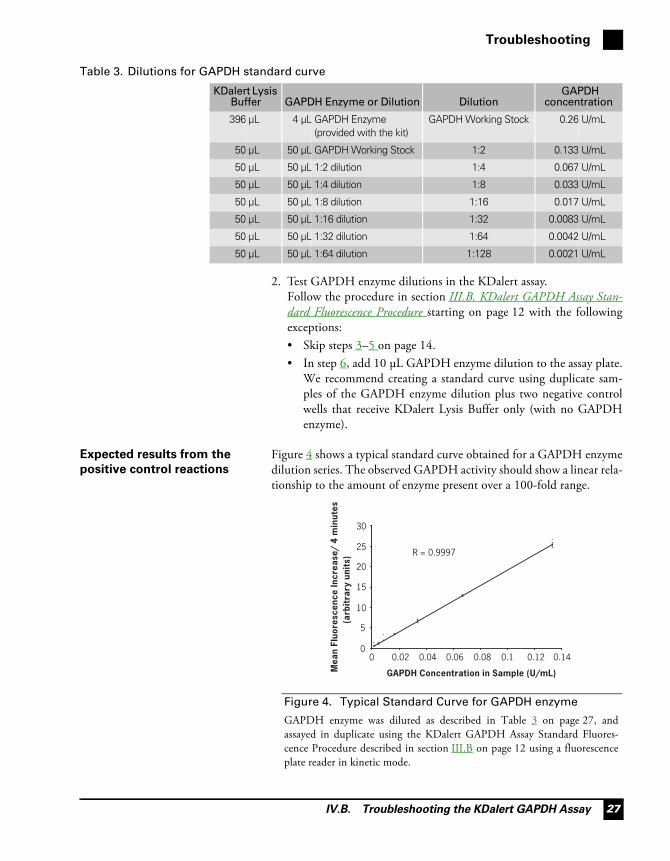
Figure 4 shows a typical standard curve obtained for a GAPDH enzymedilution series. The observed GAPDH activity should show a linear rela-tionship to the amount of enzyme present over a 100-fold range.
Table 3. Dilutions for GAPDH standard curve
KDalert Lysis Buffer GAPDH Enzyme or Dilution Dilution
GAPDH concentration
396 μL 4 μL GAPDH Enzyme (provided with the kit)
GAPDH Working Stock 0.26 U/mL
50 μL 50 μL GAPDH Working Stock 1:2 0.133 U/mL
50 μL 50 μL 1:2 dilution 1:4 0.067 U/mL
50 μL 50 μL 1:4 dilution 1:8 0.033 U/mL
50 μL 50 μL 1:8 dilution 1:16 0.017 U/mL
50 μL 50 μL 1:16 dilution 1:32 0.0083 U/mL
50 μL 50 μL 1:32 dilution 1:64 0.0042 U/mL
50 μL 50 μL 1:64 dilution 1:128 0.0021 U/mL
Figure 4. Typical Standard Curve for GAPDH enzyme
GAPDH enzyme was diluted as described in Table 3 on page 27, andassayed in duplicate using the KDalert GAPDH Assay Standard Fluores-cence Procedure described in section III.B on page 12 using a fluorescenceplate reader in kinetic mode.
R = 0.9997
0
5
10
15
20
25
30
0 0.02 0.04 0.06 0.08 0.1 0.12 0.14
GAPDH Concentration in Sample (U/mL)Mea
n Fl
uore
scen
ce In
crea
se/
4 m
inut
es
(arb
itra
ry u
nits
)
siRNA Starter Kit.book Page 27 Thursday, August 19, 2010 11:54 AM

Silencer® siRNA Starter Kit
IV.B. Troubleshooting the KDalert GAPDH Assay28
No GAPDH activity seen in
positive control reactions
If none of the diluted GAPDH enzyme reactions acquire a magentacolor after 15 min, there was most likely an error in preparing theKDalert Master Mix. We suggest repeating the positive control experi-ment.
No fluorescence increase
observed for either standard
curve or samples
If several of the diluted GAPDH enzyme reactions acquire a magentacolor after 15 min, but no fluorescence increase is detected by the fluo-rometer, check the settings for the appropriate excitation and emissionwavelengths.
Low GAPDH activity If the standard curve produces the expected result, but a low signal isseen from lysates that are expected to have relatively high GAPDHactivity (i.e., lysates from nontransfected cells or cultures transfectedwith Negative Control #1 siRNA), there is probably not enoughGAPDH in the cell extract used in the assay. • For the fluorescence assay:
– Increase the amount of cell lysate to 20 μL per reaction instep III.B.6 on page 14, and use only 80 μL of KDalert Master Mixin step III.B.7.
– If fewer than 2,000 cells were plated per well, decrease the volumeof KDalert Lysis Buffer used to lyse the cells step III.B.4 onpage 14 proportionately.
• For the colorimetric assay: If fewer than 4,000 cells were plated perwell, decrease the volume of KDalert Lysis Buffer used to lyse thecells proportionately.
No GAPDH gene knockdown If the standard curve produces the expected result, and all cell lysatesshow GAPDH activity, even those from cultures transfected with theGAPDH siRNA, consider the following possible causes and sugges-tions:• Problems with transfection: see next section C. No Detectable Gene
Silencing.• Too much cell lysate used in the KDalert reaction; this is indicated
when reactions from Negative Control #1-transfected samples turnmagenta in <5 min.– For the fluorescence assay: Reduce the lysate volume in the reaction
to 2 μL.– For the colorimetric assay: Reduce the lysate volume to 10 μL.
siRNA Starter Kit.book Page 28 Thursday, August 19, 2010 11:54 AM

IV.C. No Detectable Gene Silencing
Troubleshooting
29
C. No Detectable Gene Silencing
The transfection procedure
requires optimization
We strongly recommend that you optimize the transfection procedurefor each cell type using the GAPDH siRNA as described in section III.Fstarting on page 22.
Problems with
siRNA/transfection agent
complex formation
Follow the instructions for transfection complex formation closely;using the appropriate incubation times is important for good transfec-tion efficiency.
Serum, polyanions, or other inhibitors were present during
complexing.
Although siPORT NeoFX is compatible with serum during transfection,it is not compatible with serum during complex formation. UseOpti-MEM I reduced serum medium for siRNA/siPORT NeoFX trans-fection agent complex formation.
Do not overmix.
It is important to gently mix the siRNA with the diluted siPORTNeoFX in step II.B.2.d on page 10 of the reverse transfection procedure,or V.A.2.d on page 31 of the traditional transfection procedure.
Inactivated siPORT NeoFX Store siPORT NeoFX at 4°C. Tightly cap tubes, because evaporationcan significantly impact the activity of the transfection agents.
siRNA is degraded due to
poor handling or storage
Check the integrity of the siRNA by running 4 μL (~2.5 μg) on a non-denaturing 15–20% acrylamide gel. Visualize the siRNA by stainingwith ethidium bromide, and verify that it is the expected size and inten-sity. The siRNA should migrate as a fairly tight band; smearing wouldindicate degradation.
Cells have been subcultured
too many times or have
undergone changes
Transfect cells within 10 passages of the optimization experiments. Since cells may gradually change in culture, we recommend transfectingcells within 10 passages of determining optimal transfection conditions.If transfection efficiency begins to drop, fresh cells should be thawed forsubsequent experiments.
siRNA Starter Kit.book Page 29 Thursday, August 19, 2010 11:54 AM

Silencer® siRNA Starter Kit
IV.D. Gene Silencing Experiments Lack Reproducibility30
D. Gene Silencing Experiments Lack Reproducibility
Transfection complexes
were not adequately mixed
with cells
Distribute transfection agent/siRNA complex by gently rocking theplate back and forth. Do not swirl plates to mix, because this can con-centrate cells and/or reagents in the center of the wells.
There were differences in
the experimental procedure
The time of transfection after cell plating, incubation times, master mixvolumes, and the order of component addition can all affect transfectionefficiency. To obtain reproducible results in experiments involvingtransfection, conduct experiments exactly the same way every time.
Cell density is too low Optimize cell density as described in section III.F.5 on page 24. Whencell density is too low, cell cultures can become unstable. This instabilitycan vary from well to well because conditions (pH, temperature, etc.)may not be uniform across a multi-well plate, and can differentiallyinfluence unstable cultures.
Cells were passaged too
many times
Repeat experiment using cells that have been subcultured fewer times. Since cells may gradually change in culture, we recommend transfectingcells within 10 passages of determining optimal transfection conditions.If transfection efficiency begins to drop, fresh cells should be thawed forsubsequent experiments.
siRNA Starter Kit.book Page 30 Thursday, August 19, 2010 11:54 AM

V.A. Traditional “Pre-plating” Transfection Procedure
Appendix
31
V. Appendix
A. Traditional “Pre-plating” Transfection Procedure
The following procedure is a traditional “pre-plating” method. Itrequires more time than reverse transfection, but may be more effectivewith some cell types.
IMPORTANT
The volumes and amounts in the following procedure are for transfection in
a 24 well plate.
1. Cell plating a. Approximately 24 hr before transfection, plate cells in normal growthmedium so that they will be 30–80% confluent after 24 hr.
b. Incubate the cells overnight under normal cell culture conditions.
2. Prepare
siRNA/transfection agent
complexes
a. Just before using it, briefly vortex the siPORT NeoFX.
b. Dilute the siPORT NeoFX into Opti-MEM I medium.
i. In a sterile, round-bottom (or V-bottom) 96-well tissue culture dish or in sterile polystyrene tube, dilute 1–3 μL of siPORT NeoFX dropwise into Opti-MEM I for a final volume of 25 μL.
ii. Vortex well, and then incubate at room temp 10–15 min.
c. Dilute 0.25–7.5 μL of 2 μM siRNA (for a final concentration of1–30 nM) into Opti-MEM I for a final volume of 25 μL.
d. Add diluted siRNA to diluted siPORT NeoFX; mix by gently flickingthe tube or pipetting.
e. Incubate at room temp for 10–15 min.
Table 4. Approximate Reagent Amounts per Well.
Procedure step Reagent 96 well 24 well 6 well
1. Cell plating 0.2–1 x 10⁴ 2–10 x 104 1–5 x 10⁵
2. Prepare siRNA/transfection agent complexes
Dilute Transfection agent 0.15–1.2 μL 0.5–4 μL 3–9 μL
in Opti-MEM I to: to 10 μL to 25 μL to 100 μL
Dilute small RNA (20 μM)* 0.005–0.15 μL 0.025–0.75 μL 0.125–3.75 μL
in Opti-MEM I to: to 10 μL to 25 μL to 100 μL
3. Transfect cells Adjust medium in wells to: 80 μL 450 μL 2300 μL
Final transfection volume 100 μL 500 μL 2500 μL
Add fresh normal growth medium after 8–48 hr
100 μL 0.5–1 mL 1–3 mL
* This gives a final concentration of 1–30 nM. If not preparing a master mix, we recommend diluting the stock siRNA to 1–2 μM using nuclease-free water or Opti-MEM I for easier handling.
siRNA Starter Kit.book Page 31 Thursday, August 19, 2010 11:54 AM

Silencer® siRNA Starter Kit
V.B. Alternate KDalert Colorimetric Assay Procedure32
3. Transfect cells a. Adjust the volume of normal growth medium in wells containingcells to 450 μL.
b. Add the siPORT NeoFX/siRNA complex from step 2.e dropwise tothe cells (the final transfection volume will be 500 μL).
c. Without swirling, gently rock the dish back and forth to evenlydistribute the complexes.
d. Incubate cells under normal cell culture conditions for 48 hr.
e. 0.5–1 mL fresh normal growth medium may be added to each wellafter 8–48 hr to maximize cell growth and prevent potentialcytotoxicity.
B. Alternate KDalert Colorimetric Assay Procedure
Notes about the colorimetric
assay
• The colorimetric assay is slightly less sensitive than the fluorescenceassay, so the reaction volume is increased from 100 μL to 200 μL toimprove the signal.
• A water + KDalert Master Mix control is used as the reference fordetermining GAPDH activity.
• This procedure is written for assays conducted in 96-well plates.
Materials not provided with
the assay
• Clear polystyrene 96 well plates. We recommend BD FalconCat #353072 or Greiner Bio-One CellStar Cat #655180.
• UV/vis plate reader.• General lab equipment and supplies such as 15 mL or microcentri-
fuge tubes, multichannel pipettors, vortex mixer, ice, etc.
Colorimetric assay
procedure
1. Set up the visible plate reader to read the absorbance of each sampleat 615 nm.
2. Prepare enough KDalert Master Mix to use 180 μL per reaction.3. 48 hr after transfection, remove the culture medium from the cells.
The plate can be stored at –80°C. Thaw frozen cells on ice beforeproceeding.
4. Add 100 μL KDalert Lysis Buffer to each sample. (For 24-well platesadd 500 μL KDalert Lysis Buffer; for 6-well plates, add 2 mLKDalert Lysis Buffer.)
5. Incubate on ice or at 4°C for 20 min.6. Pipet the cell lysate up and down 4–5 times (or shake plate for 20 sec
at room temp) to homogenize the lysate.7. Transfer 20 μL aliquots of each cell extract, GAPDH Enzyme dilu-
tion including Working Stock (for positive control reac-tion–described in section IV.B starting on page 26), or water(water + Master Mix control) to wells in a clean 96-well microplate.
siRNA Starter Kit.book Page 32 Thursday, August 19, 2010 11:54 AM

V.B. Alternate KDalert Colorimetric Assay Procedure
Appendix
33
We recommend using duplicate aliquots of each GAPDH Enzymedilution or water + Master Mix control.
8. Add 180 μL of KDalert Master Mix, and allow the reaction to pro-ceed at room temp for 15 min.
9. Measure the absorbance of each reaction at 615 nm. Use a water ref-erence blank in the measurement.GAPDH activity is indicated by a reduction in A615.Some samples may acquire a magenta color over 15 min.
Determining optimal
transfection conditions
using colorimetric data
Calculating GAPDH activity using colorimetric data
During the assay, GAPDH activity causes a reduction in A615 of thesamples, although they may eventually acquire a magenta color. For thecolorimetric data, subtract the A615 for each sample (A615-sample) from theA615 of the water + Master Mix control (A615-WMM) to determine aGAPDH activity, ΔA615-sample, for that sample:
ΔA615-sample = A615-WMM – A615-sample
For example, the GAPDH activity ΔA615-Neg is obtained by subtractingA615-WMM from A615 for negative control siRNA-transfected cells(A615-Neg):
ΔA615-Neg #1 = A615-WMM – A615-Neg #1
Construction of the GAPDH Enzyme standard curve
A graph of ΔA615, calculated as described above, for each enzyme dilu-tion should yield a line with a positive slope similar to that yielded byfluorescence data, e.g., Figure 4 on page 27.
Calculating % remaining gene expression and % knockdown of
GAPDH expression
Knockdown is calculated in the same way as for the fluorescence-basedassay, once the GAPDH activity, ΔA615, is calculated. For each transfec-tion condition, the % remaining expression and % knockdown is calcu-lated from the ratio of ΔA615 for samples transfected with GAPDHsiRNA to ΔA615 for samples transfected with negative control siRNA, asfollows.
% remaining expression 100 x ΔA615-GAPDH
ΔA615 -Neg #1----------------------=
% knockdown 100 100 x ΔA615-GAPDH
ΔA615 -Neg #1----------------------( )–=
siRNA Starter Kit.book Page 33 Thursday, August 19, 2010 11:54 AM
Silencer® siRNA Starter Kit
V.C. Quality Control34
Measuring transfection associated toxicity
For each transfection condition, calculate the GAPDH activity, ΔA615,as described above. Then compare the GAPDH activity in cells trans-fected with Negative Control #1 siRNA (ΔA615-Neg #1) to that in non-transfected cells (ΔA615-Nontransfected) to examine the cytotoxic effects ofthe transfection conditions. Transfection conditions which do not causeany cytotoxicity would result in a ratio of 1, therefore, the closer thisratio is to 1, the less cytotoxic the transfection conditions.
Calculating the best transfection condition tested
Typically, there is an inverse relationship between transfection effi-ciency (and associated gene knockdown) and cell viability. A useful wayto describe the balance of transfection efficiency and cytotoxicity is theterm Optimal Balance Factor, or OBF. The OBF for each transfectioncondition is calculated with colorimetric data in the same way as for flu-orescence data:
OBF = % knockdown X cytotoxicity ratio
Typically, optimal transfection condition(s) are those which exhibit thehighest OBF value.
C. Quality Control
Functional testing Cells were transfected with GAPDH siRNA or Negative ControlsiRNA using siPORT NeoFX. When measuring protein activity usingKDalert, cells transfected with the GAPDH siRNA are shown to have a50% reduction in GAPDH RNA signal compared to cells transfectedwith the Negative Control siRNA. A GAPDH Enzyme standard curvewith an R-value of >0.94 was obtained.
RNase activity testing The siPORT NeoFX and siPORT Amine are tested for RNase usingAmbion RNaseAlert® assay.
Nuclease testing Relevant kit components are tested in the following nuclease assays:
RNase activity
Meets or exceeds specification when a sample is incubated with labeledRNA and analyzed by PAGE.
cytotoxicity ratio =
ΔA615-Neg #1
ΔA615-Nontransfected
OBF = 100 100 x ΔA615-GAPDHΔA615 -Neg #1---------------------------------⎝ ⎠
⎛ ⎞ ×–ΔA615-Neg #1
ΔA615-Nontransfected
siRNA Starter Kit.book Page 34 Thursday, August 19, 2010 11:54 AM

V.D. Safety Information
Appendix
35
D. Safety Information
Chemical safety guidelines To minimize the hazards of chemicals:• Read and understand the Material Safety Data Sheets (MSDS) pro-
vided by the chemical manufacturer before you store, handle, orwork with any chemicals or hazardous materials.
• Minimize contact with chemicals. Wear appropriate personal protec-tive equipment when handling chemicals (for example, safety gog-gles, gloves, or protective clothing). For additional safety guidelines,consult the MSDS.
• Minimize the inhalation of chemicals. Do not leave chemical con-tainers open. Use only with adequate ventilation (for example, fumehood). For additional safety guidelines, consult the MSDS.
• Check regularly for chemical leaks or spills. If a leak or spill occurs,follow the manufacturer’s cleanup procedures as recommended onthe MSDS.
• Comply with all local, state/provincial, or national laws and regula-tions related to chemical storage, handling, and disposal.
About MSDSs Chemical manufacturers supply current Material Safety Data Sheets(MSDSs) with shipments of hazardous chemicals to new customers.They also provide MSDSs with the first shipment of a hazardous chem-ical to a customer after an MSDS has been updated. MSDSs provide thesafety information you need to store, handle, transport, and dispose ofthe chemicals safely.
Each time you receive a new MSDS packaged with a hazardous chemi-cal, be sure to replace the appropriate MSDS in your files.
Obtaining the MSDS To obtain Material Safety Data Sheets (MSDSs) for any chemical prod-uct supplied by Applied Biosystems or Ambion:• At www.appliedbiosystems.com, select Support, then MSDS.
Search by chemical name, product name, product part number, orMSDS part number. Right-click to print or download the MSDS ofinterest.
• At www.ambion.com, go to the web catalog page for the product ofinterest. Click MSDS, then right-click to print or download.
• E-mail ([email protected]) or tele-phone (650-554-2756; USA) your request, specifying the catalog orpart number(s) and the name of the product(s). We will e-mail theassociated MSDSs unless you request fax or postal delivery. Requestsfor postal delivery require 1–2 weeks for processing.
For the MSDSs of chemicals not distributed by Applied Biosystems orAmbion, contact the chemical manufacturer.
siRNA Starter Kit.book Page 35 Thursday, August 19, 2010 11:54 AM