siadh, di and cerebral salt...
TRANSCRIPT

SIADH, DI and Cerebral Salt Wasting
Karim Rafaat, MD
ADH
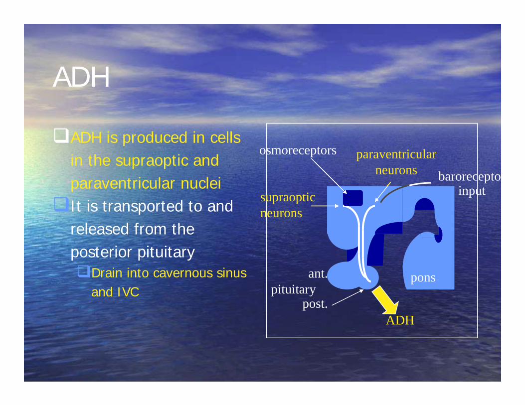
ADH is produced in cells in the supraoptic and paraventricular nuclei
It is transported to and released from the posterior pituitaryDrain into cavernous sinus
and IVC
supraoptic neurons
paraventricular neurons
osmoreceptors
pons
baroreceptorinput
ant.
ADH
pituitarypost.

Secretion of ADH
• Most important variable is osmotic pressure of plasma and ECF.– Largely a function of the
concentration of Na• Osmoregulation mediated
by osmoreceptors in the anteromedial hypothalamus near the suraoptic nucleus– Blood supply is via
fenestrated capillaries, and so they are outside BBB
supraoptic neurons
paraventricular neurons
osmoreceptors
pons
baroreceptorinput
ant.
ADH
pituitarypost.

ADH secretion
• Osmoreceptors are sensitive to small changes in plasma osmolality
• A decrease in osmolality of 1-2% will maximally and rapidly suppress ADH levels to allow diuresis
• The osmostat set point in adults is a plasma osmolality of 275-295 mOsm/kg
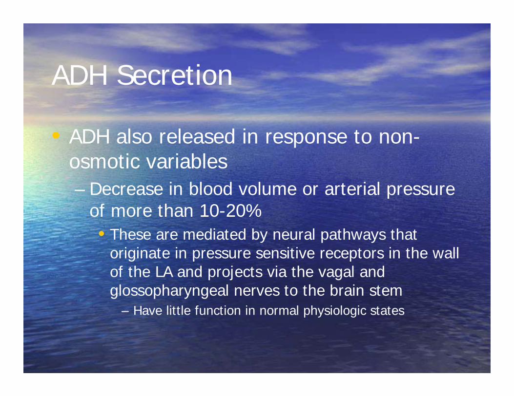
ADH Secretion
• ADH also released in response to non-osmotic variables– Decrease in blood volume or arterial pressure
of more than 10-20%• These are mediated by neural pathways that
originate in pressure sensitive receptors in the wall of the LA and projects via the vagal and glossopharyngeal nerves to the brain stem
– Have little function in normal physiologic states

ADHcAMP
ATP
Protkinase
H2OH2O
H2OH2O
RecAC
Tubule
ADH Function In the collecting duct blood-borne ADH binds to a V2 receptor on the baso-lateral surface of the principal tubule cell.
This stimulates adenylyl cyclase to generate cAMP and activate protein kinases.
This increases the insertion of water channels, aquaporins, into the apical surface of the cell and so increase water permeability
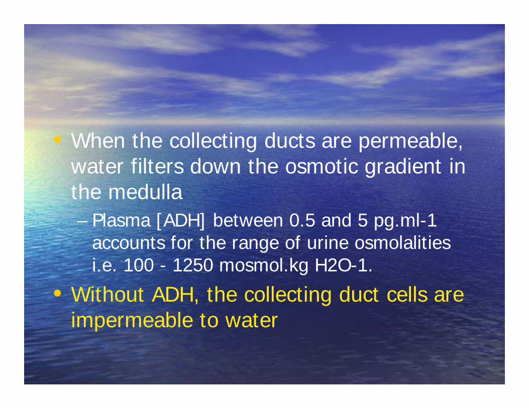
• When the collecting ducts are permeable, water filters down the osmotic gradient in the medulla– Plasma [ADH] between 0.5 and 5 pg.ml-1
accounts for the range of urine osmolalities i.e. 100 - 1250 mosmol.kg H2O-1.
• Without ADH, the collecting duct cells are impermeable to water
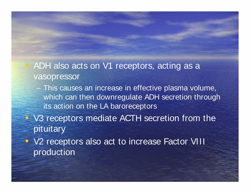
• ADH also acts on V1 receptors, acting as a vasopressor– This causes an increase in effective plasma volume,
which can then downregulate ADH secretion through its action on the LA baroreceptors
• V3 receptors mediate ACTH secretion from the pituitary
• V2 receptors also act to increase Factor VIII production

Distribution and Clearance of ADH
• ADH circulates in an unbound state and equilibrates within minutes between plasma and ECF
• Average half life is 20 minutes• 15-20% is filtered by glomerulus and then
either destroyed or excreted.• The rest is degraded in the liver

ADH and Thirst: salt and water balance
• ADH function is limited, because it cannot prevent water loss in excess of that needed to excrete a certain solute load.– Another mechanism is needed to ensure that
these, and other losses are replaced…• THIRST

ADH and Thirst: salt and water balance
Plasma osmolality ≈ 290 mosmoles. kg H2O-1.
water lackincreased osmolality
excessive water intakedecreased osmolality
drinking
osmoreceptors
supraopticPVN nuclei
supraopticPVN nuclei
lateral pre-optic area
lateral pre-optic area
ADH releaseincreased
ADH releasereduced
thirst enhanced
thirst depressed
collecting ductmore permeable
collecting ductless permeable
water retentionby kidneys
water lossby kidneys
osmoreceptors
Disorders of ADH secretion or action

Diabetes Insipidus
Diabetes Insipidus
• Characterized by – A large volume of urine (diabetes)– That is hypotonic, dilute and tasteless
(insipid)• Versus the sweet urine of diabetes mellitus
(honey)
Etiology
• Four fundamentally distinct defects– Central DI, a primary deficiency of ADH– Nephrogenic DI, caused by an inappropriate
renal response to ADH– Transient DI of pregnancy, caused by
vasopressinase produced by placenta– Primary Polydipsia, in which pathology is
secondary to ingestion, rather than elimination of, fluid
Central DI
• Most common• Usually secondary to irreversible
destruction of >80% ADH producing neurons
• Genetic forms caused by accumulation of poorly folded precursor proteins
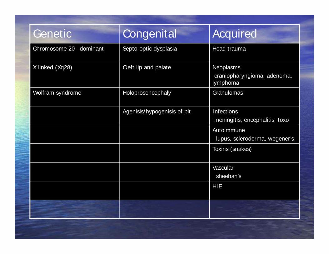
Genetic Congenital AcquiredChromosome 20 –dominant Septo-optic dysplasia Head trauma
X linked (Xq28) Cleft lip and palate Neoplasmscraniopharyngioma, adenoma,
lymphoma
Wolfram syndrome Holoprosencephaly Granulomas
Agenisis/hypogenisis of pit Infectionsmeningitis, encephalitis, toxo
Autoimmunelupus, scleroderma, wegener’s
Toxins (snakes)
Vascularsheehan’s
HIE
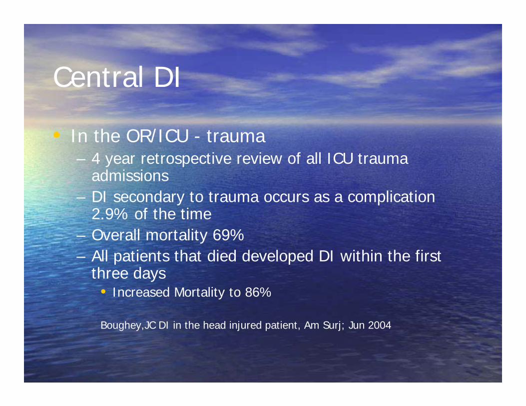
Central DI
• In the OR/ICU - trauma– 4 year retrospective review of all ICU trauma
admissions– DI secondary to trauma occurs as a complication
2.9% of the time– Overall mortality 69%– All patients that died developed DI within the first
three days• Increased Mortality to 86%
Boughey,JC DI in the head injured patient, Am Surj; Jun 2004
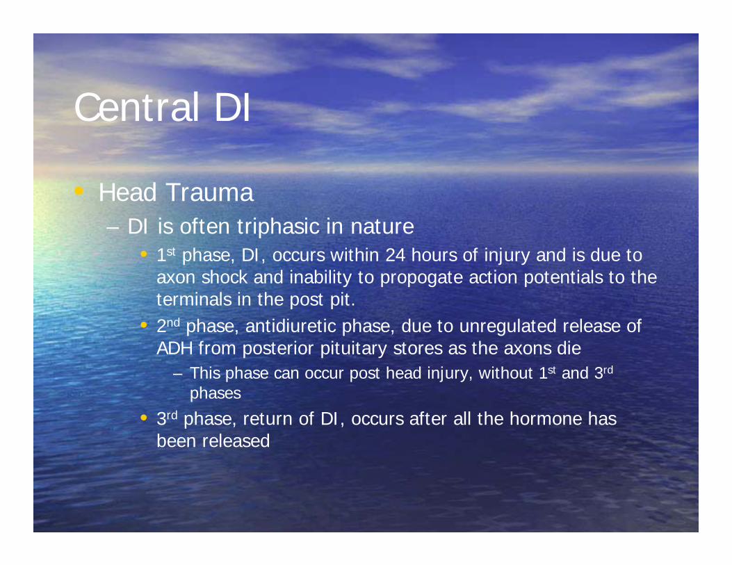
Central DI
• Head Trauma– DI is often triphasic in nature
• 1st phase, DI, occurs within 24 hours of injury and is due to axon shock and inability to propogate action potentials to the terminals in the post pit.
• 2nd phase, antidiuretic phase, due to unregulated release of ADH from posterior pituitary stores as the axons die
– This phase can occur post head injury, without 1st and 3rd
phases
• 3rd phase, return of DI, occurs after all the hormone has been released
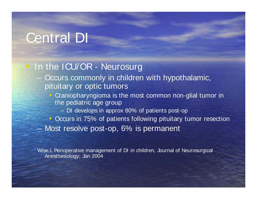
Central DI
• In the ICU/OR - Neurosurg– Occurs commonly in children with hypothalamic,
pituitary or optic tumors• Craniopharyngioma is the most common non-glial tumor in
the pediatric age group– DI develops in approx 80% of patients post-op
• Occurs in 75% of patients following pituitary tumor resection– Most resolve post-op, 6% is permanent
Wise,L Perioperative management of DI in children, Journal of Neurosurgical Anesthesiology; Jan 2004

Nephrogenic DI
• Caused by a poor response of the kidney to ADH
• Most common form is congenital• Other forms are acquired

Genetic Acquired
X linked (V2 gene) Drugslithium, Ampho B, rifampin, cisplatin,
foscarnet
Recessive (aquaporin gene) Metabolichypercalcemia, hypercalcuria,
hypokalemia
Vascularsickle cell disease, ischemia
Granuloma
Neoplasm
Pathophysiology
• Lack of ADH, or ADH response, leads to a deficiency in urine concentration– 1-2% decrease in TBW, and a rise in plasma
osmolality and Na• The latter cause a thirst response, which, if intact,
stabilizes osmo’s and Na
• Polyuria further impairs concentrating ability through washout of medullary concentration gradient
Diagnosis
• Lots of stuff about fluid restriction, ADH levels, DDAVP trials, MRI’s….not useful for us– Useful for distinguishing between nephrogenic
and central forms• Endocrine consult
• There is a much easier way to get what we need, and is based on simple pathophysiology
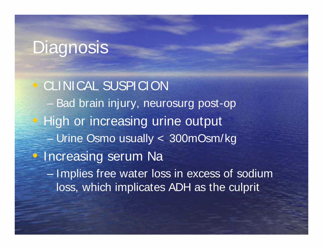
Diagnosis
• CLINICAL SUSPICION – Bad brain injury, neurosurg post-op
• High or increasing urine output– Urine Osmo usually < 300mOsm/kg
• Increasing serum Na– Implies free water loss in excess of sodium
loss, which implicates ADH as the culprit

Treatment
• Vasopressin– 5-10 mUnits/kg/hr– Titrate to a level that normalizes urine output and stabilizes
serum Na• DDAVP
– For chronic correction– More resistant than vasopressin to degradation– 5-20 micrograms/day by nasal spray
• Oral and SQ forms available• If nephrogenic
– Sodium restriction and mild diuresis to encourage Na elimination– Gene therapy in the works…..

SIADH

SIADH
• Characterized by– Hypotonic hyponatremia – impaired urinary dilution in the face of hypoosmolality
• Urine osmolality >100mOsm/kg
– In the absence of:• Hypovolemia• Hypotension• Adrenal insufficiency• Protracted emesis

SIADH
• One of the most common causes of hyponatremia in children and adults, in the hospital setting
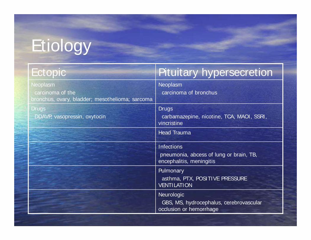
EtiologyEctopic Pituitary hypersecretionNeoplasm
carcinoma of the bronchus, ovary, bladder; mesothelioma; sarcoma
Neoplasmcarcinoma of bronchus
DrugsDDAVP, vasopressin, oxytocin
Drugscarbamazepine, nicotine, TCA, MAOI, SSRI,
vincristine
Head Trauma
Infectionspneumonia, abcess of lung or brain, TB,
encephalitis, meningitis
Pulmonaryasthma, PTX, POSITIVE PRESSURE
VENTILATION
NeurologicGBS, MS, hydrocephalus, cerebrovascular
occlusion or hemorrhage
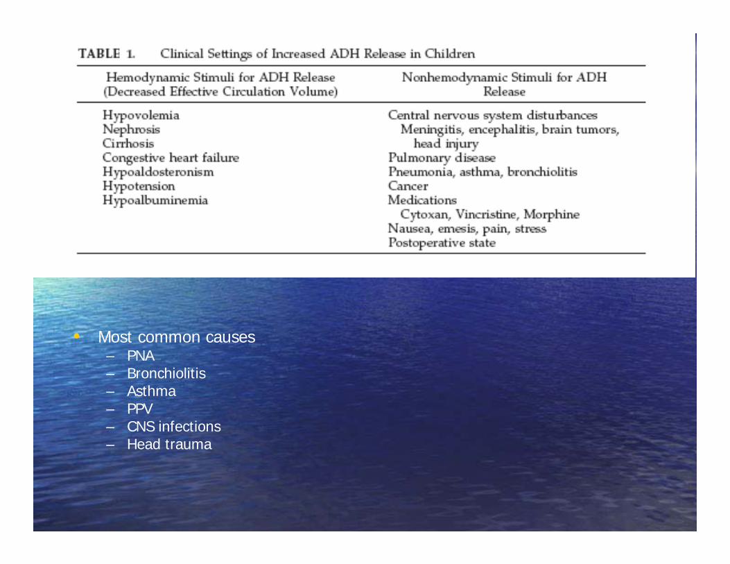
• Most common causes– PNA– Bronchiolitis– Asthma– PPV– CNS infections– Head trauma

Pathophysiology
• ADH cannot significantly lower plasma Na, unless water input > output
• When this occurs, the excess ADH makes a making a dilute urine difficult
• Water accumulates in ECF and ICF compartments• Conc of Na and other solutes decreases• Renin and aldosterone activity decreases
– Increases natriuresis as a response to expansion of ECF volume• Natriuresis aggravates dilutional hyponatremia
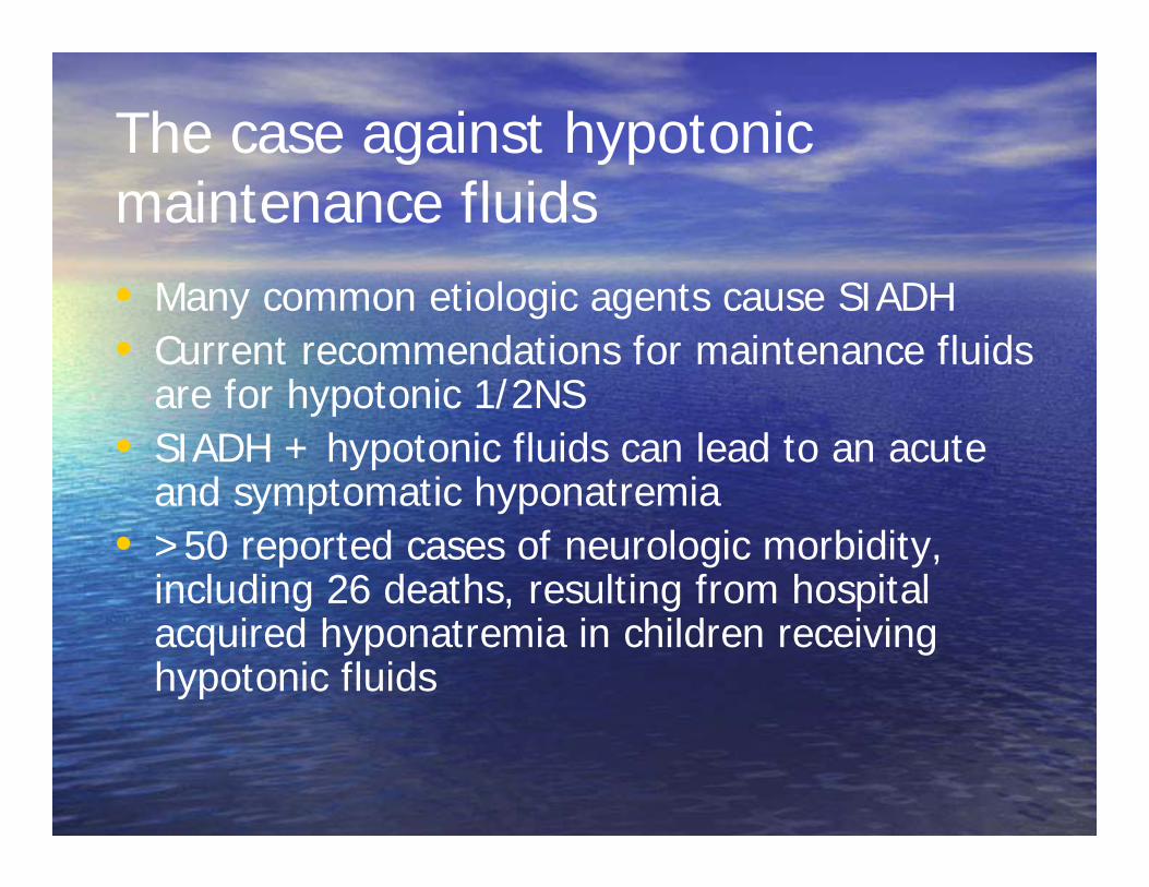
The case against hypotonic maintenance fluids• Many common etiologic agents cause SIADH• Current recommendations for maintenance fluids
are for hypotonic 1/2NS• SIADH + hypotonic fluids can lead to an acute
and symptomatic hyponatremia• >50 reported cases of neurologic morbidity,
including 26 deaths, resulting from hospital acquired hyponatremia in children receiving hypotonic fluids

The case against hypotonic maintenance fluids
• Children at greater risk for symptomatic hyponatremia than adults– Encephalopathy occurs at higher values in
kids• Higher brain to skull ratio leaves less room for
expansion

The case against hypotonic maintenance fluids• Moritz et al, Prevention of hospital acquired
hyponatremia: a case for using isotonic saline, Pediatrics Aug 2003– “isotonic saline in 5% dextrose is the safest fluid composition”
• Acute Hyponatremia Related to IV fluid administration in hospitalized children: An observational study– 1586 patients admitted to hospital; 40 developed hyponatremia
– 2 with major neurologic sequalae, 1 death– These patients received sig. more free water than age matched
controls– Support isotonic fluids for maintenance
Differential Diagnosis
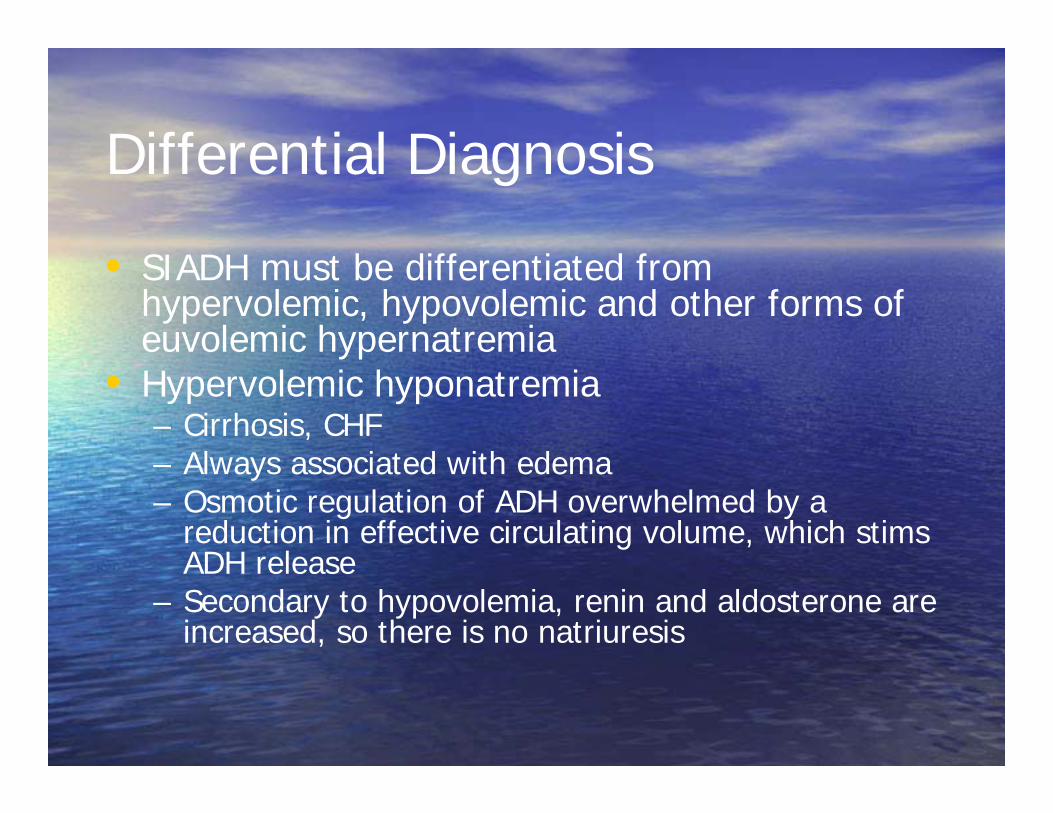
• SIADH must be differentiated from hypervolemic, hypovolemic and other forms of euvolemic hypernatremia
• Hypervolemic hyponatremia– Cirrhosis, CHF– Always associated with edema– Osmotic regulation of ADH overwhelmed by a
reduction in effective circulating volume, which stims ADH release
– Secondary to hypovolemia, renin and aldosterone are increased, so there is no natriuresis

Differential Diagnosis
• Hypovolemic hyponatremia– Diuretic abuse, AGE, mineralocorticoid
deficiency– Excessive loss of Na and H2O– Associated with signs of hypovolemia:
tachycardia, hypotension

Diagnosis
•Urine osmolality >100mOsm/kg of water during hypotonicity•Urine sodium concentration >40 mEq/L with normal dietary salt intake•Euvolemic hyponatremia <134, Plasma Osmolarity <275mOsm/L
Treatment
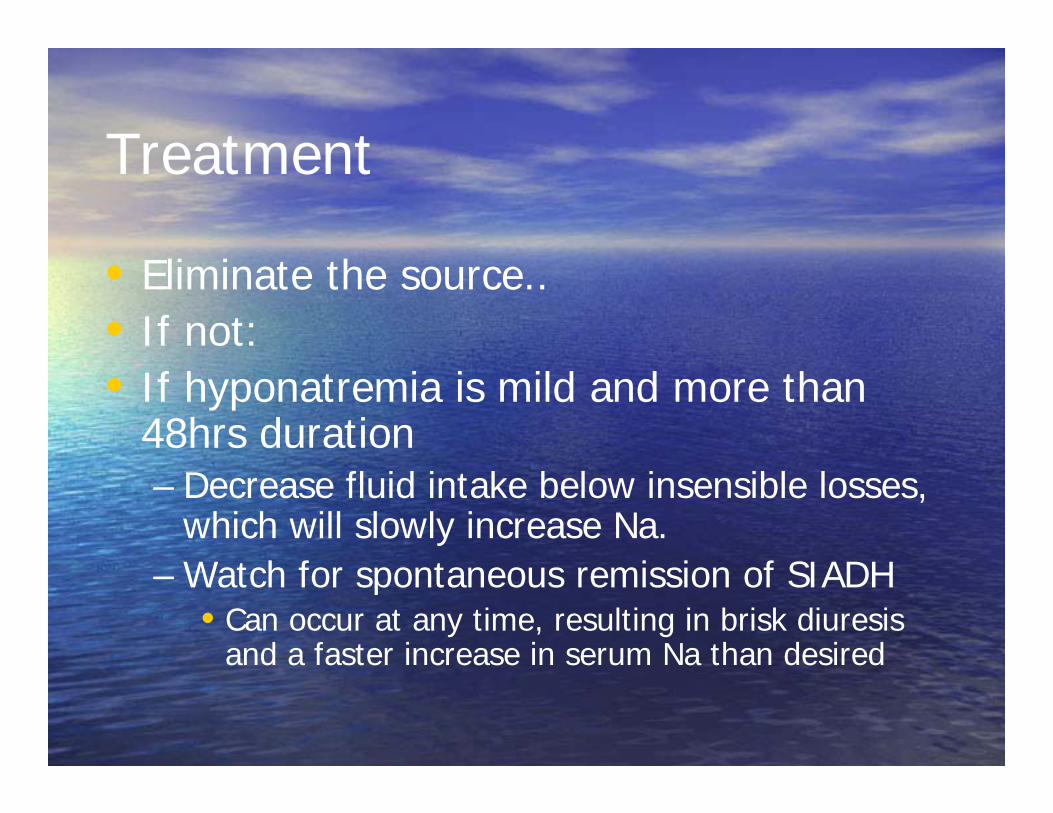
• Eliminate the source..• If not:• If hyponatremia is mild and more than
48hrs duration– Decrease fluid intake below insensible losses,
which will slowly increase Na.– Watch for spontaneous remission of SIADH
• Can occur at any time, resulting in brisk diuresis and a faster increase in serum Na than desired

Treatment
• If hyponatremia is severe (accompanied with neuro symptoms) or of less than 48 hours duration– Slow 3% infusion– Raise Na to point of cessation of symptoms– After above, no faster than 2 mEq/L per hour

Cerebral Salt Wasting
Harrigan, Mark Cerebral salt wasting syndrome Critical Care Clinics, Jan 2001
Definition
• A diagnosis of exclusion• Requires:
– A cerebral lesion– Renal Na and Cl wasting
• Implies that above were excreted WITHOUT physiologic stimulus
– Which means you need to have:
– A contracted effective arterial blood volume
• So patient must not have – A physiologic cause for Na and Cl excretion
• an expanded effective circulating arterial volume
– A condition that may cause a deficiency of Na resorption • Aldosterone deficiency• diuretics

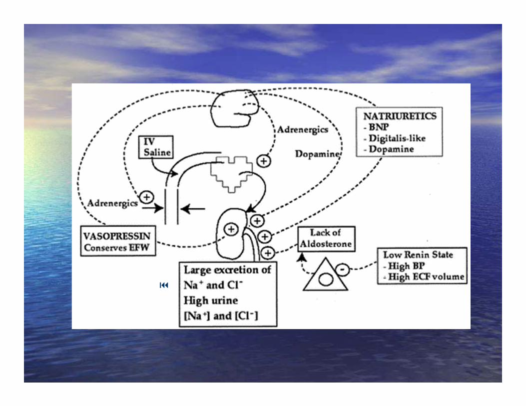
Pathophysiology
• Natriuretic Factors• Direct Neural effects
Pathophysiology –Natriuretic Factors• ANP
– 28 amino acid polypeptide– Effects:
• Natriuresis• Diuresis• Vasodilation• Suppression of renin and aldosterone secretion

Pathophysiology –Natriuretic Factors• ANP
– Released in response to atrial stretch– CNS modulates ANP secretion
• lesions of specific areas in hypothalamus decrease cardiac secretion of ANP in response to volume expansion
• So, intracranial disease may lead to a disturbance in its control over ANP secretion
– 6/8 neurosurg pts had elevated ANP levels, with a correlation between ANP level and degree of hyponatremia
– After SAH, ADH and ANP levels elevated on days 0-2, ANP levels remained high after one week, when ADH levels declined SIADH and CSW may co-exist, complicating diagnosis
Pathophysiology –Natriuretic Factors• ANP
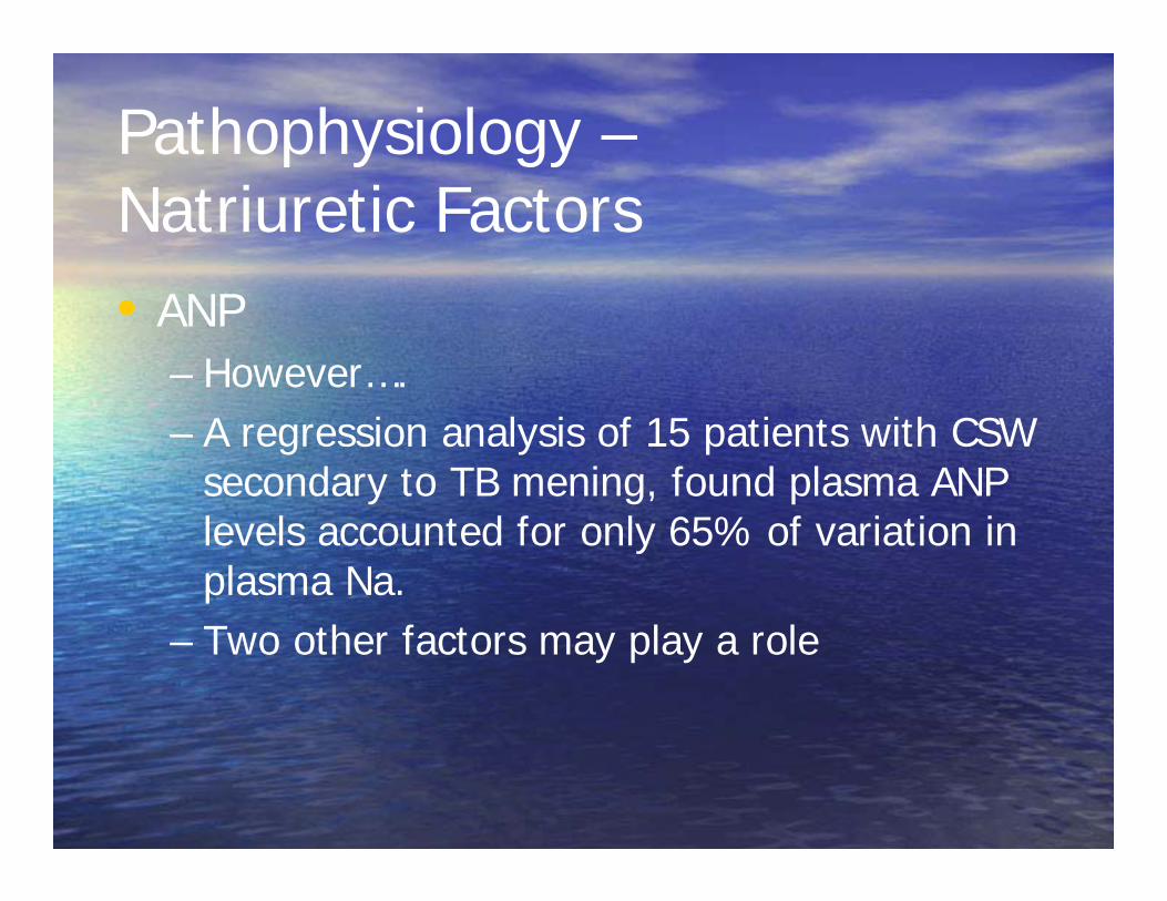
– However….– A regression analysis of 15 patients with CSW
secondary to TB mening, found plasma ANP levels accounted for only 65% of variation in plasma Na.
– Two other factors may play a role
• BNP– Of cardiac ventricular origin
• Also localized in hypothalamus– Secreted in response to increased pressure or stretch– Similar effects to ANP– BNP levels are elevated in patients with SAH– Acute intracranial disease may cause release of
cardiac BNP, and hypothalamic damage may also release BNP
• Plasma Natriuretic factor– ?

A little more on BNP……
• 40 patients with SAH, BNP levels measured– Change in, not absolute value of, BNP level independently
associated with hyponatremia– In those in whom vasospasm developed, BNP levels increased
5.4x within 24 hours, and 11.2x within 3 days• BNP level independently associated with vasospasm• Increase in BNP did not precede vasospasm, but increased
concurrently– May be a good marker….
– Patients with increasing BNP levels after admission had NO CHANGE in their admit GCS after two weeks
– Patients with no change, or decrease in BNP, had a 3 point improvement in GCS 2 weeks after admission
McGirt, MJ CORRELATION OF SERUM BRAIN NATRIURETIC PEPTIDE WITH HYPONATREMIA AND DELAYED ISCHEMIC NEUROLOGICAL DEFICITS AFTER SUBARACHNOID HEMORRHAGE, Neurosurgery June 2004
Direct Neural Effects
• Generalized hyperactivity of the sympathetic nervous system occurs after SAH– Sustained activity leads to a decrease in plasma
volume and blood volume
• Acute brain injury may lead to an interruption in sympathetic flow to kidneys– Increases glomerular blood flow, GFR and thus
diuresis– Decreases renin release, and promotes natriuresis

Diagnosis
• Because those patients at risk for CSW, are also at risk for SIADH, the two must be distinguished…

CSW SIADH*Hypovolemiaclinical dehydrationlow CVP
*Euvolemia or hypervolemia
Negative salt balanceMarkedly elevated Una Variable UnaElevated K with hypoNa Normal KSerum ADH and ANP not helpful
• Likely that SIADH and CSW, in the setting of head injury/surgery, represent two opposite ends of a spectrum:
• pt’s probably have both exaggerated natriuresis and elevated ADH release, and which effect predominates depends on relative intensities

Treatment
• Treat underlying process• Volume and sodium replacement• NS, 3% NaCl, or enteral salt for restoring NaCl
balance– Enteral salt preferable if euvolemia already restored
• Volume expansion can lead to increased aldosterone, which can then promote more Na loss
• Restore Euvolemia• Fludrocortisone
– Works on renal tubule to increase Na reabsorption– 0.05-0.1 mg/day