sherlock’s diseases of the liver and biliary...
TRANSCRIPT


SHERLOCK’S DISEASES OF THE LIVER AND BILIARY SYSTEM

Companion Website
This book has a companion website
www.wiley.com/go/sherlock/liver
with:
• All 700 fi gures and captions in the book as Powerpoints for downloading

Sherlock’s Diseases of the Liver and Biliary SystemEDITED BY
JAMES S. DOOLEYCentre for HepatologyUniversity College London Medical School andRoyal Free Sheila Sherlock Liver CentreRoyal Free HospitalLondonUK
ANNA S. F. LOKDivision of GastroenterologyUniversity of Michigan Health SystemAnn ArborUSA
ANDREW K. BURROUGHSRoyal Free Sheila Sherlock Liver CentreRoyal Free Hospital;University College LondonLondonUK
E. JENNY HEATHCOTEDivision of Gastroenterology University Health Network University of TorontoTorontoOntarioCanada
12TH EDITION
A John Wiley & Sons, Ltd., Publication

This edition fi rst published 2011, © 1963, 1968, 1975, 1981, 1985, 1989, 1993, 1997, 2002, 2011 by Blackwell Publishing Ltd
Blackwell Publishing was acquired by John Wiley & Sons in February 2007. Blackwell’s publishing program has been merged with Wiley’s global Scientifi c, Technical and Medical business to form Wiley-Blackwell.
First published 1955Second edition 1958Third edition 1963Fourth edition 1968Fifth edition 1975Sixth edition 1981Seventh edition 1985Eighth edition 1989Ninth edition 1993Tenth edition 1997Eleventh edition 2002
Registered offi ce: John Wiley & Sons Ltd, The Atrium, Southern Gate, Chichester, West Sussex, PO19 8SQ, UK
Editorial offi ces: 9600 Garsington Road, Oxford, OX4 2DQ, UKThe Atrium, Southern Gate, Chichester, West Sussex, PO19 8SQ, UK111 River Street, Hoboken, NJ 07030-5774, USA
For details of our global editorial offi ces, for customer services and for information about how to apply for permission to reuse the copyright material in this book please see our website at www.wiley.com/wiley-blackwell
The right of the author to be identifi ed as the author of this work has been asserted in accordance with the Copyright, Designs and Patents Act 1988.
All rights reserved. No part of this publication may be reproduced, stored in a retrieval system, or transmitted, in any form or by any means, electronic, mechanical, photocopying, recording or otherwise, except as permitted by the UK Copyright, Designs and Patents Act 1988, without the prior permission of the publisher.
Wiley also publishes its books in a variety of electronic formats. Some content that appears in print may not be available in electronic books.
Designations used by companies to distinguish their products are often claimed as trademarks. All brand names and product names used in this book are trade names, service marks, trademarks or registered trademarks of their respective owners. The publisher is not associated with any product or vendor mentioned in this book. This publication is designed to provide accurate and authoritative information in regard to the subject matter covered. It is sold on the understanding that the publisher is not engaged in rendering professional services. If professional advice or other expert assistance is required, the services of a competent professional should be sought.
The contents of this work are intended to further general scientifi c research, understanding, and discussion only and are not intended and should not be relied upon as recommending or promoting a specifi c method, diagnosis, or treatment by physicians for any particular patient. The publisher and the author make no representations or warranties with respect to the accuracy or completeness of the contents of this work and specifi cally disclaim all warranties, including without limitation any implied warranties of fi tness for a particular purpose. In view of ongoing research, equipment modifi cations, changes in governmental regulations, and the constant fl ow of information relating to the use of medicines, equipment, and devices, the reader is urged to review and evaluate the information provided in the package insert or instructions for each medicine, equipment, or device for, among other things, any changes in the instructions or indication of usage and for added warnings and precautions. Readers should consult with a specialist where appropriate. The fact that an organization or Website is referred to in this work as a citation and/or a potential source of further information does not mean that the author or the publisher endorses the information the organization or Website may provide or recommendations it may make. Further, readers should be aware that Internet Websites listed in this work may have changed or disappeared between when this work was written and when it is read. No warranty may be created or extended by any promotional statements for this work. Neither the publisher nor the author shall be liable for any damages arising herefrom.
Library of Congress Cataloging-in-Publication Data
Sherlock’s diseases of the liver and biliary system / edited by James S. Dooley ... [et al.]. – 12th ed. p. ; cm. Diseases of the liver and biliary system Rev. ed. of: Diseases of the liver and biliary system / Sheila Sherlock. 11th ed. 2002. Includes bibliographical references and index. ISBN 978-1-4051-3489-7 (hardcover : alk. paper) 1. Liver–Diseases. 2. Biliary tract–Diseases. I. Dooley, James (James S.) II. Sherlock, Sheila, Dame. Diseases of the liver and biliary system. III. Title: Diseases of the liver and biliary system. [DNLM: 1. Liver Diseases. 2. Biliary Tract Diseases. WI 700] RC845.S52 2011 616.3’6–dc22 2010039149
A catalogue record for this book is available from the British Library.
This book is published in the following electronic formats: ePDF 9781444341263; Wiley Online Library 9781444341294; ePub 9781444341270; Mobi 9781444341287
Set in 9.5/12 pt Palatino by Toppan Best-set Premedia Limited
01 2011

v
List of Contributors, xi
Preface to the Twelfth Edition, xv
Preface to the First Edition, xvi
1 Anatomy and Function, 1Jay H. Lefkowitch
Development of the liver and bile ducts, 1Anatomy of the liver, 1Functional liver anatomy: sectors and segments,
3Anatomical abnormalities of the liver, 4Anatomy of the biliary tract, 5Surface marking, 6Methods of examination, 6Microanatomy of the liver, 7Hepatic ultrastructure (electron microscopy) and
organelle functions, 11Functional heterogeneity of the liver, 15Dynamics of the hepatic microenvironment in
physiology and disease, 16Hepatocyte death and regeneration, 17References, 18
2 Assessment of Liver Function, 20Sandeep Mukherjee & John L. Gollan
Selection of biochemical tests, 20Bile pigments, 21Serum enzyme tests, 22Quantitative assessment of hepatic function,
25Lipid and lipoprotein metabolism, 25Bile acids, 27Amino acid metabolism, 30Plasma proteins, 30Carbohydrate metabolism, 33Effects of ageing on the liver, 33References, 34
3 Biopsy of the Liver, 36David Patch & Amar Paul Dhillon
Selection and preparation of the patient, 36Techniques, 37Risks and complications, 40Sampling variability, 41Naked-eye appearances, 43Preparation of the specimen, 43Interpretation, 43Indications, 43Special methods, 45References, 46
4 Haematological Disorders of the Liver, 48Pramod K. Mistry & Dhanpat Jain
The liver and blood coagulation, 50Haemolytic jaundice, 53The liver in haemolytic anaemias, 54The liver in myelo- and lymphoproliferative disease, 57Leukaemia, 57Bone marrow transplantation, 57Lymphoma, 58Lipid storage diseases, 62References, 66
5 Acute Liver Failure, 70Shannan R. Tujios & William M. Lee
Defi nition, 70Epidemiology and aetiologies, 71Clinical features, 74Initial investigations, 75Complications and management of acute liver failure, 77Specifi c therapies, 84Prognosis, 86Liver transplantation, 86Liver support systems, 88Conclusion, 88References, 89
Contents
vi Contents
6 Hepatic Fibrogenesis, 94Meena B. Bansal & Scott L. Friedman
Introduction, 94Natural history of hepatic fi brosis, 94Cellular and molecular features of hepatic fi brosis, 95Clinical aspects of hepatic fi brosis, 100Emerging antifi brotic targets and strategies, 101References, 101
7 Hepatic Cirrhosis, 103P. Aiden McCormick
Defi nition, 103Causes of cirrhosis, 103Anatomical diagnosis, 104Reversible cirrhosis, 106Clinical cirrhosis: compensated versus decompensated,
106Vasodilatation and hyperdynamic circulation, 108Prognosis (Child–Pugh score, MELD, UKELD), 110Clinical and pathological associations, 111Management, 117References, 118
8 Hepatic Encephalopathy in Patients with Cirrhosis, 121Marsha Y. Morgan
Classifi cation, 121Diagnosis, 124Differential diagnosis, 130Hepatic encephalopathy and liver transplantation, 131Prognosis, 131Pathogenesis, 131Management of hepatic encephalopathy, 139Prevention, 146References, 146
9 The Hepatic Artery, Portal Venous System and Portal Hypertension: the Hepatic Veins and Liver in Circulatory Failure, 152Andrew K. Burroughs
The hepatic artery, 152The portal venous system, 156Haemodynamics of portal hypertension, 160Clinical features of portal hypertension, 162Diagnosis of varices, 163Imaging the portal venous system, 166Classifi cation of portal hypertension, 171Extrahepatic portal venous obstruction, 171Presinusoidal intrahepatic and sinusoidal portal
hypertension, 176
Bleeding oesophageal varices, 179Management of acute variceal bleeding, 181The hepatic veins, 189Budd–Chiari (hepatic venous obstruction) syndrome,
191Circulatory failure, 197References, 202
10 Ascites, 210Guadalupe Garcia-Tsao
Mechanisms of ascites formation, 210Clinical features, 213Differential diagnosis, 215Spontaneous bacterial peritonitis, 216Treatment of cirrhotic ascites, 218Hyponatraemia, 222Refractory ascites, 223Hepatorenal syndrome, 224Prognosis, 228References, 228
11 Jaundice and Cholestasis, 234Elwyn Elias
Introduction, 234Classifi cation of jaundice, 234Physiology and pathophysiology, 235Syndrome of cholestasis, 240Investigation of the jaundiced patient, 245Differential diagnosis, 247Treatment, 249Familial non-haemolytic hyperbilirubinaemias, 250References, 254
12 Gallstones and Benign Biliary Diseases, 257James S. Dooley
Imaging, 258Composition of gallstones, 261Formation of cholesterol stones, 261Factors in cholesterol stone formation, 264Pigment gallstones, 266Natural history of gallbladder stones, 266Acute calculous cholecystitis, 267Empyema of the gallbladder, 269Emphysematous cholecystitis, 269Chronic calculous cholecystitis, 269Acalculous cholecystitis, 270Cholecystectomy, 271Postcholecystectomy bile duct damage, 273Postcholecystectomy syndromes, 275Non-surgical treatment of gallstones in the gallbladder,
276

Contents vii
Other gallbladder pathology, 277Biliary fi stulae, 279Gallstone ileus, 280Bile peritonitis, 280Association between cholecystectomy and colorectal
cancer, 281Common duct stones, 281Management of duct stones, 282Haemobilia, 285Bile duct–bowel anastomotic stricture, 285Chronic pancreatitis, 286Primary sclerosing cholangitis and autoimmune
pancreatitis, 287Bile duct pathology following liver transplantation, 287References, 287
13 Malignant Biliary Diseases, 294Rahul S. Koti & Brian R. Davidson
Carcinoma of the gallbladder, 294Carcinoma of the bile duct (cholangiocarcinoma), 296Intrahepatic cholangiocarcinoma, 302Other biliary malignancies, 302Metastases at the hilum, 302Periampullary carcinoma, 302Conclusions, 308References, 308
14 Cysts and Congenital Biliary Abnormalities, 312Giorgina Mieli-Vergani & Nedim Hadžic
Fibropolycystic diseases, 312Adult polycystic disease, 314Congenital hepatic fi brosis, 316Caroli’s disease, 318Microhamartoma (von Meyenberg complexes), 319Choledochal cysts, 320Congenital anomalies of the biliary tract, 322References, 326
15 Primary Biliary Cirrhosis, 329Margaret F. Bassendine
Clinical features, 329Diagnosis, 332Aetiology, 335Epidemiology and genetics, 336Treatment, 337Prognosis, 338References, 338
16 Sclerosing Cholangitis, 342Simon Rushbrook & Roger W. Chapman
Introduction, 342Primary sclerosing cholangitis, 342
Secondary sclerosing cholangitis, 348References, 350
17 Enterically Transmitted Viral Hepatitis: Hepatitis A and Hepatitis E, 353Peter Karayiannis & Howard C. Thomas
General features of enterically transmitted viral hepatitis, 353
Hepatitis A virus, 358Hepatitis E virus, 362References, 364
18 Hepatitis B, 367Anna S. F. Lok
Introduction, 367Hepatitis B virus, 367Immune response and mechanisms of hepatic injury,
369Epidemiology, 370Prevention, 371Diagnosis, 374Clinical manifestations, 376Natural history, 377Treatment, 380References, 389
19 Hepatitis D, 393Patrizia Farci
History, 393Hepatitis D virus, 393Epidemiology, 395Pathogenesis, 396Modes of infection and clinical course, 396Diagnosis, 399Treatment, 400Prevention, 403References, 403
20 Hepatitis C, 406Geoffrey Dusheiko
Introduction, 406Epidemiology, 406Virology, 408Pathology and pathogenesis, 409Diagnostic tests for hepatitis C, 410Acute hepatitis C, 411Chronic hepatitis C, 412References, 424

viii Contents
21 Hepatitis due to Non-A–E Viruses, 427Antonio Craxì & Rosa Di Stefano
General features of non-A–E hepatitides, 427Hepatotropic viruses, 429Systemic viral infections that often cause transient liver
involvement, 431References, 435
22 HIV and the Liver, 438Marion G. Peters & Vincent Soriano
Viral hepatitis and human immunodefi ciency virus (HIV) infection, 438
Cirrhosis and liver transplantation, 444HIV-associated opportunistic infections and the liver, 444HIV-associated neoplasms of the liver, 446Antiretroviral-related liver injury in HIV, 446References, 448
23 Autoimmune Hepatitis and Overlap Syndromes, 452Gideon M. Hirschfi eld & E. Jenny Heathcote
Introduction, 452Disease overview, 452Biological determinants of disease, 454Disease presentation, 455Laboratory features, 457Imaging, 459Liver biopsy and histological features, 459Differential diagnosis, 461Diagnostic dilemmas, 463Making a diagnosis in practice, 463Management strategies, 464Pregnancy and autoimmune hepatitis, 468Contraception choices for patients with autoimmune
hepatitis, 469The elderly and autoimmune hepatitis, 469Childhood-onset autoimmune hepatitis, 469Autoimmune hepatitis and liver transplantation, 471Overlap syndromes, 471Conclusion, 475References, 475
24 Drug-Induced Liver Injury, 478Leonard B. Seeff & Robert J. Fontana
Introduction, 478Worldwide epidemiology, 479Expressions of hepatotoxicity, 481Classifi cation of hepatotoxicity, 482Predictors of susceptibility and outcome in drug-
induced liver injury, 483Mechanisms of injury, drug metabolism and
pharmacokinetics, 484
Diagnostic approaches and causality assessment of drug-induced liver injury, 487
Clinical and biochemical presentations of drug-induced liver disease, 488
Assessment of suspected drug-induced liver disease, 489
Assessing causality for drug-induced liver disease, 489Medical management, 491Liver injury from specifi c drugs, 491References, 499
25 Alcohol and the Liver, 507Stephen Stewart & Chris Day
Introduction, 507Alcohol metabolism, 507Pathogenesis, 508Susceptibility, 510Histological features, 511Clinical features, 513Clinical syndromes, 516Prognosis, 517Treatment, 517References, 519
26 Iron Overload States, 521Paul Adams
Normal iron metabolism, 521Iron overload and liver damage, 523Genetic haemochromatosis, 523Other iron storage diseases, 530References, 531
27 Wilson’s Disease, 534Eve A. Roberts
Molecular genetics: pathogenesis, 534Pathology, 536Clinical picture, 537Genetic strategies, 539Diagnostic diffi culties, 540Treatment, 540Prognosis, 542Indian childhood cirrhosis, 543References, 543
28 Non-alcoholic Fatty Liver Disease and Nutrition, 546Stephen H. Caldwell & Curtis K. Argo
Introduction, 546Clinical features, 548Laboratory testing, 549Mitochondriopathies and lipodystrophy, 549Epidemiology of non-alcoholic fatty liver disease, 549

Contents ix
Pathogenesis of non-alcoholic fatty liver disease and non-alcoholic steatohepatitis, 550
The natural history of non-alcoholic fatty liver disease (non-alcoholic steatohepatitis and non-NASH fatty liver), 556
Therapy of non-alcoholic fatty liver disease, 558Other forms of non-alcoholic fatty liver, 560References, 561
29 The Liver in the Neonate, in Infancy and Childhood, 568Deirdre A. Kelly
Investigation of liver disease in children, 568Neonatal jaundice, 569Neonatal liver disease (conjugated hyperbilirubinaemia),
571Neonatal hepatitis syndrome, 574Inherited disease in the neonate, 576Genetic cholestatic syndromes, 578Structural abnormalities: biliary atresia and choledochal
cyst, 580Acute liver failure in infancy, 583Liver disease in older children, 585Metabolic disease in older children, 587Cirrhosis and portal hypertension, 594Liver transplantation, 594Tumours of the liver, 595References, 596
30 The Liver in Pregnancy, 602Andrew K. Burroughs & E. Jenny Heathcote
Normal pregnancy, 602Liver disease in pregnancy, 602Diseases specifi c to pregnancy, 602Diseases of late pregnancy, 603Pregnancy in those with acute or chronic liver disease, 608Hepatotoxic drugs and the pregnant woman, 609Pre-existing liver disease, 610Pregnancy in liver transplant recipients, 611References, 611
31 The Liver in Systemic Disease, 615Humphrey J. F. Hodgson
Collagen-vascular and autoimmune disorders, 615Hepatic granulomas, 616The liver in diabetes mellitus, 622Liver and thyroid, 622Liver and adrenal, 623Liver and growth hormone, 623Amyloidosis, 623Porphyrias, 626Non-metastatic complications of malignancy, 628
Bone-marrow/stem cell transplantation; graft-versus-host disease, 629
References, 629
32 The Liver in Infections, 632Christopher C. Kibbler
Introduction, 632Jaundice of infections, 632Pyogenic liver abscess, 632Hepatic amoebiasis, 635Tuberculosis of the liver, 637Hepatic actinomycosis, 638Syphilis of the liver, 639Perihepatitis, 640Leptospirosis, 640Relapsing fever, 643Lyme disease, 643Rickettsial infections, 643Fungal infections, 644Schistosomiasis (bilharzia), 645Malaria, 647Kala-azar (visceral leishmaniasis), 648Hydatid disease, 648Ascariasis, 652Strongyloides stercoralis, 654Trichinosis, 654Toxocara canis (visceral larva migrans), 654Liver fl ukes, 654References, 656
33 Space-Occupying Lesions: the Diagnostic Approach, 660Neil H. Davies & Dominic Yu
Ultrasound, 660Computed tomography, 661Magnetic resonance imaging, 663Radioisotope scanning, 666Positron emission tomography, 667MR spectroscopy, 668Conclusions and choice of imaging technique,
669References, 669
34 Benign Liver Tumours, 671Ian R. Wanless
Diagnosis of focal liver lesions, 671Hepatocellular tumours, 671Biliary and cystic lesions, 676Mesenchymal tumours, 677References, 678
x Contents
35 Primary Malignant Neoplasms of the Liver, 681Morris Sherman
Hepatocellular carcinoma, 681Cholangiocarcinoma, 696Other malignant neoplasms of the liver, 698References, 698
36 Hepatic Transplantation, 704Andrew K. Burroughs & James O’Beirne
Selection of patients, 704Candidates: outcome, 706Absolute and relative contraindications,
712General preparation of the patient, 713Donor selection and operation, 713The recipient operation, 714
Immunosuppression, 716Postoperative course, 717Post-transplantation complications, 718Conclusion, 726References, 726
37 Liver Transplantation in Patients with Hepatitis B, C or HIV Infection, 731Norah Terrault
Introduction, 731Hepatitis B and liver transplantation, 731Hepatitis C and liver transplantation, 735HIV and liver transplantation, 740References, 741
Index, 747
Companion Website
This book has a companion website
www.wiley.com/go/sherlock/liver
with:
• All 700 fi gures and captions in the book as Powerpoints for downloading

xi
List of Contributors
Paul Adams MD Professor of Medicine Chief of Gastroenterology University Hospital University of Western Ontario London, Ontario, Canada
Curtis K. Argo MD, MS Assistant Professor of Medicine Division of Gastroenterology and Hepatology Department of Internal Medicine University of Virginia Health System Charlottesville, VA, USA
Meena B. Bansal MD Assistant Professor of Medicine Division of Liver Diseases Mount Sinai School of Medicine New York, NY, USA
Margaret F. Bassendine BSc, MBBS, FRCP, FRCP(E), DSc(Med) Professor of Hepatology Institute of Cellular Medicine Medical School Newcastle University Newcastle upon Tyne, UK
Andrew K. Burroughs FRCP, FMedSci Consultant Physician and Professor of Hepatology Royal Free Sheila Sherlock Liver Centre Royal Free Hospital University College London London,UK
Stephen H. Caldwell MD Professor and Director of Hepatology Division of Gastroenterology and Hepatology Department of Internal Medicine University of Virginia Health System Charlottesville, VA, USA
Roger W. Chapman MD, FRCP Consultant Hepatologist,Department of Translational Gastroenterology, John Radcliffe Hospital Oxford, UK
Antonio Craxi MD Professor of Internal Medicine and Gastroenterology University of Palermo Palermo, Italy
Brian R. Davidson MD, FRCS Professor of Surgery Academic Department of Surgery University College London Medical School Royal Free Hospital London, UK
Neil H. Davies MB BS, FRCS, FRCRConsultant Interventional Radiologist Department of Radiology Royal Free Hampstead NHS Trust London, UK
Chris Day FMedSci Pro - Vice Chancellor and Professor of Liver Medicine Faculty of Medical Sciences Newcastle University Medical School Newcastle upon Tyne, UK
Amar Paul Dhillon MD, FRCP, FRCPath Professor of Histopathology Department of Cellular Pathology University College London Medical School Royal Free Campus London, UK
Rosa Di Stefano PhD Virologist Department of Virology University of Palermo Palermo, Italy
James S. Dooley MD, FRCP Reader and Honorary Consultant in Medicine Centre for Hepatology University College London Medical School; Royal Free Sheila Sherlock Liver Centre Royal Free Hospital London, UK

xii List of Contributors
Geoffrey Dusheiko FCP(SA), FRCP, FRCP(Edin) Professor of Medicine Centre for Hepatology University College London Medical School; Royal Free Sheila Sherlock Liver Centre Royal Free Hospital London, UK
Elwyn Elias MD, FRCP Honorary Professor of Hepatology University of Birmingham Birmingham, UK
Patrizia Farci MD Chief, Hepatic Pathogenesis Section Laboratory of Infectious Diseases National Institute of Allergy and Infectious Diseases National Institutes of Health Bethesda, MD, USA
Robert J. Fontana MD Professor of Medicine Division of Gastroenterology Department of Internal Medicine University of Michigan Medical School Ann Arbor, MI, USA
Scott L. Friedman MD Fishberg Professor of Medicine Chief, Division of Liver Diseases Mount Sinai School of Medicine New York, NY, USA
Guadalupe Garcia - Tsao MD Professor of Medicine Section of Digestive Diseases Yale School of Medicine New Haven, Connecticut; Veterans Affairs Connecticut Healthcare System West Haven, Connecticut, USA
John L. Gollan MD, PhD, FRCP, FRACP Dean and Stokes - Shackleford Professor of Medicine University of Nebraska Medical Center Omaha, NE, USA
Nedim Had ž i c MD Reader in Paediatric Hepatology King ’ s College London School of Medicine King ’ s College Hospital London, UK
E. Jenny Heathcote MB BS, MD, FRCP, FRCP(C) Frances Family Chair in Hepatology Research Professor of Medicine University of Toronto Head, Patient Based Clinical Research Toronto Western Hospital Research Institute Toronto, Ontario, Canada
Gideon M. Hirschfi eld MBBChir, MRCP, PhD Assistant Professor of Medicine Liver Centre Toronto Western Hospital Toronto, Ontario, Canada
Humphrey J. F. Hodgson FRCP, DM, FMedSci Sheila Sherlock Chair of Medicine Centre for Hepatology University College London School of Medicine; Royal Free Sheila Sherlock Liver Centre Royal Free Hospital London, UK
Dhanpat Jain MD Associate Professor of Pathology Yale School of Medicine New Haven, CT, USA
Peter Karayiannis BSc, PhD, FIBMS, FRCPath Reader in Molecular Virology Imperial College London, UK
Deirdre A. Kelly MD, FRCP, FRCPI, FRCPCH Professor of Paediatric Hepatology Liver Unit Birmingham Children ’ s Hospital University of Birmingham Birmingham, UK
Christopher C. Kibbler MA, FRCP, FRCPath Professor of Medical Microbiology Centre for Clinical Microbiology University College London Medical School; Department of Medical Microbiology Royal Free Hampstead NHS Trust London, UK
Rahul S. Koti MD, FRCS Honorary Lecturer in Surgery Academic Department of Surgery University College London Medical School Royal Free Hospital London, UK
William M. Lee MD, FACP Professor of Internal Medicine University of Texas Southwestern Medical Center at Dallas Dallas, TX, USA
Jay H. Lefkowitch MD Professor of Clinical Pathology College of Physicians and Surgeons Columbia University New York, NY, USA

List of Contributors xiii
Anna S. F. Lok MBBS, MD, FRCP Alice Lohrman Andrews Research Professor in Hepatology Director of Clinical Hepatology Division of Gastroenterology University of Michigan Health System Ann Arbor, MI, USA
P. Aiden McCormick MD, FRCP, FRCPI Consultant Hepatologist and Newman Clinical Research Professor, St Vincent ’ s University Hospital and University College Dublin, Dublin Ireland
Giorgina Mieli - Vergani MD, PhD Alex Mowat Chair of Paediatric Hepatology King ’ s College London School of Medicine King ’ s College Hospital London, UK
Pramod K. Mistry MD, PhD, FRCP Professor of Pediatrics and Medicine Chief, Pediatric Gastroenterology and Hepatology Yale University School of Medicine New Haven, CT, USA
Marsha Y. Morgan FRCP Reader in Medicine and Honorary Consultant Physician Centre for Hepatology Royal Free Campus University College London Medical School London, UK
Sandeep Mukherjee MB BCh, MPH, FRCPC Associate Professor of Internal Medicine Nebraska Medical Center Section of Gastroenterology and Hepatology Omaha, NE, USA
James O ’ Beirne MB BS, MD, MRCP Consultant Physician and Hepatologist Royal Free Sheila Sherlock Liver Centre Royal Free Hospital London, UK
David Patch MB BS, FRCP Hepatologist Royal Free Sheila Sherlock Liver Centre Royal Free Hospital London, UK
Marion G. Peters MD, FRACP John V. Carbone MD Endowed Chair in Medicine Division of Gastroenterology University of California, San Francisco San Francisco, CA, USA
Eve A. Roberts MD, MA, FRCPC Departments of Paediatrics, Medicine and Pharmacology University of Toronto Toronto, Ontario, Canada
Simon Rushbrook MD, MRCP Consultant Gastroenterologist, Department of Gastroenterology, Norfolk and Norwich Hospital, Norwich, UK
Leonard B. Seeff MD Former Senior Scientifi c Offi cer National Institute of Diabetes and Digestive and Kidney Diseases National Institutes of Health Bethesda, MD, USA
Morris Sherman MB BCh, PhD, FRCP(C) Associate Professor of Medicine University of Toronto Toronto, Ontario, Canada
Vincent Soriano MD, PhD Assistant Director Hospital Carlos III Department of Infectious Diseases Madrid, Spain
Stephen Stewart MBChB, PhD Consultant Hepatologist and Director of Liver Centre Mater Misericordiae University Hospital Dublin
Norah Terrault MD, MPH Professor of Medicine and Surgery Division of Gastroenterology University of California San Francisco San Francisco, CA, USA
Howard C. Thomas BSc, PhD, FRCP, FRCPath, FMedSci Liver Unit Department of Hepatology and Gastroenterology Imperial College London London, UK
Shannan R. Tujios MD Fellow, Division of Digestive Diseases Department of Internal Medicine Southwestern Medical Center at Dallas Dallas, TX, USA

xiv List of Contributors
Ian R. Wanless MD, CM, FRCPC Professor of Pathology Department of Pathology Dalhousie University Queen Elizabeth II Health Services Centre Halifax, Canada
Dominic Yu MB BS, MRCPI, FRCR Consultant Radiologist Department of Radiology Royal Free Hampstead NHS Trust London, UK

xv
Preface to the Twelfth Edition
The 11 th edition marked the end of an era. Professor Dame Sheila Sherlock died in December 2001, having a month before seen and enjoyed an advanced copy of her latest textbook. Her journey in Hepatology began in the 1940s, and she was instrumental in its development and recognition as a major specialty. In 1955 she published the fi rst edition of what was to become a classic text-book. Single handed she updated the script on a regular basis and it became an infl uential instrument for the development of Hepatology. There were many transla-tions of the editions over subsequent 50 years. Recognising the growth and complexity of the subject, she involved a co - author from 1993. Many attribute their career in liver disease to reading and enjoying her approach to Hepatology through her book.
The question of a 12 th edition was raised on several occasions over the subsequent years. Although some wondered whether it should cease with her passing, many others constantly asked when the next edition would be — a refl ection of the special content, presenta-tion and readability — an accessible source to relevant information for student to specialist physician.
Continuing a two author book was not thought prac-ticable. The growth of Hepatology as a speciality demanded a greater pool of expertise, in viral, immune and genetic diseases, as well as the management of the complications of acute and chronic liver disease, and of course, liver transplantation.
Dame Sheila always promoted the internationalism of Hepatology and therefore it was a short step to draw together editors and contributors from the UK, Europe and North America. The challenge — apart from updat-ing the previous edition with pertinent data — was to keep the ethos of the book. The style of English, the lay out of text and the clarity of fi gures and tables were hallmarks. With this in mind contributors were approached with expertise in particular areas; most had trained or worked with Dame Sheila. It is a tribute to her infl uence that the resultant text comes from such an international community, many of whom had close links with her.
Apart from updating the previous chapters, there have been other changes. New chapters have been com-
missioned including those on fi brogenesis, non alco-holic fatty liver disease, HIV and the liver, and transplantation in patients with hepatitis B, C or HIV infection. Some previous chapters, which have stood the test of time on their own, have been removed or com-bined with others. Thus Budd Chiari syndrome joins the portal hypertension chapter, and biliary imaging that on gallstones and benign bile duct diseases.
The 12 th edition contains more than 2240 new refer-ences and over 130 new fi gures. Each chapter begins with learning points. The previous artwork has been reformatted, alongside the new fi gures and tables. As before the book is intended for a wide readership across students, trainees, general and specialist physicians.
We are most grateful to the production team at Wiley Blackwell, in particular Rebecca Huxley (whose 3rd edition this is). Anne Bassett and Annette Abel have enthusiastically taken on the challenge of collecting manuscripts and proofs and chasing the large number of contributors, working beyond the call of duty to produce the book rapidly. We are grateful to Jane Fallows for the new artwork and reworking of the old. As before the publishers have allowed the latest important publi-cations to be included at the proofi ng stage.
We dedicate this edition to the memory of Sheila Sherlock and to Geraint James, her husband of 50 years who died in October 2010. He knew of the development of the new edition and took pleasure in its anticipation. We hope that their two daughters, Amanda and Auriole, always referenced in previous prefaces with their life stories, will take pleasure from seeing the legacy of their mother ’ s exceptional life preserved in this textbook.
The science and practice of Hepatology continue to move on at breathtaking speed. This progress is refl ected in the 12 th edition of Sherlock ’ s Diseases of the Liver and Biliary System , in a manner which we hope will continue to enthuse its readers.
James S. Dooley Anna S.F. Lok
Andrew K. Burroughs E. Jenny Heathcote
March 2011

xvi
Preface to the First Edition
My aim in writing this book has been to present a com-prehensive and up - to - date account of diseases of the liver and biliary system, which I hope will be of value to physicians, surgeons and pathologists and also a ref-erence book for the clinical student. The modern litera-ture has been reviewed with special reference to articles of general interest. Many older more specialized classi-cal contributions have therefore inevitably been excluded.
Disorders of the liver and biliary system may be clas-sifi ed under the traditional concept of individual dis-eases. Alternatively, as I have endeavoured in this book, they may be described by the functional and morpho-logical changes which they produce. In the clinical man-agement of a patient with liver disease, it is important to assess the degree of disturbance of four functional and morphological components of the liver — hepatic cells, vascular system (portal vein, hepatic artery and hepatic veins), bile ducts and reticulo - endothelial system. The typical reaction pattern is thus sought and recognized before attempting to diagnose the causative insult. Clinical and laboratory methods of assessing each of these components are therefore considered early in the book. Descriptions of individual diseases follow as illustrative examples. It will be seen that the features of hepatocellular failure and portal hypertension are described in general terms as a foundation for subse-quent discussion of virus hepatitis, nutrition liver disease and the cirrhoses. Similarly blood diseases and infections of the liver are included with the reticulo - endothelial system, and disorders of the biliary tract follow descriptions of acute and chronic bile duct obstruction.
I would like to acknowledge my indebtedness to my teachers, the late Professor J. Henry Dible, the late Professor Sir James Learmonth and Professor Sir John McMichael, who stimulated my interest in hepatic disease, and to my colleagues at the Postgraduate Medical School and elsewhere who have generously invited me to see patients under their care. I am grateful to Dr A. G. Bearn for criticizing part of the typescript and to Dr A. Paton for his criticisms and careful proof reading. Miss D. F. Atkins gave much assistance with proof reading and with the bibliography. Mr Per
Saugman and Mrs J. M. Green of Blackwell Scientifi c Publications have co - operated enthusiastically in the production of this book.
The photomicrographs were taken by Mr E. V. Willmott, frps , and Mr C. A. P. Graham from section prepared by Mr J. G. Griffi n and the histology staff of the Postgraduate Medical School. Clinical photographs are the work of Mr C. R. Brecknell and his assistants. The black and white drawings were made by Mrs H. M. G. Wilson and Mr D. Simmonds. I am indebted to them all for their patience and skill.
The text includes part of unpublished material included in a thesis submitted in 1944 to the University of Edinburgh for the degree of MD, and part of an essay awarded the Buckston – Browne prize of the Harveian Society of London in 1953. Colleagues have allowed me to include published work of which they are jointly responsible. Dr Patricia P. Franklyn and Dr R. E. Steiner have kindly loaned me radiographs. Many authors have given me permission to reproduce illustrations and detailed acknowledgments are given in the text. I wish also to thank the editors of the following journals for permission to include illustrations: American Journal of Medicine, Archives of Pathology, British Heart Journal, Circulation, Clinical Science, Edinburgh Medical Journal, Journal of Clinical Investigation, Journal of Laboratory and Clinical Investigation, Journal of Pathology and Bacteriology, Lancet, Postgraduate Medical Journal, Proceedings of the Staff Meetings of the Mayo Clinic, Quarterly Journal of Medicine, Thorax and also the following publishers: Butterworth ’ s Medical Publications, J. & A. Churchill Ltd, The Josiah Macy Junior Foundation and G. D. Searle & Co.
Finally I must thank my husband, Dr D. Geraint James, who, at considerable personal inconvenience, encouraged me to undertake the writing of this book and also criticized and rewrote most of it. He will not allow me to dedicate it to him.
SHEILA SHERLOCK 1955

Development of the l iver and b ile d ucts
The liver begins as a hollow endodermal bud from the foregut (duodenum) during the third week of gestation. The bud separates into two parts — hepatic and biliary. The hepatic part contains bipotential progenitor cells that differentiate into hepatocytes or ductal cells, which form the early primitive bile duct structures (bile duct plates). Differentiation is accompanied by changes in cytokera-tin type within the cell [1] . Normally, this collection of rapidly proliferating cells penetrates adjacent mesoder-mal tissue (the septum transversum) and is met by ingrowing capillary plexuses from the vitelline and umbilical veins, which will form the sinusoids. The con-nection between this proliferating mass of cells and the
foregut, the biliary part of the endodermal bud, will form the gallbladder and extrahepatic bile ducts. Bile begins to fl ow at about the 12th week. Connective tissue cells of portal tracts are derived from the mesoderm of the septum transversum. Kupffer cells derive from cir-culating monocytes and possibly yolk sac macrophages. Hepatic stellate cells appear to be mesodermal deriva-tives from submesothelial cells located beneath the surface of the developing liver [2] . The fetal liver is the main site of haemopoiesis by the 12th week; this sub-sides in the fi fth month coincident with the onset of bone marrow haemopoietic activity, so that only a few haemopoietic cells remain at birth.
Anatomy of the l iver
The liver, the largest organ in the body, weighs 1200 – 1500 g and comprises one - fi ftieth of the total adult body weight. It is relatively larger in infancy, comprising one - eighteenth of the birth weight. This is mainly due to a large left lobe.
Sheltered by the ribs in the right upper quadrant, the upper border lies approximately at the level of the nipples. There are two anatomical lobes, the right being about six times the size of the left (Figs 1.1 – 1.3 ). Lesser segments of the right lobe are the caudate lobe on the posterior surface and the quadrate lobe on the inferior surface. The right and left lobes are separated anteriorly by a fold of peritoneum called the falciform ligament, posteriorly by the fi ssure for the ligamentum venosum and inferiorly by the fi ssure for the ligamentum teres.
The liver has a double blood supply. The portal vein brings venous blood from the intestines and spleen and the hepatic artery , coming from the coeliac axis, supplies the liver with arterial blood. These vessels enter the liver through a fi ssure, the porta hepatis , which lies far back on the inferior surface of the right lobe. Inside the porta, the portal vein and hepatic artery divide into branches to the right and left lobes, and the right and left hepatic
Sherlock’s Diseases of the Liver and Biliary System, Twelfth Edition. Edited by James S. Dooley, Anna S.F. Lok, Andrew K. Burroughs, E. Jenny Heathcote.© 2011 by Blackwell Publishing Ltd. Published 2011 by Blackwell Publishing Ltd.
1
CHAPTER 1
Anatomy and Function
Jay H. Lefkowitch College of Physicians and Surgeons, Columbia University, New York, NY, USA
Learning p oints
• The liver is derived from a foregut endodermal bud which develops in the third week of gestation and divides into two parts: hepatic and biliary.
• The Couinaud classifi cation subdivides the liver into eight segments (segments I – IV in the left lobe, segments V – VIII in the right lobe) based on vascular and biliary anatomical landmarks.
• The lobule described by Kiernan is the most widely used unit of liver microanatomy, consisting of a hexagon - like region of liver parenchyma with a central vein as its hub and portal tracts located in the periphery of the hexagon.
• Hepatocytes are functionally heterogeneous within the lobular parenchyma, whereby centrilobular cells sub-serve different functions (e.g. drug metabolism) from periportal cells (e.g. bile salt - dependent bile formation).
• Uncomplicated regeneration of hepatocytes and/or bile duct epithelium usually occurs by cell division of the indigenous cells; however, when normal regenerative capacity is overwhelmed there may be activation of pro-genitors cells located in the region of the canals of Hering.

2 Chapter 1
Lymphatic vessels terminate in small groups of glands around the porta hepatis. Efferent vessels drain into glands around the coeliac axis. Some superfi cial hepatic lymphatics pass through the diaphragm in the falciform ligament and fi nally reach the mediastinal glands. Another group accompanies the inferior vena cava into the thorax and ends in a few small glands around the intrathoracic portion of the inferior vena cava.
The inferior vena cava makes a deep groove to the right of the caudate lobe about 2 cm from the midline.
The gallbladder lies in a fossa extending from the infe-rior border of the liver to the right end of the porta hepatis.
The liver is completely covered with peritoneum, except in three places. It comes into direct contact with the diaphragm through the bare area which lies to the right of the fossa for the inferior vena cava. The other areas without peritoneal covering are the fossae for the inferior vena cava and gallbladder.
The liver is kept in position by peritoneal ligaments and by the intra - abdominal pressure transmitted by the tone of the muscles of the abdominal wall.
bile ducts join to form the common hepatic duct. The hepatic nerve plexus contains fi bres from the sympathetic ganglia T7 – T10, which synapse in the coeliac plexus, the right and left vagi and the right phrenic nerve. It accom-panies the hepatic artery and bile ducts into their fi nest ramifi cations, even to the portal tracts and hepatic parenchyma [3] .
The ligamentum venosum , a slender remnant of the ductus venosus of the fetus, arises from the left branch of the portal vein and fuses with the inferior vena cava at the entrance of the left hepatic vein. The ligamentum teres , a remnant of the umbilical vein of the fetus, runs in the free edge of the falciform ligament from the umbilicus to the inferior border of the liver and joins the left branch of the portal vein. Small veins accompanying it connect the portal vein with veins around the umbili-cus. These become prominent when the portal venous system is obstructed inside the liver.
The venous drainage from the liver is into the right and left hepatic veins which emerge from the back of the liver and at once enter the inferior vena cava very near its point of entry into the right atrium.
Fig. 1.1. Anterior view of the liver.
Right lobe Diaphragm
Left lobe
Falciform ligament
Ligamentum teres
Gallbladder
Fig. 1.2. Posterior view of the liver.
Caudatelobe
Leftlobe
Fissure forligamentumvenosum
Inferior vena cava
Gallbladder
Rightlobe
Bare area
Fig. 1.3. Inferior view of the liver.
Gastric impressionPortal veinPorta hepatis
Hepatic artery
Bile duct
Ligamentum teres
Quadrate lobe
Renal impression
Duodenal impression
Colonic impression
Gallbladder
Fissure forligamentum venosum

Anatomy and Function 3
the equivalent of the caudate lobe, is separate from the other segments and does not derive blood directly from the major portal branches or drain by any of the three major hepatic veins.
This functional anatomical classifi cation allows inter-pretation of radiological data and is of importance to the
Fig. 1.4. The sectors of the human liver.
posterior
anterior
medial
Left side
Right side
Right Left
lateral
Fig. 1.5. Schematic representation of the functional anatomy of the liver. Three main hepatic veins (dark blue) divide the liver into four sectors, each of them receiving a portal pedicle; hepatic veins and portal veins are intertwined as the fi ngers of two hands [5] .
VIIVIII
V VI
IVI
II
III
Functional l iver a natomy: s ectors and s egments
Based on the external appearances described above, the liver has a right and left lobe separated along the line of insertion of the falciform ligament. This separation, however, does not correlate with blood supply or biliary drainage. A functional anatomy is now recognized based upon vascular and biliary anatomy. The Couinaud classifi cation [4] defi nes eight segments (segments I-IV in the left lobe, V-VIII in the right lobe), while the Bismuth classifi cation [5] divides the liver into four sectors. These can be correlated with results seen with imaging techniques.
The main portal vein divides into right and left branches and each of these supplies two further subu-nits (variously called sectors). The sectors on the right side are anterior and posterior and, in the left lobe, medial and lateral — giving a total of four sectors (Fig. 1.4 ). Using this defi nition, the right and left side of the liver are divided not along the line of the falciform ligament, but along a slightly oblique line to the right of this, drawn from the inferior vena cava above to the gallbladder bed below. The right and left side are inde-pendent with regard to portal and arterial blood supply, and bile drainage. Three planes separate the four sectors and contain the three major hepatic vein branches.
Closer analysis of these four hepatic sectors produces a further subdivision into segments (Fig. 1.5 ). The right anterior sector contains segments V and VIII; right pos-terior sector, VI and VII; left medial sector, IV; left lateral sector, II and III. There is no vascular anastomosis between the macroscopic vessels of the segments but communications exist at the sinusoidal level. Segment I,

4 Chapter 1
just below the costal margin [12] . The mechanism is unknown, but it affects elderly women who have worn corsets for many years. It presents as an abdominal mass in front of and below the liver and is isodense with the liver. It may be confused with a hepatic tumour.
Lobar a trophy. Interference with the portal supply or biliary drainage of a lobe may cause atrophy. There is usually hypertrophy of the opposite lobe. Left lobe atrophy found at post - mortem or during scanning is not uncommon and is probably related to reduced blood supply via the left branch of the portal vein. The lobe is decreased in size with thickening of the capsule, fi brosis and prominent biliary and vascular markings. The vas-cular problem may date from the time of birth. Loss of left lobe parenchyma in this instance develops by the process of ischaemic extinction due to impaired fl ow from the affected large portal vein branch. Replacement fi brosis ensues. This large vessel extinction process should be distinguished from cirrhosis in which the entire liver is affected by numerous intrahepatic and discrete extinction lesions, which affect small hepatic veins and portal vein branches during the course of infl ammation and fi brosis. Hence, in cirrhosis the entire liver surface is diffusely converted to regenerative parenchymal nodules surrounded by fi brosis.
Obstruction to the right or left hepatic bile duct by benign stricture or cholangiocarcinoma is now the most common cause of lobar atrophy [13] . The alkaline phos-phatase is usually elevated. The bile duct may not be dilated within the atrophied lobe. Relief of obstruction may reverse the changes if cirrhosis has not developed. Distinction between a biliary and portal venous aetiol-ogy may be made using technetium - labelled imino-diacetic acid (IDA) and colloid scintiscans. A small lobe with normal uptake of IDA and colloid is compatible with a portal aetiology. Reduced or absent uptake of both isotopes favours biliary disease.
Agenesis of the r ight l obe [14] . This rare lesion may be an incidental fi nding associated, probably coincidentally, with biliary tract disease and also with other congenital abnormalities. It can cause presinusoidal portal hyper-tension. The other liver segments undergo compensa-tory hypertrophy. It must be distinguished from lobar atrophy due to cirrhosis or hilar cholangiocarcinoma.
Situs i nversus ( SI ). In the exceedingly rare SI totalis or abdominalis the liver is located in the left hypochon-drium and may be associated with other anomalies including biliary atresia, polysplenia syndrome, aber-rant hepatic artery anatomy and absent portal vein. Hepatic surgery (partial hepatectomy, liver transplanta-tion) is feasible, but complex. Other conditions associ-ated with displacement of the liver from its location in
surgeon planning a liver resection. There are wide variations in portal and hepatic vessel anatomy which can be demonstrated by spiral computed tomography (CT) and magnetic resonance imaging (MRI) reconstruc-tion [6] .
Anatomical a bnormalities of the l iver
These are being increasingly diagnosed with more widespread use of CT and ultrasound scanning.
Accessory l obes. The livers of the pig, dog and camel are divided into distinct and separate lobes by strands of connective tissue. Occasionally, the human liver may show this reversion and up to 16 lobes have been reported. This abnormality is rare and without clinical signifi cance. The lobes are small and usually on the undersurface of the liver so that they are not detected clinically but are noted incidentally at scanning, opera-tion or necropsy. Rarely they are intrathoracic [7] . An accessory lobe may have its own mesentery containing hepatic artery, portal vein, bile duct and hepatic vein. This may twist and demand surgical intervention.
Ectopic l iver. Small nodules of normal liver derived from the embryologic hepatic bud may be found in less than 1% of laparoscopies and autopsies near the gallbladder, hepatic ligaments, gastrorenal ligament, omentum, ret-roperitorneum and thorax. These may give rise to hepa-tocellular carcinoma [8,9] .
Riedel ’ s l obe. This is fairly common and is a downward tongue - like projection of the right lobe of the liver [10] . It is a simple anatomical variation; it is not a true acces-sory lobe. The condition is more frequent in women. It is detected as a mobile tumour on the right side of the abdomen which descends with the diaphragm on inspi-ration. It may come down as low as the right iliac region. It is easily mistaken for other tumours in this area, espe-cially a visceroptotic right kidney. It does not cause symptoms and treatment is not required. Rarely, it is a site for metastasis or primary hepatocellular carcinoma. Scanning may be used to identify Riedel ’ s lobe and other anatomical abnormalities.
Cough f urrows on the l iver. These are vertical grooves on the convexity of the right lobe. They are one to six in number and run anteroposteriorly, being deeper pos-teriorly. These represent diaphragmatic sulci and fi s-sures produced by pressure exerted by diaphragmatic muscle on peripheral structurally weak liver parenchy-mal zones associated with watershed vascular distribu-tion [11] . Chronic cough produces such pressure.
Corset l iver. This is a horizontal fi brotic furrow or pedicle on the anterior surface of one or both lobes of the liver

Anatomy and Function 5
less than 11 mm, although after cholecystectomy it may be more in the absence of obstruction.
The duodenal portion of the common bile duct is sur-rounded by a thickening of both longitudinal and circu-lar muscle fi bres derived from the intestine. This is called the sphincter of Oddi ( c . 1887).
The gallbladder is a pear - shaped bag 9 cm long with a capacity of about 50 mL. It always lies above the trans-verse colon, and is usually next to the duodenal cap overlying, but well anterior to, the right renal shadow. The fundus is the wider end and is directed anteriorly; this is the part palpated when the abdomen is examined. The body extends into a narrow neck which continues into the cystic duct. The valves of Heister are spiral folds of mucous membrane in the wall of the cystic duct and neck of the gallbladder. Hartmann ’ s pouch is a saccula-tion at the neck of the gallbladder; this is a common site for a gallstone to lodge.
The mucosa is in delicate, closely woven folds; instead of glands there are indentations of mucosa which usually lie superfi cial to the muscle layer. Increased intralumi-nal pressure in chronic cholecystitis results in formation of branched, diverticula - like invaginations of the mucosa which reach into the muscular layer, termed Rokitansky – Aschoff sinuses. There is no submucosa or muscularis mucosae. The gallbladder wall consists of a loose connective tissue lamina propria and muscular layer containing circular, longitudinal and oblique muscle bundles without defi nite layers, the muscle being particularly well developed in the neck and fundus. The outer layers are the subserosa and serosa. The distensible normal gallbladder fi lls with bile and bile acids secreted by the liver, concentrates the bile through absorption of water and electrolytes and with meals contracts under the infl uence of cholecystokinin (acting through preganglionic cholinergic nerves) to empty bile into the duodenum.
Blood s upply. The gallbladder receives blood from the cystic artery . This branch of the hepatic artery is large, tortuous and variable in its anatomical relationships. Smaller blood vessels enter from the liver through the gallbladder fossa. The venous drainage is into the cystic vein and thence into the portal venous system. Attention to the vascular - biliary anatomy in the reference area known as Calot ’ s triangle (bordered by the cystic duct, common hepatic duct and lower edge of the liver) reduces the risk of vascular injuries and potential biliary strictures. Most bile duct injuries occur at cholecystec-tomy (incidence of < 1.3% for either open or laparoscopic cholecystectomy). After liver transplantation 10 – 33% of patients may develop biliary complications, of which biliary stricture is the most important.
The arterial blood supply to the supraduodenal bile duct is generally by two main (axial) vessels which run
the right upper quadrant include congenital diaphrag-matic hernias , diaphragmatic eventration and omphalocoele.
Anatomical abnormalities of the gallbladder and biliary tract are discussed in Chapter 12 .
Anatomy of the b iliary t ract (Fig. 1.6 )
The right and left hepatic ducts emerge from the liver and unite in the porta hepatis to form the common hepatic duct . This is soon joined by the cystic duct from the gall-bladder to form the common bile duct.
The common bile duct runs between the layers of the lesser omentum, lying anterior to the portal vein and to the right of the hepatic artery. Passing behind the fi rst part of the duodenum in a groove on the back of the head of the pancreas, it enters the second part of the duodenum. The duct runs obliquely through the pos-teromedial wall, usually joining the main pancreatic duct to form the ampulla of Vater ( c . 1720). The ampulla makes the mucous membrane bulge inwards to form an eminence, the duodenal papilla . In about 10 – 15% of sub-jects the bile and pancreatic ducts open separately into the duodenum.
The dimensions of the common bile duct depend on the technique used. At operation it is about 0.5 – 1.5 cm in diameter. Using ultrasound the values are less, the common bile duct being 2 – 7 mm, with values greater than 7 mm being regarded as abnormal. Using endo-scopic cholangiography, the duct diameter is usually
Fig. 1.6. Gallbladder and biliary tract.
Sphincter of Oddi
Commonhepaticduct
Commonbileduct
Hepaticducts
Pancreatic duct
Ampulla of VaterDuodenal papilla
Hartmann'spouch
Body
Fundus
Neck
Cystic duct
Heister's spiral valve
Duodenum

6 Chapter 1
an obese subject it may be diffi cult to identify the outer border of the rectus sheath and the gallbladder may then be located by the Grey – Turner method. A line is drawn from the left anterior superior iliac spine through the umbilicus; its intersection with the right costal margin indicates the position of the gallbladder. These guidelines depend upon the individual ’ s build. The fundus may occasionally be found below the iliac crest.
Methods of e xamination
Liver. The lower edge should be determined by palpa-tion just lateral to the right rectus muscle. This avoids mistaking the upper intersection of the rectus sheath for the liver edge.
The liver edge moves 1 – 3 cm downwards with deep inspiration. It is usually palpable in normal subjects
beside the bile duct. These are supplied predominantly by the retroduodenal artery from below, and the right hepatic artery from above, although many other vessels contribute. This pattern of arterial supply would explain why vascular damage results in bile duct stric-turing [15] .
Lymphatics. There are many lymphatic vessels in the submucous and subperitoneal layers. These drain through the cystic gland at the neck of the gallbladder to glands along the common bile duct, where they anastomose with lymphatics from the head of the pancreas.
Nerve s upply. The gallbladder and bile ducts are liber-ally supplied with nerves, from both the parasympa-thetic and the sympathetic system.
Surface m arking (Figs 1.7 , 1.8 )
Liver. The upper border of the right lobe is on a level with the 5th rib at a point 2 cm medial to the right mid-clavicular line (1 cm below the right nipple). The upper border of the left lobe corresponds to the upper border of the 6th rib at a point in the left midclavicular line (2 cm below the left nipple). Here only the diaphragm separates the liver from the apex of the heart.
The lower border passes obliquely upwards from the 9th right to the 8th left costal cartilage. In the right nipple line it lies between a point just under to 2 cm below the costal margin. It crosses the midline about midway between the base of the xiphoid and the umbili-cus and the left lobe extends only 5 cm to the left of the sternum.
Gallbladder. Usually the fundus lies at the outer border of the right rectus abdominis muscle at its junction with the right costal margin (9th costal cartilage) (Fig. 1.8 ). In Fig. 1.7. The surface marking of the liver.
Fig. 1.8. Surface markings of the gallbladder. Method I: the gallbladder is found where the outer border of the right rectus abdominis muscle intersects the 9th costal cartilage. Method II: a line drawn from the left anterior superior iliac spine through the umbilicus intersects the costal margin at the site of the gallbladder.
Method I
Gallbladder
Outer borderright rectussheath
Method II
Costalmargin Gallbladder
Umbilicus
Anteriorsuperiorspine

Anatomy and Function 7
gallbladder is then driven against the fi ngers and the pain causes the patient to catch their breath.
The enlarged gallbladder must be distinguished from a visceroptotic right kidney. This, however, is more mobile, can be displaced towards the pelvis and has the reso-nant colon anteriorly. A regenerative or malignant nodule feels much fi rmer.
Imaging. A plain fi lm of the abdomen, including the diaphragms, may be used to assess liver size and in particular to decide whether a palpable liver is due to actual enlargement or to downward displacement. On moderate inspiration the normal level of the diaphragm, on the right side, is opposite the 11th rib posteriorly and the 6th rib anteriorly.
Ultrasound, CT or MRI can be used to study liver size, shape and content.
Microanatomy of the l iver
For over a century, many models of liver substructure have been proposed [17] . The most popular of these is the lobule introduced by Kiernan in 1833 as the basic architectural unit, based on pig dissections [18] . He described circumscribed, hexagonal lobules consisting of a central tributary of the hepatic vein (central vein) and at the periphery a portal tract containing the bile duct, portal vein radicle and hepatic artery branch. Cords (plates) of liver cells and blood - containing sinu-soids extend between these two systems. The lobule has foundations in pig, camel, raccoon and polar bear livers, in which such hexagonal units are surrounded by inter-lobular connective tissue septa [19] . Such septa have no counterparts in human liver.
Stereoscopic reconstructions and scanning electron microscopy have shown the human liver as cords of liver cells radiating from a central vein, and interlaced in orderly fashion by sinusoids (Figs 1.9 , 1.10 ). The ter-minal branches of the portal vein discharge their blood into the sinusoids and the direction of fl ow is deter-mined by the higher pressure in the portal vein than in the central vein (or terminal hepatic venule) — see below.
The portal tracts are small connective tissue islands containing triads composed of the portal vein radicle, the hepatic arteriole and bile duct (Fig. 1.11 ). Portal tracts are surrounded by a limiting plate of liver cells. Histological sections of normal liver show portal tracts containing dyads as frequently as triads, with the portal vein being the most frequently absent element. Within each linear centimetre of liver tissue obtained at biopsy there are usually two interlobular bile ducts, two hepatic arteries and one portal vein per portal tract, with six full portal triads [20] .
The liver has to be divided functionally . Tradition-ally, the unit is based on a central hepatic vein and its
inspiring deeply. The edge may be tender, regular or irregular, fi rm or soft, thickened or sharp. The lower edge may be displaced downwards by a low diaphragm, for instance in emphysema. Movements may be particu-larly great in athletes or singers. Some patients with practice become very effi cient at ‘ pushing down ’ the liver. The normal spleen can become palpable in similar fashion. Common causes of a liver palpable below the umbilicus are malignant deposits, polycystic or Hodgkin ’ s disease, amyloidosis, congestive cardiac failure and gross fatty change. Rapid change in liver size may occur when congestive cardiac failure is corrected, cholestatic jaundice relieved, or when severe diabetes is controlled. The surface can be palpated in the epigas-trium and any irregularity or tenderness noted. An enlarged caudate lobe, as in the Budd – Chiari syndrome or with some cases of cirrhosis, may be palpated as an epigastric mass.
Pulsation of the liver, usually associated with tricus-pid valvular incompetence, is felt by manual palpation with one hand behind the right lower ribs posteriorly and the other anteriorly on the abdominal wall.
The upper edge is determined by fairly heavy percus-sion passing downwards from the nipple line. The lower edge is recognized by very light percussion passing upwards from the umbilicus towards the costal margin. Percussion is a valuable method of determining liver size and is the only clinical method of determining a small liver.
The anterior liver span is obtained by measuring the vertical distance between the uppermost and lowermost points of hepatic dullness by percussion in the right midclavicular line. This is usually 12 – 15 cm. Direct per-cussion is as accurate as ultrasound in estimating liver span [16] .
Friction may be palpable and audible, usually due to recent biopsy, tumour or perihepatitis. The venous hum of portal hypertension is audible between the umbilicus and the xiphisternum. An arterial murmur over the liver may indicate a primary liver cancer or acute alcoholic hepatitis.
Gallbladder. The gallbladder is palpable only when it is distended. It is felt as a pear - shaped cystic mass usually about 7 cm long. In a thin person, the swelling can some-times be seen through the anterior abdominal wall. It moves downwards on inspiration and is mobile later-ally but not downwards. The swelling is dull to percus-sion and directly impinges on the parietal peritoneum, so that the colon is rarely in front of it. Gallbladder dull-ness is continuous with that of the liver.
Abdominal tenderness should be noted. Infl ammation of the gallbladder causes a positive Murphy ’ s sign . This is the inability to take a deep breath when the examining fi ngers are hooked up below the liver edge. The infl amed

8 Chapter 1
Fig. 1.10. Normal hepatic histology. H, terminal hepatic vein; P, portal tract. (H & E, × 60.)
P
P
P
P
H
H
P
Fig. 1.9. The structure of the normal human liver.
Perisinusoidalspace of Disse
Sinusoids
Arterial capillary emptying intopara-portal sinusoid
Arterial capillary emptying intopara-portal sinusoid
Portalvein
Limitingplate
Peri-portalconnective
tissue
Central(hepatic)
veinsLymph vessel
Sub-lobularvein
Central (hepatic)veins
Intra-lobularcholangiole
Bile canaliculi on the surfaceof liver plates (not frequent)
Cholangioles inportal canals
Hepaticartery
Bileduct
Portalvein Limiting
platePortal tract
Inletvenules
Arterial capillaryemptying into intra-lobular sinusoid
Central(hepatic)
veins
Perisinusoidalspace of Disse
Central(hepatic)
veins
Sinusoid
Sieveplate withfenestrae

Anatomy and Function 9
The liver cells ( hepatocytes ) comprise about 60% of the liver. They are polygonal and approximately 30 μ m in diameter. The nucleus is single or, less often, multiple and divides by mitosis. The lifespan of liver cells is about 150 days in experimental animals. The hepatocyte has three surfaces: one facing the sinusoid and space of Disse, the second facing the canaliculus and the third facing neighbouring hepatocytes (Fig. 1.14 ). There is no basement membrane.
The sinusoids are lined by endothelial cells with small pores (fenestrae) for macromolecule diffusion from blood to hepatocytes. On the vascular side of the sinusoids are the phagocytic cells of the reticuloen-dothelial system (Kupffer cells) and pit cells with natural killer function.
There are approximately 202 × 10 3 cells in each milli-gram of normal human liver, of which 171 × 10 3 are parenchymal and 31 × 10 3 littoral (sinusoidal, including Kupffer cells).
The space of Disse between hepatocytes and sinusoidal endothelial cells contains a few collagen fi brils and the hepatic stellate cells, which have also been called fat - storing cells, Ito cells and lipocytes. These cells store vitamin A and when activated in disease become collagen - synthesizing myofi broblasts. The hepatic lym-phatics are found in the periportal connective tissue and are lined throughout by endothelium. Tissue fl uid seeps through the endothelium into the lymph vessels.
The branch of the hepatic arteriole forms a plexus around the bile ducts and supplies the structures in the portal tracts. It empties into the sinusoidal network at different levels. There are no direct hepatic arteriolar – portal venous anastomoses.
The excretory system of the liver begins with the bile canaliculi (Figs 1.14 , 1.15 ). These are formed by
surrounding liver cells. However, Rappaport [21] envis-ages a series of functional acini , each centred on the portal tract with its terminal branch of portal vein, hepatic artery and bile duct (zone 1) (Figs 1.12 , 1.13 ). These interdigitate, mainly perpendicularly, with termi-nal hepatic veins of adjacent acini. The circulatory peripheries of acini (adjacent to terminal hepatic veins) (zone 3) suffer most from injury, whether viral, toxic or anoxic. Bridging necrosis may extend from the periph-ery (acinar zone 1) to zone 3. The regions closer to the axis formed by afferent vessels and bile ducts survive longer and may later form the core from which regen-eration will proceed. The contribution of each acinar zone to liver cell regeneration depends on the acinar location of damage [21] .
Fig. 1.11. Normal portal tract. A, hepatic artery; B, bile duct; P, portal vein. (H & E.)
P
A
B
Fig. 1.12. The complex acinus according to Rappaport. Zone 1 is adjacent to the entry (portal venous) system. Zone 3 is adjacent to the exit (hepatic venous) system.
1 2 3Efferent vein
Simple acinus
Preterminalvessel
Terminal vessel

10 Chapter 1
Fig. 1.13. Blood supply of the simple liver acinus, zonal arrangements of cells and the microcirculatory periphery. The acinus occupies adjacent sectors of the neighbouring hexagonal fi elds. Zones 1, 2 and 3, respectively, represent areas supplied with blood of fi rst, second and third quality with regard to oxygen and nutrient content. These zones centre on the terminal afferent vascular branches, bile ductules, lymph vessels and nerves (PS) and extend into the triangular portal fi eld from which these branches crop out. Zone 3 is the microcirculatory periphery of the acinus since its cells are as remote from their own afferent vessels as from those of adjacent acini. The perivenular area is formed by the most peripheral portions of zone 3 of several adjacent acini. In injury progressing along this zone, the damaged area assumes the shape of a starfi sh (darker tint around a terminal hepatic venule, THV, in the centre). 1 – 3, microcirculatory zones; 1 ′ – 3 ′ , zones of neighbouring acinus [21] .
Microcirculatory periphery
THV
PS
PS
PS
LIVER ACINUS
3
3'
2 2
2'
1
1'
1
Fig. 1.14. The organelles of the liver cell.
Hepatic stellate cellEndothelial cell
Sinusoid
Space of DisseFenestrae
Lysosome
Peroxisome
Vacuole
Nucleolus
Chromatin
Lipid
Rough endoplasmicreticulum
Smoothendoplasmicreticulum
Kupffer cell
Reticulin fibre
Cell membrane
Desmosome
Gap junction
Tight junction
Biliary canaliculus
Golgi apparatus
Mitochondrion
Glycogen

Anatomy and Function 11
The mitochondria also have a double membrane, the inner being invaginated to form grooves or cristae. An enormous number of energy - providing processes take place within them, particularly those involving oxi-dative phosphorylation. They contain many enzymes,
modifi cations of the contact surfaces of liver cells and are covered by microvilli. The plasma membrane is rein-forced by microfi laments forming a supportive cytoskel-eton. The canalicular surface is sealed from the rest of the intercellular surface by junctional complexes includ-ing tight junctions, gap junctions and desmosomes. The intralobular canalicular network drains into the canals of Hering lined by low cuboidal epithelium which connect via short bile ductules to the larger terminal bile ducts within the portal tracts. Bile ducts are classifi ed into small (less than 100 μ m in diameter), medium (about 100 μ m) and large (more than 100 μ m) calibre types.
Hepatic u ltrastructure ( e lectron m icroscopy) and o rganelle f unctions
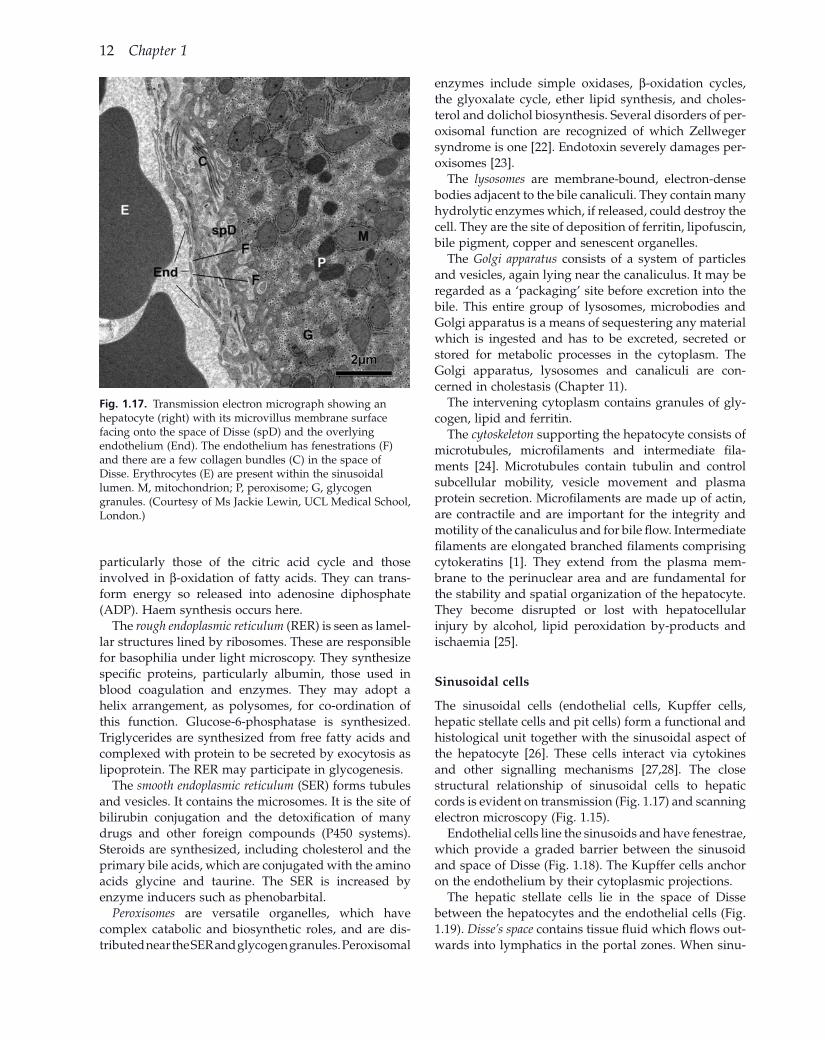
Hepatocytes (Figs 1.14 – 1.17 )
The liver cell margin is straight except for a few anchoring pegs (desmosomes). From it, equally sized and spaced microvilli project into the lumen of the bile canaliculi. Along the sinusoidal border, irregularly sized and spaced microvilli project into the perisinusoidal tissue space. The microvillous structure indicates active secretion or absorption, mainly of fl uid.
The nucleus has a double contour with pores allowing interchange with the surrounding cytoplasm. Human liver after puberty contains tetraploid nuclei and, at about age 20, in addition, octoploid nuclei are found. Increased polyploidy has been regarded as precancer-ous. In the chromatin network one or more nucleoli are embedded.
Fig. 1.15. Colourized scanning electron micrograph of liver showing hepatocytes in green, sinusoids (S) in light pink, erythrocytes (E), Kupffer cells (K) and bile canaliculi (BC). (Courtesy of Ms Jackie Lewin, UCL Medical School, London.)
Fig. 1.16. Electron microscopic appearances of part of a normal human liver cell. N, nucleus; M, mitochondrion; P, peroxisome; L, lysosome; ER, rough endoplasmic reticulum. (Courtesy of Ms. Jackie Lewin, UCL Medical School, London).
12 Chapter 1
Fig. 1.17. Transmission electron micrograph showing an hepatocyte (right) with its microvillus membrane surface facing onto the space of Disse (spD) and the overlying endothelium (End). The endothelium has fenestrations (F) and there are a few collagen bundles (C) in the space of Disse. Erythrocytes (E) are present within the sinusoidal lumen. M, mitochondrion; P, peroxisome; G, glycogen granules. (Courtesy of Ms Jackie Lewin, UCL Medical School, London.)
enzymes include simple oxidases, β - oxidation cycles, the glyoxalate cycle, ether lipid synthesis, and choles-terol and dolichol biosynthesis. Several disorders of per-oxisomal function are recognized of which Zellweger syndrome is one [22] . Endotoxin severely damages per-oxisomes [23] .
The lysosomes are membrane - bound, electron - dense bodies adjacent to the bile canaliculi. They contain many hydrolytic enzymes which, if released, could destroy the cell. They are the site of deposition of ferritin, lipofuscin, bile pigment, copper and senescent organelles.
The Golgi apparatus consists of a system of particles and vesicles, again lying near the canaliculus. It may be regarded as a ‘ packaging ’ site before excretion into the bile. This entire group of lysosomes, microbodies and Golgi apparatus is a means of sequestering any material which is ingested and has to be excreted, secreted or stored for metabolic processes in the cytoplasm. The Golgi apparatus, lysosomes and canaliculi are con-cerned in cholestasis (Chapter 11 ).
The intervening cytoplasm contains granules of gly-cogen, lipid and ferritin.
The cytoskeleton supporting the hepatocyte consists of microtubules, microfi laments and intermediate fi la-ments [24] . Microtubules contain tubulin and control subcellular mobility, vesicle movement and plasma protein secretion. Microfi laments are made up of actin, are contractile and are important for the integrity and motility of the canaliculus and for bile fl ow. Intermediate fi laments are elongated branched fi laments comprising cytokeratins [1] . They extend from the plasma mem-brane to the perinuclear area and are fundamental for the stability and spatial organization of the hepatocyte. They become disrupted or lost with hepatocellular injury by alcohol, lipid peroxidation by - products and ischaemia [25] .
Sinusoidal c ells
The sinusoidal cells (endothelial cells, Kupffer cells, hepatic stellate cells and pit cells) form a functional and histological unit together with the sinusoidal aspect of the hepatocyte [26] . These cells interact via cytokines and other signalling mechanisms [27,28] . The close structural relationship of sinusoidal cells to hepatic cords is evident on transmission (Fig. 1.17 ) and scanning electron microscopy (Fig. 1.15 ).
Endothelial cells line the sinusoids and have fenestrae, which provide a graded barrier between the sinusoid and space of Disse (Fig. 1.18 ). The Kupffer cells anchor on the endothelium by their cytoplasmic projections.
The hepatic stellate cells lie in the space of Disse between the hepatocytes and the endothelial cells (Fig. 1.19 ). Disse ’ s space contains tissue fl uid which fl ows out-wards into lymphatics in the portal zones. When sinu-
particularly those of the citric acid cycle and those involved in β - oxidation of fatty acids. They can trans-form energy so released into adenosine diphosphate (ADP). Haem synthesis occurs here.
The rough endoplasmic reticulum (RER) is seen as lamel-lar structures lined by ribosomes. These are responsible for basophilia under light microscopy. They synthesize specifi c proteins, particularly albumin, those used in blood coagulation and enzymes. They may adopt a helix arrangement, as polysomes, for co - ordination of this function. Glucose - 6 - phosphatase is synthesized. Triglycerides are synthesized from free fatty acids and complexed with protein to be secreted by exocytosis as lipoprotein. The RER may participate in glycogenesis.
The smooth endoplasmic reticulum (SER) forms tubules and vesicles. It contains the microsomes. It is the site of bilirubin conjugation and the detoxifi cation of many drugs and other foreign compounds (P450 systems). Steroids are synthesized, including cholesterol and the primary bile acids, which are conjugated with the amino acids glycine and taurine. The SER is increased by enzyme inducers such as phenobarbital.
Peroxisomes are versatile organelles, which have complex catabolic and biosynthetic roles, and are dis-tributed near the SER and glycogen granules. Peroxisomal