shashikant kulkarni, m.s (medicine)., ph.d., facmg head … 2 - latest... · ~15,000 sq ft...
TRANSCRIPT

Shashikant Kulkarni, M.S (Medicine)., Ph.D., FACMG Head of Clinical Genomics
Medical Director of Cytogenomics and Molecular Pathology Associate Professor of Pediatrics, Genetics, Pathology and Immunology
http://clinicalgenomics.wustl.edu

Dislosures (compensated and non-compensated)
• Scientific Advisory Board/Consultant – National Institute of General Medical Sciences (NIGMS)
Coriell cell repositories
– Chromosome Disorder Outreach
– Genomequest
– Agilent technologies
• Speaker honorarium – National Cancer Institute (NCI), American College of
Medical Genetics, Association of Molecular Pathology, University of Minnesota, University of Florida, CDC, Molecular Medicine- tricon, OMICS revolution, Illumina, Novartis, Affymetrix, Agilent

Genomics and Pathology Services at Washington University
Research & Panel Development
Genomic Technologies
and Innovation
Clinical Genomics
Biomedical Informatics
Training and Education
Over 150 faculty and staff support GPS function
Computational biologists, bench scientists
Software engineers, informaticians
Biostatisticians, IT administrators
Board-certified clinical genomocists and pathologists (ABMG,
ABP)
~15,000 sq ft dedicated labs; majority CAP/CLIA
Chromosomes to base pairs
Next generation sequencing
11/11/11: Clinical Launch – Cancer Panel
Clinical exome sequencing
Targeted assays for inherited disorders
Pharmacogenetic assays
Clinical Genomics
Pathology Consulting Services
Biomedical
Informatics

Clinical Genomics
• Current state of diagnostic testing
– Constitutional
• Chromosomal microarrays
• Karyotyping (?)/phenotype directed FISH tests (?)
• Single gene molecular testing
• Next Generation sequencing (NGS) disease panels
• NGS- exome and whole genome sequencing
– Cancer
• FISH (rapid)
• Karyotyping (whole genome view)
• Chromosomal microarrays (?)
• Single gene molecular testing
• Next Generation sequencing (NGS) cancer specific panels
• NGS- exome and whole genome sequencing
Washington University School of Medicine, St Louis

Clinical Genomics
• Chromosomal Microarrays (CMA)-aCGH and SNP arrays
– Increasing our understanding of genomic aberrations at very high resolution
• Next Generation sequencing (NGS)
– Revolutionizing, paradigm shifting look at the genome
• Fluorescence in situ hybridization (FISH) is key in integrated clinical genomic analyses

FISH
• Most common method for verifying CMA findings • Visualization in intact cellular context
– Positional and orientational information of chromosome structures
• Rapid turn around time • Better detection of low-level mosaicism • Best method for rapid detection of translocations,
inversions, amplifications, deletions and duplications
• CMA and Next generation sequencing still lacks most of the above

FISH
• Clone based methods • FISH probes typically generated from genomic
DNA, bacterial artificial chromosomes (BAC), fosmids, PAC (P1-derived artificial chromosome), YAC (yeast artificial chromosome), PCR templates
• Most FISH probes are between 150-300 Kb • Not useful for visualization/verification of smaller
abnormalities • PCR template generated probes
– Not ideal, time consuming, multiple steps

Cases to demonstrate limitations of clone based FISH methods

Case-1 • Three year old boy with global developmental
delay, hypotonia, speech problems
• Chromosomal Microarray (CMA) reveals ~70Kb deletion on 6q22.33 disrupting LAMININ, ALPHA-2; (LAMA2)
– Laminin is a heterotrimeric extracellular matrix protein consisting of 3 chains: alpha-1, beta-1, and gamma-1.
– Several isoforms of each chain have been identified. Laminin-2 (merosin) is a heterotrimer composed of laminin subunits alpha-2, beta-1, and gamma-1.
– It is the main laminin found in muscle fibers

Case-1
• Clone based FISH methods failed to help verify/visualize LAMA2 deletion
– BAC, fosmids
• Parental studies performed by CMA
• Deletion maternal in origin
• Mutation of the other allele

Case-2
A 39 year-old woman with acute myeloid leukemia (AML)
referred for an allogeneic stem cell transplant
Atypical promyelocytes with invaginated nuclei
(dense primary granules)

Does the patient have APL, or does she have AML with
unfavorable-risk cytogenetics?
RARA-PML fusion

Schematic representation of ins(15;17)
identified by WGS and resulting in
PML-RARA fusion
FISH identifies a fusion event on
der(17), consistent with ins(15;17)
Impact of NGS on Cancer Genomics
et al

Oligonucleotide FISH
• Uses high complexity oligonucleotide libraries as starting point for probe generation
• Bioinformatic approaches and various algorithms are used for probe selection based on in silico predictions
• Repetitive elements can be avoided
• Precise genomic coordinates from reference genomes are used to generate probes

Oligonucleotide FISH
• Involved in early strategic design and selection of probes utilizing knowledge generated by genome sequencing
• Understanding of detailed genomic structure very useful in carefully excluding noise generating repetitive elements
• Preliminary experimental data presented here on results from pilot research studies
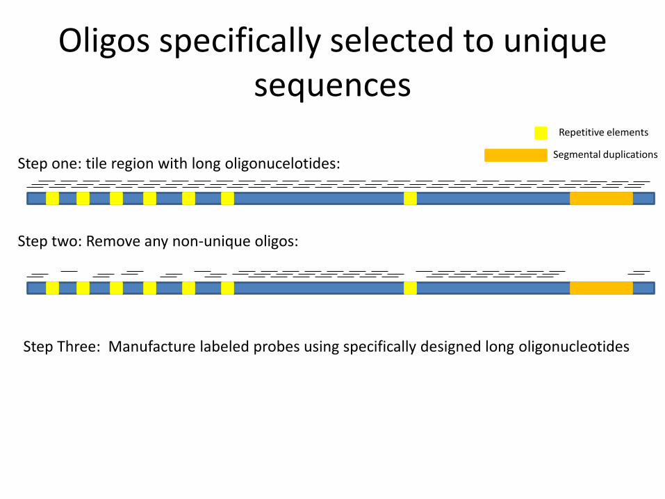
Oligos specifically selected to unique sequences
Step one: tile region with long oligonucelotides:
Step two: Remove any non-unique oligos:
Repetitive elements
Segmental duplications
Step Three: Manufacture labeled probes using specifically designed long oligonucleotides

Oligo FISH BAC-FISH
Minimum region targeted <50kb ~100kb
Requires available clone? No Yes
Need Cot-1 DNA ? No Yes
Can specifically target regions
that have a high degree of
homology/repetitive
elements?
Yes No
Probe Signal to noise +++ ++
Detect chromosome
rearrangements? Yes Yes
Hybridization time 4-14 hours ~14 hours
Advantages of Oligo FISH Over BAC-
FISH

Detection of Smaller Regions Region
Size (kb)
Sequence
Tiled (kb)
1 23.3 14.1
2 20.0 14.1
3 27.9 14.1
4 27.6 14.1
5 31.4 14.1
6 23.6 13.6 c-met locus divided into 6 regions
Repeat Gaps
Region 6
Region 2 Region 1
Red : SureFISH probe
Green: BAC CEP
Region 3
Region 5
Region 4
Regions 1-6 20/20 metaphase and 20/20 interphase cells showed this staining

Detection of Difficult Regions
Region
Size
% Repeat Num Gaps Median Gap Size Max Gap
Size
Tiled Region % GC
23 kb 61% 5 660bp 1.2 kb 8.6kb 62%
Green: BAC CEP Red: SureFISH Probe

A 6.7-kb region at 6p22.2 (110,219,652–110,316,643) is detected using oligonucleotide-based FISH, shown by the red signals. The same FISH image is shown with DAPI counterstain (left), and inverted DAPI stain or ‘pseudo G-banding’ confirming the chromosomal location (right). Arrows indicate chromosome 6
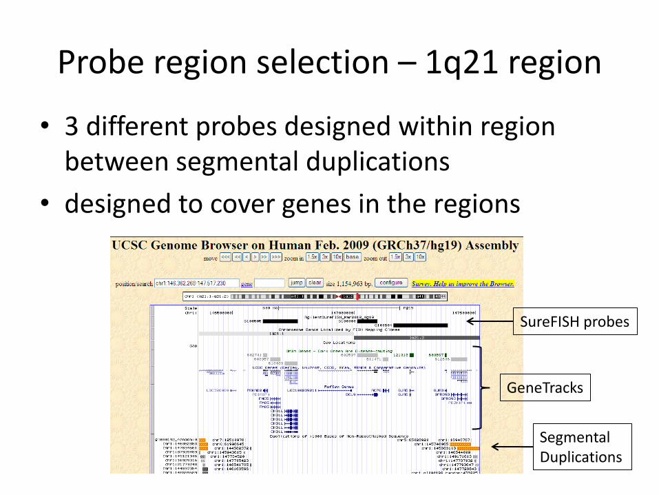
Probe region selection – 1q21 region
• 3 different probes designed within region between segmental duplications
• designed to cover genes in the regions
SureFISH probes
GeneTracks
Segmental Duplications

Probe design – 1q21region
• Focus on 224kb region of interest
• Oligos in region target specific sequences
SureFISH probe location
GeneTracks
Segmental Duplications
Oligo coverage within probe
Repeat Masked Region

Research Pilot study
• OFISH (4 hour) compared to overnight BAC based FISH
• OFISH performed on cases with smaller genomic aberrations not easily detected by chromosomal microarray (CMA)
• Preliminary data

BCR ABL NEGATIVE
O-FISH BAC probe BCR=Red; ABL=Green BCR=Green; ABL=Red
BCR ABL POSITIVE
O-FISH BAC probe BCR=Red; ABL=Green BCR=Green; ABL=Red

PML RARA POSITIVE
O-FISH BAC probe PML=Green; RARA=Red PML=Red; RARA=Green
PML RARA NEGATIVE
O-FISH BAC probe PML=Green; RARA=Red PML=Red; RARA=Green

CEP 8 POSITIVE Trisomy
O-FISH BAC probe
CEP 8 NEGATIVE
O-FISH BAC probe

EGR1 POSITIVE
O-FISH BAC probe
EGR1=Red; CEP5=Green EGR1 NEGATIVE
O-FISH BAC probe
EGR1=Red; CEP5=Green
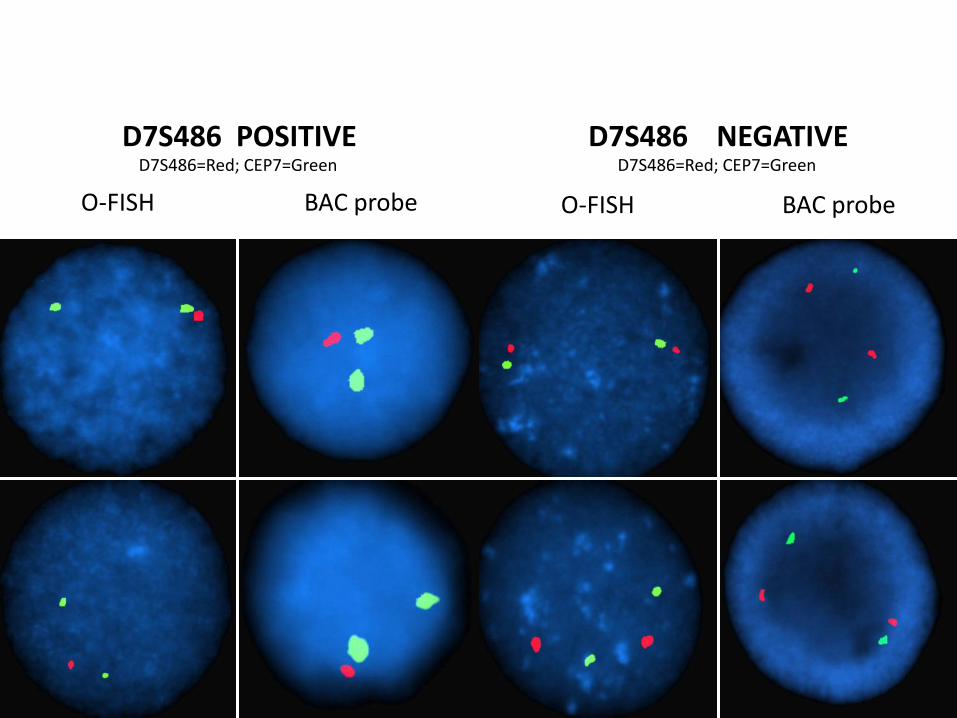
D7S486 POSITIVE
O-FISH BAC probe
D7S486=Red; CEP7=Green
D7S486 NEGATIVE
O-FISH BAC probe
D7S486=Red; CEP7=Green
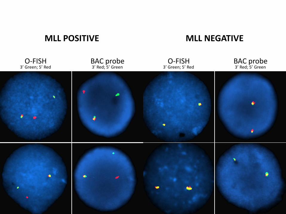
MLL NEGATIVE
O-FISH BAC probe 3’ Green; 5’ Red 3’ Red; 5’ Green
MLL POSITIVE
O-FISH BAC probe 3’ Green; 5’ Red 3’ Red; 5’ Green

O-FISH for RP11-414N15 Region
Deletion Control No Cross-hybridization
Chr15:31775207-31899230

O-FISH for RP11-433J22 Region
Duplication Control
Chr1:147162143-147386375
duplicated signal

Preliminary results
• Signal intensity, sensitivity, specificity, reproducibility of OFISH probes determined
• High intensity, robust signals could be generated from regions that are smaller to detect by clone based methods
• 100% concordance between clone based FISH methods and OFISH
• 100% concordance between CMA findings and OFISH

Summary
• OFISH is a powerful alternative to clone based FISH methods
• Use of genomic information to design probes helps in generation of highly reproducible robust FISH probes
• Additional studies are underway • Ability to detect smaller aberrations not previously
visualized by traditional FISH probes is very valuable as we enter the high resolution, fine scale clinical genomics era
• Availability of OFISH probes with sequence level information and confirmation of chromosomal localization and performance quality metrics will markedly improve study of genome complexities
Karen Seibert, John Pfiefer, Skip Virgin,
Jeffrey Millbrandt, Rob Mitra, Rich Head
Rakesh Nagarajan and his Bioinf. team
David Spencer, Eric Duncavage, Andy Bredm.
Hussam Al-Kateb, Cathy Cottrell
Dorie Sher, Jennifer Stratman
Tina Lockwood, Jackie Payton
Mark Watson, Seth Crosby, Don Conrad
Andy Drury, Kris Rickoff, Karen Novak
Mike Isaacs and his IT Team
Norma Brown, Cherie Moore, Bob Feltmann
Heather Day, Chad Storer, George Bijoy
Dayna Oschwald, Magie O Guin, GTAC team
Jane Bauer and Cytogenomics &Mol path team
MANY MORE!
