session 2 cardiovascular & hematology
TRANSCRIPT

AACN PCCN Webinar
Session 2
Cardiovascular & Hematology
Presenter: Carol A. Rauen, RN, MS, CCNS, CCRN, PCCN, CEN
Independent Clinical Nurse Specialist & Education Consultant [email protected]

Session 2: Cardiovascular & Hematology
1
Table of Contents Cardiac Pharmacology – Vasoactive & Inotropic Agents ................................................................ 2
Cardiac Complications..................................................................................................................... 7
Dysrhythmias ................................................................................................................................ 20
Hematology/Immunology/Oncology ............................................................................................ 29

Session 2: Cardiovascular & Hematology
2
Cardiac Pharmacology – Vasoactive & Inotropic Agents
I. PHYSIOLOGICAL PRINCIPLES
Physiology of the Autonomic Nervous System
Sympathetic Parasympathetic
Purpose to Regulate Autonomic Function
Activation: Flight or Fight Responses Conservation: Maintain Organ Function and Conserve Energy
Motor Neurons Large and Diffuse Number of Postganglionic Stimulation
Narrow and Specific Postganglionic Stimulation
Neurotransmitters Norepinephrine
Epinephrine
Acetylcholine
Receptors Adrenergic: o Alpha o Beta o Throughout body
Cholinergic: o Nicotinic o Muscarinic o Specific areas
Innervation Heart, Blood Vessels, Glands, Visceral Organs & Smooth Muscles
Heart, Glands, & Visceral Organs
Autonomic Receptor Stimulation
ORGAN SYMP Receptor ADRENERGIC Response PARA Receptor CHOLINERGIC Response
Heart Beta 1 Increase Conduction Velocity (rate) &
Increase Contractility
Muscarinic 2 Decrease Conduction Velocity & Contractility
Lungs Beta 2 Bronchial Dilation
Decrease Secretions
Muscarinic 2 Bronchial constriction
Increase Secretions Vessels Alpha 1
Beta 2 Constriction
Dilation
Skeletal Muscle
Beta 2 Increase Contractility
Bladder Sphincter
Alpha 1 Contraction Muscarinic 3 Relaxes
GI: Motility Sphincter
Alpha1,2 B2 Alpha 1
Decrease
Contraction
Muscarinic 3
Increase
Relax
Kidney Beta 1 Rennin Secreted
Liver Alpha 1, B2 Increase Glucose

Session 2: Cardiovascular & Hematology
3
II. COMMON VASOPRESSOR & INOTROPIC AGENTS
Common Vasopressor & Inotropic Agents
Inotropes Vasopressors Vasodilators Beta Agonist & Antagonists
Digoxin (Lanoxin)
Amrinone Lactate (Inocor)
Milrinone (Primacor)
Dobutamine Hydrochloride (Dobutrex)
Epinephrine
Norepinephrine Bitartrate (Levophed)
Dopamine Hydrochloride (Intropin)
Phenylephrine (Neosynephrine)
Vasopressin
Sodium Nitroprusside (Nipride)
Nitroglycerin
Calcium Channel Blockers
Isoproterenol Hydrochloride (Isuprel)
Normodyne (labetalol) Brevibloc (esmolol)
Dopamine Hydrochloride (Dopamine)
Therapeutic Use Naturally occurring catecholamine and precursor to norephinephrine, also serves as a central and peripheral neurotransmitter. First line agent for many types of shocks states. Versatile drug secondary to different actions depending on delivered concentration. The stimulation of dopaminergic receptors is a unique property of this agent.
Pharmacokinetics IV administration only with short half life
Pharmacodynamics a. Central and peripheral nervous system neurotransmitter and precursor of norepiphrine b. Low concentration: vascular DA2 – dopaminergic receptors primarily in renal, mesenteric,
coronary and cerebral beds – cause vasodilation. D1 receptors mediate a mild natriuresis. Current research has demonstrated that even with 1mcg most people will also get some alpha or beta stimulation.
c. Moderate concentrations: beta1 adrenergic receptor agonist – positive inotropic effect d. High concentrations: alpha1 adrenergic receptor agonist – potent vasoconstriction
Hemodynamics (dose dependent) a. Low concentration: increase in UO, maybe some increase in HR or SBP (current research has
not shown this to be renally protective) b. Moderate concentrations: increase in HR, SBP, CO (mild) c. High concentrations: increase in SBP, DBP, SVR
Mixing and Dosing a. Typical 400mg/250 D5W or NS b. Dosed in mcg/kg/min c. 1-3mcg/kg/min low dose d. 3-5mcg/kg/min mid dose e. 5-10mcg/kg/min high dose

Session 2: Cardiovascular & Hematology
4
f. > 10 pure alpha dose
Norepinephrine Bitartrate (Levophed)
Therapeutic Use Endogenous catecholamine with powerful inotropic and peripheral vasoconstriction effects. Typically not utilized as first line drug due to strong vasoconstrictive properties.
Pharmacokinetics IV administration only with short half life
Pharmacodynamics: a. Potent α1 & α2 agonist b. Mild β1 agonist c. No effect on β2 d. Systemic arterial and venous constriction e. Coronary flow increases slightly
Hemodynamics a. Increase in SBP & DBP b. Increase in SVR and PVR c. Cardiac output unchanged or decreased (increase in afterload) d. Heart rate may slow from compensatory vagal reflex
Dosing and Mixing a. Typical 4mg/250 D5W b. Dosed in mcg/min c. 2-10mcg/min
Epinephrine Hydrochloride
Therapeutic Use Endogenous catecholamine with powerful inotropic, peripheral vasoconstriction effects and inotropic properties. Typically not utilized as first line drug due to profound vasoconstrictive and subsequent side effects.
Pharmacokinetics Short half life with rapid onset
Pharmacodynamics a. Alpha and Bata agonist b. Increases myocardial contractility c. Vasoconstriction (all beds) d. Increases myocardial 02 consumption

Session 2: Cardiovascular & Hematology
5
Hemodynamics a. Increases HR, MAP, CO, SVR, PVR b. Pro-arrhythmic
Mixing and Dosing a. Typical 2mg/250 D5W or NS up to 8mg/250 b. Dosed in mcg/min c. 1-4mcg/min
Vasopressin
Therapeutic Use Is a naturally occurring antidiuretic hormone. In unnaturally high doses it functions as a non-adrenergic peripheral vasoconstrictor. Major use is as a first line agent in ACLS for pulseless VT/V-fib. Shown to reduce or eliminate the need for catecholamine administration.
Pharmacokinetics a. IV administration only b. Half life 10-20 min
Pharmacodynamics a. Direct stimulation of smooth muscle V1 receptors b. Smooth muscle constriction: pallor of skin, nausea, intestinal cramps, desire to defecate,
bronchial constriction, uterine contraction c. Less constriction of coronary and renal vascular beds and vasodilation of cerebral
vasculature d. No skeletal muscle vasodilation or increased myocardial 02 consumption during CPR
because there is no Beta-adrenergic activity e. May enhance platelet aggregation in septic shock
Hemodynamics a. Increase in SBP, MAP and SVR b. Increase UO
Mixing and Dosing a. Typical 200U/250 D5W or NS b. Dosed in unit/min c. 0.2-0.9U/min
Dobutamine
Therapeutic Use Synthetic catecholamine which has selective beta adrenergic agonist properties. Effective as a positive inotropic for both preload and afterload reduction. Used for its positive inotropic

Session 2: Cardiovascular & Hematology
6
properties when vasoconstriction is not preferable. Also used commonly as a combination therapy with another catecholamine or vasodilator
Pharmacokinetics a. IV administration only b. half life 2 minutes – rapid onset
Pharmacodynamics a. β1 adrenergic receptor agonists: increases contractility and stroke volume, increases sinus
node automaticity and AV conduction, increases in myocardial oxygen demand b. Mild β2 adrenergic receptor agonist: mild vasodilation, increased perfusion c. Mild α1 vasoconstriction properties are counter balanced by β2 properties d. Does not cause release of endogenous norepinephrine e. Infusions of > 72 hrs have shown tolerance 20 to down regulation of β adrenergic receptors f. Less effective in patients receiving β blocking agents or with chronic heart failure
Hemodynamics a. Increase CO b. Mild decrease in SVR c. Mild increase in HR (sometimes more than mild)
Mixing and Dosing a. Typical 500mg/250 D5W or NS b. Dosed in mcg/kg/min c. 2-10mcg/kg/min
Milrinone (Primacor)
Therapeutic Use Synthetic noncatecholamine agent that does not stimulate or block adrenergic receptors. Inhibits the phosphodiesterase III enzyme. Effective as a positive inotrope and vasodilator.
Pharmacokinetics a. IV administration only b. Hepatically cleared c. Half life 2-3 hours
Pharmacodynamics a. Phophodiesterase III enzyme inhibitor – increases cyclic adenosine monophosphase (cAMP)
which enhances calcium entry into the cell and improves myocardial contractility, and inhibiting vasoconstriction (vasodilator).
b. Increased cardiac output by positive inotropic action and reduction in preload and afterload c. Most effective with patients who have over stimulated sympathetic system d. Effective in patients with beta receptor down regulation

Session 2: Cardiovascular & Hematology
7
Hemodynamics a. Increase in CO b. Decrease in CVP, SVR, PAOP c. No significant effect on HR or BP (unless compensatory)
Mixing and Dosing a. Mix with NS ONLY b. Dosed in mcg/kg/min c. Loading dose 50mcg/kg over 10 min d. 0.375-0.75mcg/kg/min
Review ALL ACLS drugs and algorithms when studying for the PCCN Exam.

Session 2: Cardiovascular & Hematology
7
Cardiac Complications
I. ACUTE PULMONARY EDEMA
Introduction A change in alveolar-capillary membrane permeability leads to pulmonary interstitial edema. The initiating pathology can be either cardiac or non-cardiac in origin.
Pathogenesis of Pulmonary Edema
Alveolar-Capillary Membrane a. Capillary Endothelial Layer: Microvascular Barrier b. Alveolar Epithelial Layer: Alveolar Barrier
Fluid Dynamics of the Alveolar-Capillary Membrane a. Hydrostatic pressure b. Osmotic pressure c. Membrane permeability
Etiology of Pulmonary Edema
Cardiogenic Pulmonary Edema Cardiogenic pulmonary edema, which is the most common type of acute pulmonary edema, occurs when there is an increase in hydrostatic pressure within the pulmonary capillary bed as a result of heart failure. Causes include: a. Heart Failure b. Myocardial Infarction c. Cardiac Ischemia d. Acute Mitral Regurgitation e. Cardiac Tamponade f. Tachy Dysrhythmias g. Hypertensive Crisis
Non-Cardiogenic Pulmonary Edema Non-cardiogenic pulmonary edema results from one of four primary abnormalities (or a combination): a. Impaired endothelial integrity b. Decreased colloidal oncotic pressure c. Elevated capillary hydrostatic pressure d. Lymphatic obstruction The impaired endothelial integrity (change in permeability) is typically caused by a direct or indirect injury to the lung tissue. Acute respiratory distress syndrome (ARDS) is a form of non-cardiogenic pulmonary edema.

Session 2: Cardiovascular & Hematology
8
Diagnosis
History a. Cardiogenic Pulmonary Edema b. Acute Cardiac Event c. Chest Pain d. Tachy-Palpitations e. New Dysrhythmia f. History of Ischemic Heart Disease g. Acute CP and/or SOB in the Absence of Any Other Pathologies h. Absence of Cardiac Hx Does Not Rule Out CPE
Physical Exam a. Dyspnea, Tachypnea & Apprehension b. Presence of S3 Heart Sound c. Jugular Venous Distension d. Breath Sounds e. Increased Frothy Sputum Production f. Laterally Displaced Point of Maximal Impulse (PMI) g. New or Louder Cardiac Murmur h. Unilateral Lung Adventitious Sounds: more commonly assessed in non-cardiogenic i. Diffuse Decreased Breath Sounds: more commonly assessed in non-cardiogenic j. Peripheral Edema is non-specific
Chest X-Ray There are chest x-ray changes that are unique to cardiogenic and non-cardiogenic pulmonary edema. If the pattern changes from day to day or significantly after treatment it is more indicative of cardiogenic.
ECG Changes Tachycardia or acute ST-T segment changes are more commonly assessed in cardiogenic pulmonary edema
Echocardiography A Transthroacic echo may be helpful to identify myocardial ischemia, wall motion abnormalities, ventricular dysfunction, valvular disease, and LV hypertrophy, all of which will suggest cardiogenic pulmonary edema.
Laboratory Data Arterial Blood Gas: a. Low Oxygen Saturation b. Respiratory Alkalosis c. Refractory Hypoxemia

Session 2: Cardiovascular & Hematology
9
Treatment Options
Cardiogenic Pulmonary Edema ACLS algorithm while treating the underlying cardiac condition: a. Diuretics: Furosemide IV 0.5 to 1.0 mg/kg b. Analgesics: Morphine IV 2 – 4mg c. Preload Reduction: Nitroglycerin SL d. Oxygen/Intubation e. Afterload Reduction if SBP > 100mmHg
IV Nitroglycerin 10-20 g/min or consider
IV Nitroprusside 0.1 – 5.0 g/kg/min f. Vasoconstriction if Hypotensive
Severe (SBP < 70mmHg) and S&S of shock
o IV Norepinephrine 0.5 – 30 g/min
Moderate (SBP 70– 100mmHg) with S&S of shock
o IV Dopamine 5 – 15 g/kg/min
Moderate without shock
o IV dobutamine 2 – 20 g/kg/min g. Further Diagnostic Considerations
Pulmonary Artery Catheter
Intra-Aortic balloon pump
Cardiac Angiography
II. RUPTURED OR DISSECTING AORTIC ANEURYSMS
Definitions These conditions can overlap at times with one leading to or increasing the risk of the other.
Aortic Aneurysm A localized dilation of the arterial wall that can be saccular, fusiform or cylindrical. The dilation frequently renders the aorta weak in that region. a. Complication of aneurysms b. Management of aneurysms
Aortic Dissection a longitudinal separation of the aortic wall between the intima and the adventitia. An acute dissection is one that is diagnosed within 14 days of the onset of symptoms. The risk of death is greatest during this acute period. A chronic dissection is one that is diagnosed after two weeks of the onset of symptoms.

Session 2: Cardiovascular & Hematology
10
Classification Systems
Stanford Classification System Type A Aortic Dissection The dissecting area involves the ascending aorta. It may be confined to only the ascending aorta or may also involve the descending as well. Typically occur in a younger patient population with a congenital weakening of the ascending aorta. Type A dissection account for 2/3 of all dissections. Type B Aortic Dissection The dissecting area involves only the descending aorta distal to the left Subclavian artery. Typically occurs in the older patient population with a history of hypertension and atherosclerosis.
DeBakey Classification System
Type I Aortic Dissection The dissection involves the ascending aorta but also extends beyond the left subclavian artery. Type II Aortic Dissection The dissection involves only the ascending aorta. Type III Aortic Dissection The dissection involves only the descending aorta. IIIa limited to the thoracic aorta, IIIb involving various degrees of the thoracic and abdominal aorta
Common Risk Factors a. Although hypertension does not appear to be the sole contributor to the occurrence of
AAD, it plays a major role in the development and/or propagation of a dissection. The etiology of AAD is believed to be a combination of something that has caused a weakening in the vessel that ‘allows’ the original tear to occur and that, in combination of HTN (70-90% of the victims have a history of HTN) triggers the filling of the false lumen and dissection of the arterial layers.
b. Marfan’s Syndrome (a chromosomal mutation with many genotypes and phenotypes) c. Annuloaortic Ectasia d. Aortic Dilatation and Wall Thinning e. Bicuspid Aortic Valve (congenital malformation) f. Spontaneous Rupture of Vasa Vasorum g. Aortic Coarctation h. Trauma (blunt, penetrating or iatrogenic) i. Aging j. Arterial Hypertension k. Cocaine Use

Session 2: Cardiovascular & Hematology
11
Diagnosis
Presenting Signs & Symptoms AAD is Known as the Great Imitator a. Sudden Severe Pain Not Relieved with Analgesics b. Initially Normal or High Blood Pressure c. Hypotension d. Acute Aortic Valvular Insufficiency: High-Pitched, Blowing Diastolic Murmur e. Audible S3 Heart Sound f. Abrupt Onset of a Pulseless Extremity g. Peripheral Vascular Insufficiency h. End-Organ Ischemia (brain, kidney, intestines, spinal, lower extremities) i. Pericardial Effusion j. Cardiac Tamponade k. Acute Myocardial Ischemia
Tests a. Chest X-ray
Wide Mediastinum
Wide Aortic Silhouette
Pleural Effusion
CHF
Pericardial Effusion (cardiomeagaly) b. ECG: Nonspecific Changes
Left Ventricular Hypertrophy
Acute Myocardial Ischemia c. Chest CT: Sensitive test. d. Transthroacic or Transesophageal (TEE) Echocardiography e. Magnetic Resonance Imaging (MRI) Scan f. Aortagraphy
Treatment
Adequate Blood Pressure Management a. Antihypertensive Agents: Nipride 0.5g/kg/min titrate up to maintain SBP below 110mgHg
or at a level to maintain perfusion
b. Negative Inotropic Agents: blockers c. Pharmacological management may be the primary treatment for a dissection involving the
descending aorta
Pain Relief Typically done with Morphine
Reduction of Environmental and Emotional Stresses May need anti-anxiety agents.

Session 2: Cardiovascular & Hematology
12
Surgical Repair a. All patients with ascending aortic dissection require immediate repair b. Descending dissections repairs have high mortality and morbidity
III. HYPERTENSIVE CRISIS
Introduction
Pathophysiology of Hypertension Definition and Current Guidelines
Definitions
Hypertensive Crisis A diastolic blood pressure greater than 120mmHg. Global term does not denote physiologic response or need for immediate treatment.
Hypertensive Emergency A diastolic blood pressure of greater than 120mmHg with acute or ongoing end organ (neurological, cardiac or renal) damage. Immediate blood pressure reduction is required within a few hours to prevent or limit target organ damage. The reduction does not necessarily need to be back to normal pressure just out of the dangerous range.
Hypertensive Urgency A diastolic blood pressure of greater than 120mmHg without end organ damage. Reduction of blood pressure is important to limit the risk of potential end organ damage but not emergent. The goal is to bring down the blood pressure within 24 – 48 hours.
Malignant Hypertension Described by Volhard and Fahr in 1914, MHT is characterized by severe accelerating hypertension with evidence of renal, neurological, vascular and retinal damage/dysfunction that can be rapidly fatal ending in heart attack, stroke or heart and renal failure. The modern criteria for MHT are severe hypertension (DBP > 120mmHg) associated with retinal hemorrhages, exudates and papilledema (group 4 Keith-Wagener-Barker retinopathy) (Laragh, 2001). Some authors define it simply as elevated BP accompanied by encephalopathy or nephropathy (Varon, 2000).
Accelerated Hypertension A more ‘mild’ form of MHT without the presence of papilledema and a group 3 Keith-Wagener-Barker retinopathy.
Post Operative Hypertension Defined as systolic blood pressure of greater than 190mmHg and/or diastolic blood pressure of greater than or equal to 100mmHg on two consecutive readings following surgery. Because of

Session 2: Cardiovascular & Hematology
13
the unique and transient physiological factors following surgery and anesthesia this clinical syndrome is separated from the other hypertensive crises.
Gestational Hypertension There are multiple names for this syndrome. A blood pressure is considered an emergency in a pregnant woman and requires immediate pharmacologic management when the systolic pressure is greater than 169mmHg or diastolic greater than 109mmHg.
Hypertensive Crisis – Pathophysiology & Management
Etiologies There is not one cause of HTN Crisis. A history of preexisting hypertension is the common denominator regardless of the secondary causative factor(s).
Pathophysiology Although the exact physiological mechanism(s) of hypertensive crisis are unknown, there appears to be a vicious cycle of increased vasoconstriction which leads to increasing pressure.
Assessment In addition to the blood pressure, the presence and relative degree of end organ damage/dysfunction is important to assess for and essential to identify before selecting the appropriate treatment option. a. Previous Diagnosis of HTN
How long?
Prescribed Medications?
Adherence to Prescription Medication?
General Level of Control or Typical Blood Pressure? b. All Other Medications: Prescription, Over the Counter, Dietary Supplements and/or Illicit
Drugs c. Cardiac Assessment d. Renal Assessment e. Neurological Assessment – Hypertensive Encephalopathy f. Laboratory Data g. Evaluate Secondary Causes
Treatment for Hypertensive Emergency The goal is to reduce the mean arterial pressure by 25% within the first two hours (preferably within the first few minutes) in a controlled, predictable and safe fashion and then toward 160/100mmHg within two to six hours. a. Nitroprusside Sodium (Nipride) b. Fenoldopam Mesylate (Corlopam) c. IV Vasodilators d. IV Adrenergic Inhibitors e. Diuretics
Session 2: Cardiovascular & Hematology
14
Treatment for Hypertensive Urgency Blood pressure should be lowered within 24-48 hours and frequently oral agents are adequate in this patient population. a. ACE inhibitors b. Calcium Channel Blockers c. Alpha2 Adrenergic Stimulators (Clonidine)
IV. VALVULAR HEART DISEASE
Pathophysiology a. Congenital Malformations b. Connective Tissue Disorders c. Degenerative Disease d. Rheumatic Heart Disease e. Infective Endocarditis f. Dysfunctional Ruptures
Specific Valvular Dysfunction a. Mitral Stenosis b. Mitral Insufficiency/Regurgitation c. Aortic Stenosis d. Aortic Insufficiency/Regurgitation
Management of Valve Disorders (pre-op and post-op) a. Oxygenation b. Hemodynamic Stability c. Dysrhythmias d. Activity e. Anticoagulation f. Antibiotic Prophylaxis g. Patient/Family Education
Surgical Management of Valve Defects a. Indications b. Valve Repairs c. Prosthetic Valve Replacement
Mechanical
Biological

Session 2: Cardiovascular & Hematology
15
Septal Defects a. Locations: Atrial and Ventricular b. Types: Congenital and Acquired
V. VASCULAR DISEASE
Peripheral Vascular Disease Note: DVT is addressed in the Pulmonary section combination with the Pulmonary Emboli
Pathophysiology a. Smoking b. HTN c. DM d. Lipid Disorders e. Hyperhomocysteinemia
Assessment a. Intermittent Claudication b. Resting Pain c. Cool Temperature d. Diminished Pulses e. Leg/Skin Changes
Diagnosis Acute occlusion
Acute Arterial Occlusion 5Ps a. Pain b. Pulselessness c. Pallor d. Paresthesian e. Paralysis
Management/Treatment a. Risk Factor Modification b. Vasodilators c. Antiplatelets d. Exercise e. Angioplasty f. Vascular Bypass Surgery g. Minimally-Invasive Interventions (stents, endografts)

Session 2: Cardiovascular & Hematology
16
Compartmental Syndrome a. Compartments are closed spaces containing muscles, nerves, and vascular structures b. Internal and external causes can increase pressure within a compartment c. Increased pressure can lead to ischemia, injury and necrosis to the contents within
the compartment d. Signs and Symptoms of CS
Throbbing Pain (localized)
Firmness of area
Altered Sensation: Numbness, tingling, sticking feelings
Pulselessness
Decreased Voluntary Limb Movement e. Treatment for CS
Eliminate the Cause
Elevation
Pain Management
Fasciotomy
Carotid Artery Disease
Pathophysiology Assessment Management/Treatment a. Risk Factor Modification b. Vasodilators c. Antiplatelets d. Neuro Monitoring e. Carotid Endarterectomy f. Carotid Stents
VI. CARDIAC TAMPONADE
Introduction A cardiac effusion is the accumulation of fluid within the pericardial space. A cardiac tamponade occurs when an effusion causes compression on the heart and the external pressure effects cardiac function.

Session 2: Cardiovascular & Hematology
17
Etiology
Any pathology that can lead to a pericardial effusion can cause a tamponade. Pericarditis is the most common cause: a. Any Type of Pericarditis (inflammatory, infectious, immunologic or physical)
Neoplastic
Renal Insufficiency ESRD
Post Acute Myocardial Infarction
Collagen Vascular Disease
Autoimmune Diseases
Chemotherapy
Radiation Therapy
Nephrotic Syndrome
Tuberculosis
Hepatic Cirrhosis b. Invasive Cardiac Procedures (cardiac surgery or biopsy) c. Indwelling Cardiac Instrumentation d. Anticoagulant or Thrombolytic Therapy e. HIV/AIDS f. Valvular Heart Disease g. Trauma h. Pregnancy i. Aortic Dissection j. Chronic Heart Failure
Pathophysiology
Pericardium The pericardium is a membrane that surrounds the heart. The pericardial space is between the visceral (next to the myocardium) and parietal layers and there is typically15-35ml of serous fluid in the space. The function of the fluid is to provide a cushion for the heart. Fifty mls or more of fluid (serous fluid, blood, pus, clots or gas) is considered an effusion. Tamponade can be classified as acute vs chronic, surgical vs medical.
Presenting Signs & Symptoms Early Physical Signs &Symptoms May occur prior to full tamponade a. Chest Pain b. Anxiety c. Tachycardic d. Tachpneic e. Diaphoretic f. Bibasilar Rales g. Fever h. Unstable Blood Pressure

Session 2: Cardiovascular & Hematology
18
i. S&S of Shock j. Specific to Pericarditis
Diffuse ST elevation
Chest pain worse when supine
Pericardial friction rub Beck’s Triad (1935) a. Muffled Heart Sounds: from the accumulation of fluid. b. Narrowing of the Pulse Pressure and Hypotension: The pulse pressure is the difference
between the SBP & the DBP. Due to the increasing pressure outside the heart there is less dilation during diastole, which causes the DBP to either stay the same or rise. There is a lower EF because of the decreased filling and contractility so the SBP drops.
c. Jugular Vein Distention (JVD): There is ‘back-up’ of blood into the venous system from a combination of the impaired filling and impaired emptying of the heart due to external pressure. Kussmaul’s sign is a pathological increase in jugular venous pressure (JVP) seen during inspiration.
Pulsus Paradoxus a. The negative pressure created in the thorax during normal inspiration limits cardiac filling,
decreases CO and causes a weaker pulse. There is typically a difference of as much as 10mmHg in SBP between inspiration and expiration.
b. During tamponade the ‘normal’ pressure is increased causing more of a compression, which is increased even more during the inspiratory phase of ventilation. A drop of >10mmHg systolic blood pressure heard during inspiration is considered a Pulsus Paradoxus. Commonly (present in 90% of cases) assessed with cardiac tamponade.
c. Also found in other pathologies that cause increased pressure on the heart like COPD. PP was described by Kussmaul in 1873.
ECG Changes a. Electrical Alternans: beat to beat change in QRS amplitude (seen with late or rapid
tamponade) b. Low Amplitude of the QRS Complex c. Absence of Ischemic Changes Despite Chest Pain d. Non-specific ST changes may be Present in Pericarditis e. In late tamponade there may be decreased coronary flow (typically in the elderly and
patients with CAD)
Diagnosis
Chest X-Ray Enlarged cardiac silhouette with clear lungs
Hemodynamic Parameters: a. Drop in Cardiac Output b. Pulsus Paradoxus Visible on Arterial Line. Note that if the patient is on positive pressure
ventilation this response will be reversed.
Session 2: Cardiovascular & Hematology
19
Echocardiography A non-invasive, relatively available test to identify the presence of a pericardial effusion. Occasionally if the effusion is primarily posterior it may be difficult to visualize on a transthroacic echo and a transesophageal echo is necessary. Of all the diagnostic tools for tamponade this is the most sensitive and specific.
Therapeutic Management Options
Pericardiocentesis Needle aspiration of effusion. Subxiphoidal approach. Procedure can be performed blind, or with ECG clamp, echo or fluoroscopy guidance. If recurrent effusion is a concern a drainage system should be left in place for a few days. a. 6 inch, 16-18 gauge, over-the-needle catheter.
Surgical Drainage a. Subxiphoid surgical incision and thoracoscopic drainage b. Video assisted thoracoscopy and drainage c. Pericardial window and drainage d. Pericardectomy: complete stripping of the pericardial sac is performed on some medical
patients with chronic effusion/tamponade. This is typically a last resort.
Medical Management ACLS Oxygen, IV, CPR- if needed, Epinephrine, diagnosis and treat cause ASAP General Management a. Support compensatory and physiologic response b. Expand Intravascular volume c. Increase or decrease SVR d. Support contractility and stroke volume

Session 2: Cardiovascular & Hematology
20
Dysrhythmias
I. INTRODUCTION Review basic dysrhythmias. Questions on PCCN exam will be related to rhythm identification, cause or appropriate treatment. ACLS is not listed on the blueprint but identifying and treating life-threatening dysrhythmias is the major reason to place a patient on telemetry.
II. CARDIAC ELECTROPHYSIOLOGY
Impulse Conduction & Pathways SA Node Internodal Pathways (atrial contraction) AV Node (delay) His-Purkinje System (ventricular contraction)
SA
AV
Intraatrial
Right Bundle
Left Bundle
Bundle of His
Purkinje Fibers
Internodal Conduction
SA

Session 2: Cardiovascular & Hematology
21
III. DYSRHYTHMIAS
Common Causes a. Decreased Coronary Perfusion (CAD) b. Impaired Myocardial Oxygen Delivery (hypoxia) c. Electrolyte Disturbances d. Cardiac Muscle Injury e. Ischemia or Infarction f. Defects in the Heart Muscle or Electrical System g. Cardiac Surgery h. Electrical Stimulation to the Heart Muscle i. Medications
Lead Selection for Monitoring
AACN Practice Alerts for ECG Monitoring a. Dysrhythmia Monitoring (4/08) b. ST Segment Monitoring (5/09)
AHA/ACCF/HRS Recommendations for Standardization & Interpretation of the Electrocardiogram a. I ECG Technology ‘07 b. II ECG Diagnostic Tests ‘07 c. Pre-hospital ECG and ACS ‘08 d. III Intraventicular Conduction Disturbances ‘09 e. ST-Segment, T, and U Wave and QT Interval ‘09 f. Hypertrophy Evaluation ‘09 g. Acute Ischemia/Infarction ‘09 h. Prevention of Torsade de Points 2/8/10
Best Leads for Monitoring
Monitoring Purpose Lead Recommendation
Dysrhythmia Detection Aberrancy vs Ectopy: V1 or V6 (if V1 not available)
AFib/flutter: II, III, aVF : Monitoring of P wave Problems; whichever lead allows best visualization of fib/flutter waves; consider atrial ECG for post-cardiac surgery patients if pacer wires in place, could try Lewis Lead RA electrode at 4th ICS Rt Sternal boarder, LA on back at Rt side of spine read Lead I
Junctional Rhythms: Lead II
Bundle Branch Blocks V1 and/or V6
ST Segment Monitoring Unknown Problem III or V3
Right coronary artery: III or aVF
Left anterior descending/Circumflex: V3
Activity-induced ischemia (no specific vessel identified): V5
Pt “Finger Print” from Known Ischemic Changes

Session 2: Cardiovascular & Hematology
22
Inferior Wall Anterior Wall Lateral Wall
II, III, AVF
V1-V4
V5, V6, I, aVL
QTc Identify 12-lead with most well-defined T wave
V3, V4, II
Best Lead Combinations
One Channel Recording V1 or V6
Two Channel Recording Arrhythmia: V1 and III
ST Segment: V3 and III
Arrhythmia + ST Segment: V1 or V6 + aVF or III
Table originally developed by Bridges and published in CCN (2008) Rauen et al. Updated for this outline by Rauen 1/13
Treatment Options Identify & Treat the Underlying Cause
Defibrillation/Cardioversion The passage of electrical current through the cardiac muscle (cells) causes a massive depolarization allowing the cells to ‘reset’ themselves and hopefully creating an environment where the SA node can ‘take back’ the pacemaker function.
Pacing For bradycardic rhythms, electrical stimulation of the heart might be necessary with a transcutaneous, transvenous or permanent pacemaker.
Pharmacology Drugs are the primary treatment if the dysrhythmia is NOT life-threatening. Meaning there is not a significant drop in blood pressure or level of consciousness. The Antidysrhythmic agents are classified using the VaughanWilliams classification system.
Drug Classification Primary Action Drug Options/Indications
CLASS I Sodium Channel Blockers
Membrane-Stabilizing
Slow/Block influx of Na into cell
Effect Stimulus Automaticity, Conduction & Excitability
IA
Slows Conduction Velocity
Negative Inotrope
Prolong Refractory Period (wide QRS & QT)
Suppress Ectopic & Reentry Foci
IA
Quinidine (Quinaglute)
Procainamide (Procan, Proestyl)
Disopyramide (Norpace) Atrial Flutter, Atrial Fib, SVT, VT

Session 2: Cardiovascular & Hematology
23
Specific Dysrhythmias
Tachycardias The major problem with the tachy dysrhythmias is that the heart chambers do not have enough time to completely fill or empty. This leads to a drop in stroke volume and subsequently cardiac output. Depending on the exact rhythm, there may also be loss of synchrony between atrial and ventricular contractions (A-fib, V-Tach), which causes a loss of atrial kick and up to 30% of cardiac output. Another potential problem is clot formation in a chamber that has incomplete emptying. In clinical terms tachycardic rhythms can cause anything from dizziness to heart failure and cardiac arrest.
CLASS I Sodium Channel Blockers
(continued)
IB
Shorten Duration of Action Potential
Shorten Refractory Period
IB
Lidocaine (Xylocaine)
Phenytoin (Dilantin)
Mexiletine (Mexitil)
Tocainide (Tonocard)
Ventricular Rhythms
IC
Slow Conduction Velocity (depress phase 0)
Increase Refractory Period
Increase QRS Duration
IC
Flecainide (Tambocor)
Propafenone (Rythmol)
Ventricular Rhythms
CLASS II Beta Blockers
Antisympathetic Effects
Inhibit Sympathetic Activity Associated with Beta-Adrenergic Stimulation
Decrease Force of Ventricular Contraction
Slow SA Node and AV Conduction
Decrease in Myocardial Oxygen Consumption
Propranolol (Inderal)
Acebutolol (Sectral)
Esmolol (Brevibloc)
Sotalol (Betapace)
Supraventricular Rhythms
CLASS III Potassium Channel Blockers
Antiadrenergics
Prolong duration of action potential
Delay repolarization
Prolong QT interval, AP & effective refractory period
Bretylium (Bretylol) dropped from ACLS
Amiodarone (Cordarone)
Sotalol (Betapace)
Ibutilide (Corvert)
Dofetilide (Tikosyn)
Ventricular and Supraventricular Rhythms
CLASS IV Calcium Channel Blockers
Calcium Antagonist
Decreased SA node firing
Slower conduction AV node
Decrease myocardial 02 demand & contractility
Diltiazem (Cardizem)
Verapamil (Isoptin, Calan)
Supraventricular Tachycardias

Session 2: Cardiovascular & Hematology
24
Narrow QRS Complex Tachycardias (supraventricular) a. Rhythms
Sinus Tachycardia (ST)
Atrial Fibrillation (A-Fib)
Atrial Flutter (AF)
Atrial Tachycardia (ectopic and reentrant) (AT)
Multifocal Atrial Tachycardia (MAT)
Junctional Tachycardia (JT)
Accessory Pathway-Mediated o Atrial tachycardia w/ accessory pathway o AV reentry tachycardia
b. Treatment (remember to evaluate ventricular function) Based on 2010 ACLS guidelines
Stable? o A-Fib or AF: Identify length of time in rhythm and consider WPW and LV
impairment before determining treatment. Control Rate, Convert Rhythm, Provide Anticoagulation
o Vagal Stimulation o Adenosine
o PSVT: -Blockers, Ca++ Channel Blockers, Dig, Antiarrhythmics and Cardioversion. If EF < 40% Start with Cardioversion
o JT: -blockers, Ca++ Channel Blockers, Amiodarone, NO Cardioversion
o MAT: -blockers, Ca++ Channel Blockers, Amiodarone, NO Cardioversion
Unstable? Immediate Cardioversion, Followed by Drugs Wide QRS Complex Tachycardias a. Criteria for Wide QRS
Rate > 120 bpm
Uniform QRS > 120 ms
No S&S or in Consciousness b. Rhythms
Ventricular Tachycardia (VT)
Ventricular Fibrillation (VF)
SVT with Aberrancy (identify and treat as SVT) c. Treatment (remember to evaluate ventricular function) Based on 2010 ACLS guidelines
Ventricular Tachycardia Stable w/ Pulse o Monomorphic: Procainamide, Sotalol, Amiodarone, Lidocaine. Amiodarone
1st if ventricle impaired
o Polymorphic: normal QT - -blockers, Lidocaine, Amiodarone, Procainamide, Sotalol, Amiodarone 1st if ventricle impaired. Long QT – Mg+, overdrive pacing, Isoproterenol, Phenytoin, Lidocaine
Ventricular Tachycardia Unstable w/ Pulse: Immediate Cardioversion
Ventricular Tachycardia Without Pulse – Treat as VF

Session 2: Cardiovascular & Hematology
25
Ventricular Fibrillation/Pulseless VT: o Assess ABCs o Basic Life Support o Defibrillation: 120-200J biphasic or 360J monophasic (one shock) o CPR o Defibrillation: 120-200J biphasic or 360J monophasic (one shock) o Vasopressin or Epinephrine o Defibrillation: 200J biphasic or 360J monophasic o Antiarrhythmics: Amiodarone, Lidocaine, Magnesium, Procainamide o Defibrillation: 200J biphasic or 360J monophasic
Long QT Syndrome: The QT represents the repolarization of the ventricle. Repolarization is an electrically unstable time. VT is a likely outcome if the next R wave were to fall on the T wave. In situations where the QT interval is long, there is an increased likelihood of an R on T to occur. Conditions that can lead to this situation include: a. Congenital Long QT Syndrome (genetic) b. Exercise Induced QT Syndrome c. Drug Induced QT Syndrome (many drugs lengthen QT)
Antiarrhythmic Agents: Class IA, IB & III
Tricyclic Antidepressants
Phenothiazine
Antimicrobials (specifically Erythromycin)
Nicardipine (Cardene)
Cisapride (Propulsid)
Haloperidol (Haldol)
Tamoxifen (Nolvadex)
Bradycardias The major problem with slow rhythms is a lack of stroke volume to sustain an adequate cardiac output. Treatment is dependent on rhythm and cause of slow rate. In the unstable patient with a slow rate: a. ABCs & BLS b. Atropine c. Transcutaneous Pacing d. Dopamine or Epinephrine e. If the rhythm is Type II 2nd Degree or 3rd Degree HB and the pt is unstable pace ASAP
(transcutaneous transvenous)
Session 2: Cardiovascular & Hematology
26
Conduction Defects
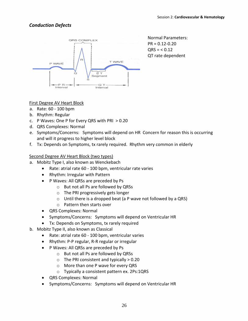
Normal Parameters: PR = 0.12-0.20 QRS = < 0.12 QT rate dependent
First Degree AV Heart Block a. Rate: 60 - 100 bpm b. Rhythm: Regular c. P Waves: One P for Every QRS with PRI > 0.20 d. QRS Complexes: Normal e. Symptoms/Concerns: Symptoms will depend on HR Concern for reason this is occurring
and will it progress to higher level block f. Tx: Depends on Symptoms, tx rarely required. Rhythm very common in elderly Second Degree AV Heart Block (two types) a. Mobitz Type I, also known as Wenckebach
Rate: atrial rate 60 - 100 bpm, ventricular rate varies
Rhythm: Irregular with Pattern
P Waves: All QRSs are preceded by Ps o But not all Ps are followed by QRSs o The PRI progressively gets longer o Until there is a dropped beat (a P wave not followed by a QRS) o Pattern then starts over
QRS Complexes: Normal
Symptoms/Concerns: Symptoms will depend on Ventricular HR
Tx: Depends on Symptoms, tx rarely required b. Mobitz Type II, also known as Classical
Rate: atrial rate 60 - 100 bpm, ventricular varies
Rhythm: P-P regular, R-R regular or irregular
P Waves: All QRSs are preceded by Ps o But not all Ps are followed by QRSs o The PRI consistent and typically > 0.20 o More than one P wave for every QRS o Typically a consistent pattern ex. 2Ps:1QRS
QRS Complexes: Normal
Symptoms/Concerns: Symptoms will depend on Ventricular HR

Session 2: Cardiovascular & Hematology
27
Tx: Depends on Symptoms, typically treated o Consider External Pacemaker o Consider Cause o Stop Digoxin o Atropine or Epinephrine
Third-degree AV Heart Block/Complete Heart Block aka AV Dissociation a. Rate: < 60 bpm b. Rhythm: P-P regular, R-R regular c. P Waves: P waves “march” out regular but have no discernible relationship to the QRS d. QRS Complexes: Slow, Wide > 0.12, “march” out, regular e. Symptoms/Concerns: Symptoms will depend on Ventricular HR and LOC f. Tx: Depends on Symptoms, tx typically required
External pacemaker
Atropine (not typically helpful because it will increase sinus node firing (P waves) but not ventricular conduction
Epinephrine
Bundle Branch Blocks QRS Complex a. Represents: Ventricular Depolarization b. Shape:
Q First Negative Deflection
R First Positive Deflection
S Second Negative Deflection c. Duration (time):
QRS > 0.12sec (3mm)
Q < 0.03sec (< 1mm)
AV Node Bundle of His 1. Septal Depolarization L R 2. Biventricular Depolarization The ventricles depolarize simultaneously. Because the LV mass is larger than the RV the mean vector of electrical current is the LV depolarization.
When there is an electrical block in the normal conduction pathway for ventricular depolarization it is a called a bundle branch block. This block can be permanent or intermittent and has a variety of causes. Depolarization occurs because of the principle of conductivity. This depolarization takes longer (QRS duration > 0.12sec) and the configuration is slightly different than the normal QRS pattern. Bundle Branch Block patterns are best evaluated in precordial leads V1 and V6.
AV
RBB
Ant. LBB Post. LBB
Septal LBB

Session 2: Cardiovascular & Hematology
28
Right Bundle Branch Block V1 rsR’ > 0.12sec V6 qRs > 0.12sec
Left Bundle Branch Block V1 rS > 0.12sec V6 R > 0.12sec
AV
Rt Bundle Block
Ant. LBB Post. LBB
Septal LBB
Ant. LBB
RBB
Left Bundle Block AV

Session 2: Cardiovascular & Hematology
29
Hematology/Immunology/Oncology
I. INTRODUCTION
PCCN Blueprint
Heme, Endocrine, Renal & GI: 18% a. Anemia b. Cancer c. Hemostasis Disorders (coagulopathies)
Heparin-Induced Thrombocytopenia (HIT)
Drug-Induced Overdose (Coumadin, Pradaxa) d. Immunosuppressive Disorders
II. PHYSIOLOGY OF HEMATOPOIETIC SYSTEM
Purpose a. Circulate b. Provide Nutrition c. Provide Oxygen d. Remove Waste Products (carbon dioxide and metabolic wastes) e. Maintain Hemostasis
Location a. Veins & Venules: 66% b. Pulmonary Loop: 12% c. Arteries & Arterioles: 11% d. Heart: 6% e. Capillaries: 5%
Composition 4-6 liters of blood a. Plasma: 55% b. Cellular Component: 45%
Erythrocytes (red blood cells)
Leukocytes (white blood cells)
Thrombocytes (platelets)
Components Hematopoiesis – blood cells come from stem cells in the bone marrow

Session 2: Cardiovascular & Hematology
30
Assessment Complete Blood Count (CBC)
Test Normal findings Red Blood Cell Male: 4.6 – 6.0 million/mm3
Female: 4.0 – 5.0 million/mm3 Mean Corpuscular Volume (MCV)
Men: 78 – 100 cubic micrometers Female: 78 – 102 cubic micrometers
Mean Corpuscular Hemoglobin (MCH) 25 – 35 pg Mean Corpuscular Hemoglobin Concentration (MCHC)
31 – 37%
RBC Distribution Width (RDW) 11.5% - 14.5% Erythrocyte Sedimentation Rate (Sed Rate)
Male: 0 – 17mm/hr Female: 1 – 25mm/hr
Hematocrit (Hct)
Male: 37 – 49%
Female: 36 – 46% Hemoglobin (Hgb)
Male: 13 – 18 g/100ml Female: 12 – 16 g/100ml
Hemoglobin Electrophoresis
Hgb A1 = 95-98%
Hgb A2 = 1.5% Hgb F < 2%
Methemoglobin < 1% of total Hemoglobin Reticulocyte Count 0.5 – 2.5% of total RBC count White Blood Cells 4,500 – 11,000/mm3 Polymorphonuclear (PMN) or Granulocytes Leukocytes
% Absolute count
Neutrophils 45 – 75% 2,000 – 7,000
Eosinophils 0 – 4% 0 – 400 Basophils 0 – 3% 0 – 200
Mononuclear Leukocytes
% Absolute count
Lymphocytes 25 – 40% 1700 – 3500 Monocytes 4 – 6% 200 – 600

Session 2: Cardiovascular & Hematology
31
Coagulation Profiles
TEST NORMAL RANGE PARAMETER MEASURED Platelet Count 150,000-400,000/mm3 # of Circulating Platelets, Measures
Amount not Functional Ability Prothrombin Time (PT) 11-15 seconds Extrinsic & Common Coagulation
Pathways International Normalized Ratio (INR)
0.7 – 1.8 Standardized Method of Reporting the PT
Partial Thromboplastin Time (PTT) Activated Partial Thromboplastin Time (APTT)
APTT 20-35 seconds PTT 60 – 70 seconds
Intrinsic & Common Coagulation Pathways
Bleeding Time Depends on system Ivy 1-8, Duke 1-3min
Normal Platelet and Tissue Function with Bleeding
Activated Clotting Time (ACT)
70 – 120 seconds Intrinsic & Common Coagulation Pathways
Fibrinogen 200 - 400mg/dL Circulating Fibrinogen Thrombin Time (TT) 14 -16 sec Common Coagulation Pathway and
Quality of the Functional Fibrinogen Fibrin Degradation (Split) Products 2-10mcg/ml Degree of Fibrinolysis D-Dimer < 2.5mcg/ml Specific Fibrin Breakdown Product
III. BLEEDING DISORDERS
Causes for Bleeding a. Vessel Integrity Disruption
Surgical
Trauma b. Platelet Disorders
Quantitative
Qualitative c. Coagulation Disorders
Acquired
Congenital
Coagulation Disorders
Acquired a. Malnutrition b. Liver Dysfunction (decrease synthesis of factors) c. Vitamin K Deficiency d. GI Dysfunction (unable to absorb Vit K) e. Uremia f. Medications (heparin, Coumadin)

Session 2: Cardiovascular & Hematology
32
g. Massive Transfusions h. Consumptive Coagulopathies (DIC)
Congenital a. Abnormal Structure or Function of Blood Vessels
Rendu-Osler-Weber Disease b. Platelet Coagulation Abnormality
Kasabach-Merrit Syndrome
vonWillebrand’s Disease
Hemophilia A or B
Afibrinogenemia c. Hyper-Coagulable Disorders
Protein C or S Deficiency
DIC - Disseminated Intravascular Coagulation
Definition DIC is a secondary disorder resulting from a primary pathophysiologic state or disease. It is complex because it presents as an over stimulation of both bleeding and thrombosis. The victim has microvascular thrombi and bleeding occurring simultaneously. The disorder can be life-threatening, acute or chronic and has a mortality rate of 50%-80%. When DIC is a complication of sepsis or shock the mortality rate can be as high as 90%. It frequently is associated with MODS.
Risk Factors There does not appear to be one common risk factor for this acquired coagulation disorder.
Session 2: Cardiovascular & Hematology
33
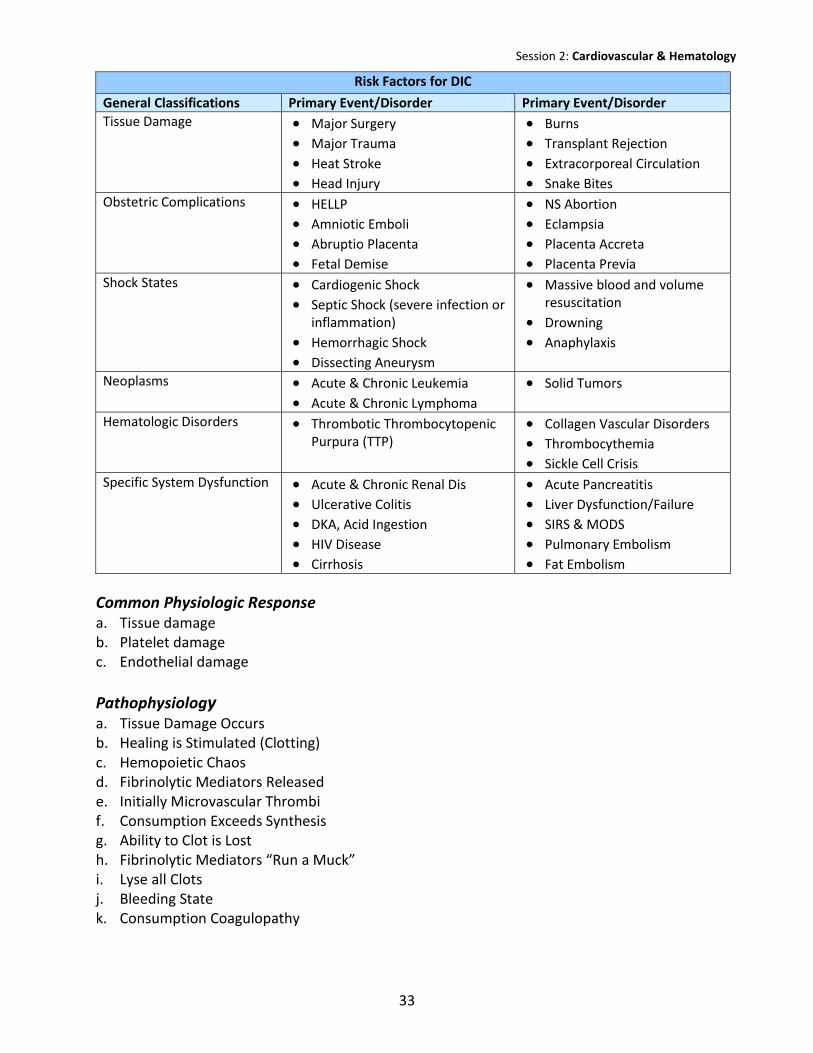
Risk Factors for DIC
General Classifications Primary Event/Disorder Primary Event/Disorder Tissue Damage Major Surgery
Major Trauma
Heat Stroke Head Injury
Burns Transplant Rejection
Extracorporeal Circulation Snake Bites
Obstetric Complications HELLP Amniotic Emboli Abruptio Placenta Fetal Demise
NS Abortion Eclampsia Placenta Accreta Placenta Previa
Shock States Cardiogenic Shock Septic Shock (severe infection or
inflammation)
Hemorrhagic Shock Dissecting Aneurysm
Massive blood and volume resuscitation
Drowning
Anaphylaxis
Neoplasms Acute & Chronic Leukemia
Acute & Chronic Lymphoma
Solid Tumors
Hematologic Disorders Thrombotic Thrombocytopenic Purpura (TTP)
Collagen Vascular Disorders Thrombocythemia Sickle Cell Crisis
Specific System Dysfunction Acute & Chronic Renal Dis Ulcerative Colitis DKA, Acid Ingestion HIV Disease Cirrhosis
Acute Pancreatitis Liver Dysfunction/Failure SIRS & MODS Pulmonary Embolism Fat Embolism
Common Physiologic Response a. Tissue damage b. Platelet damage c. Endothelial damage
Pathophysiology a. Tissue Damage Occurs b. Healing is Stimulated (Clotting) c. Hemopoietic Chaos d. Fibrinolytic Mediators Released e. Initially Microvascular Thrombi f. Consumption Exceeds Synthesis g. Ability to Clot is Lost h. Fibrinolytic Mediators “Run a Muck” i. Lyse all Clots j. Bleeding State k. Consumption Coagulopathy

Session 2: Cardiovascular & Hematology
34
Physical Assessment and Findings The primary problem and pre-existing condition certainly play a major role in the presentation. All systems are at risk for dysfunction. The most common problems occur in the pulmonary, renal and hematopoietic systems. Any bleeding patient who does not have a history of or “reason” to bleed should be suspected of DIC.
Laboratory Findings
Test Elevated Decreased
Hgb
HCT
Platelet Ct
PT
PTT
Fibrinogen
FDP/FSP
D-Dimer
Treatment No definitive treatment exists for DIC. The major goal is to treat primary disorder – stopping the hemapoietic chaos. In addition patient and family emotional support is paramount for quality nursing care. a. Support/Treat the Primary Problem – Eradicate the Cause of DIC b. Early Recognition c. Decrease Bleeding Risk d. Treat Pain e. Transfusion Therapy – PRBC, FFP, Platelets, Cyro f. Vit K g. Anticoagulation Therapy – Heparin h. General Critical Care Management
Heparin Induced Thrombocytopenia (HIT) & Thrombus a. Acquired Allergy to Heparin b. Antibodies are Produced to Heparin c. With Heparin Admin the Antibodies ‘attack’ Heparin and Thrombocytes d. Pt’s Platelet Count Drops (typically by 50% from baseline) e. Some Patients Will Develop Thrombi f. Treatment is to Stop all Heparin g. Admin Non-Heparin Anticoagulant h. Admin Platelets ONLY if Needed

Session 2: Cardiovascular & Hematology
35
Thrombotic Thrombocytopenic Purpura (TTP) a. Drop in Platelet Ct b. Hemolytic Anemia c. Classically Presents with Neuro Symptoms or Renal Dysfunction and Fever d. Difficult Diagnosis e. Causes: Drugs or BMT, Autoimmune Dis, AIDS, Depressed Bone Marrow, DIC, WCS,
Bleeding, Extracorporeal Cir., Medications, Artificial Heart Valve, Hemodilution f. Treatment
Stop Cause
Admin Platelets or Neumega
Plasmapheresis
Idiopathic Thrombocytopenic Purpura (ITP) a. Thrombocytopenia < 150,000 b. Unable to Determine Cause
Drug Induced Coagulopathies
The Physiology of Coagulation & Fibrinolysis (review) Hemostatic Mechanisms The actual forming of a blood clot is a complex integration of mechanisms of the blood vessels, thrombocytes, erythrocytes, coagulation factors, endothelial cells, leukocytes and a myriad of chemical mediators Physiological Clotting Process a. Local Vascular Response - vasoconstriction b. Activation of Platelets & Formation of a Platelet Plug c. Formation of a Blood Clot Fibrinolytic Mechanisms a. Enzymatic degradation of fibrin clot by plasmin b. Hemostasis/Fibrinolysis Control Mechanisms
Anticoagulants Principle indication for anticoagulant therapy is to prevent or decrease the risk of venous thrombosis. Indirect Thrombin Inhibitors a. Unfractionated Heparin: Monitor aPTT, reversal agent Protamine b. Low-Molecular-Weight Heparins: Monitor Antifactor Xa, reversal agent Protamine c. Arixtra (Fondaparinux) synthetic – no reversal d. Xarelto (Rivaroxaban) oral – no reversal

Session 2: Cardiovascular & Hematology
36
Direct Thrombin Inhibitors a. Hirudin & Derivatives – IV: leech saliva – no reversal b. Argatroban – synthetic IV – no reversal c. Pradax (Dabigatran) oral – no reversal Vitamin K Antagonist a. Coumadin (Warfarin), Monitor PT/INR, reversal agent vit K
Antiplatelet Agents Principle indication for antiplatelet therapy is to prevent or decrease the risk of arterial thrombosis. Bleeding time is the primary monitoring test and no reversal agent has been identified a. Aspirin b. Non-Aspirin NSAIDs c. Adenosine Diphosphate Receptor Antagonists
Plavix (Clopidogrel)
Prasugrel (Effient)
IV. ANEMIA The primary problem with anemia, decrease number of Red Blood Cells, is that the body will not have adequate oxygen delivery.
Etiologies a. Blood Loss b. Lack of or Underproduction of Red Cells
Malnutrition
Chronic Illness
Cancer or Cancer Treatments
Liver or Renal Dysfunction
Macrocytic
Microcytic c. Destruction of Red Cells or Hemolysis
Cardiopulmonary Bypass Machine
Immune Response
Sickle Cell Disease
TTP

Session 2: Cardiovascular & Hematology
37
Clinical Presentation Directly result from lack of oxygen delivery a. Tachycardia b. Rapid Respiratory Rate c. Weak Pulses d. Orthostatic Hypotension e. Decreased Urinary Output f. Decreased LOC g. Hypovolemic Shock
Treatment Option a. Identify and Treat the Underlining Cause b. Administer Packed Red Blood Cells c. Recombinant Human Erythropoietin d. Supplemental Vitamins & Minerals e. Blood Conservation Procedures f. Maintain Hgb 7-9 (non-bleeding patient)
V. IMMUNOLOGY – ONCOLOGY
Etiology of Immunosuppression a. Primary Neutropenia b. Immunosuppressive Agents (chemo, anti-rejection) c. Radiation Therapy d. Autoimmune Disorders e. Viral Infections (HIV/AIDS) f. Genetic Disorders g. Diseases/Disorders (DM, ETOH abuse) h. Chronically Critically Ill and Septic
Care Goal Priorities a. Safety b. Prevention of Opportunistic Infection c. Monitoring and Treatment if Infection d. General Support

Session 2: Cardiovascular & Hematology
38
HIV/AIDS A virus spread by blood and body fluids. Incubation period can be 45 days 6 months. Pt feels “flu like” with some lymphadenopathy in the early stages. The virus destroys the CD4 lymphocyte causing immunosuppression. The first concern with HIV is prevention and education. The second is caring for the Immunosupressed individual. Currently infection to AIDS is about 10 – 12 years. In the ED setting assess for opportunistic infections, neutropenic precautions, education and emotional and psychological support.
HIV Testing The target cells for the virus are the T helper cells (CD4). Antibodies will not be detectable immediately after exposure. Enzyme-Linked Immunosorbent Assay (ELISA) Antibody test with 94 – 99% sensitivity. It may take between 6 weeks – 6 months for antibody tests to be positive. Confirmed with a Western blot. CD4 Levels The overall T cell count will decrease secondary to the drop in CD4 cells. Levels of CD4 are more valuable than total count, a CD4 below 200/mm3 is reflective of HIV. The CD4 is also used to monitor the effectiveness of therapy or disease progression. Viral Load Measurement of the HIV-RNA present in the blood. Levels of < 10,000 are considered low risk for disease progression, > 100,000 are considered high risk for progression to AIDS. This measurement is also used to evaluate the effectiveness of therapy.
HIV/AIDS Education a. Medication Education b. Infection Prevention and Early Identification c. Safe Sex Practices d. Communicate with Intimate Partners e. Do Not Share Personal Hygiene Items f. Community Resources g. Avoid Smoking and ETOH h. Encourage Healthy Living (eating, sleeping, exercise…)
Prevention & Early Detection Patients and Families a. Smoking Cessation b. Low Fat Diets c. Ideal Body Wt d. Exercise e. Cancer Screening (colonoscopy at 50yo, mammograms…) f. Sun Screen Use