sensory receptors; neuronal circuits for processing...
TRANSCRIPT

University of Jordan 1
Sensory Receptors; Neuronal
Circuits For Processing Information
Faisal I. Mohammed, MD, PhD

University of Jordan 2
Objectives➢ Define receptors (Transducers) and classify them
➢ Describe the generator (receptor) potential and its importance in sensory coding
➢ List the types of somatic receptors in the skin
➢ Explain the mechanism of sensory coding
➢ Interpret the mechanism of receptor adaptation and classify the types of receptors accordingly (Phasic and Tonic receptors)
➢ Describe sensory neuronal processing and its functional importance

University of Jordan 3
Types of Sensory Receptors: Classification
by Modality (Stimulus they transduce)
➢ Mechanoreceptors
➢ detect deformation, Touch and Prssure
➢ Thermoreceptors
➢ detect change in temperature
➢ Nociceptors
➢ detect tissue damage (pain receptors)
➢ Electromagnetic (Photoreceptors)
➢ detect light (Rods and Cones)
➢ Chemoreceptors
➢ taste, smell, CO2, O2, etc.

University of Jordan 4
Classification by Location➢ Exteroceptors – sensitive to stimuli arising from outside the
body
➢ Located at or near body surfaces
➢ Include receptors for touch, pressure, pain, and temperature
➢ Interoceptors – (visceroceptors) receive stimuli from internal viscera
➢ Monitor a variety of stimuli (distension of viscera, pain)
➢ Proprioceptors – sense of position- monitor degree of stretch
➢ Located in musculoskeletal organs (muscle, tendons and skin around joints)

University of Jordan 5
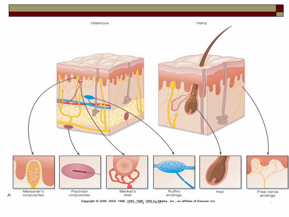
Types of Sensory Receptors

University of Jordan 6
University of Jordan 7
University of Jordan 8
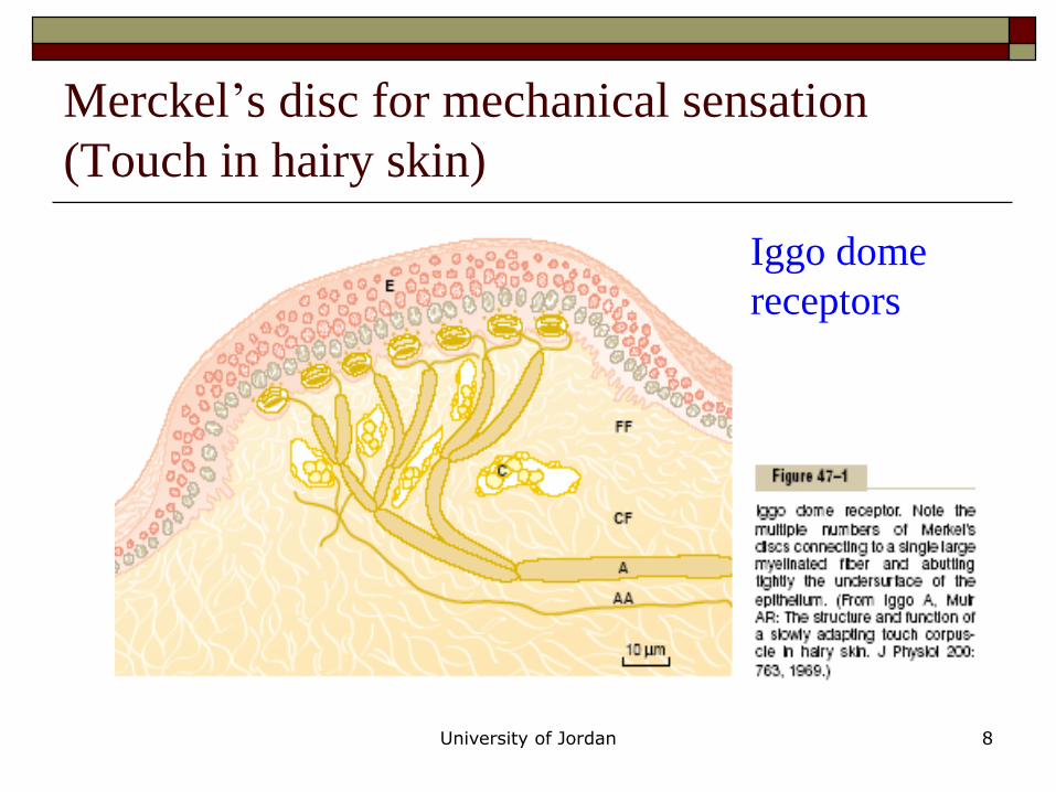
Merckel’s disc for mechanical sensation
(Touch in hairy skin)
Iggo dome
receptors

University of Jordan 9
Tactile Receptors
Free nerve endings (A and C fibers)
detect touch and pressure
found everywhere in the skin and other tissues
Meissner’s corpuscles (A)
rapidly adapting (within a fraction of a second) and detect movement of light objects over skin
found on nonhairy skin (glabrous skin), fingertips and lips
Merkel’s discs (A)
respond rapidly at first and then slowly adapt, detect the “steady state”
found on hairy as well a glabrous (non hairy) skin

University of Jordan 10
Tactile Receptors
Hair end organ
adapts rapidly and detects movement over the body
Ruffini’s end organ
slowly adapting and respond to continual
deformation of the skin and joint rotation
Pacinian corpuscle
very rapidly adapting and is stimulated only by rapid
movement
detects vibration and other rapid changes in the skin

University of Jordan 11
Tactile Sense Transmission
Meissner’s corpuscles, hair receptors,
Pacinian corpuscles and Ruffini’s end
organs transmit signals in type A nerve
fibers at 30-70 m/sec.
Free nerve endings transmit signals in type
A nerve fibers at 5-30 m/sec, some by
type C unmyelinated fibers at 0.5-2 m/sec.
The more critical the information the faster
the rate of transmission.

University of Jordan 12
Sensory Receptors: General structure
Receptor area is
None-excitable
region so as it can
discriminate
different intensities,
otherwise it will not
be able to
differentiate
strengths of stimuli

Conversion of Receptor and Generator
Potentials into Action Potentials
Receptor Potential Generator Potential
University of Jordan 14
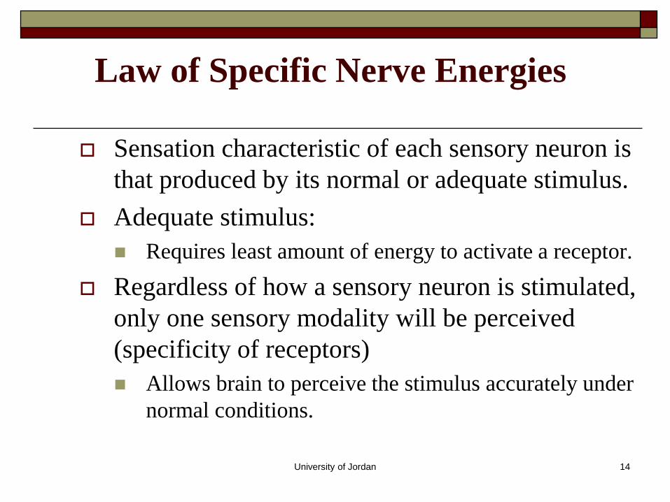
Law of Specific Nerve Energies
Sensation characteristic of each sensory neuron is
that produced by its normal or adequate stimulus.
Adequate stimulus:
Requires least amount of energy to activate a receptor.
Regardless of how a sensory neuron is stimulated,
only one sensory modality will be perceived
(specificity of receptors)
Allows brain to perceive the stimulus accurately under
normal conditions.

University of Jordan 15
Sensation
➢ Each of the principle types sensation; touch, pain, sight, sound, is called a modality of sensation.
➢ Each receptor is responsive to one type of stimulus energy. Specificity is a key property of a receptor, it underlines the most important coding mechanism, the labeled line principle
➢ How the sensation is perceived is determined by the characteristics of the receptor and the central connections of the axon connected to the receptor.

University of Jordan 16
Receptor Excitation
➢ mechanical deformation which stretches the membrane and opens ion channels
➢ application of chemicals which also opens ion channels
➢ change in temperature which alters the permeability of the membrane through changing the metabolic rate
➢ electromagnetic radiation that changes the membrane characteristics

University of Jordan 17
Receptor Region
Spike Generating
Region
Conducting
Region
General Structure of Receptors

University of Jordan 18
Receptor Excitation

University of Jordan 19
Receptor Potential
➢ The membrane potential of the receptor
➢ Excitation of the receptor results from a change in
this potential.
➢ When the receptor potential rises above the
threshold, action potentials appear and the
receptor is active.
➢ The greater the intensity of the stimulus, the
greater the receptor potential, and the greater the
rate of action potential generation.

University of Jordan 20
Generator Potentials In response to stimulus,
sensory nerve endings
produce a local graded
change in membrane
potential.
Potential changes are called
receptor or generator
potential.
Analogous to EPSPs.

University of Jordan 21
Relationship between Receptor Potential and
Action Potentials

University of Jordan 22
Stimulus Strength
Am
plitu
de o
f
Recep
tor p
oten
tial
The effect of stimulus strength on RP
amplitude
Slop of the curve
Greater
sensitivity
region Lesser sensitivity
region

University of Jordan 23
The effect of the amplitude of RP on
the frequency of impulses generated

University of Jordan 24
Adaptation of Receptors
➢ When a continuous stimulus is applied, receptors respond
rapidly at first, but response declines until all receptors
stop firing.
University of Jordan 25
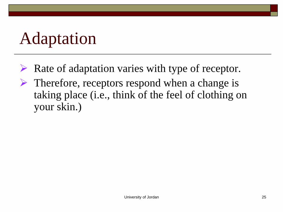
Adaptation
➢ Rate of adaptation varies with type of receptor.
➢ Therefore, receptors respond when a change is taking place (i.e., think of the feel of clothing on your skin.)

University of Jordan 26
Adaptation of Sensory Receptors
Receptors responding to pressure, touch, and smell
adapt quickly
Receptors responding slowly include Merkel’s discs,
Ruffini’s corpuscles, and interoceptors that respond
to chemical levels in the blood
Pain receptors and proprioceptors do not exhibit
adaptation

University of Jordan 27
Slowly Adapting (Tonic) Receptors
➢ continue to transmit impulses to the brain for long periods of time while the stimulus is present
➢ keep brain apprised of the status of the body with respect to its surroundings
➢ will adapt to extinction as long as the stimulus is present, however, this may take hours or days
➢ these receptors include: muscle spindle, golgi tendon apparatus, Ruffini’s endings, Merkels discs, Macula, chemo- and baroreceptors
University of Jordan 28
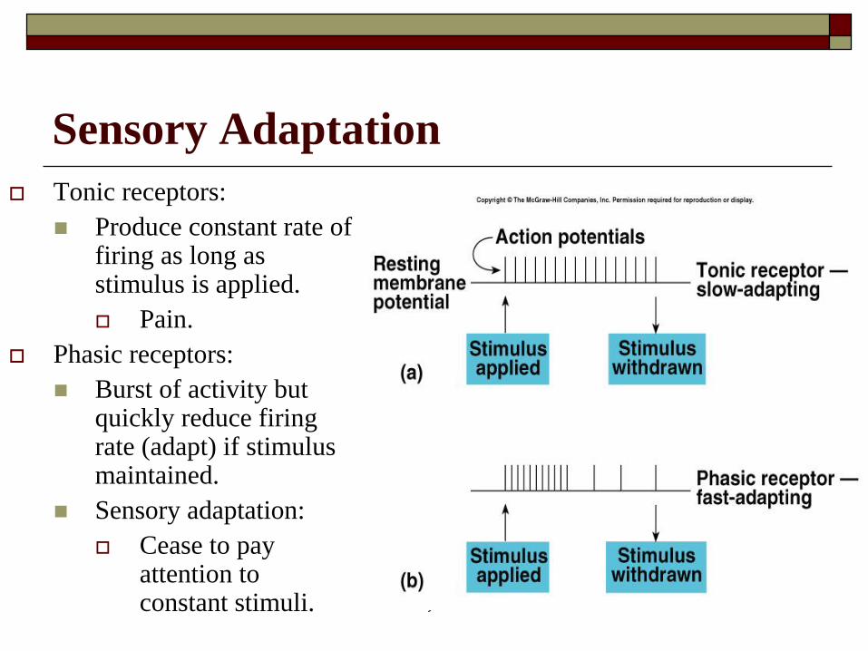
Sensory Adaptation
Tonic receptors:
Produce constant rate of firing as long as stimulus is applied.
Pain.
Phasic receptors:
Burst of activity but quickly reduce firing rate (adapt) if stimulus maintained.
Sensory adaptation:
Cease to pay attention to constant stimuli.

University of Jordan 29
Rapidly Adapting (Phasic) Receptors
➢ respond only when change is taking place
➢ Rate and Strength of the response is related to the Rate and Intensity of the stimulus
➢ important for predicting the future position or condition of the body
➢ very important for balance and movement
➢ types of rapidly adapting receptors: pacinian corpuscle, semicircular canals in the inner ear
University of Jordan 30
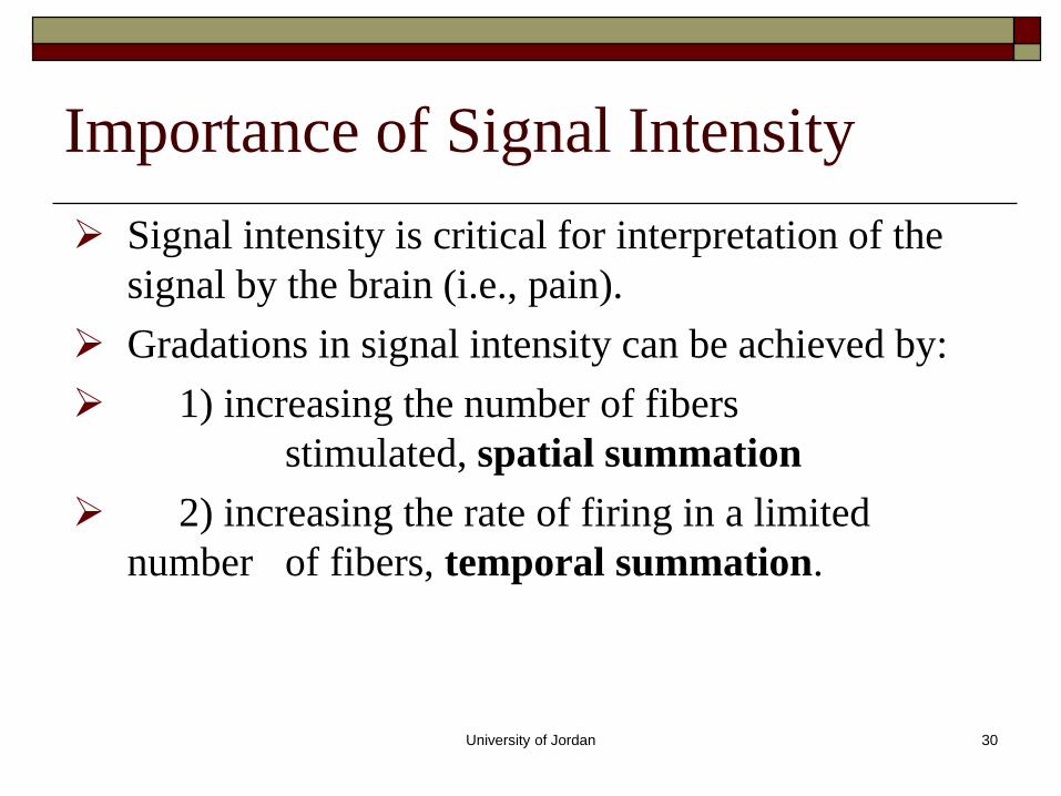
Importance of Signal Intensity
➢ Signal intensity is critical for interpretation of the
signal by the brain (i.e., pain).
➢ Gradations in signal intensity can be achieved by:
➢ 1) increasing the number of fibers
stimulated, spatial summation
➢ 2) increasing the rate of firing in a limited
number of fibers, temporal summation.
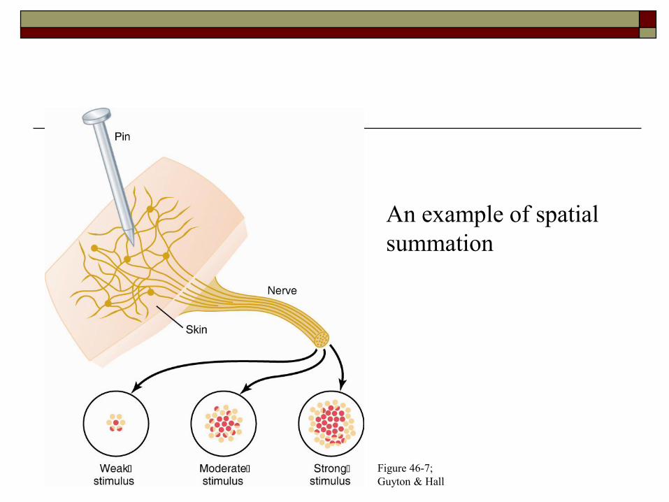
Figure 46-7;
Guyton & Hall
Signal Intensity
An example of spatial
summation
University of Jordan 32
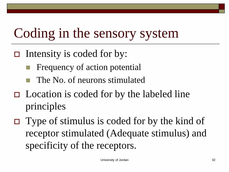
Coding in the sensory system
Intensity is coded for by:
Frequency of action potential
The No. of neurons stimulated
Location is coded for by the labeled line
principles
Type of stimulus is coded for by the kind of
receptor stimulated (Adequate stimulus) and
specificity of the receptors.


➢ Mapping of the
postcentral gyrus.
➢ Size of the cortical
region representing a
body part depends on
density of receptors
on that part and the
sensory impulses
received from that
part.
Mapping of the Primary
Somatosensory Area
University of Jordan 35
Receptive Fields
Area of skin whose stimulation results in changes
in the firing rate of the neuron.
Area of each receptor field varies inversely with the
density of receptors in the region.
Back and legs have few sensory endings.
Receptive field is large.
Fingertips have large # of cutaneous receptors.
Receptive field is small.

Figure 46-7;
Guyton & Hall
Signal Intensity
An example of spatial
summation
University of Jordan 37
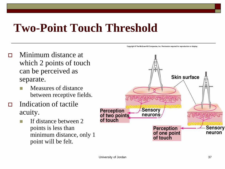
Two-Point Touch Threshold
Minimum distance at which 2 points of touch can be perceived as separate.
Measures of distance between receptive fields.
Indication of tactile acuity.
If distance between 2 points is less than minimum distance, only 1 point will be felt.

University of Jordan 38
Neuronal Processing

University of Jordan 39
Relaying Signals through Neuronal
Pools

University of Jordan 40
Neuronal Pools
➢ groups of neurons with special characteristics
of organization
➢ comprise many different types of neuronal
circuits
➢ converging
➢ diverging
➢ reverberating

University of Jordan 41
Neuronal Pools: Localization of sensory
Information modification

University of Jordan 42
University of Jordan 43
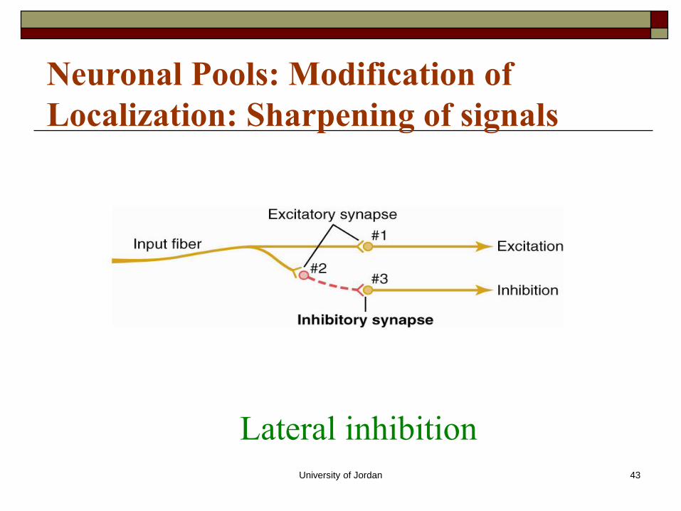
Lateral inhibition
Neuronal Pools: Modification of
Localization: Sharpening of signals

University of Jordan 44
Lateral Inhibition Sharpening of sensation.
When a blunt object touches the skin, sensory neurons in the center areas are stimulated more than neighboring fields.
Stimulation will gradually diminish from the point of greatest contact, without a clear, sharp boundary.
Will be perceived as a single touch with well defined borders.
Occurs within CNS.

Lateral Inhibition
in the sensory
System as a way
of sharpening of the
stimulus

University of Jordan 46
Reverberating Circuits: prolongation of
Time of the signals

Copyright © 2004 Pearson Education, Inc., publishing as Benjamin Cummings
The Organization of Neuronal Pools


49
Neural Circuits
University of Jordan 50
Other mechanisms for prolongation of
time
Synaptic afterdischarge: since the time of
EPSP (15-20 msec) is longer than the time of
AP(0.1 – 10 msec) then more No. of AP per
one EPSP
Parallel circuits

University of Jordan 51
Stabilization of neuronal discharge
Synaptic fatigue: short term and acute adjustment of sensitivity
Neuronal inhibitory circuits:
Gross inhibition –Basal ganglia inhibits muscle tone
Feed back inhibition-Cortico-fugal fibers from cerebral cortex descending fibers to control the intensity and sharpness
Downregulation and upregulation- Long term stabilization through modification of the receptor availability (internalization or externalization)

University of Jordan 52