introduction to histology -...
TRANSCRIPT
Introduction to Histology
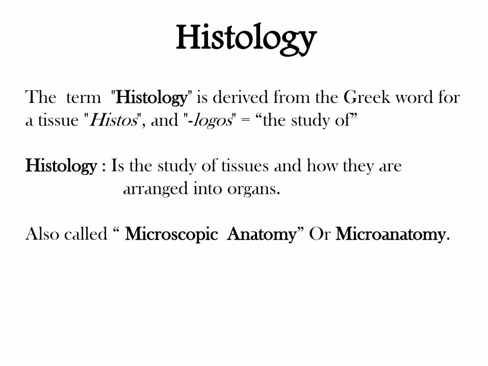
Histology The term "Histology" is derived from the Greek word for
a tissue "Histos", and "-logos" = “the study of”
Histology : Is the study of tissues and how they are
arranged into organs.
Also called “ Microscopic Anatomy” Or Microanatomy.
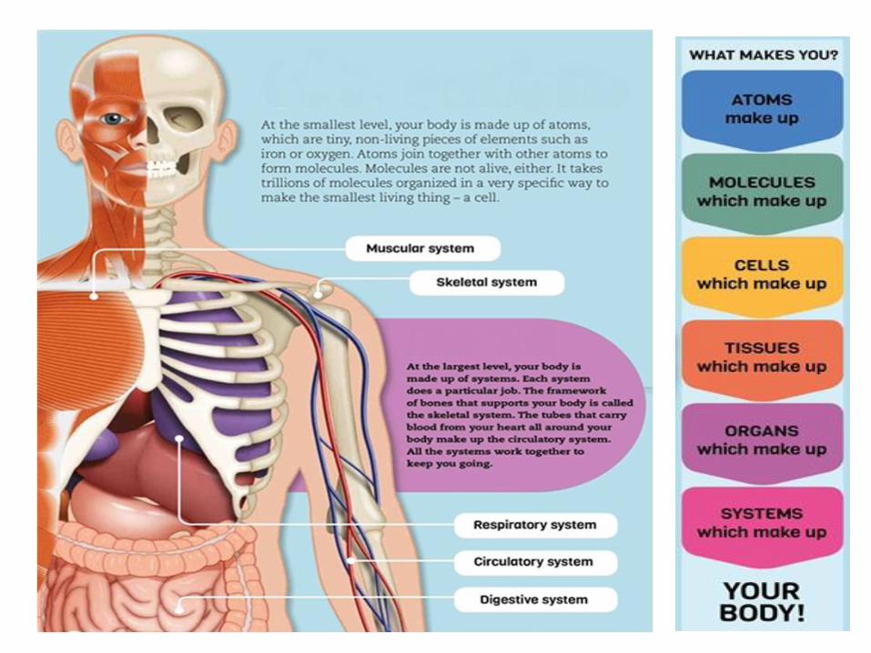
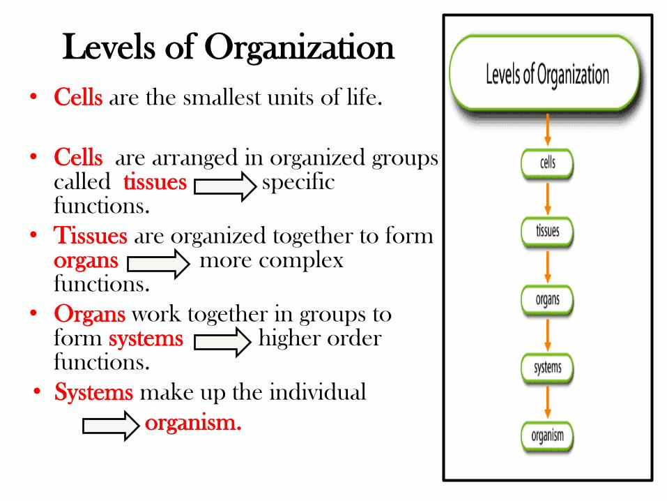
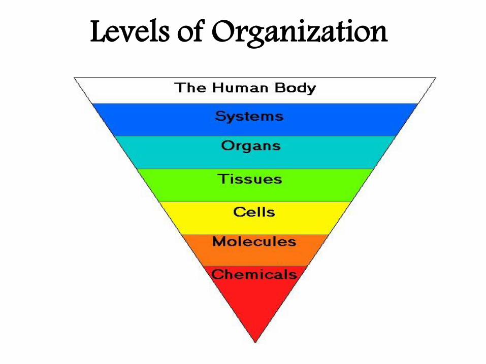
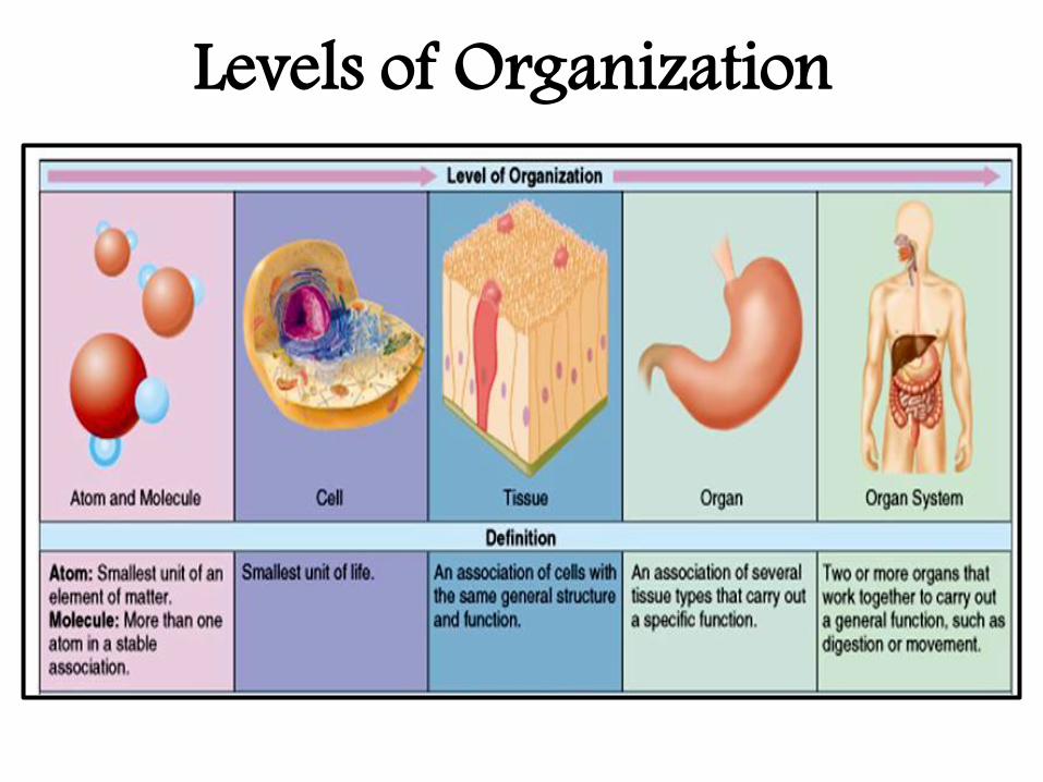
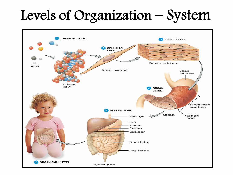
• Cells are the smallest units of life.
• Cells are arranged in organized groups called tissues specific functions.
• Tissues are organized together to form organs more complex functions.
• Organs work together in groups to form systems higher order functions.
• Systems make up the individual
organism.
Levels of Organization
Levels of Organization
Levels of Organization
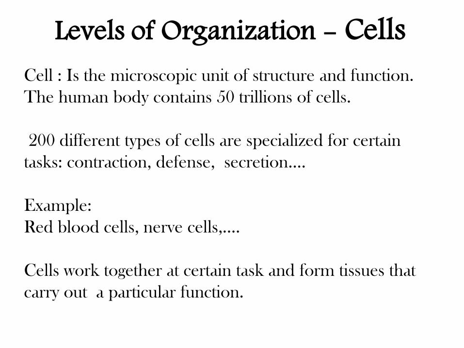
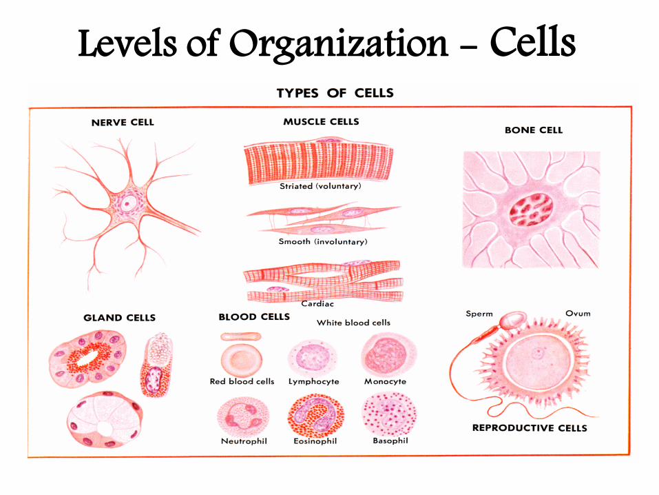
Cell : Is the microscopic unit of structure and function.
The human body contains 50 trillions of cells.
200 different types of cells are specialized for certain
tasks: contraction, defense, secretion….
Example:
Red blood cells, nerve cells,….
Cells work together at certain task and form tissues that
carry out a particular function.
Levels of Organization - Cells
Levels of Organization - Cells
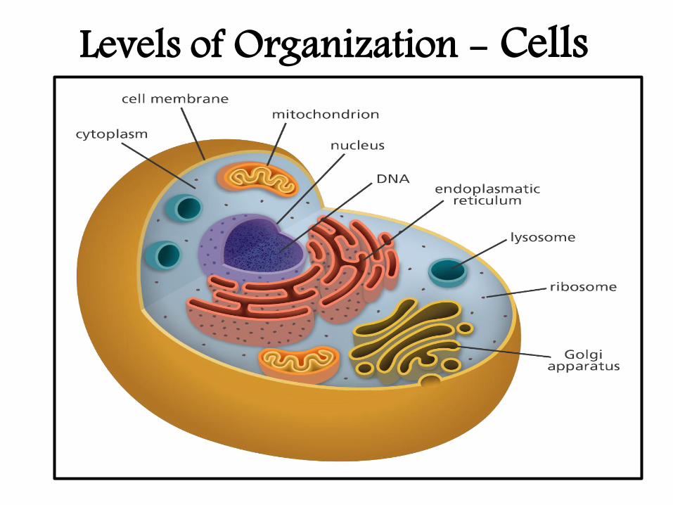
Levels of Organization - Cells
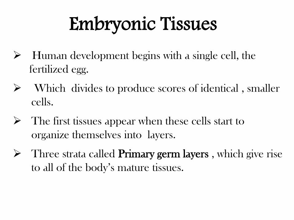
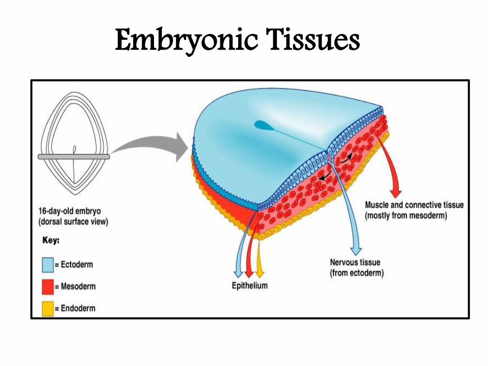
Human development begins with a single cell, the
fertilized egg.
Which divides to produce scores of identical , smaller
cells.
The first tissues appear when these cells start to
organize themselves into layers.
Three strata called Primary germ layers , which give rise
to all of the body’s mature tissues.
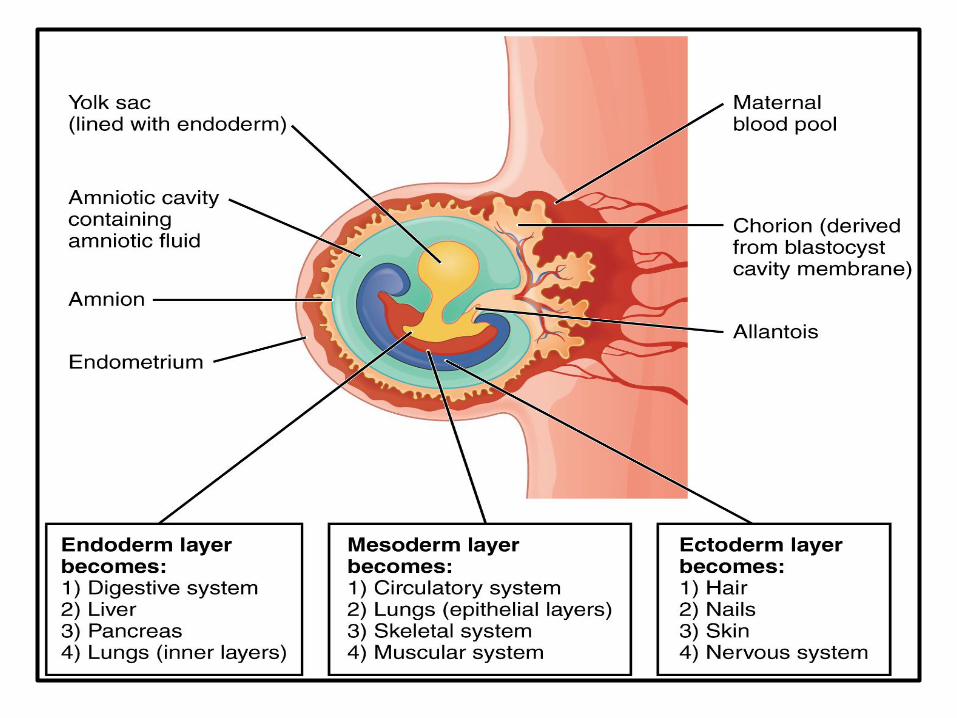
Embryonic Tissues
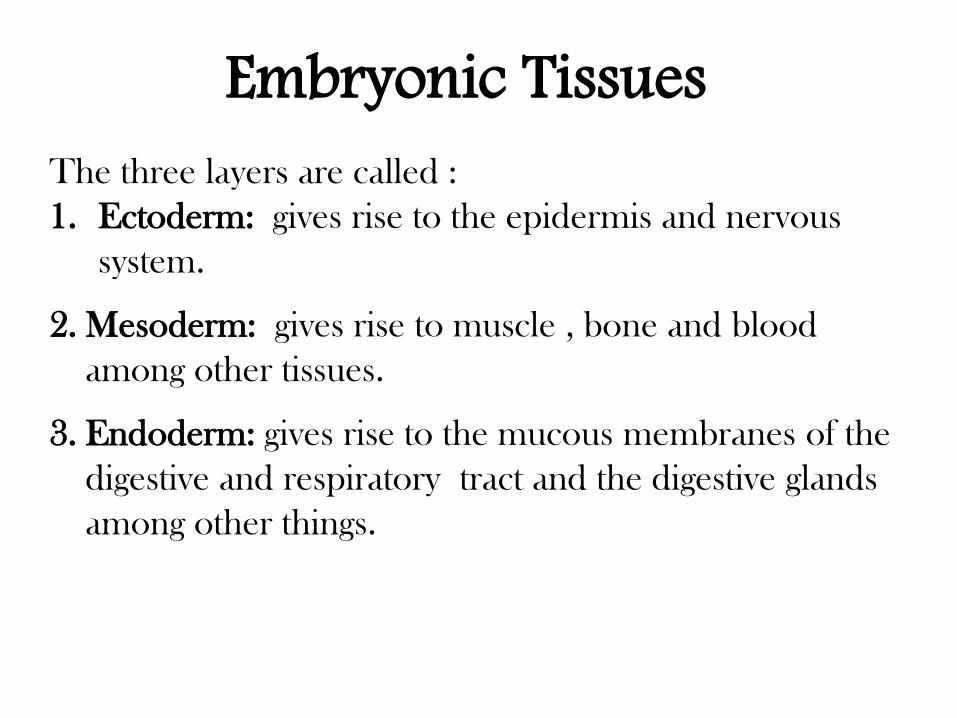
The three layers are called :
1. Ectoderm: gives rise to the epidermis and nervous
system.
2. Mesoderm: gives rise to muscle , bone and blood
among other tissues.
3. Endoderm: gives rise to the mucous membranes of the
digestive and respiratory tract and the digestive glands
among other things.
Embryonic Tissues
Embryonic Tissues
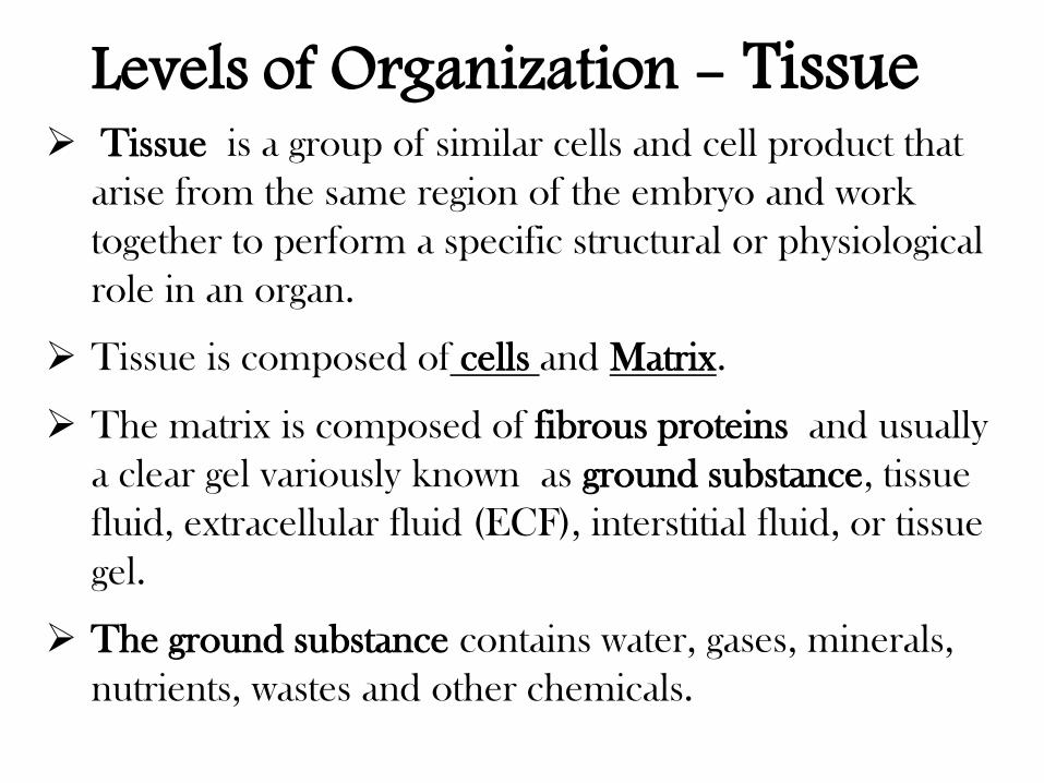
Tissue is a group of similar cells and cell product that
arise from the same region of the embryo and work
together to perform a specific structural or physiological
role in an organ.
Tissue is composed of cells and Matrix.
The matrix is composed of fibrous proteins and usually
a clear gel variously known as ground substance, tissue
fluid, extracellular fluid (ECF), interstitial fluid, or tissue
gel.
The ground substance contains water, gases, minerals,
nutrients, wastes and other chemicals.
Levels of Organization - Tissue
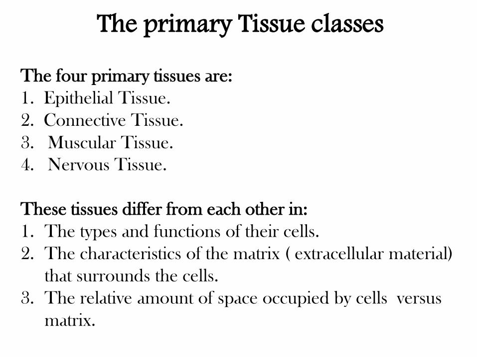
The primary Tissue classes The four primary tissues are:
1. Epithelial Tissue.
2. Connective Tissue.
3. Muscular Tissue.
4. Nervous Tissue.
These tissues differ from each other in:
1. The types and functions of their cells.
2. The characteristics of the matrix ( extracellular material)
that surrounds the cells.
3. The relative amount of space occupied by cells versus
matrix.
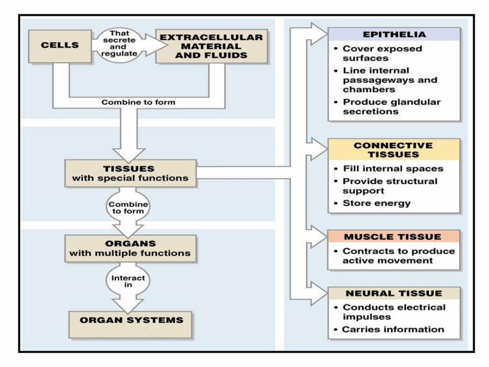
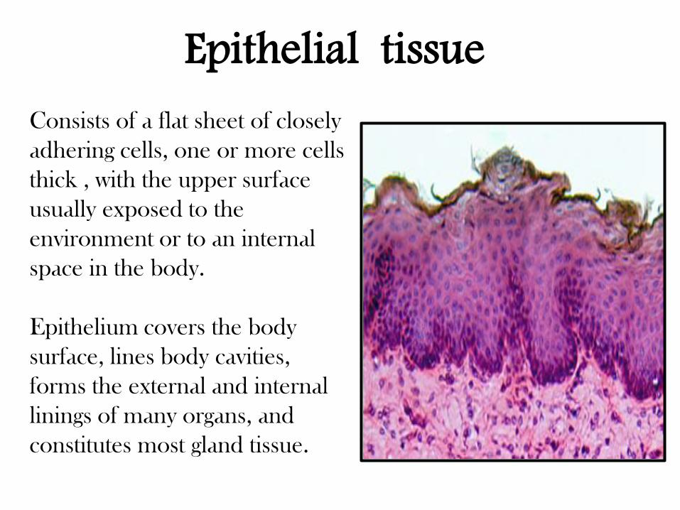
Consists of a flat sheet of closely
adhering cells, one or more cells
thick , with the upper surface
usually exposed to the
environment or to an internal
space in the body.
Epithelium covers the body
surface, lines body cavities,
forms the external and internal
linings of many organs, and
constitutes most gland tissue.
Epithelial tissue
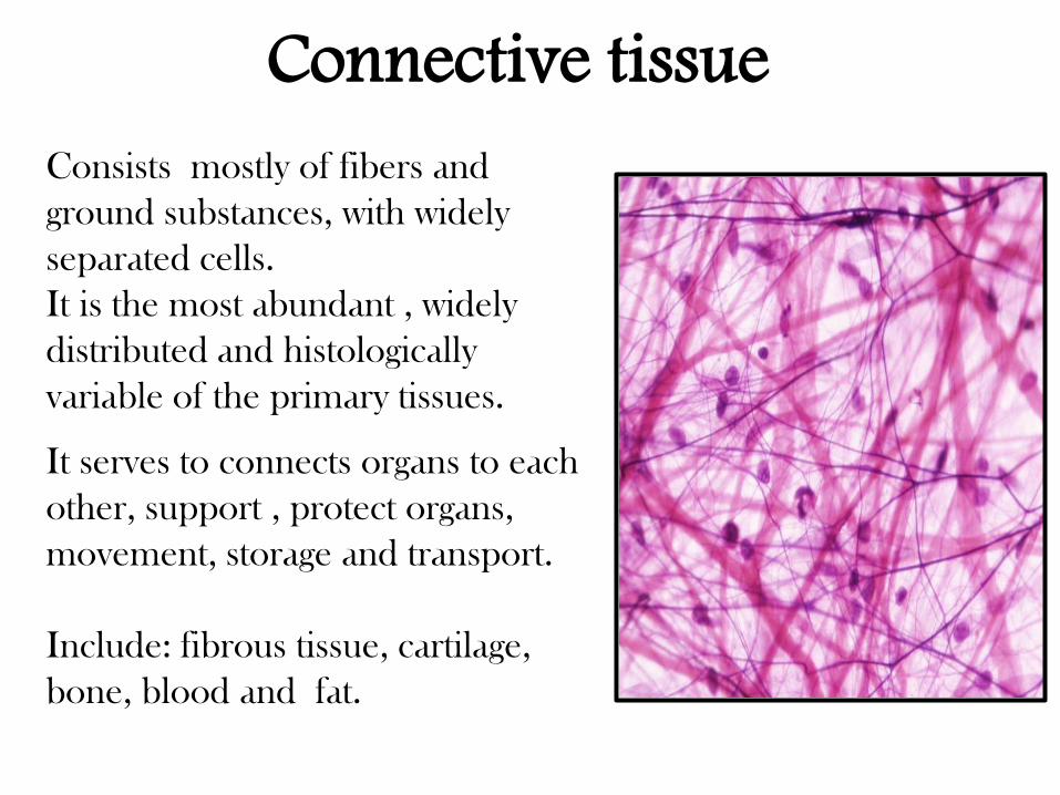
Consists mostly of fibers and
ground substances, with widely
separated cells.
It is the most abundant , widely
distributed and histologically
variable of the primary tissues.
It serves to connects organs to each
other, support , protect organs,
movement, storage and transport.
Include: fibrous tissue, cartilage,
bone, blood and fat.
Connective tissue
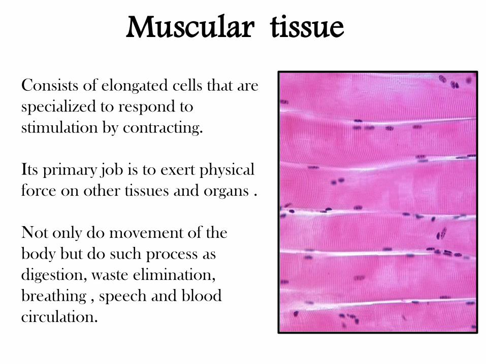
Consists of elongated cells that are
specialized to respond to
stimulation by contracting.
Its primary job is to exert physical
force on other tissues and organs .
Not only do movement of the
body but do such process as
digestion, waste elimination,
breathing , speech and blood
circulation.
Muscular tissue
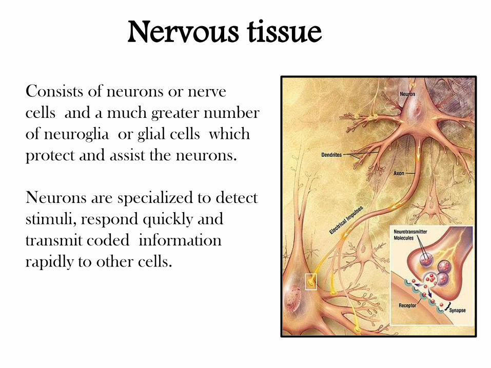
Consists of neurons or nerve
cells and a much greater number
of neuroglia or glial cells which
protect and assist the neurons.
Neurons are specialized to detect
stimuli, respond quickly and
transmit coded information
rapidly to other cells.
Nervous tissue
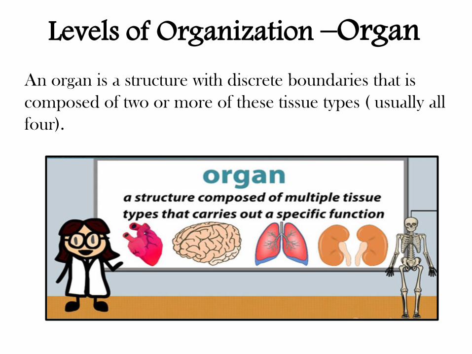
An organ is a structure with discrete boundaries that is
composed of two or more of these tissue types ( usually all
four).
Levels of Organization –Organ
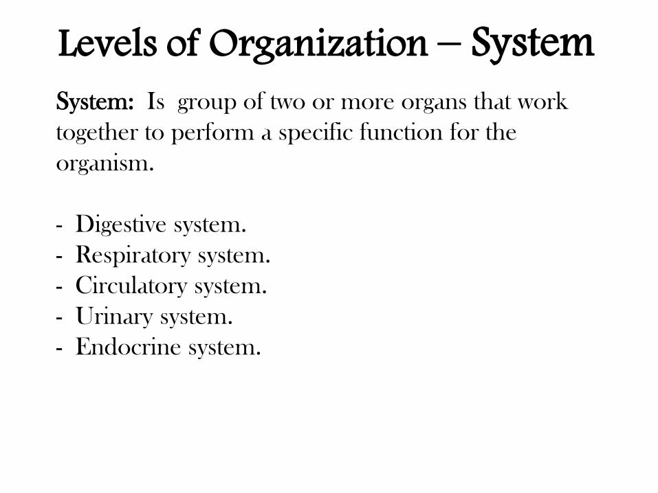
System: Is group of two or more organs that work
together to perform a specific function for the
organism.
- Digestive system.
- Respiratory system.
- Circulatory system.
- Urinary system.
- Endocrine system.
Levels of Organization – System
Levels of Organization – System
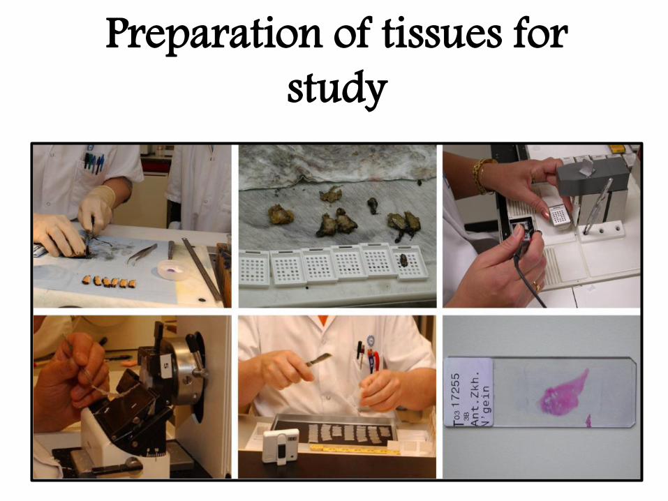
Preparation of tissues for study
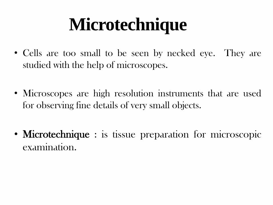
• Cells are too small to be seen by necked eye. They are
studied with the help of microscopes.
• Microscopes are high resolution instruments that are used
for observing fine details of very small objects.
• Microtechnique : is tissue preparation for microscopic
examination.
Microtechnique
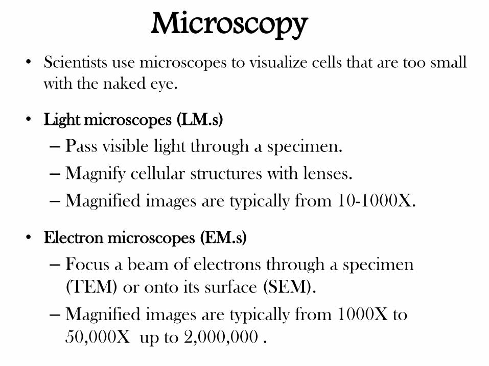
• Scientists use microscopes to visualize cells that are too small
with the naked eye.
• Light microscopes (LM.s)
– Pass visible light through a specimen.
– Magnify cellular structures with lenses.
– Magnified images are typically from 10-1000X.
• Electron microscopes (EM.s)
– Focus a beam of electrons through a specimen
(TEM) or onto its surface (SEM).
– Magnified images are typically from 1000X to
50,000X up to 2,000,000 .
Microscopy
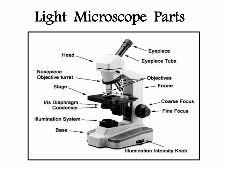
Light Microscope Parts
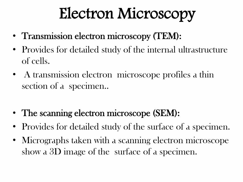
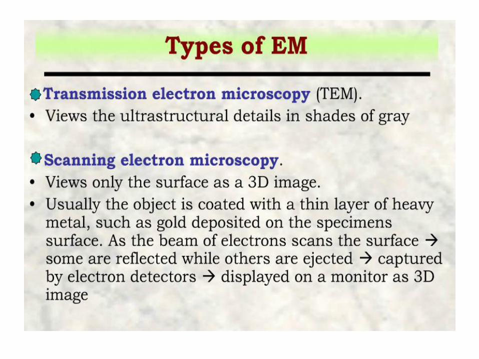
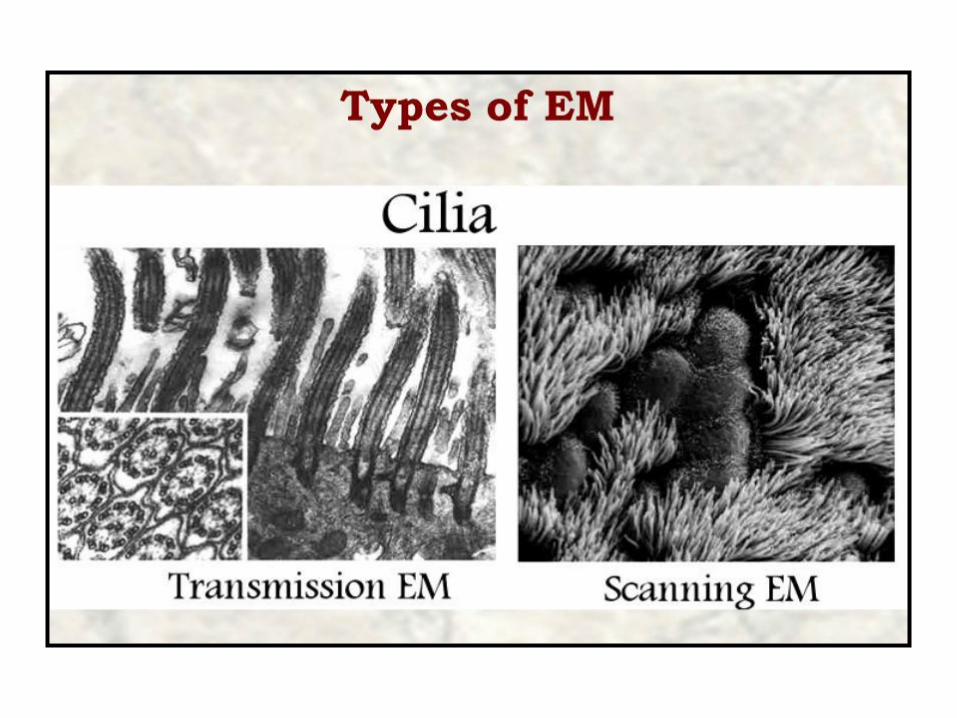
• Transmission electron microscopy (TEM):
• Provides for detailed study of the internal ultrastructure
of cells.
• A transmission electron microscope profiles a thin
section of a specimen..
• The scanning electron microscope (SEM):
• Provides for detailed study of the surface of a specimen.
• Micrographs taken with a scanning electron microscope
show a 3D image of the surface of a specimen.
Electron Microscopy
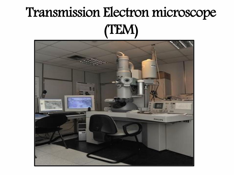
Transmission Electron microscope (TEM)
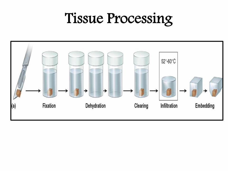
Preparation of tissue For Light Microscopy
( Tissue Processing) • The aim of tissue processing is to embed the tissue in
a solid medium firm enough to support the tissue and
give it sufficient rigidity to enable thin sections to be
cut, and yet soft enough not to damage the knife or
tissue with preservation of the structure with the least
possible alteration.
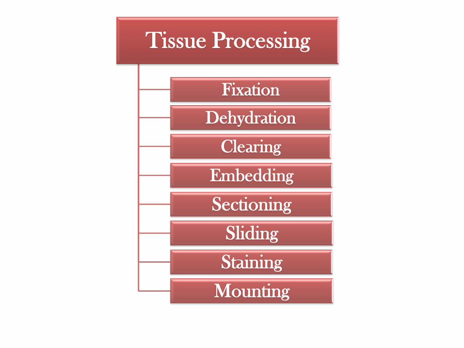
Tissue Processing
Fixation
Dehydration
Clearing
Embedding
Sectioning
Sliding
Staining
Mounting
Tissue Processing
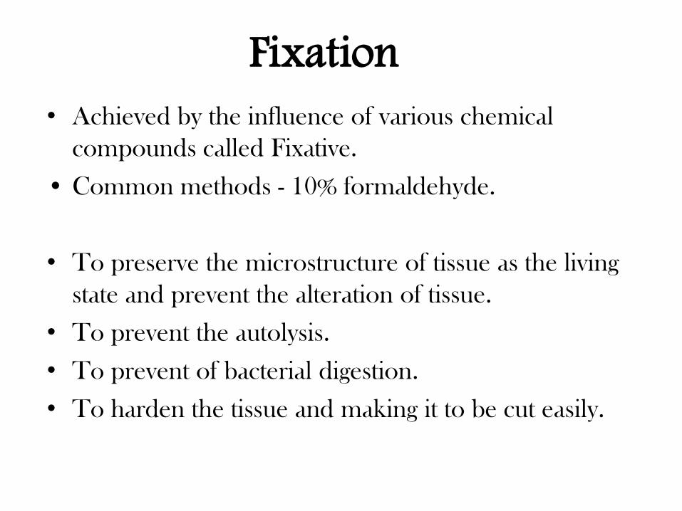
Fixation • Achieved by the influence of various chemical
compounds called Fixative.
• Common methods - 10% formaldehyde.
• To preserve the microstructure of tissue as the living
state and prevent the alteration of tissue.
• To prevent the autolysis.
• To prevent of bacterial digestion.
• To harden the tissue and making it to be cut easily.
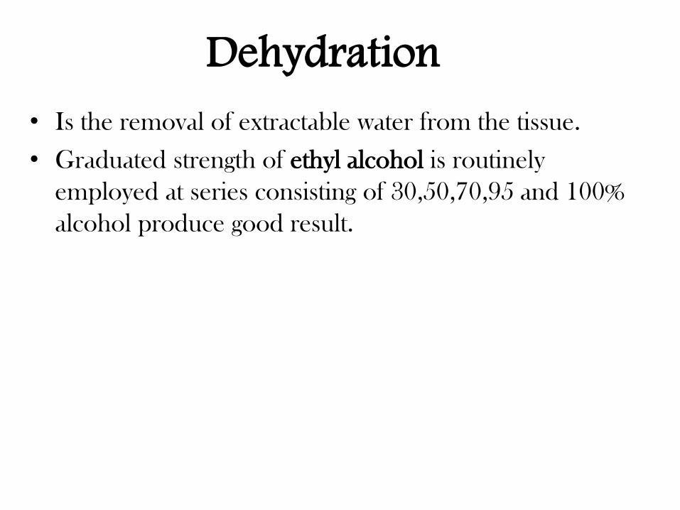
Dehydration • Is the removal of extractable water from the tissue.
• Graduated strength of ethyl alcohol is routinely
employed at series consisting of 30,50,70,95 and 100%
alcohol produce good result.
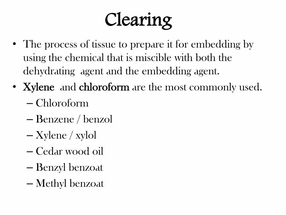
Clearing • The process of tissue to prepare it for embedding by
using the chemical that is miscible with both the
dehydrating agent and the embedding agent.
• Xylene and chloroform are the most commonly used.
– Chloroform
– Benzene / benzol
– Xylene / xylol
– Cedar wood oil
– Benzyl benzoat
– Methyl benzoat
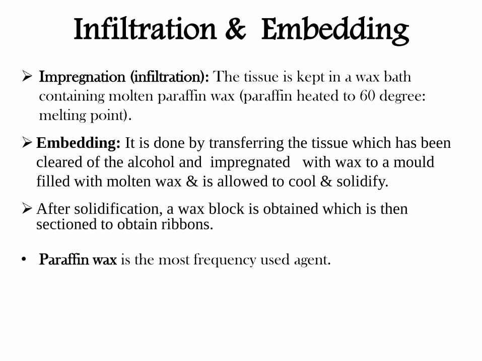
Infiltration & Embedding Impregnation (infiltration): The tissue is kept in a wax bath
containing molten paraffin wax (paraffin heated to 60 degree:
melting point).
Embedding: It is done by transferring the tissue which has been
cleared of the alcohol and impregnated with wax to a mould
filled with molten wax & is allowed to cool & solidify.
After solidification, a wax block is obtained which is then sectioned to obtain ribbons.
• Paraffin wax is the most frequency used agent.
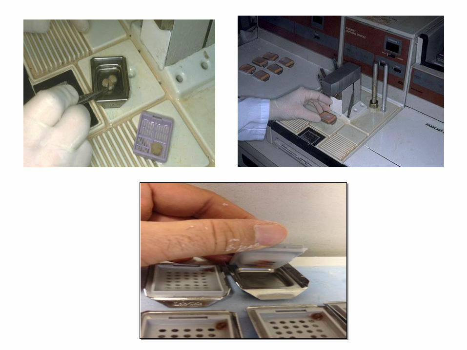
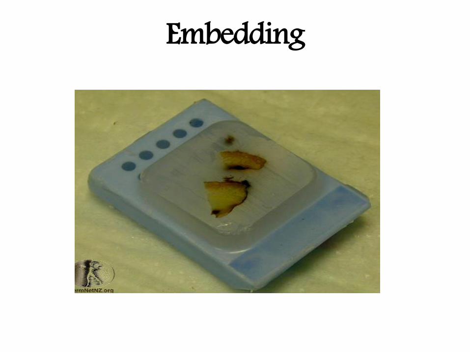
Embedding
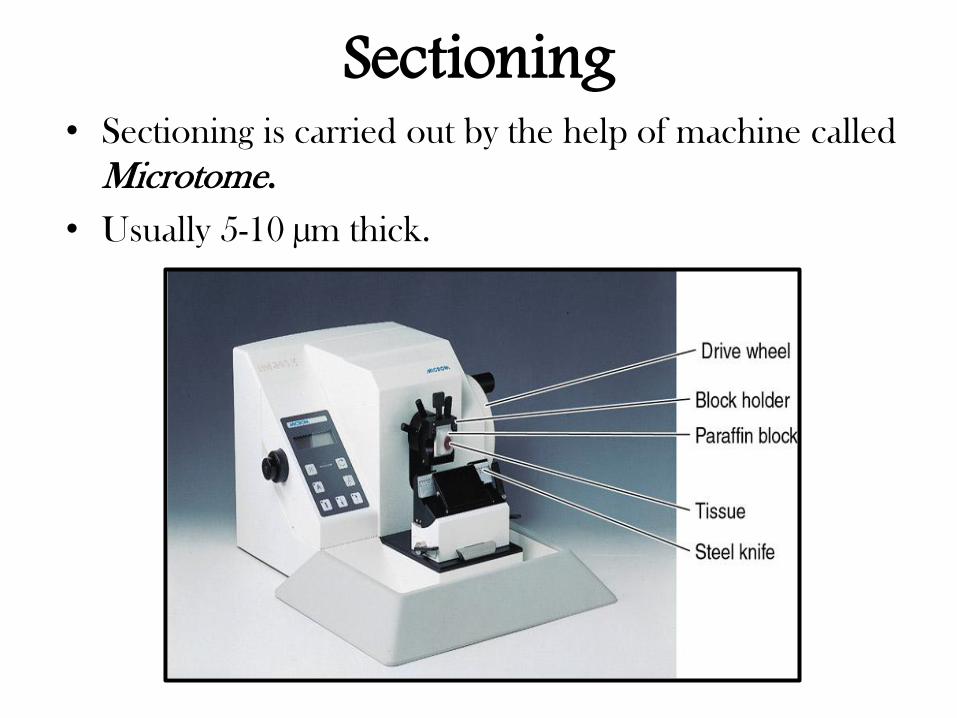
Sectioning • Sectioning is carried out by the help of machine called
Microtome.
• Usually 5-10 µm thick.
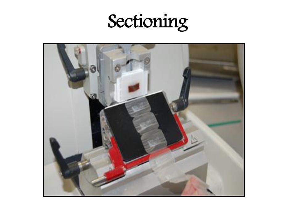
Sectioning
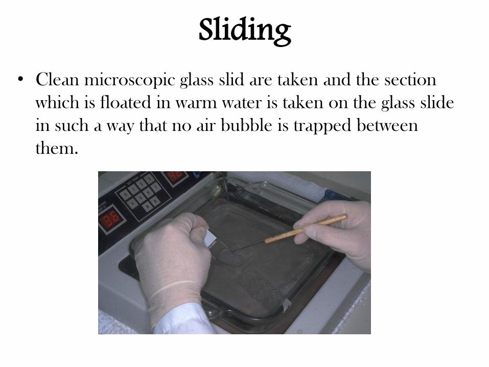
Sliding • Clean microscopic glass slid are taken and the section
which is floated in warm water is taken on the glass slide
in such a way that no air bubble is trapped between
them.
Staining • Because the tissues of the body are colourless and are
difficult to study their details. The staining technique
enhance natural contrast and permits distinction to be made
between them.
• The most commonly used dye is a combination of
HEMATOXYLIN and EOSIN ( H&E).
• By this method the nuclear structures are stained dark
purple or blue .
• All cytoplasmic structures and intracellular substances are
stained pink or red.
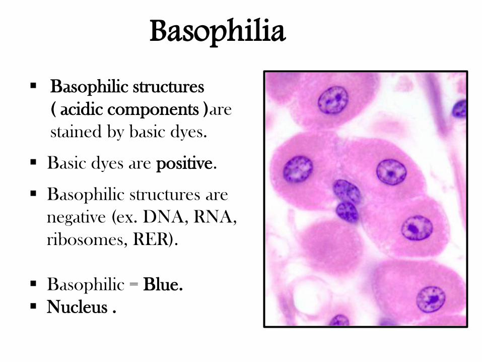
Basophilic structures
( acidic components )are
stained by basic dyes.
Basic dyes are positive.
Basophilic structures are
negative (ex. DNA, RNA,
ribosomes, RER).
Basophilic = Blue.
Nucleus .
Basophilia
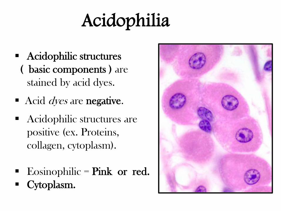
Acidophilic structures
( basic components ) are
stained by acid dyes.
Acid dyes are negative.
Acidophilic structures are
positive (ex. Proteins,
collagen, cytoplasm).
Eosinophilic = Pink or red.
Cytoplasm.
Acidophilia
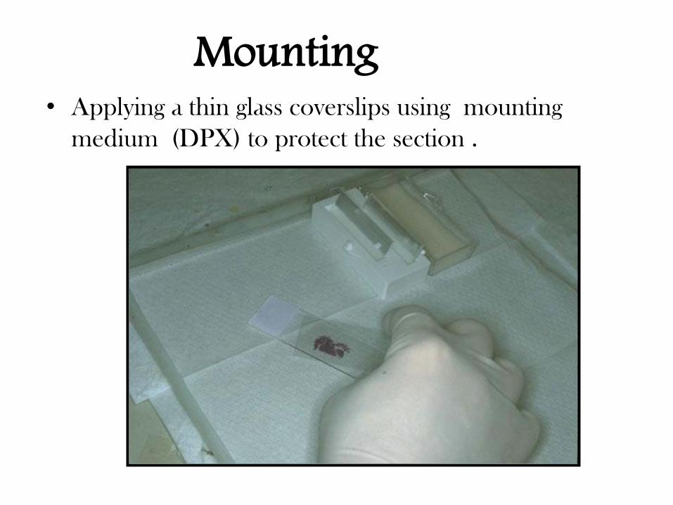
Mounting • Applying a thin glass coverslips using mounting
medium (DPX) to protect the section .
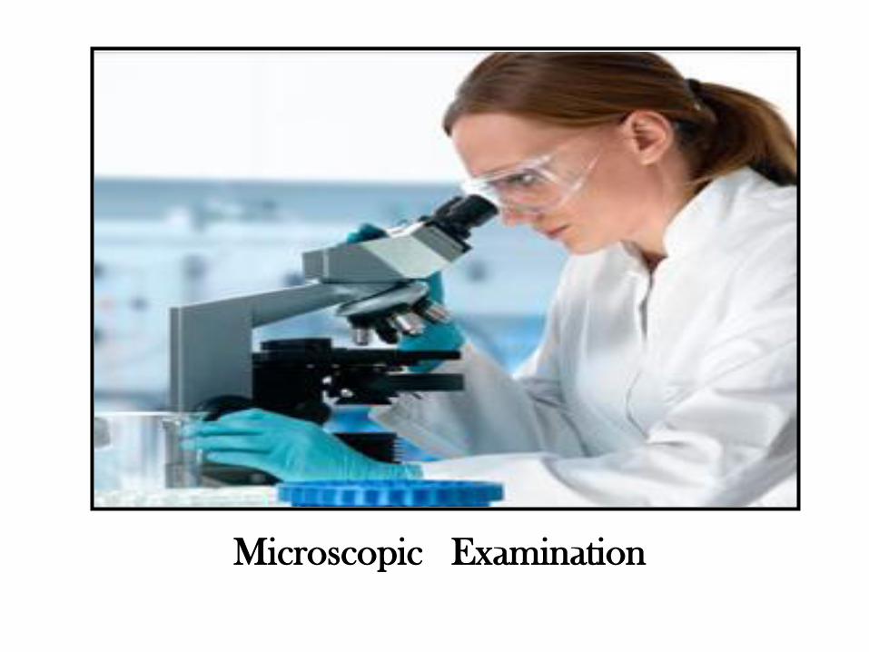
Microscopic Examination
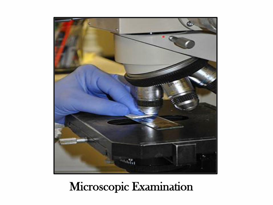
Microscopic Examination