senescent wi-38 cells fail to express epc-1, a gene ... · senescent wi-38 cells fail to express...
TRANSCRIPT

THE JOURNAL OF BIOLOGICAL CHEMISTRY 0 1993 by The American Society for Biochemistry and Molecular Biology, Inc.
V O l . 268, No. 12, Issue of April 25, pp. 8949-8957,1993 Printed in U. S. A.
Senescent WI-38 Cells Fail to Express EPC-1, a Gene Induced In Young Cells upon Entry into the Go State*
(Received for publication, November 2, 1992)
Robert J. Pignolo, Vincent J. CristofaloS, and Mitch 0. Rotenberg From the Center for Gerontological Research and the Department of Physiology and Biochemistry, The Medical College of Pennsylvania, Philadelphia, Pennsylvania 19129
Recently we reported the isolation of cDNAs for a number of genes that are differentially expressed be- tween nonproliferating early (young) and late (senes- cent) population doubling level (PDL) WI-38 human, fetal lung-derived, fibroblast-like cells. We now dem- onstrate that one of these isolates, EPC-1 (early PDL cDNA- l), derives from an approximately 1.4-kilobase mRNA species that is expressed at a 2 100-fold higher level in serum-starved, confluent, young versus simi- larly treated senescent WI-38 cells. Complete nucleo- tide sequence analysis of this cDNA confirms its iden- tity with that of a cDNA encoding a secreted, retinal pigmented epithelium differentiation factor as well as similarity of the encoded protein with a number of mammalian serine protease inhibitors. We show that EPC-1 expression is associated with Go growth arrest in WI-38 cells. The mRNA readily accumulates in den- sity-arrested and/or serum-starved young cells but not in log phase, low density young cells. In contrast, EPC- I transcript abundance and expression of the encoded, secreted protein are both negligible in senescent WI- 38 cells under all culture conditions tested. These find- ings support the hypothesis that senescent WI-38 cells exhibit a state of growth arrest fundamentally distinct from that of quiescent (GO) young cells.
Normal human diploid fibroblast-like (HDF)’ cells exhibit a limited proliferative life span when serially cultured (Hay- flick and Moorhead, 1961; Hayflick, 1965). Repeated subcul- tivation is accompanied by a progressive decline in the pro- portion of rapidly dividing cells (Cristofalo and Sharf, 1973) and an increase in the average length of the GI phase of the cell cycle (Macieira-Coehlo et al., 1966; Grove and Cristofalo, 1977). These cells eventually arrest in a viable, nonprolifera-
* This work was supported by National Institutes of Health Grant AGO0378 (to V. J. C.) and a grant from the American Federation for Aging Research (to M. 0. R.). The costs of publication of this article were defrayed in part by the payment of page charges. This article must therefore be hereby marked “aduertisement” in accordance with 18 U.S.C. Section 1734 solely to indicate this fact.
The nucleotide sequence(s) reported in this paper has been submitted to the GenBankTM/EMBL Data Bank with accession number(s) M90439.
$To whom correspondence should be addressed Audrey Meyer Mars Professor and Director, Center for Gerontological Research, The Medical College of Pennsylvania, 3300 Henry Ave., Philadelphia, PA 19129.
The abbreviations used are: HDF, human diploid fibroblast-like; PDL, population doubling level; EPC, early PDL cDNA; PEDF, pigmented epithelial differentiation factor; MEM, minimum essential medium; FBS, fetal bovine serum; kb, kilobase(s); PAGE, polyacryl- amide gel electrophoresis; RPE, retinal pigmented epithelial; gas, growth-arrest specific.
tive state in which they cannot be induced to synthesize DNA by subcultivation or by refeeding with fresh medium-contain- ing serum or any combination of growth factors. Limited in vitro replicative life spans have been described for a variety of cell types (Ponten, 1973; Rheinwald and Green, 1975; Bierman, 1978; Tassin et al., 1979; Tice et al., 1979; Mueller et al., 1980) and shown to be inversely related to donor age (Martin et al., 1970; Schneider and Mitsui, 1976) as well as directly related to the maximum life span of the donor species (Rohme, 1981). These findings suggest that aging in vivo is expressed in culture (for a review see Cristofalo and Stanulis- Praeger, 1982).
I t is unlikely that the inability of senescent or late popula- tion doubling level (PDL) cells to synthesize DNA in response to mitogenic stimulation is due to a diminished capacity for peptide growth factor signal transduction alone. Senescent and early PDL cells exhibit approximately the same number of growth factor receptors per unit cell surface area, as well as similar binding affinities, for epidermal growth factor (Phil- lips et al., 1983), insulin-like growth factor (Phillips et al., 1987), and platelet-derived growth factor (Gerhard et al., 1991). Furthermore, the transcription of several growth-reg- ulated genes is fully induced upon treatment of senescent cells with serum growth factors (Rittling et al., 1986; Seshadri and Campisi, 1990).
In HDF cells, growth arrest can be achieved through serum deprivation or through density-dependent contact inhibition. This reversible growth-arrested condition is referred to as the Go or quiescent state (for review see Baserga, 1985). Some studies have provided evidence for biochemical and molecular similarities between quiescent and senescent HDF cells (Ra- binovitch and Norwood, 1980; Pereira-Smith et al., 1985; Wang, 1985; Stein et al., 1986). These similarities, however, may be common to both senescence and quiescence simply because of a shared “nongrowth” phenotype. With few excep- tions (e.g. Wang and Tomaszewski, 1991), relatively little effort has been focused on defining differences between these cell types which could serve to identify which characteristics are specific to, and perhaps causally involved with, cellular senescence. Of particular significance to the current study is the observation of Gorman and Cristofalo (1986) that, based upon nuclear quinacrine dihydrochloride fluorescence stain- ing patterns, and regardless of cell culture conditions, senes- cent cells have characteristics consistent with their being blocked at or near the GI/S interface. Thus, despite the occurrence of some apparent similarities between quiescent and senescent cells, the latter appear to exhibit a state of growth arrest fundamentally distinct from that of Go-arrested cells.
Although cell fusion (Norwood et al., 1974; Burmer et al., 1982, 1983, 1984; Drescher-Lincoln and Smith, 1983, 1984) and mRNA microinjection studies (Lumpkin et al., 1986)
8949

8950 Age and Growth State-dependent Expression of EPC-1
provided evidence that senescence is dominant to the prolif- erating phenotype in normal mammalian cells and that se- nescence may occur through the elaboration of one or more factors that inhibit DNA synthesis, no such inhibitory factors unique to senescence have yet been identified. In fact, both increases and decreases in the expression of specific genes may be important to the establishment of the senescent phenotype. In this regard, we reported recently the isolation of cDNAs for a number of genes that appear to be differen- tially expressed in quiescent early PDL uersus similarly treated late PDL WI-38 human diploid fibroblast-like cells (Doggett et al., 1992). Several of these transcripts are more abundant in early PDL cells, whereas others are more highly expressed in late PDL cells.
In the present study, we provide a detailed characterization of one of these cDNA isolates, EPC-1 (early PDL cDNA-l), including determination of its identity with a cDNA for a retinal pigmented epithelial differentiation factor (PEDF) and sequence similarity of its encoded protein to several mammalian serine protease inhibitors. We illustrate that the EPC-1 transcript is highly expressed in density-arrested and/ or serum-starved (ie. quiescent (Go)) early PDL HDF cells but is virtually undetectable in both log phase, sparsely cul- tured, early PDL HDF cells and in late PDL HDF cells under all culture conditions tested. Further, we demonstrate that senescent HDF cells fail to secrete EPC-1 protein into the culture medium. We explore the possible roles of the EPC-1 gene product and the relationship of the current findings to the growth arrest of late PDL cells.
EXPERIMENTAL PROCEDURES
Materials-Enzymes, molecular biological reagents and kits, and chemical reagents were obtained from the following sources: restric- tion enzymes, random-primer DNA labeling kits, calf intestine alka- line phosphatase, Escherichia coli T4 DNA ligase, and proteinase K (Boehringer Mannheim); Sequenase DNA polymerase (U. S. Bio- chemical Corp.); Vectastain alkaline phosphatase kits (Vector Labo- ratories); prestained low molecular weight protein standards (Bio- Rad); M13 universal sequencing primers (New England Biolabs); Nytran and nitrocellulose filters (Schleicher & Schuell); [cY-~'P]~CTP (>3,000 Ci/mmol), C Y - ~ ~ S - ~ A T P (>1,000 Ci/mmol), and [methyL3H] thymidine (2 Ci/mmol) (Du Pont-New England Nuclear); acetone (Fisher).
Cell Culture-Stock cultures of WI-38 fetal human diploid fibro- blast-like cells (Hayflick, 1965) and fetal human AG 5337 pigmented retinal epithelial cells (Aronson, 1983) were grown in Autopow min- imum essential medium (MEM), supplemented with basal medium Eagle vitamins and 10% fetal bovine serum (FBS) (Flow Laboratories, McLean, VA), as described previously (Cristofalo and Charpentier, 1980). Cultures were defined as "phased out," i.e. at the end of their proliferative life spans or 100% life span completed, when they were unable to complete one population doubling during a 4-week period that included 3 consecutive weeks of refeeding with fresh medium containing 10% FBS (Cristofalo and Charpentier, 1980). Since the phase out point could be predicted a priori from each culture's [3H] thymidine labeling index under our standard culture conditions (Cris- tofalo and Sharf, 1973; Cristofalo, 1976), we were able to calculate the percent life span completed either prospectively from the [3H] thymidine labeling index, or retrospectively from the phase out point. Early PDL cells were defined as those that had progressed through e 50% of their replicative life span, whereas late PDL (senescent) cells were defined as those that had completed ? 95% of their replicative life span.
The percentages of cells capable of entering into the S phase of the cell cycle under the various culture conditions used for analysis of EPC-1 mRNA expression (see below) were determined by growing cultures in parallel on glass coverslips and then adding [3H]thymidine (1 pCi/ml; 2 Ci/mmol) to the medium for the 24-h period immediately prior to harvest. Following this 24-h pulse the coverslips were col- lected, fixed for autoradiography, and scored for the percentage of labeled nuclei as described by Cristofalo and Sharf (1973). All assays were done in triplicate.
were seeded at a density of 1 X lo4 cells/cm2 in MEM plus 10% FBS RNA Extraction-For the initial characterization of EPC-1, cells
and grown to confluence (7 days for early PDL cells or 10 days for late PDL cells). The medium was then replaced with serum-free MCDB-104 (GIBCO; buffered with NaHC03 instead of Hepes) for 2 days to further ensure synchronization in quiescence. Total cellular RNA was extracted via the guanidinium thiocyanate procedure of Chirgwin et al. (1979), purified over a cushion of CsCl as described by Glisin et al. (1974), and subjected to blot hybridization analysis as described below. In subsequent analyses, cell seeding densities and incubation times in the presence and absence of serum were varied
the expression of EPC-1. to assess the effects of cell density and/or serum deprivation upon
Northern Analysis-Total cellular RNA was isolated as described above, precipitated twice with ethanol, and quantified by spectropho- tometry. Twenty-microgram aliquots of each sample were glyoxylated and then fractionated on 1.2% agarose gels. The gels were then incubated 2 X 22 min in 50 mM NaOH, 100 mM NaC1, and 2 X 30 min in 20 X SSPE (3.6 M NaC1,0.2 M Na2HP04, 0.2 M EDTA) before transfer to Nytran membranes by capillary blotting in 20 X SSPE. Following transfer, the membranes were washed for 10 min with 2 X SSPE, air dried, and baked for 3 h at 80 "C in uacw.
Blots were prehybridized for 18 h at 45 "C in 50% formamide, 4 X SSPE, 1.0% SDS, 0.1 mg/ml denatured herring sperm DNA, and 0.5% Blotto (10% nonfat powdered milk, 0.2% sodium azide, 0.02% antifoam A). Hybridizations were for 18 h at 45 "C in 47% formamide, 3 X SSPE, 1.0% SDS, 10% dextran sulfate, 0.1 mg/ml denatured herring sperm DNA and 0.5% Blotto plus 1-2 X lo6 cpm/ml of the 32P-labeled full-length EPC-1 cDNA (prepared via random primer labeling according to the manufacturer's instructions (Boehringer Mannheim) and purified free of unincorporated nucleotides by pas- sage over a NucTrap (Stratagene), gel filtration, push column). A 32P-labeled, 0.6-kb PstI cDNA fragment of the human &-microglob- ulin gene (Suggs et al., 1981) and/or a 32P-labeled, 2.1-kb BamHI full- length cDNA clone of the human p-actin gene (Gunning et al., 1983) were included as probes for internal standardization of all Northern blot hybridization assays. Following hybridization, the blots were washed 3 X 15 min in 2 X S s c (1 X SSC is 0.15 M NaCl, 0.015 M sodium citrate), 0.2% SDS at 45 "C, and 1 X 15 min in 0.2 X SSC, 0.2% SDS at 45 "C, then subjected to autoradiography at -70 "C using Kodak XAR-5 film and an intensifying screen. Autoradi- ographic signal intensities were quantified via laser densitometry in the near linear range of the film using a Hoefer GS300 transmittance/ reflectance scanning densitometer and Hewlett-Packard HP3396A integrator.
Expression and Purification of Recombinant EPC-1 Protein-EPC- 1 was expressed in E. coli as a recombinant fusion protein bearing a hexahistidine (Hiss) amino-terminal affinity label to facilitate puri- fication of this protein via nickel-chelate affinity chromatography. The EPC-1 coding sequence occurring downstream of the MscI site within codon 86 (PEDF; GenBank accession no. M76979) was in- serted, in its entirety, into plasmid pQE-10 (Qiagen, Inc., Chatsworth, CA) such that it was positioned immediately downstream and in- frame with the His6 coding sequences occurring in this vector. Follow- ing introduction of the resulting plasmid into E. coli M15 cells harboring plasmid pREP4 (Qiagen, Inc.), expression of the Hi%- EPC-1 protein was induced and the protein purified by affinity chromatography as described by Stiiber et al. (1990).
Preparation of EPC-1 Polyclonnl Antisera-Female New Zealand White rabbits were immunized once with 100 pg of affinity-purified His6-EPC-1 protein in complete Freund's adjuvant and then boosted twice, at 2-week intervals, with 50 pg of the purified protein in incomplete Freund's adjuvant. The animals were bled 2 weeks after the final injection and at regular intervals thereafter. Preimmune and immune sera thus obtained were used in the immunoblotting studies described below. All manipulations involving animals for the prepa- ration of polyclonal antisera were performed by CoCaLiCo Biolog- icals, Inc. (Reamstown, PA).
Western Analysis-Proteins secreted into serum-free MCDB-104 medium by density-arrested monolayers of WI-38 or AG 5337 cells were precipitated with 5 volumes of acetone at -20 "C for at least 18 h, pelleted at 10,000 X g for 30 min at 4 "C, and resuspended in TE (10 mM Tris-HC1, 1 mM EDTA, pH 7.5). Cells were allowed to condition serum-free MCDB-104 for at least 24 h prior to harvest for acetone precipitation. Final protein concentrations of samples were determined using the Bradford assay (Bradford, 1976) according to the manufacturer's instructions (Bio-Rad). Equal amounts of secreted protein (50 pg) were separated by 10% SDS-polyacrylamide gel elec-
Age and Growth State-dependent Expression of EPC-1 8951
trophoresis (PAGE) as described previously (Laemmli, 1970) and electroblotted onto nitrocellulose in 25 mM Tris, 192 mM glycine, and 20% methanol (Towbin, 1979) for 2 h at 120 V followed by 1 h at 60 V, or at 30 V for 18 h. Analysis of specific proteins was performed using EPC-1 antiserum at a 1:1,000 dilution and a secondary anti- body/alkaline phosphatase detection system (Vector Laboratories). Before incubation with primary antiserum, blots were incubated with Tris-buffered saline containing 0.1% (v/v) Tween-20 (TTBS) and 1% (w/v) bovine serum albumin. All other steps were carried out exactly according to the manufacturer’s instructions (Vector Laboratories).
DNA Sequencing-To facilitate sequence analysis of isolate EPC- 1, the intact approximately 1.2-kb cDNA was excised from vector pCDM8 (Seed, 1987) and recloned into the XbaI site of plasmid pUC19 (Yanisch-Perron et al., 1985). Double-stranded DNA sequenc- ing was then performed by the method of Chen and Seeburg (1985) using M13 universal primers, CY-~~S-~ATP, and Sequenase DNA po- lymerase (U. S. Biochemical Corp.). Reaction samples were fraction- ated on 7% acrylamide, 8 M urea gels; fixed 15 min in 5% methanol, 5% acetic acid; dried under vacuum at 80 “C; and subjected to auto- radiography at room temperature for 16-18 h. Smaller fragments of EPC-I were subcloned into pUC19, as needed, to permit complete sequencing of the entire cDNA. Computer analyses of assembled sequence data were performed using software obtained through the University of Wisconsin Genetics Computer Group (Devereux et al., 1984).
Genomic DNA Isolation and Analysis-High molecular weight ge- nomic DNA was isolated from 1.4 X lo8 WI-38 cells at PDL 30 (50% life span completed) via proteinase K digestion at 50 “C in the presence of 0.5% SDS, followed by repeated extraction with pheno1:chloroform:isoamyl alcohol (25:24:1), and ethanol precipita- tion, as described previously (Ausubel et al., 1987). Thirty-microgram aliquots of the isolated genomic DNA were then digested for 18 h with a 2-fold excess of BamHI, HindIII, or XbaI, fractionated on a 0.8% agarose gel, capillary blotted to a Nytran filter via standard methods (Sambrook et al., 1989) using 10 X SSC, and subjected to blot hybridization analysis using a 3ZP-labeled -0.5-kb XhoI frag- ment, derived from the 5’ end of the EPC-1 cDNA, as the probe.
Prehybridization was for 4.25 h at 45 “C in a buffer composed of 50% formamide, 25 mM potassium phosphate, pH 7.4, 5 X SSC, 5 X Denhardt’s solution (1 X Denhardt’s solution is 0.02% polyvinylpyr- rolidone, 0.02% bovine serum albumin, and 0.02% Ficoll), 1.0% SDS, and 0.05 mg/ml denatured herring sperm DNA. Hybridization was for 18 h at 45 “C in the same buffer plus 10% dextran sulfate and 1.2 X lo6 cpm/ml of the 32P-labeled probe. The filter was then washed as follows: 2 X 10 min at room temperature with 2 X SSC, 0.1% SDS; 1 X 15 min at room temperature with 0.5 X SSC, 0.1% SDS; 1 X 15 min at 45 “C with 0.1 X SSC, 0.1% SDS; and 1 X 15 min at 45 “C with 0.1 X SSC, 1.0% SDS. After washing, the filter was subjected to autoradiography at -70 “C as described above for Northern analysis.
RESULTS
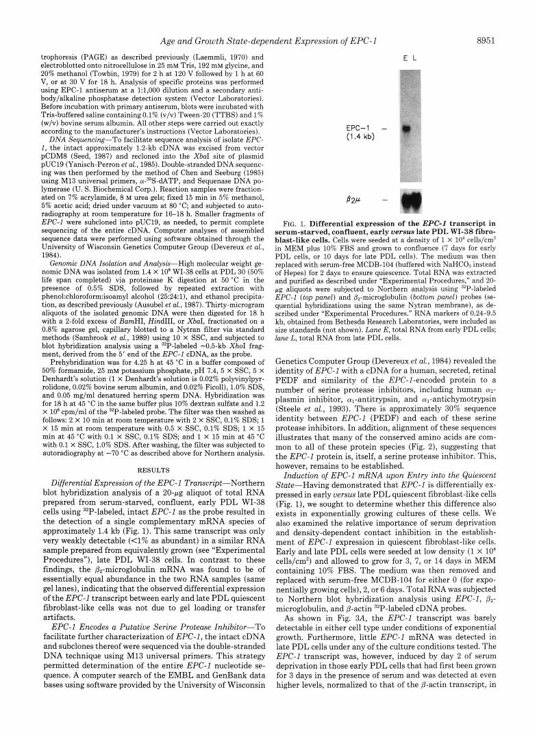
Differential Expression of the EPC-1 Transcript-Northern blot hybridization analysis of a 20-pg aliquot of total RNA prepared from serum-starved, confluent, early PDL WI-38 cells using 32P-labeled, intact EPC-1 as the probe resulted in the detection of a single complementary mRNA species of approximately 1.4 kb (Fig. 1). This same transcript was only very weakly detectable (<1% as abundant) in a similar RNA sample prepared from equivalently grown (see “Experimental Procedures”), late PDL WI-38 cells. In contrast to these findings, the Pz-microglobulin mRNA was found to be of essentially equal abundance in the two RNA samples (same gel lanes), indicating that the observed differential expression of the EPC-1 transcript between early and late PDL quiescent fibroblast-like cells was not due to gel loading or transfer artifacts.
EPC-1 Encodes a Putative Serine Protease Inhibitor-To facilitate further characterization of EPC-1, the intact cDNA and subclones thereof were sequenced via the double-stranded DNA technique using M13 universal primers. This strategy permitted determination of the entire EPC-1 nucleotide se- quence. A computer search of the EMBL and GenBank data bases using software provided by the University of Wisconsin
E L
EPC-1 - e (1.4 kb)
FIG. 1. Differential expression of the EPC-1 transcript in serum-starved, confluent, early versus late PDL WI-38 fibro- blast-like cells. Cells were seeded at a density of 1 X 10‘ cells/cm2 in MEM plus 10% FBS and grown to confluence (7 days for early PDL cells, or 10 days for late PDL cells). The medium was then replaced with serum-free MCDB-104 (buffered with NaHC03 instead of Hepes) for 2 days to ensure quiescence. Total RNA was extracted and purified as described under “Experimental Procedures,” and 20- pg aliquots were subjected to Northern analysis using 32P-labeled EPC-I (top panel) and &-microglobulin (bottom panel) probes (se- quential hybridizations using the same Nytran membrane), as de- scribed under “Experimental Procedures.” RNA markers of 0.24-9.5 kb, obtained from Bethesda Research Laboratories, were included as size standards (not shown). Lane E, total RNA from early PDL cells; lane L, total RNA from late PDL cells.
Genetics Computer Group (Devereux et al., 1984) revealed the identity of EPC-1 with a cDNA for a human, secreted, retinal PEDF and similarity of the EPC-I-encoded protein to a number of serine protease inhibitors, including human a2- plasmin inhibitor, al-antitrypsin, and a,-antichymotrypsin (Steele et al., 1993). There is approximately 30% sequence identity between EPC-1 (PEDF) and each of these serine protease inhibitors. In addition, alignment of these sequences illustrates that many of the conserved amino acids are com- mon to all of these protein species (Fig. 2), suggesting that the EPC-1 protein is, itself, a serine protease inhibitor. This, however, remains to be established.
Induction of EPC-1 mRNA upon Entry into the Quiescent State-Having demonstrated that EPC-1 is differentially ex- pressed in early versus late PDL quiescent fibroblast-like cells (Fig. l), we sought to determine whether this difference also exists in exponentially growing cultures of these cells. We also examined the relative importance of serum deprivation and density-dependent contact inhibition in the establish- ment of EPC-1 expression in quiescent fibroblast-like cells. Early and late PDL cells were seeded a t low density (1 X lo4 cells/cm2) and allowed to grow for 3, 7, or 14 days in MEM containing 10% FBS. The medium was then removed and replaced with serum-free MCDB-104 for either 0 (for expo- nentially growing cells), 2, or 6 days. Total RNA was subjected to Northern blot hybridization analysis using EPC-1, P2-
microglobulin, and P-actin 32P-labeled cDNA probes. As shown in Fig. 3A, the EPC-1 transcript was barely
detectable in either cell type under conditions of exponential growth. Furthermore, little EPC-1 mRNA was detected in late PDL cells under any of the culture conditions tested. The EPC-1 transcript was, however, induced by day 2 of serum deprivation in those early PDL cells that had first been grown for 3 days in the presence of serum and was detected a t even higher levels, normalized to that of the P-actin transcript, in

8952
EPC-1 305 N ~ D ~ A G T T P S P G L Q P A H L T F P L D - - - - Y H ~ ~ ~ I ~ R D T D ~ ~ I ~ I L ~ ~ R G P 358 PEDF 365 N~D@AGTTPSPGLQPAHLTFPLD----YHL&Q&I&LRDTD$W&&$II$~ILD~RGP 418
al-AT 369 D # K C T E A A G A M F L E A I P M S I P P E - - - - V K F B ~ K ~ ~ I E Q N & S ~ ~ ~ Q K 422 a2-~1 395 S~~~AAAATSIAMSRMSLS-S----FSV~~IFED~L~~~S~~S 448
al-~c 345 F~EWEASAATAVKITLLSALVETRTIVRF@&&LMIIVPTDTQNIFJ~IS~ITNRKQA ..... .. . . . . 398
FIG. 2. Aligned amino acid sequences of EPC-1, PEDF, and three human serine protease inhibitors. The amino acid sequence of the EPC-1 protein (predicted from the cDNA nucleotide sequence) was optimally aligned with those of retinal PEDF, human a2-plasmin inhibitor (a2-PI), human al-antitrypsin (a l -AT) , and human a,-antichymotrypsin ( a l - A C ) . Those amino acids conserved between at least four (including EPC-1) of the five proteins are marked by inclusion within the shaded regions. Positions of amino acid residues within each protein sequence are denoted by the numbers on either side of the figure. The protein sequence of EPC-1 begins with the first complete codon of the near full-length cDNA.
all other serum-starved early PDL cultures (Fig. 3, A and B ) . Autoradiographic analysis of parallel cultures of early PDL fibroblast-like cells, labeled with ['Hlthymidine for the 24-h period immediately prior to RNA harvest, revealed a steady decline in the percentage of cells in S phase (DNA synthesis) with increasing time in culture (Fig. 3B). This effect, which likely reflects density-dependent growth inhibition, was en- hanced by serum deprivation, with 6 days of serum starvation consistently having a greater inhibitory effect upon cellular DNA synthesis than 2 days of starvation. Together, these findings suggest that EPC-1 mRNA begins to accumulate as the early PDL cells enter the quiescent state and that expres- sion of this transcript increases as more cells enter Go. p2- Microglobulin and /3-actin expression, however, were observed to be relatively constant in both cell types under all culture conditions and time points tested (Fig. 3A) .
Induction of EPC-1 mRNA by Density-dependent Growth Arrest or Serum Staruation-The above findings indicated that density-dependent contact inhibition and serum starva- tion might each play a significant role in the modulation of EPC-I expression. Two approaches were therefore under- taken to determine whether either of these factors alone could promote EPC-1 expression. In the first study, early PDL cultures were seeded at a density of 1 x lo4 cells/cm2 in MEM plus 10% FBS (day 0), and on each subsequent day, through day 7, cells were harvested for total RNA extraction. Blot hybridization analysis of the isolated RNAs revealed that the EPC-1 mRNA level was high on day 1 but that the abundance of this transcript then rapidly declined and remained low
through day 4 (Fig. 4A). By day 5 , however, as the population approached the plateau phase of growth, EPC-1 transcript abundance began to increase and continued to do so through day 7.
In a parallel experiment (Fig. 4B) in which the percentage of cells in S phase was determined for days 1, 3, 7, and 10 after seeding, we found a strict inverse relationship between EPC-1 transcript abundance and the fraction of cells bearing 'H-labeled nuclei ( ie. synthesizing DNA). Although these cultures had not been deprived of serum, by day 7 only 14% of the cells were in S phase (because of density-dependent inhibition), whereas duplicate 7 day cultures accumulated EPC-1 mRNA to high levels. From this we conclude that density-dependent growth inhibition is sufficient for the in- duction of EPC-1 expression and that serum-deprivation is not required for this induction. The high level of EPC-1 mRNA detected in day 1 cultures may constitute the remain- der of that which had accumulated in their confluent (7-day) parent cultures prior to their harvest and reseeding. Alterna- tively, this mRNA may have derived from that fraction of the day 1 cell population which had not yet reentered the cell cycle at the time of RNA harvest; we note that only 17% of the cells had entered S phase during the 24-h period imme- diately following reseeding (Fig. 4R).
Our second approach, which was designed to evaluate the capacity of serum deprivation to promote EPC-1 expression in the absence of density arrest, involved seeding early PDL cells at a 10-fold lower initial density (1 X IO3 cells/cm2). These cultures were first grown for 4 days in MEM plus 10%

Age and Growth State-dependent Expression of EPC-1 8953
A
PDL E L E L E L E L E L E L E L
DAYS w\SERUM 3 3 3 3 3 3 7 7 7 7 1 4 1 4 1 4 1 4 DAYS w\o SERUM 0 0 2 2 6 6 2 2 6 6 2 2 6 6
€PC- 1 m * - - ~ - m v ”’
B 2 P
@-Actin DOO**Oi I ) O ~ o - o m U
B
2. 3dws + serum. 2don - mrn 1. 3daym+wrum
-16
LLL 2 3 [L CELL CULTURE CoNmONs
FIG. 3. Combined effects of serum deprivation and cell density upon EPC-I mRNA abundance and DNA synthetic capacity. Panel A, early and late PDL WI-38 cells were seeded a t 1 X lo‘ cells/cmz and grown for 3, 7, or 14 days in MEM containing 10% FBS. The medium was then removed and replaced with serum-free MCDB-104 for 0 (for exponentially growing cells), 2, or 6 days. Total RNA was isolated, and 20-pg aliquots were subjected to Northern analysis as described under “Experimental Procedures” using ”P-labeled EPC-I, 82- microglobulin, and 8-actin cDNA probes. Incubation periods in the presence and absence of serum were as indicated at the top of the figure. E denotes total RNA from early PDL cells; L denotes total RNA from late PDL cells. Panel B, the EPC-ZIP-actin signal ratios shown for the early PDL RNA samples in panel A were quantified via laser densitometry and are plotted here (filled bars) versus the culture conditions tested. The proportions of cells entering into S phase (percent labeled nuclei) during the 24-h period immediately preceding RNA harvest were determined for identically treated sister cultures (see “Experimental Procedures”) and are also plotted here (open bars) uersus the culture conditions tested.
FBS and then deprived of serum for 0, 2, or 6 days, after which time total RNA was extracted and subjected to blot hybridization analysis. As shown in Fig. 5, EPC-1 mRNA was undetectable in sparse cultures that had not been deprived of serum and was only weakly detectable in sparse cultures that were deprived of serum for only 2 days. However, when these same cells were deprived of serum for 6 days or were allowed to grow for 10 full days in the presence of serum, they accumulated the EPC-1 transcript to substantial levels. Early passage WI-38 cells that were been serum deprived for 6 days showed kinetic characteristics of having entered a Go state (Baserga, 1985) in that 16-18 h were required for entry into DNA synthesis following serum stimulation (data not shown). Together, these findings indicate that either extended serum deprivation or density-dependent growth inhibition is suffi- cient for the induction of high level EPC-1 expression.
Late PDL WI-38 Cells Fail to Secrete EPC-1 Protein- Above, we showed that senescent WI-38 cells have a much diminished capacity to accumulate the EPC-1 transcript un- der all cell culture conditions tested. To compare relative levels of EPC-1 protein produced by young and senescent WI-
38 cells, polyclonal antisera were prepared against a purified, recombinant form of the EPC-1 protein (see “Experimental Procedures”). Given the similarity between EPC-1 and several known serine protease inhibitors, all of which are secreted, we assayed medium conditioned by WI-38 cells for the pres- ence of EPC-1. Proteins secreted into the medium of density- arrested, serum-deprived senescent and young WI-38 cells were separated by SDS-PAGE and the presence of EPC-1 specifically detected by Western analysis as shown in Fig. 6A. EPC-1 antiserum recognizes a 45-50-kDa protein species, present in medium conditioned by early, but not late, PDL cells. Preimmune antiserum does not recognize this protein species (data not shown). Although the inability to detect EPC-1 protein in the medium conditioned by late PDL cells could point to a defect in a specific secretory function, this scenario is unlikely given the low steady-state level of EPC-1 mRNA displayed by senescent WI-38 cells.
We also used EPC-1 antiserum to confirm that the EPC-I protein expressed by early PDL WI-38 cells is the same secreted protein produced by retinal pigmented epithelial (RPE) cells. Fig. 6B shows a Western analysis of proteins

8954 Age and Growth State-dependent Expression of EPC-1
A
DAYS 1 2 3 4 5 6 7
EPC- 1 - -0
B-Actin - ”.)w I .
B
lolloo
DAYS IN CULTURE
FIG. 4. Density-dependent growth inhibition is sufficient for EPC-I induction in early PDL WI-38 cells. Panel A, early PDL cells were harvested from a confluent parent culture and re- seeded on day 0 a t a density of 1 X lo4 cells/cm2 in MEM containing 10% FBS. On each subsequent day through day 7, total RNA was extracted, and 20-pg aliquots of each were then subjected to Northern blot hybridization analysis using 32P-labeled EPC-I and &actin cDNA probes. The number of days that the cells were in culture is indicated at the top of the figure. Panel B, in a parallel experiment, cells were again seeded at a density of 1 X lo4 cells/cm2 and grown for 1,3,7, or 10 days in MEM containing 10% FBS. Total RNA was extracted and analyzed for each time point as in panel A, except that 02-microglobulin replaced 0-actin as the internal standard in the hybridization assay. EPC-I/µglobulin signal ratios were quan- tified via laser densitometry and are plotted (filled bars) versus the number of days in culture. The proportions of cells entering into S phase (percent labeled nuclei) during the 24-h period immediately preceding RNA harvest were determined for identically treated sister cultures (see “Experimental Procedures”) and are also plotted here (open bars) versus the number of days in culture.
DAYS w\ SERUM 4 4 4 1 0 DAYS w\o SERUM 0 2 6 0
EPC-1 -
,%Actin - “ FIG. 5. Serum-deprivation is sufficient for EPGI induction
in early PDL WI-38 cells. Early PDL cells were seeded at a 10- fold lower density (1 X IO3 cells/cm2) than that used for the experi- ments in all other figures and were grown for 4 days in MEM plus 10% FBS. The cells were then deprived of serum for 0, 2, or 6 days, after which time total RNA was harvested and subjected to blot hybridization analysis using 32P-labeled EPC-I and 0-actin cDNA probes. A control sample was also prepared and analyzed for cells seeded a t the same low density but grown for 10 days in MEM plus 10% FBS without serum deprivation. Incubation periods in the pres- ence and absence of serum were as indicated at the top of the figure.
secreted by density-arrested, serum-deprived AG 5337 RPE cells and identically treated early PDL WI-38 cells using EPC-1-specific antiserum. EPC-1 antiserum recognizes a 45- 50 kD protein species, present in medium conditioned by both cell types, which migrates as a doublet when resolved by SDS- PAGE. The above results are consistent with the finding that PEDF purified from RPE-conditioned medium also migrates to a similar size by SDS-PAGE and with an analgous migra- tion pattern (Tombran-Tink et al., 1991). For our study,
106 - 80 -
49.5 -
32.5 - 27.5 - 18.5 -
B - 9 v
2 106 - 80 -
49.5 -
32.5 - 27.5 - 18.5 -
1 2
- EPC-1
1 2
- EPC-1
FIG. 6. Western analysis of proteins secreted into the me- dium of young and senescent WI-38 cells as well as AG 5337 retinal pigmented epithelial cells using EPC-1-specific anti- serum. Panel A, acetone-precipitated conditioned medium proteins from density-arrested, serum-deprived late PDL (lane I ) and early PDL (lane 2) WI-38 cells were separated by 10% SDS-PAGE and prepared for Western analysis with EPC-I-specific antiserum as described under “Experimental Procedures.” Molecular mass stand- ards and EPC-1 species are indicated. Panel E , acetone-precipitated conditioned medium proteins from density-arrested, serum-deprived AG 5337 young (lane I ) and WI-38 cells (lane 2) were separated by 10% SDS-PAGE and prepared for Western analysis using EPC-1- specific antiserum as described under “Experimental Procedures.” Molecular mass standards and EPC-1 species are indicated.
conditioned medium proteins were generated from each cell type after normalization with respect to growth state and in vitro age; i.e. the cell lines had reached the plateau phase of growth and had completed essentially equivalent percentages of their replicative life spans (data not shown).
EPC-1 Is Encoded by a Single-copy Gene-To determine whether the EPC-I transcript derives from a single copy gene, we first digested 3 0 - ~ g aliquots of high molecular weight WI- 38 genomic DNA with restriction endonuclease BamHI, HindIII, or XbaI, none of which cut within the EPC-1 cDNA sequence. The digested DNAs were then fractionated on a 0.8% agarose gel and subjected to Southern blot hybridization analysis using a “P-labeled, -0.5-kb XhoI fragment derived from the 5’ end of the EPC-I cDNA as the hybridization probe. As shown in Fig. 7, only one genomic DNA fragment was detected for each digest, suggesting that EPC-1 is encoded by a single copy gene. We have not, however, ruled out the formal possibility that this gene is duplicated in tandem.

Age and Growth State-dependent Expression of EPC-1 8955
1 2 3
23.1
9.4
6.55
4.4
2.3 2.0
0 .56
FIG. 7. Detection of the EPC-1 gene in WI-38 genomic DNA. High molecular weight genomic DNA was purified from early PDL WI-38 fibroblast-like cells, and 30-pg aliquots of this DNA were then digested for 18 h with a 2-fold excess of BamHI, HindIII, or XbaI. The samples were then fractionated on a 0.8% agarose gel, capillary blotted to a Nytran filter via standard methods (Sambrook et al., 1989), and subjected to blot hybridization analysis using a 32P- labeled -0.5-kb XhoI fragment derived from the 5’ end of the EPC- 1 cDNA, as the probe. The resulting autoradiogram is shown.
DISCUSSION
Progression of cells through the cell cycle is known to be dependent upon the complex interplay of a network of both positively and negatively acting regulatory factors. Although substantial evidence has accumulated indicating that the expression of one or more, perhaps novel, inhibitory factors plays a dominant role in establishment of the senescent phenotype (for review see Goldstein, 1990), it has also been shown that senescence is accompanied by the reduced capac- ity, under certain conditions, of cells to express the c-fos proto-oncogene product, a growth-promoting factor, in re- sponse to serum stimulation (Seshadri and Campisi, 1990). Kovary and Bravo (1991) showed recently that proteins of both the fos and jun transcription factor families are required for cell cycle progression in Swiss 3T3 cells, and Phillips et al. (1992) showed that transient transfection of senescent (90- 95% life span completed) WI-38 cells with a Zn2+-inducible murine c-fos gene results in the restoration of a t least one
round of DNA synthesis in a fraction of the transfected cell population following c-fos induction. It is therefore likely that the expression of other positively acting regulatory factors is similarly reduced in senescent HDF. Indeed, the inability of senescent cells to respond fully to mitogenic stimulation may, in part, result from their not being properly “primed” for such a response. With this possibility in mind, we sought to identify and characterize those genes, such as EPC-1, that are ex- pressed more highly in quiescent early uersw late PDL WI- 38 fibroblast-like cells (Doggett et al., 1992).
We showed that EPC-1 encodes a single, approximately 1.4-kb transcript and corresponding 50-kDa secreted protein species, which is readily detected in density-arrested and/or serum-starved, early PDL WI-38 cells or in medium condi- tioned by these cells, respectively. EPC-1 mRNA expression is negligible in exponentially growing, low density, early PDL cells and in late PDL cells under all culture conditions tested. In addition, density-arrested, serum-deprived late PDL cells fail to secrete the EPC-1 protein. In early PDL cells, EPC-1 mRNA accumulation generally exhibits an inverse relation- ship with the percentage of cells in the S phase of the cell cycle. Quiescence achieved through either density arrest or serum deprivation is sufficient for the induction of EPC-1 mRNA expression.
At present, the mechanisms by which growth arrest condi- tions promote EPC-1 mRNA expression/stabilization remain to be determined. However, we found that the EPC-I tran- script is undetectable in either the presence or absence of serum (data not shown), in confluent cultures of SV40-trans- formed WI-38 cells (Girardi et al., 1965; Ponten et al., 1963). Little true growth arrest is observed for these transformed cells under these conditions, suggesting that the capacity to exhibit such negative growth control is required for the in- duction of EPC-1 expression. An alternative explanation is that EPC-1 expression is in some way required for the occur- rence of Go growth arrest.
Computer searches of the EMBL and GenBank data bases with the complete EPC-1 nucleotide sequence indicate that this cDNA is identical to that of a secreted RPE differentia- tion factor (PEDF), which has been characterized previously. Other known proteins displaying the greatest similarity (-30% sequence identity) to the EPC-1 -encoded polypeptide include a host of extracellular serine protease inhibitors such as human cy*-plasmin inhibitor, crl-antitrypsin, and a,-anti- chymotrypsin. Interestingly, these proteins similarly share only -30% sequence identity with one another (Sumi et al., 1986), suggesting that EPC-1 may be another member of this gene family. This possibility is further supported by the finding that a significant fraction of the conserved amino acids common to these serine protease inhibitors is also found in the EPC-1 protein.
Preliminary characterization of PEDF revealed that it is a protein of approximately 50 kDa which is secreted by RPE cells and which can potentiate the differentiation of retino- blastoma cells into neuronal-like cells (Tombran-Tink et al., 1991). Medium conditioned by human RPE cells can produce effects similar to those induced by purified PEDF, although an extended time interval (and usually the addition of lami- nin) is required to stimulate the appearance of extensive neurite-like processes and biochemical markers associated with neuronal cells (Tombran-Tink and Johnson, 1989). It is unclear if the role of EPC-1 in fibroblast-like cells is that of a differentiation factor and/or perhaps that of a serine pro- tease inhibitor. Its expression is not limited to fibroblast-like cells (Fig. 6B and data not shown), so it may not have a similar function in different cell types, although its physiolog-

8956 Age and Growth State-dependent Expression of EPC-1
ical role may still be mediated by its putative activity as a protease inhibitor. If its role is that of a differentiation factor, its effects may be exerted upon cell types other than those that are fibroblast-like, since prolonged exposure of fibro- blasts to their own conditioned medium does not induce differentiation, at least by morphological criteria such as the presence of neurite-like processes or the formation of cell aggregates. However, this does not rule out the possibility that EPC-1 is affecting a type of differentiation process in fibroblast-like cells. For example, senescence may be viewed as a type of terminal differentiation which results in or is influenced by the loss of EPC-1 expression.
Protease inhibitors have been shown to play important roles in blood clotting, fibrinolysis, and modulation of both immune and antiinflammatory, i.e. acute phase, responses (Koj, 1985). In addition, protease inhibitors can affect the growth of both normal and transformed cell lines by modu- lating a series of cell surface-related events involving pro- teases (for review see Carrell, 1988; Baker et al., 1986). For example, protease nexin-1, a serine protease inhibitor, has been shown to promote the outgrowth of neurites from neu- ronal cells (Gloor et al., 1986; Cunningham and Gurwitz, 1989; Wagner et al., 1989), whereas down-regulation of myeloblas- tin, a novel serine protease, inhibits proliferation and induces differentiation of HL-60 promyelocytic leukemia cells (Bories et al., 1989). A domain containing a serine protease inhibitor sequence has recently been identified within the precursor of the amyloid @-protein characteristic of Alzheimer’s disease (Kitaguchi et al., 1988; Ponte et al., 1988; Tanzi et al., 1988; Oltersdorf et al., 1989). This domain may serve as a promoter of neurite outgrowth and may thus be involved in the devel- opment of the interwoven neuritic component of amyloid plaques (Carrell, 1988). If EPC-1 does indeed encode a serine protease inhibitor, it could, as illustrated by these examples, play an important role in the establishment or maintenance of density-dependent and/or nutritional growth arrest (see above) or in priming early PDL cells for escape from the quiescent (Go) state.
The induction of EPC-1 upon entry of fibroblast-like cells into the Go state bears striking similarity with the induction of the growth-arrest-specific (gas) genes. Either serum dep- rivation or density-dependent contact inhibition will cause both EPC-1 and the gas genes to be abundantly expressed (Schneider et al., 1988; Manfioletti et al., 1990; Brancolini et al., 1992; Del Sal, 1992). In most cases, however, the gas genes are down-regulated within 10 h of growth induction via serum stimulation. In contrast to this, when low density, serum- deprived WI-38 cells are stimulated with serum, a marked reduction in the EPC-1 steady-state mRNA level occurs only after approximately 24 h of stimulation (data not shown). Interestingly, when WI-38 cells are permitted to become contact-inhibited prior to serum deprivation, substantial re- ductions in EPC-1 mRNA abundance do not occur even after 36 h of serum stimulation (data not shown). Additional evi- dence suggests, however, that it is the growth state, rather than cell densityper se, which acts to enhance EPC-1 mRNA expression in early PDL cells.2
Previous studies (Cristofalo, 1973; Olashaw et al., 1983; Rittling et al., 1986) indicated that senescent cells can com- plete at least some of the biochemical and molecular pathways through Go/G1 up to the G1/S interface but are unable to initiate DNA synthesis. In addition, Gorman and Cristofalo (1986) showed through quinacrine dihydrochloride staining that the compaction of senescent cell nuclear chromatin is
* R. J. Pignolo, M. 0. Rotenberg, and V. J. Cristofalo, manuscript in preparation.
much more like that of young cells at the G1/S boundary than that of young cells in Go. Both of these observations are consistent with the idea that senescent cells are arrested at the GI/S boundary. Thus, growth arrest in senescent cells may occur in a physiologic state fundamentally distinct from that of the Go, quiescent state that is achieved by nonproli- ferating young cells. Our current findings suggest that EPC- 1 expression is characteristic of the Go state, and the failure of EPC-1 to be expressed in senescent cells further supports the hypothesis that senescent cells are unable to enter this state. A full response to serum or growth factor addition, leading from quiescence to DNA synthesis, may require cells to initiate this traverse from a true Go state. If so, senescent cells would thus be excluded from this pathway.
Acknowledgments-We thank Dr. David L. Doggett and Dr. Paul D. Phillips for helpful advice and comments.
REFERENCES Aronson, J. F. (1983) In Vitro 19,642-650 Baker, J. B., Knauer, D. J., and Cunningham, D. D. (1986) in The Receptors
(Conn, P. M., ed) Vol. 3, DD. 153-172, Academic Press, London Baserga,. R. (1985) The B6logy of Cell Reproduction; pp. 23-25, Harvard
Bierman, E. L. (1978) In Vitro 14,951-955 Bories, D., Raynal, M. C., Solomon, D. H., Darzynkiewicz, Z., and Cayre, Y. E.
Bradford, M. M. (1976) Anal. Biochem. 72,248-254 Brancolini, C., Bottega, S., and Schneider, C. (1992) J. Cell Biol. 117 , 1251-
Burmer, G. C., Zeigler, C. J., and Norwood, T. H. (1982) J. Cell Bwl. 9 4 , 187-
Burmer, G. C., Motulsky, H., Zeigler, C. J., and Norwood, T. H. (1983) Exp.
Burmer, G. C., Rabinovitch, P. S., and Norwood, T. H. (1984) J. Cell. Physiol.
Carrell, R. W. (1988) Nature 331,478-479 Chen, E. Y., and Seeburg, P. H. (1985) DNA (N. Y . ) 4 , 165-170 Chirgwin, J. M., Przybyla, A. E., MacDonald, R. J., and Rutter, W. J. (1979)
Cristofalo. V. J. (1973) INSERM 27.65-92
University Press, Cambridge, MA
(1989) Cell 69,959-968
1261
192
Cell Res. 1 4 6 , 7 9 4 4
118,97-103
BiochemistTy 18,5294-5299
Cristofalo; V. J. (1976) Gerontology 2 2 , 9-27 Cristofalo, V. J., and Charpentier, R. (1980) J. Tissue Culture Methods 6, 117-
Cristofalo, V. J., and Sharf, B. B. (1973) Exp. Cell Res. 76 , 419-427 Cristofalo, V. J., and Stanulis-Praeger, B. M. (1982) Adu. Cell Culture 2 , 1-61 Cunningham, D. D., and Gurwitz, D. (1989) J. Cell Biochem. 39,55-64 Del Sal, G., Ruaro, M. E., Philipson, L., and Schneider, C. (1992) Cell 70,595-
Devereux, J., Haeberli, P., and Smithies, 0. (1984) Nucleic Acids Res. 12,387-
Dosxett. D. L.. Rotenhere. M. 0.. Pienolo. R. J.. PhilliDs. P. D.. and Cristofalo.
121
607
395 ~. ~ ~ . ~ ~ , ~ ~
V. J. (1992) Mech. Ageing Deu.’ 6g, 239-255 ‘ . I I
Drescher-Lincoln, C. K., and Smith, J. R. (1983) Exp. Cell Res. 144,455-462 Drescher-Lincoln. C. K.. and Smith. J. R. (1984) EXD. Cell Res. 163.208-217 Gerhard, G. S., Phillips,’P. D., and Cristofalo, V. J. 71991) Exp. CellRes. 193 ,
Girardi, A. J., Jensen, F. C., and Koprowski, H. (1965) J. Cell. Comp. Physiol.
Glisin, V., Crkvenjakov, R., and Byers, C. (1974) Biochemistry 13,2633-2637 Gloor, S., Odnik, K., Guenther, J., Nick, K., and Monard, D. (1986) Cell 4 7 ,
Goldstein, S. (1990) Science 249,1129-1133 Gorman, S. D., and Cristofalo, V. J. (1986) Exp. Cell Res. 167,87-94 Grove, G. L., and Cristofalo, V. J. (1977) J. Cell. Physiol. 90,415-422 Gunning, P., Ponte, P., Okayama, H., Engel, J., Blau, H., and Kedes, L. (1983)
Hayflick, L. (1965) Exp. Cell Res. 37,614-636 Hayflick, L., and Moorhead, P. S. (1961) Exp. Cell Res. 26,585-621 Kitarmchi, N.. Takahashi, Y.. Tokushima, Y., Shiojini, S., and Ito, H. (1988)
87-92
66,69-83
687-693
Mol. Cell. Biol. 3 , 787-795
Niture 331,530-532
A. H., and Koj, A,, e&) Elsevier Science Publishers, New York Koj, A. (1985) in The Acute-Phose Respome to Injury and Infection (Gordon,
Kovary, K., and Bravo, R. (1991) Mol. Cell. Biol. 11,4466-4472 Laemmli, U. K. (1970) Nature 227,680-685 Lumpkin, C. K., Jr., McClung, J. K., Pereira-Smith, 0. M., and Smith, J. R.
Macieira-Coelho, A., Ponten, J., and Philipson, L. (1966) Exp. Cell Res. 4 2 ,
Manfioletti, G., Ruaro, M. E., Del Sal, G., Philipson, L., and Schneider, C
Martin, G. M., Sprague, C. A., and Epstein, C. J. (1970) Lab. Inuest. 23,86-92 Mueller, S. N., Rosen, E. M., and Levine, E. M. (1980) Science.207, 889-891 Norwood, T. H., Pendergrass, W. R., Sprague, C. A., and Martin, G. M. (1974)
Olashaw, N. E., Kress, E. D., and Cristofalo, V. J. (1983) Exp. Cell Res. 1 4 9 ,
(1986) Science 232,393-395
673-684
(1990) Mol. Cell Biol. 10 , 2924-2930
Proc. Natl. Acad. Sei. U. S. A. 71,2231-2235
Oltersdorf, T., Fritz, L. C., Schenk, D. B., Lieberburg, I., Johnson-Wood, K. L., 547-554
Beattie, E. C., Ward, P. J., Blacher, R. W., Dovey, H. F., Sinka, S. (1989) Nature 341,144-147

Age and Growth State-dependent Expression of EPC-1 8957 Pereira-Smith, 0. M., Fisher, S. F., and Smith, J. R. (1985) Exp. Cell Res. 160, 297-306
Phillips, P. D., Kuhnle, E., and Cristofalo, V. J. (1983) J. Cell. Physiol. 114, 311-316
Phillips, P. D., Pignolo, R. J., and Cristofalo, V. J. (1987) J. Cell. Physiol. 133, 135-143
Phillips., P. D., Pignolo, R. J., Nishikura, K., and Cristofalo, V. J. (1992) J . Cell. Phystol. 161,206-212
Ponte, P., Gonzalez-Dewhitt, P., Schilling, J., Miller, J., Hsu, D., Greenberg, B., Davis, K., Wallace, W., Lieberburg, I., Fuller, F., and Cordell, B. (1988)
Ponten, J. (1973) INSERM 27,53-64 Ponten, J., Jensen, F., and Koprowski, H. (1963) J. Cell Comp. Physiol. 61,
Nature 331,525-527
145-1 fi.? Rabinovitch, P. S., and Norwood T. H. (1980) Exp. Cell Res. 130,101-109 Rheinwald, J. G., and Green, H. (1975) Cell 6,331-344 Rittling, S. R., Brooks, K. M. Cristofalo, V. J., and Baserga, R. (1986) Proc.
Rohme, D. (1981) Proc. Natl. Acad. Sci. U. S. A. 78,5009-5013 Sambrook, J., Fritsch, E. F., and Maniatis, T. (1989) in Molecular Cloning: A
Labomtory Manual, 2nd ed., pp. 9.38-9.44, Cold Spring Harbor Laboratory, Cold Spring Harbor, NY
"_ -_-
NatL Acad. Sci. U. S. A. 83,'3316-3320
Schneider, C., King, R. M., and Philipson, L. (1988) Cell 64, 787-793 Schneider, E. L., and Mitsui, Y. (1976) Pm. Natl. Acad. Sci. U. S. A. 73,3584-
Seed, B. (1987) Nature 329,840-842 Seshadri, T., and Cam isi, J (1990) Science 247,205-209 Steele, F. R., Chader, 8. J., Johnson, L. V., and Tombran-Tink, J. (1993) Proc.
3588
Natl. Acad. Sci. U. S. A. 90,1526-1530
Stein, G. H., Atkins, L., Beeson, M., and Gordon, L. (1986) Exp. Cell Res. 162, 255-260
Stuber, D., Bannwarth, W., Pink, J. R. L., Moloen, R. H., and Matile, H. (1990)
Strauss, WM (1987) in Current Protocols in Molecular Biology (Ausubel, F. M., Eur. J . Immunol. 20,819-824
Brent, R., Kingston, R. E., Moore, D. D., Smith, J. A., Seidman, J. G., and Struhl, K., eds) pp. 2.2.1-2.2.3, John Wiley and Sons, New York
Suggs, S. V., Wallace, R. B., Hirose, T., Kawashima, E. H., and Itakura, K. (1981) Proc. Natl. Acad. Sci. U. S. A. 78,6613-6617
Sumi, Y., Nakamura, Y., Aoki, N., Sakai, M., and Muramatsu, M. (1986) J. Biochern. 100, 1399-1402
Tanzi, R. E., McClatechey, A. I., Lampeoti, E. D., Villa-Komaroff, L., Gusella, J. F., and Neve, R. L. (1988) Nature 331,528-530
Tassin, J., Malaise, E. and Courtois, Y. (1979) Exp. Cell Res. 123, 388-392 Tice, R. R., Schneider,'E. L., Kram, D., and Thorne, P. (1979) Exp. Med. 149,
Tomhran-Tink, J., and Johnson, L. V. (1989) Inuest. Ophthalmol. & Visual Sci. 1029-1041
Tombran-Tink, J., Chader, G . G., and Johnson, L. V. (1991) Exp. Eye Res. 60, 30,1700-1707
Towbin, H. T., Staehelin, T., Gordon, J. (1979) Proc. Natl. Acad. Sci. U. S. A. 411-414
Wagner, S. L., Geddes, J. W., Cotman, C. W., Lau, A. L., Gurwitz, D., Isackson, 76,4350-4354
P. J., and Cunningham, D. D. (1989) Proc. Natl. Acad. Sci. U. S. A. 86,8284- 8288
". ".
Wang, E. (1985) J. CellEiol. 100,545-551 Wang, E., and Tomaszewski, G. (1991) J. Cell. Physiol. 147, 514-522 Yanisch-Perron, C., Vieira, J., and Messing, J. (1985) Gene ( A m t . ) 33, 103- 119