clearance of p16ink4a-positive senescent cells delays ... · letter doi:10.1038/nature10600...
TRANSCRIPT
LETTERdoi:10.1038/nature10600
Clearance of p16Ink4a-positive senescent cells delaysageing-associated disordersDarren J. Baker1,2,3, Tobias Wijshake1,4, Tamar Tchkonia3, Nathan K. LeBrasseur3,5, Bennett G. Childs1, Bart van de Sluis4,James L. Kirkland3 & Jan M. van Deursen1,2,3
Advanced age is the main risk factor for most chronic diseases andfunctional deficits in humans, but the fundamental mechanismsthat drive ageing remain largely unknown, impeding the develop-ment of interventions that might delay or prevent age-related dis-orders and maximize healthy lifespan. Cellular senescence, whichhalts the proliferation of damaged or dysfunctional cells, is animportant mechanism to constrain the malignant progression oftumour cells1,2. Senescent cells accumulate in various tissues andorgans with ageing3 and have been hypothesized to disrupt tissuestructure and function because of the components they secrete4,5.However, whether senescent cells are causally implicated in age-related dysfunction and whether their removal is beneficial hasremained unknown. To address these fundamental questions, wemade use of a biomarker for senescence, p16Ink4a, to design a noveltransgene, INK-ATTAC, for inducible elimination of p16Ink4a-positive senescent cells upon administration of a drug. Here weshow that in the BubR1 progeroid mouse background, INK-ATTAC removes p16Ink4a-positive senescent cells upon drug treat-ment. In tissues—such as adipose tissue, skeletal muscle and eye—in which p16Ink4a contributes to the acquisition of age-relatedpathologies, life-long removal of p16Ink4a-expressing cells delayedonset of these phenotypes. Furthermore, late-life clearance attenuatedprogression of already established age-related disorders. These dataindicate that cellular senescence is causally implicated in generat-ing age-related phenotypes and that removal of senescent cells canprevent or delay tissue dysfunction and extend healthspan.
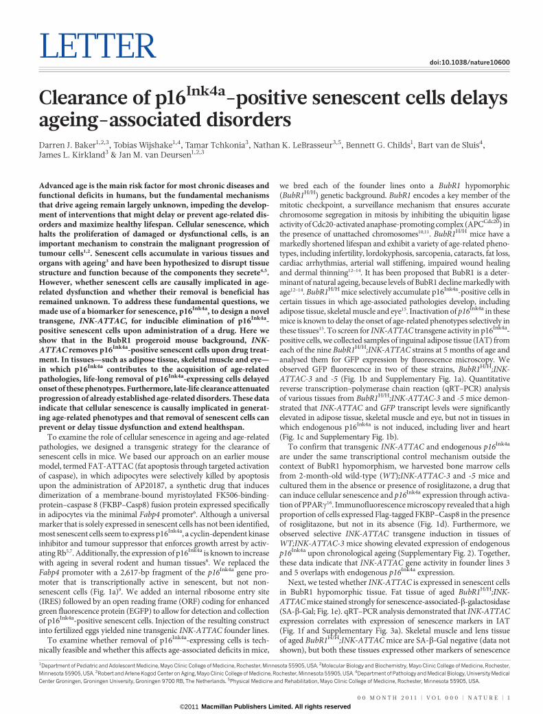
To examine the role of cellular senescence in ageing and age-relatedpathologies, we designed a transgenic strategy for the clearance ofsenescent cells in mice. We based our approach on an earlier mousemodel, termed FAT-ATTAC (fat apoptosis through targeted activationof caspase), in which adipocytes were selectively killed by apoptosisupon the administration of AP20187, a synthetic drug that inducesdimerization of a membrane-bound myristoylated FK506-binding-protein–caspase 8 (FKBP–Casp8) fusion protein expressed specificallyin adipocytes via the minimal Fabp4 promoter6. Although a universalmarker that is solely expressed in senescent cells has not been identified,most senescent cells seem to express p16Ink4a, a cyclin-dependent kinaseinhibitor and tumour suppressor that enforces growth arrest by activ-ating Rb5,7. Additionally, the expression of p16Ink4a is known to increasewith ageing in several rodent and human tissues8. We replaced theFabp4 promoter with a 2,617-bp fragment of the p16Ink4a gene pro-moter that is transcriptionally active in senescent, but not non-senescent cells (Fig. 1a)9. We added an internal ribosome entry site(IRES) followed by an open reading frame (ORF) coding for enhancedgreen fluorescence protein (EGFP) to allow for detection and collectionof p16Ink4a-positive senescent cells. Injection of the resulting constructinto fertilized eggs yielded nine transgenic INK-ATTAC founder lines.
To examine whether removal of p16Ink4a-expressing cells is tech-nically feasible and whether this affects age-associated deficits in mice,
we bred each of the founder lines onto a BubR1 hypomorphic(BubR1H/H) genetic background. BubR1 encodes a key member of themitotic checkpoint, a surveillance mechanism that ensures accuratechromosome segregation in mitosis by inhibiting the ubiquitin ligaseactivity of Cdc20-activated anaphase-promoting complex (APCCdc20) inthe presence of unattached chromosomes10,11. BubR1H/H mice have amarkedly shortened lifespan and exhibit a variety of age-related pheno-types, including infertility, lordokyphosis, sarcopenia, cataracts, fat loss,cardiac arrhythmias, arterial wall stiffening, impaired wound healingand dermal thinning12–14. It has been proposed that BubR1 is a deter-minant of natural ageing, because levels of BubR1 decline markedly withage12–14. BubR1H/H mice selectively accumulate p16Ink4a-positive cells incertain tissues in which age-associated pathologies develop, includingadipose tissue, skeletal muscle and eye15. Inactivation of p16Ink4a in thesemice is known to delay the onset of age-related phenotypes selectively inthese tissues15. To screen for INK-ATTAC transgene activity in p16Ink4a-positive cells, we collected samples of inguinal adipose tissue (IAT) fromeach of the nine BubR1H/H;INK-ATTAC strains at 5 months of age andanalysed them for GFP expression by fluorescence microscopy. Weobserved GFP fluorescence in two of these strains, BubR1H/H;INK-ATTAC-3 and -5 (Fig. 1b and Supplementary Fig. 1a). Quantitativereverse transcription–polymerase chain reaction (qRT–PCR) analysisof various tissues from BubR1H/H;INK-ATTAC-3 and -5 mice demon-strated that INK-ATTAC and GFP transcript levels were significantlyelevated in adipose tissue, skeletal muscle and eye, but not in tissues inwhich endogenous p16Ink4a is not induced, including liver and heart(Fig. 1c and Supplementary Fig. 1b).
To confirm that transgenic INK-ATTAC and endogenous p16Ink4a
are under the same transcriptional control mechanism outside thecontext of BubR1 hypomorphism, we harvested bone marrow cellsfrom 2-month-old wild-type (WT);INK-ATTAC-3 and -5 mice andcultured them in the absence or presence of rosiglitazone, a drug thatcan induce cellular senescence and p16Ink4a expression through activa-tion of PPARc16. Immunofluorescence microscopy revealed that a highproportion of cells expressed Flag-tagged FKBP–Casp8 in the presenceof rosiglitazone, but not in its absence (Fig. 1d). Furthermore, weobserved selective INK-ATTAC transgene induction in tissues ofWT;INK-ATTAC-3 mice showing elevated expression of endogenousp16Ink4a upon chronological ageing (Supplementary Fig. 2). Together,these data indicate that INK-ATTAC gene activity in founder lines 3and 5 overlaps with endogenous p16Ink4a expression.
Next, we tested whether INK-ATTAC is expressed in senescent cellsin BubR1 hypomorphic tissue. Fat tissue of aged BubR1H/H;INK-ATTAC mice stained strongly for senescence-associated-b-galactosidase(SA-b-Gal; Fig. 1e). qRT–PCR analysis demonstrated that INK-ATTACexpression correlates with expression of senescence markers in IAT(Fig. 1f and Supplementary Fig. 3a). Skeletal muscle and lens tissueof aged BubR1H/H;INK-ATTAC mice are SA-b-Gal negative (data notshown), but both these tissues expressed other markers of senescence
1Department of Pediatric and Adolescent Medicine, Mayo Clinic College of Medicine, Rochester, Minnesota 55905, USA. 2Molecular Biology and Biochemistry, Mayo Clinic College of Medicine, Rochester,Minnesota 55905, USA. 3Robert and Arlene Kogod Center on Aging, Mayo Clinic College of Medicine, Rochester, Minnesota 55905, USA. 4Department of Pathology and Medical Biology, University MedicalCenter Groningen, Groningen University, Groningen 9700 RB, The Netherlands. 5Physical Medicine and Rehabilitation, Mayo Clinic College of Medicine, Rochester, Minnesota 55905, USA.
0 0 M O N T H 2 0 1 1 | V O L 0 0 0 | N A T U R E | 1
Macmillan Publishers Limited. All rights reserved©2011
(Fig. 1f and Supplementary Fig. 3a). Senescence markers were notelevated in 3-week-old BubR1H/H;INK-ATTAC mice (SupplementaryFig. 3b, c). To obtain additional evidence for selective expression ofINK-ATTAC in senescent cells, we collected IAT from agedBubR1H/H;INK-ATTAC animals, prepared single-cell suspensions bycollagenase treatment, separated GFP1 and GFP– cell populations byfluorescence activated cell sorting (FACS; Fig. 1g), and analysed eachpopulation for expression of INK-ATTAC and senescence markers byqRT–PCR. GFP1 cells not only expressed much higher levels ofp16Ink4a than GFP– cells but also had elevated levels of other keysenescence markers (Fig. 1h and Supplementary Fig. 3d). Furthermore,two conditions that induce p16Ink4a expression and senescence inprimary mouse embryonic fibroblasts (MEFs), ectopic expression ofoncogenic Ras and serial passaging12,17,18, produced a subpopulation of
GFP1 WT;INK-ATTAC-3 MEFs that, in contrast to the remainingGFP– cells, stained positively for SA-b-Gal (Fig. 1i). Taken together,these results indicate that INK-ATTAC is selectively expressed inp16Ink4a-positive senescent cells.
To determine whether INK-ATTAC can eliminate senescent cells,we cultured bone marrow cells of WT;INK-ATTAC transgenic lines 3and 5 in the presence of rosiglitazone to induce senescence and thenmonitored cell survival after activating the FKBP–Casp8 fusionprotein by AP20187 treatment. We found that the vast majority ofcells from both transgenic lines were either dead or in the process ofdying 48 h after adding AP20187 (Fig. 2a). In contrast, parallel culturesthat remained untreated consisted almost entirely of viable SA-b-Gal-positive cells. These data show that FKBP–Casp8 activation efficientlyeliminates p16Ink4a-positive senescent cells in vitro.
H/H;ATTAC-3WT;ATTAC-3
FKB
P-C
asp
8 re
lative e
xp
ressio
n
H/H;ATTAC-3WT;ATTAC-3
SkM
IAT
Eye
Liv
er
Heart
FKB
P-C
asp
8 re
lative e
xp
ressio
n
/ ; C 3
SkM
IAT
Eye
Liv
er
Heart
EG
FP r
ela
tive e
xp
ressio
n
SkM
IAT
Eye
Liv
er
b
c
a
M
Casp8–Flag Myristoylation
FKBP
M
Inactive Casp8
Cell membrane
+ AP20187
Apoptosis
Active Casp8
M M M M Casp8–Flag IRES EGFP
p16Ink4a
promoter
–2,617 FKBP
INK-ATTAC
DNA FKBP–Casp8–Flag
WT;ATTAC-3 bone marrow cells
– Rosiglitazone + Rosiglitazone
d
Heart
e
Rela
tive e
xp
ressio
n in IAT
Rela
tive e
xp
ressio
n in S
kM
Rela
tive e
xp
ressio
n in e
ye
h H/H;ATTAC-3 GFP+
Rela
tive e
xp
ressio
n in IAT
H/H;ATTAC-3 GFP–
H/H;ATTAC-3
WT;ATTAC-3 f
H/H;ATTAC-3i
H/H;ATTAC-3WT;ATTAC-3
SA- -Gal stained WT;ATTAC-3 MEFs
+ H-ras Serial passaging
GF
P–
GF
P+
**
**
*
***
**
*
**
*
**
p21
Pai
1 Il6
Mm
p13
p19
g WT;ATTAC-3
;
Fluorescence intensity
Co
unts
x 1
02
3
2
1
GF
P+
GF
P–
Rela
tive
exp
ressio
n in IAT
Rela
tive
exp
ressio
nin
SkM
Rela
tive
exp
ressio
nin
SkM
pS
Rela
tive
exp
ressio
nin
eye
Rela
tive
exp
ressio
nin
eye
C-3 GFP3 – i SA- l-Gal
p21
Pai
1 Il6
Igfb
p2
p21
Pai
1 Il6
Mm
p13
M
mp
3
p19
p19
3 w
eeks
5 m
on
ths
p16
Ink4a rela
tive e
xp
ressio
n
35
30
25
20
15
10
5
0
35
30
25
20
15
10
5
0
30
25
20
15
10
5
0
80
60
10
8
6
4
2
0
75
55
25
20
15
10
5
0
8
6
4
2
0
βH/H;ATTAC-3 GFP
p
50
40
30
20
10
0
p21
Pai
1 p
19
p16
Il6
Figure 1 | Generation and characterization of INK-ATTAC transgenicmice. a, Schematic of the INK-ATTAC construct and the mechanism ofapoptosis activation. b, GFP intensity of IAT. c, qRT–PCR analysis of theindicated tissues of 10-month-old mice. ATTAC, INK-ATTAC; H/H,BubR1H/H; SkM, skeletal muscle (gastrocnemius). d, Bone marrow cellsharvested from 2-month-old mice immunostained for Flag after culture inthe absence or presence of rosiglitazone for 48 h. e, SA-b-Gal stained IATcollected from 9-month-old mice of the indicated genotypes. f, Expression ofsenescence markers in tissues of 10-month-old mice measured by qRT–PCR.
All increases are statistically significant (P , 0.05). g, FACS profile of single-cellsuspensions from IAT of 10-month-old mice. Brackets indicate sorting gates.h, GFP1 and GFP– cell populations from IAT analysed for relative expressionof senescence markers by qRT–PCR. All increases are statistically significant(P , 0.01). i, Bright field images of MEFs sorted into GFP1 and GFP–
populations after induction of senescence and then stained for SA-b-Gal. For allexperiments, n 5 3 untreated females per genotype. Error bars, s.d. Scale bars inb, d and i, 20mm. *P , 0.05, **P , 0.01, ***P , 0.001.
RESEARCH LETTER
2 | N A T U R E | V O L 0 0 0 | 0 0 M O N T H 2 0 1 1
Macmillan Publishers Limited. All rights reserved©2011
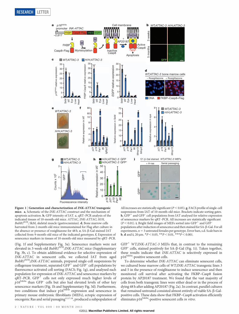
Next, we examined whether clearance of p16Ink4a-expressing cellsfrom BubR1H/H mice prevents or delays the onset of age-relatedphenotypes in this progeroid background. To this end, we establishedcohorts of BubR1H/H;INK-ATTAC-3 and -5 mice, which were eithertreated with AP20187 every third day beginning at 3 weeks of age orleft untreated. Both treated and untreated mice were monitored for thedevelopment of age-associated deficits known to accompany p16Ink4a
induction, including sarcopenia, cataracts and loss of adipose tissue15.Remarkably, treated mice of both BubR1H/H;INK-ATTAC lines hadsubstantially delayed onset of lordokyphosis (a measure of sarcopeniaonset in this model15) and cataracts compared to untreated mice(Fig. 2b, c). Consistent with decreased lordokyphosis, muscle fibrediameters of AP20187-treated BubR1H/H;INK-ATTAC animals werelarger than those of untreated counterparts (Fig. 2d). In addition tomuscle retention, treadmill exercise tests revealed that duration ofexercise, distance travelled and overall amount of work performedwere all significantly increased in the animals treated with AP20187(Fig. 2e), indicating preservation of muscle function. Dual-energyX-ray absorptiometry (DEXA) scans of BubR1H/H;INK-ATTAC miceconfirmed that AP20187 treatment prevented loss of adipose tissue
(Fig. 2f). All major fat deposits were larger in AP20187-treatedBubR1H/H;INK-ATTAC animals (Fig. 2f) and individual adipocyteswere markedly increased in size (Fig. 2g). Consistent with this gen-erally increased adiposity, lateral skin contained significantly moresubdermal adipose tissue (Fig. 2h). The above age-related phenotypeswere not delayed upon AP20187 treatment of BubR1H/H mice lackingINK-ATTAC (Fig. 2b and Supplementary Fig. 4).
Age-related phenotypes of BubR1H/H mice that arise in a p16Ink4a-independent fashion, such as cardiac arrhythmias and arterial wallstiffening14, were not attenuated in AP20187-treated BubR1H/H;INK-ATTAC mice (Supplementary Fig. 5a, b). This correlated with lack ofINK-ATTAC induction in heart and aorta (Fig. 1c and SupplementaryFig. 5c). Cardiac failure is presumably the main cause of death inBubR1H/H mice (data not shown), which could explain why the overallsurvival of AP20187-treated BubR1H/H;INK-ATTAC mice was notsubstantially extended (Supplementary Fig. 5d). To examine whetherclearance of p16Ink4a-positive cells might have any overtly negative sideeffects, WT;INK-ATTAC mice were continuously treated withAP20187 until 8 months of age; however, no such effects were observed(data not shown). Taken together, these results indicate that continu-ous removal of p16Ink4a-expressing cells from BubR1H/H;INK-ATTAC
0
10
20
30
40
50
60
Ing 0
50
100
150
200
250
300
350
D Ad
a b WT;ATTAC-3
+ Rosiglitazone –AP
+ Rosiglitazone +AP
SA
-β-G
al
H/H;ATTAC-3 +AP (n = 25) H/H;ATTAC-3 –AP (n = 18)
H/H;ATTAC-5 +AP (n = 36) H/H;ATTAC-5 –AP (n = 18) H/H +AP (n = 52)
Lo
rdo
kyp
ho
sis
incid
ence (%
)
Age (days)
SA
-β-G
al
H/H;ATTAC-3 +AP
H/H;ATTAC-3 –AP
c
Thic
kness (μm
)
Cell
dia
mete
r (μ
m)
H/H;ATTAC-3 –AP H/H;ATTAC-3 +AP H/H;ATTAC-5 –AP H/H;ATTAC-5 +AP
Fib
re d
iam
ete
r (μm
)
IAT Dermis Adipose
f
25
30
35
40
Gastro Abdominastro Abdomina
H/H;A;; TTTT AC-3 TT –H/H;A;; TTTT AC-3TT +H/H;A;; TTTT AC-5TT –H/H;A;; TTTT AC-5TT +
*
***
d
Gastro ABD
P
P
H/H;ATTT AC 3TT AP (n(( = 18) H/H+AH P (n(( = 52)
Lo
rdo
kyp
ho
sis
incid
ence (%
)
Age (days)
h
*
** **
Imp
rovem
ent
over
untr
eate
d (%
)
e
***
**
**
*
* ***
*
** **
***
g **
***
****
*
** **
***
0
50
100
150
200
250
300
R D WTime Distance Work
Mouse (treatment) Weight (g) Fat (%) POV (g) Peri (g) IAT (g) Mes (g) SSAT (g) Brown (g)
H/H;ATTAC-3 (–AP)17.4 (1.8)
15.6 (3.4)
0.054 (0.05)
0.016 (0.02)
0.058 (0.02)
0.063 (0.05)
0.038 (0.01)
0.084 (0.005)
H/H;ATTAC-3 (+AP)21.3* (3.8)
28.6*** (4.1)
0.334* (0.28)
0.069 (0.06)
0.192* (0.12)
0.151 (0.09)
0.095** (0.03)
0.119 (0.053)
H/H;ATTAC-5 (–AP)16.7 (2.2)
15.4 (2.5)
0.137 (0.13)
0.020 (0.01)
0.075 (0.05)
0.061 (0.04)
0.050 (0.02)
0.104 (0.030)
H/H;ATTAC-5 (+AP)24.0*** (2.1)
27.4*** (4.2)
0.433** (0.11)
0.071** (0.02)
0.225** (0.10)
0.262* (0.17)
0.115* (0.06)
0.159** (0.029)
Age (days)
Cata
ract
incid
ence (%
) 100
75
50
25
00 100 200 300
100
75
50
25
00 100 200 300
***************************************
*****************
Figure 2 | BubR1H/H;INK-ATTAC mice treated with AP20187 fromweaning age on show delayed onset of p16Ink4a-mediated age-relatedphenotypes. a, Bone marrow cells cultured in rosiglitazone for 5 days and thentreated or not treated with AP20187 (AP) for 2 days before SA-b-Gal staining.Scale bar, 50mm. b, Incidence of lordokyphosis and cataracts. c, Representativeimages of 9-month-old mice. d, Mean skeletal muscle fibre diameters of 10-month-old mice. ABD, abdominal muscle; Gastro, gastrocnemius muscle.e, Exercise ability of 10-month-old AP20187-treated mice relative to age-matched untreated mice. Time is running time to exhaustion; distance isdistance travelled at time of exhaustion; work is the energy expended toexhaustion. f, Body and fat depot weights of 10-month-old mice. Parentheses,s.d. Mes, mesenteric; Peri, perirenal; POV, paraovarian; SSAT, subscapularadipose tissue. g, Average fat cell diameters in IAT of 10-month-old mice.h, Dermis and subdermal adipose layer thickness of 10-month-old mice.Colour codes in e, g and h are as indicated in d. Error bars, s.e.m. For all analysisn 5 6 female mice per genotype (per treatment). *P , 0.05, **P , 0.01,***P , 0.001.
Inguinal adiposeGastro
Rela
tive e
xp
ressio
n in
IA
T
Rela
tive e
xp
ressio
n in
SkM
p21
Pai
1
Mm
p13
Cas
p8
GFP
p16
Rela
tive e
xp
ressio
n in
eye
H/H;ATTAC-3 –APWT;ATTAC-3 –AP
H/H;ATTAC-3 +AP
Brd
U p
ositiv
e c
ells
(%
)
IAT SkM
H/H;ATTAC-3 –AP H/H;ATTAC-3 +AP H/H;ATTAC-5 –AP H/H;ATTAC-5 +AP
b c
d e
SA
-β-G
al
H/H;ATTAC-3–AP
H/H;ATTAC-3+AP
H/H;ATTAC-5–AP
H/H;ATTAC-5+AP
a
p19
*
*
p21
Pai
1 Il6
Mm
p3
Cas
p8
GFP p16
Mm
p13
p19
Rela
tive e
xp
ressio
n in
IAA
T
A
H/H;A;; TTT AC-3TT –APH/H;A;; TTTT AC-3TT +AP
p21
Pai
1 Il6
Igfb
p2
Cas
p8
GFP
p16
p19
75
60
30
25
20
15
10
5
0
70
60
50
40
30
20
10
0
8
6
4
2
0
5
4
3
2
1
0
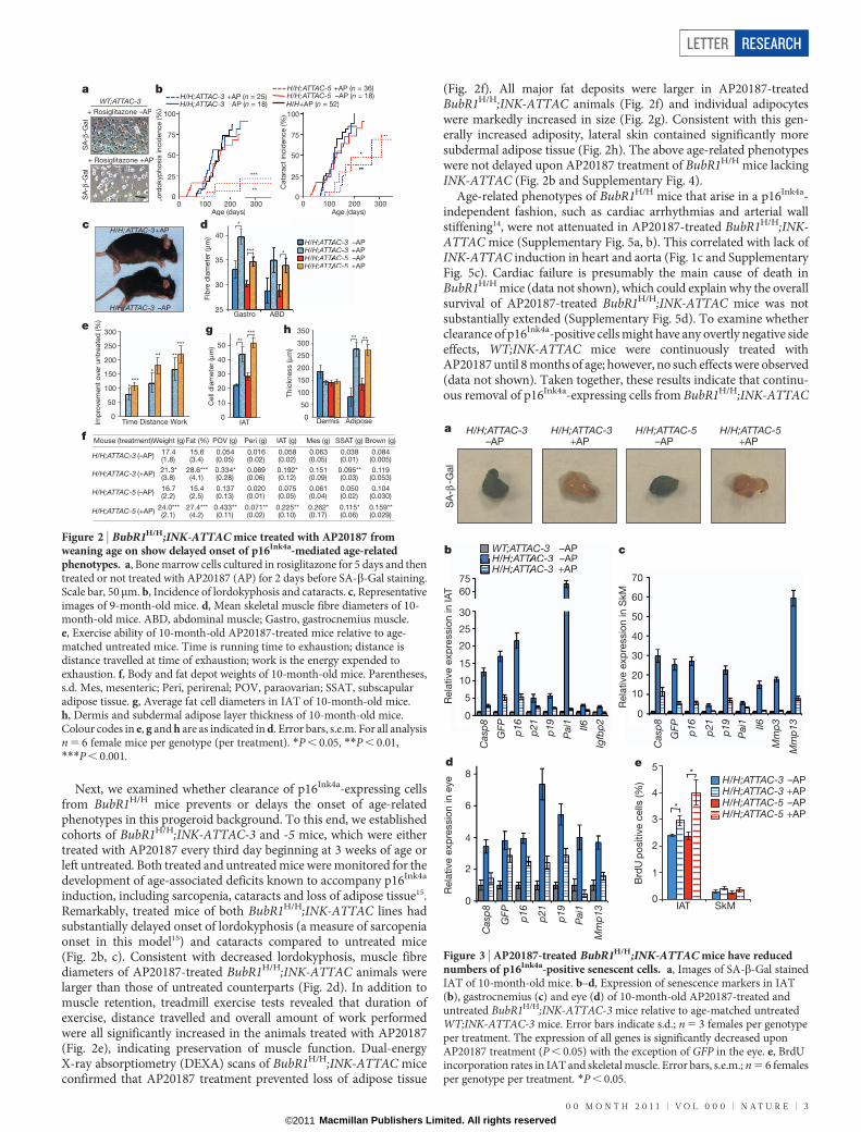
Figure 3 | AP20187-treated BubR1H/H;INK-ATTAC mice have reducednumbers of p16Ink4a-positive senescent cells. a, Images of SA-b-Gal stainedIAT of 10-month-old mice. b–d, Expression of senescence markers in IAT(b), gastrocnemius (c) and eye (d) of 10-month-old AP20187-treated anduntreated BubR1H/H;INK-ATTAC-3 mice relative to age-matched untreatedWT;INK-ATTAC-3 mice. Error bars indicate s.d.; n 5 3 females per genotypeper treatment. The expression of all genes is significantly decreased uponAP20187 treatment (P , 0.05) with the exception of GFP in the eye. e, BrdUincorporation rates in IAT and skeletal muscle. Error bars, s.e.m.; n 5 6 femalesper genotype per treatment. *P , 0.05.
LETTER RESEARCH
0 0 M O N T H 2 0 1 1 | V O L 0 0 0 | N A T U R E | 3
Macmillan Publishers Limited. All rights reserved©2011
mice selectively delays age-related phenotypes that depend on p16Ink4a
induction.Next, we determined whether the delayed onset of age-related
pathologies coincided with a reduction in the number of senescentcells in these tissues. The IAT of AP20187-treated BubR1H/H;INK-ATTAC mice showed a marked decrease in SA-b-Gal staining com-pared with the IAT of untreated counterparts (Fig. 3a). Correspondingdecreases in other senescence-associated markers were also observed,as well as expected reductions in INK-ATTAC and GFP (Fig. 3b andSupplementary Fig. 6a). Skeletal muscle and eye had a similar reduc-
tion in senescence indicators (Fig. 3c, d and Supplementary Fig. 6b, c).BrdU incorporation was lower in IAT and muscle tissue of untreatedthan treated animals (Fig. 3e), supporting the contention thatsenescence-associated replicative arrest is decreased upon administra-tion of AP20187 in BubR1H/H;INK-ATTAC transgenic animals.Together, these data indicate that senescent cells were cleared fromtissues and that this delays acquisition of age-related dysfunction inBubR1 hypomorphic mice.
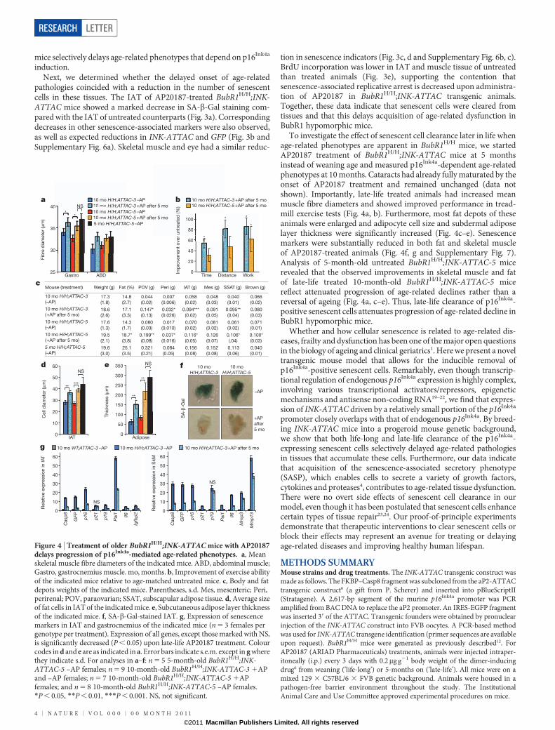
To investigate the effect of senescent cell clearance later in life whenage-related phenotypes are apparent in BubR1H/H mice, we startedAP20187 treatment of BubR1H/H;INK-ATTAC mice at 5 monthsinstead of weaning age and measured p16Ink4a-dependent age-relatedphenotypes at 10 months. Cataracts had already fully maturated by theonset of AP20187 treatment and remained unchanged (data notshown). Importantly, late-life treated animals had increased meanmuscle fibre diameters and showed improved performance in tread-mill exercise tests (Fig. 4a, b). Furthermore, most fat depots of theseanimals were enlarged and adipocyte cell size and subdermal adiposelayer thickness were significantly increased (Fig. 4c–e). Senescencemarkers were substantially reduced in both fat and skeletal muscleof AP20187-treated animals (Fig. 4f, g and Supplementary Fig. 7).Analysis of 5-month-old untreated BubR1H/H;INK-ATTAC-5 micerevealed that the observed improvements in skeletal muscle and fatof late-life treated 10-month-old BubR1H/H;INK-ATTAC-5 micereflect attenuated progression of age-related declines rather than areversal of ageing (Fig. 4a, c–e). Thus, late-life clearance of p16Ink4a-positive senescent cells attenuates progression of age-related decline inBubR1 hypomorphic mice.
Whether and how cellular senescence is related to age-related dis-eases, frailty and dysfunction has been one of the major open questionsin the biology of ageing and clinical geriatrics1. Here we present a noveltransgenic mouse model that allows for the inducible removal ofp16Ink4a-positive senescent cells. Remarkably, even though transcrip-tional regulation of endogenous p16Ink4a expression is highly complex,involving various transcriptional activators/repressors, epigeneticmechanisms and antisense non-coding RNA19–22, we find that expres-sion of INK-ATTAC driven by a relatively small portion of the p16Ink4a
promoter closely overlaps with that of endogenous p16Ink4a. By breed-ing INK-ATTAC mice into a progeroid mouse genetic background,we show that both life-long and late-life clearance of the p16Ink4a-expressing senescent cells selectively delayed age-related pathologiesin tissues that accumulate these cells. Furthermore, our data indicatethat acquisition of the senescence-associated secretory phenotype(SASP), which enables cells to secrete a variety of growth factors,cytokines and proteases4, contributes to age-related tissue dysfunction.There were no overt side effects of senescent cell clearance in ourmodel, even though it has been postulated that senescent cells enhancecertain types of tissue repair23,24. Our proof-of-principle experimentsdemonstrate that therapeutic interventions to clear senescent cells orblock their effects may represent an avenue for treating or delayingage-related diseases and improving healthy human lifespan.
METHODS SUMMARYMouse strains and drug treatments. The INK-ATTAC transgenic construct wasmade as follows. The FKBP–Casp8 fragment was subcloned from the aP2-ATTACtransgenic construct6 (a gift from P. Scherer) and inserted into pBlueScriptII(Stratagene). A 2,617-bp segment of the murine p16Ink4a promoter was PCRamplified from BAC DNA to replace the aP2 promoter. An IRES-EGFP fragmentwas inserted 39 of the ATTAC. Transgenic founders were obtained by pronuclearinjection of the INK-ATTAC construct into FVB oocytes. A PCR-based methodwas used for INK-ATTAC transgene identification (primer sequences are availableupon request). BubR1H/H mice were generated as previously described12. ForAP20187 (ARIAD Pharmaceuticals) treatments, animals were injected intraper-itoneally (i.p.) every 3 days with 0.2mg g21 body weight of the dimer-inducingdrug6 from weaning (‘life-long’) or 5-months on (‘late-life’). All mice were on amixed 129 3 C57BL/6 3 FVB genetic background. Animals were housed in apathogen-free barrier environment throughout the study. The InstitutionalAnimal Care and Use Committee approved experimental procedures on mice.
0
10
20
30
40
50
60 d e
0
50
100
150
200
250
300
350
25
30
35
40
Fib
re d
iam
ete
r (μ
m)
Mouse (treatment) Weight (g) Fat (%) POV (g) Peri (g) IAT (g) Mes (g) SSAT (g) Brown (g)
10 mo H/H;ATTAC-3(–AP)
17.3
(1.8)
14.8
(2.7)
0.044
(0.02)
0.007
(0.006)
0.058
(0.02)
0.048
(0.03)
0.040
(0.01)
0.066
(0.02)
10 mo H/H;ATTAC-3 (+AP after 5 mo)
18.6
(2.6)
17.1
(3.3)
0.147*
(0.13)
0.032*
(0.026)
0.094***
(0.02)
0.091
(0.05)
0.095**
(0.04)
0.080
(0.03)
10 mo H/H;ATTAC-5(–AP)
17.6
(1.3)
14.3
(1.7)
0.080
(0.03)
0.017
(0.010)
0.070
(0.02)
0.081
(0.02)
0.061
(0.02)
0.071
(0.01)
10 mo H/H;ATTAC-5 (+AP after 5 mo)
19.5
(2.1)
18.7*
(3.8)
0.199**
(0.08)
0.037*
(0.016)
0.116*
(0.05)
0.126
(0.07)
0.106*
(.04)
0.100*
(0.03)
5 mo H/H;ATTAC-5 (–AP)
19.6
(3.0)
25.1
(3.5)
0.321
(0.21)
0.084
(0.05)
0.156
(0.08)
0.152
(0.08)
0.113
(0.06)
0.040
(0.01)
Gastro ABD 0
20
40
60
80
100
Time Distance Work
Imp
rovem
en
t o
ver
un
treate
d (%
)
*
* *
*
10 mo WT;ATTAC-3 –AP
0
10
20
30
40
50
60
Rela
tive e
xp
ressio
n in S
kM
10 mo H/H;ATTAC-3 –AP 10 mo H/H;ATTAC-3 +AP after 5 mo
SA
-β-G
al
–AP
+AP
after
5 mo
10 mo
H/H;ATTAC-3
10 mo H/H;ATTAC-3 +AP after 5 mo 10 mo H/H;ATTAC-5 +AP after 5 mo
60ver *
80
100
un
treate
d (%
)
10 mo H/H;ATTA10 mo H/H;ATTA
a b
c
f
g
p21
Pai
1 Il6
Mm
p3
Cas
p8
GFP
p16
Mm
p13
p19
*
*
10 mo H/H;ATTAC-3 –AP
10 mo H/H;ATTAC-3 +AP after 5 mo
10 mo H/H;ATTAC-5 –AP
10 mo H/H;ATTAC-5 +AP after 5 mo
0
5
0
**
10 mo H/H;ATTA10 mo10 mo H/H;ATTAH/H;10 mo ;ATTAH/H;10 mo ;ATTAH/H;
NS
5 mo H/H;ATTAC-5 –AP
Thic
kness (μm
)
Cell
dia
mete
r (μ
m)
Adipose IAT
** ***
**
**
0
10
20
30
40
50
60
Rela
tive e
xp
ressio
n in IAT
p21
Pai
1 Il6
Igfb
p2
Cas
p8
GFP
p16
p19
NS
NS
NS
NS
10 mo
H/H;ATTAC-5
Figure 4 | Treatment of older BubR1H/H;INK-ATTAC mice with AP20187delays progression of p16Ink4a-mediated age-related phenotypes. a, Meanskeletal muscle fibre diameters of the indicated mice. ABD, abdominal muscle;Gastro, gastrocnemius muscle. mo, months. b, Improvement of exercise abilityof the indicated mice relative to age-matched untreated mice. c, Body and fatdepots weights of the indicated mice. Parentheses, s.d. Mes, mesenteric; Peri,perirenal; POV, paraovarian; SSAT, subscapular adipose tissue. d, Average sizeof fat cells in IAT of the indicated mice. e, Subcutaneous adipose layer thicknessof the indicated mice. f, SA-b-Gal-stained IAT. g, Expression of senescencemarkers in IAT and gastrocnemius of the indicated mice (n 5 3 females pergenotype per treatment). Expression of all genes, except those marked with NS,is significantly decreased (P , 0.05) upon late-life AP20187 treatment. Colourcodes in d and e are as indicated in a. Error bars indicate s.e.m. except in g wherethey indicate s.d. For analyses in a–f: n 5 5 5-month-old BubR1H/H;INK-ATTAC-5 –AP females; n 5 9 10-month-old BubR1H/H;INK-ATTAC-3 1APand –AP females; n 5 7 10-month-old BubR1H/H;INK-ATTAC-5 1APfemales; and n 5 8 10-month-old BubR1H/H;INK-ATTAC-5 –AP females.*P , 0.05, **P , 0.01, ***P , 0.001. NS, not significant.
RESEARCH LETTER
4 | N A T U R E | V O L 0 0 0 | 0 0 M O N T H 2 0 1 1
Macmillan Publishers Limited. All rights reserved©2011
Full Methods and any associated references are available in the online version ofthe paper at www.nature.com/nature.
Received 8 May; accepted 30 September 2011.
Published online 2 November 2011.
1. Campisi, J. Cellular senescence: putting the paradoxes in perspective. Curr. Opin.Genet. Dev. 21, 107–112 (2011).
2. Kuilman, T., Michaloglou, C., Mooi, W. J. & Peeper, D. S. The essence of senescence.Genes Dev. 24, 2463–2479 (2010).
3. Campisi, J. Senescent cells, tumor suppression, and organismal aging: goodcitizens, bad neighbors. Cell 120, 513–522 (2005).
4. Coppe, J. P. et al. Senescence-associated secretory phenotypes reveal cell-nonautonomous functions of oncogenicRASand the p53 tumor suppressor. PLoSBiol. 6, e301 (2008).
5. Rodier, F.& Campisi, J. Four faces of cellular senescence. J. Cell Biol. 192, 547–556(2011).
6. Pajvani, U. B. et al. Fat apoptosis through targeted activation of caspase 8: a newmouse model of inducible and reversible lipoatrophy. Nature Med. 11, 797–803(2005).
7. Kim, W. Y. & Sharpless, N. E. The regulation of INK4/ARF in cancer and aging. Cell127, 265–275 (2006).
8. Krishnamurthy, J. et al. Ink4a/Arf expression is a biomarker of aging. J. Clin. Invest.114, 1299–1307 (2004).
9. Wang, W., Wu, J., Zhang, Z. & Tong, T. Characterization of regulatory elements onthe promoter region of p16INK4a that contribute to overexpression of p16 insenescent fibroblasts. J. Biol. Chem. 276, 48655–48661 (2001).
10. Malureanu, L. A. et al. BubR1 N terminus acts as a soluble inhibitor of cyclin Bdegradation by APC/CCdc20 in interphase. Dev. Cell 16, 118–131 (2009).
11. Kulukian, A., Han, J. S. & Cleveland, D. W. Unattached kinetochores catalyzeproduction of an anaphase inhibitor that requires a Mad2 template to primeCdc20 for BubR1 binding. Dev. Cell 16, 105–117 (2009).
12. Baker, D. J. et al. BubR1 insufficiency causes early onset of aging-associatedphenotypes and infertility in mice. Nature Genet. 36, 744–749 (2004).
13. Hartman, T. K., Wengenack, T. M., Poduslo, J. F. & van Deursen, J. M. Mutant micewith small amounts of BubR1 display accelerated age-related gliosis. Neurobiol.Aging 28, 921–927 (2007).
14. Matsumoto, T. et al. Aging-associated vascular phenotype in mutant mice with lowlevels of BubR1. Stroke 38, 1050–1056 (2007).
15. Baker, D. J. et al. Opposing roles for p16Ink4a and p19Arf in senescence and ageingcaused by BubR1 insufficiency. Nature Cell Biol. 10, 825–836 (2008).
16. Gan, Q. et al. PPARc accelerates cellular senescence by inducing p16INK4a
expression in human diploid fibroblasts. J. Cell Sci. 121, 2235–2245 (2008).
17. Serrano, M., Lin, A. W., McCurrach, M. E., Beach, D. & Lowe, S. W. Oncogenic rasprovokes premature cell senescence associated with accumulation of p53 andp16INK4a. Cell 88, 593–602 (1997).
18. Kim, H. et al. Expression profiles of p53-, p16INK4a-, and telomere-regulating genesin replicative senescent primary human, mouse, and chicken fibroblast cells. Exp.Cell Res. 272, 199–208 (2002).
19. Popov, N. & Gil, J. Epigenetic regulation of the INK4b–ARF–INK4a locus: in sicknessand in health. Epigenetics 5, 685–690 (2010).
20. Gil, J. & Peters, G. Regulation of the INK4b–ARF–INK4a tumour suppressor locus:all for one or one for all. Nature Rev. Mol. Cell Biol. 7, 667–677 (2006).
21. Burd, C. E. et al. Expression of linear and novel circular forms of an INK4/ARF-associated non-coding RNA correlates with atherosclerosis risk. PLoS Genet. 6,e1001233 (2010).
22. Li, J., Poi, M. J. & Tsai, M. D. Regulatory mechanisms of tumor suppressor P16INK4A
and their relevance to cancer. Biochemistry 50, 5566–5582 (2011).23. Krizhanovsky, V. et al. Senescence of activated stellate cells limits liver fibrosis. Cell
134, 657–667 (2008).24. Jun, J. I. & Lau, L. F. The matricellular protein CCN1 induces fibroblast senescence
and restricts fibrosis in cutaneous wound healing. Nature Cell Biol. 12, 676–685(2010).
Supplementary Information is linked to the online version of the paper atwww.nature.com/nature.
Acknowledgements We thank W. Zhou, D. Norris, T. Mann, U. Moedder, T. Pirtskhalavaand S. Yamada for assistance; S. Khosla, T. von Zglinicki, L. Malureanu, R. Ricke andP. Galardy, and members of the J.M.v.D. laboratory for helpful discussions; andP.Scherer for thegift of theaP2-ATTACplasmid. Thisworkwas supportedby the EllisonMedical Foundation (J.M.v.D.), the Noaber Foundation (J.M.v.D. and J.L.K.), the Robertand Arlene Kogod Center on Aging, and the National Institutes of Health (CA96985,J.M.v.D. and AG13925, J.L.K.).
Author Contributions D.J.B., T.T., J.L.K., and J.M.v.D designed the INK-ATTAC strategy.D.J.B. and T.W. performed most of the experiments, T.T. did the rosiglitazoneexperiments, N.K.L. and B.G.C. assisted with the analysis of muscle functionality and invitro senescence, respectively, and B.v.d.S. helped supervise T.W. The manuscript waswritten by D.J.B. and J.M.v.D. All authors discussed results, made figures and edited themanuscript. J.M.v.D. directed and supervised all aspects of the study.
Author Information Reprints and permissions information is available atwww.nature.com/reprints. The authors declare no competing financial interests.Readers are welcome to comment on the online version of this article atwww.nature.com/nature. Correspondence and requests for materials should beaddressed to J.M.v.D. ([email protected]).
LETTER RESEARCH
0 0 M O N T H 2 0 1 1 | V O L 0 0 0 | N A T U R E | 5
Macmillan Publishers Limited. All rights reserved©2011
METHODSMouse strains and drug treatments. The INK-ATTAC transgenic construct wasmade as follows. The FKBP–Casp8 fragment was subcloned from the aP2-ATTACtransgenic construct6 (a gift from P. Scherer) and inserted into pBlueScriptII(Stratagene). A 2,617-bp segment of the murine p16Ink4a promoter was PCR amp-lified from BAC DNA to replace the aP2 promoter. An IRES-EGFP fragment wasinserted 39 of the ATTAC. Transgenic founders were obtained by pronuclear injec-tion of the INK-ATTAC construct into FVB oocytes. A PCR-based method wasused for INK-ATTAC transgene identification (primer sequences are available uponrequest). BubR1H/H mice were generated as previously described12. For AP20187(ARIAD Pharmaceuticals) treatments, animals were injected intraperitoneally (i.p.)every 3 days with 0.2mg g21 body weight of the dimer-inducing drug6 from weaning(‘life-long’) or 5-months on (‘late-life’). All mice were on a mixed 129 3 C57BL/6 3
FVB genetic background. Animals were housed in a pathogen-free barrier environ-ment throughout the study. The Institutional Animal Care and Use Committeeapproved experimental procedures on mice.Statistical analysis. Prism software was used for the generation of all survivalcurves and statistical analyses. Two-tailed unpaired t tests were used for pairwisesignificance analysis in the following figures: Fig. 1c, f and h; Fig. 2d–h; Fig. 3b–e;Fig. 4a–e, g; Supplementary Fig. 1b; Supplementary Fig. 2; Supplementary Fig. 3;Supplementary Fig. 4; Supplementary Fig. 5a–c; Supplementary Fig. 6; andSupplementary Fig. 7. Log-rank tests were used to determine overall and pairwisesignificance for incidence curves in Fig. 2b and survival curves in SupplementaryFig. 5d. For consistency in these comparisons, the following identifies the signifi-cance values: *P , 0.05, **P , 0.01, ***P , 0.001.Cell culture. Culture of bone marrow cells was as previously described25. Briefly,tibia and femur bones of 2-month-old WT;INK-ATTAC transgenic mouse lineswere collected and flushed with DMEM containing 15% FBS. After centrifugationat 400g for 10 min and counting of viable cells with trypan blue, cells were resus-pended in DMEM containing 15% FBS to a final concentration of 5 3 106 viablecells per ml. Initially, cells were plated in 6-well tissue culture dishes at 3.5 mlwell21 (1.9 3 106 cells cm22). Cultures were kept in a humidified 5% CO2 incubatorat 37 uC for 72 h, when non-adherent cells were removed by changing the medium.Assays were performed on cells that had been trypsinized and seeded to confluencyin 24-well plates. To induce senescence and evaluate expression of the INK-ATTACtransgene, cells were treated with 1mM rosiglitazone (Cayman Chemical Company)or with vehicle. The accumulation of GFP1 cells was observed by fluorescencemicroscopy and transgene expression was verified by immunofluorescence stainingfor Flag (Origene) as described26. After 5 days of rosiglitazone treatment, cells werewashed with PBS and treated with vehicle, 1mM rosiglitazone, 10 nM AP20187, orboth. After 48 h, cultures were fixed and stained for SA-b-Gal activity as described27.WT;INK-ATTAC MEFs were generated and cultured as previously described12. Forinduction of replicative senescence, WT;INK-ATTAC MEF cultures were main-tained in 20% O2 for 12–15 passages. For oncogene-induced senescence, earlypassage MEFs were infected with concentrated pBABE puro H-rasG12V retrovirus(Addgene plasmid 9051) for 48 h. MEFs were then cultured in DMEM containingpuromycin (2mg ml21) for 5 days. Cells from serial passage and H-ras inducedsenescence were sorted into GFP1 and GFP– populations using a FACS Aria CellSorter (BD Biosciences) running FACSDiva software (serial passaging and H-rasexpression yielded cultures with approximately 90% and 50% GFP1 cells, respect-ively). Sorted cells were transferred to polyethylenimine-coated chambered slidesand stained for SA-b-Gal according to manufacturer’s instructions (Cell Signaling).
qRT–PCR and flow cytometry. RNA extraction, cDNA synthesis and qRT–PCRfrom whole-mouse tissue were performed as previously described15. To performqRT–PCR on GFP1 and GFP– cell populations of IAT, single-cell suspensions ofstromal vascular fraction were prepared from ,50 mg IAT as described28. Cellsorting was performed as described above. RNA was extracted from the collectedcells using an RNeasy Micro Kit (Qiagen) and cDNA synthesized using a WT-Ovation RNA Amplification kit (NuGEN Technologies), according to the manu-facturers’ protocols. qRT–PCR primers were as follows: FKBP–Casp8 forward,GAATCACAGACTTTGGACAAAGTT; FKBP–Casp8 reverse, GGTCAAAGCCCCTGCATCCAAG; EGFP forward, CAAACTACAACAGCCACAA CG;EGFP reverse, GGTCACGAACTCCAGCAG. Sequences of other primers usedwere as previously described15.Analysis of progeroid phenotypes. Bi-weekly checks for lordokyphosis andcataracts were performed as described15. Skeletal muscle fibre diameter measure-ments were performed on cross-sections of gastrocnemius and abdominal musclesof female mice as described15. Fifty total fibres per sample were measured using acalibrated computer program (Olympus MicroSuite Five). Fat cell diameter mea-surements were performed on IAT according to the same method. Dissection,histology and measurements of dermal and adipose layers of skin were performedas described previously12, although the lateral skin between the front and hind limbwas used because this adipose layer is nearly three times thicker than dorsal skin.Measurements of body weight, length, gastrocnemius muscle and assorted adiposedepots were performed on 10-month-old females. Bone mineral content, bonemineral density and total body adipose tissue were analysed by DEXA scanning aspreviously described6. Exercise measurements were performed on 10-month-oldmice as previously described29. Animals were acclimated for 3 days for 5 min at aspeed of 5 m min21 before experimentation. For the experiment, the speed of thetreadmill began at 5 m min21 and was increased to 8 m min21 after 2 min.Thereafter, the speed was increased at a rate of 2 m min21 every 2 min and thetime (in seconds) and distance (in metres) to exhaustion, as defined by an inabilityto move along the treadmill with stimulation, were determined. The formula todetermine the amount of work (J) performed was: mass (kg) 3 g (9.8 m s22) 3
distance (m) 3 sin(h) (with an incline of h5 5u). Cardiac arrhythmia measure-ments were performed using a Vevo2100 ultrasound system (Visualsonics) aspreviously described30.In vivo BrdU incorporation and SA-b-Gal staining. Analyses for in vivo BrdUincorporation were performed in 10-month-old female mice as described15.Adipose tissue depots were stained for SA-b-Gal activity as previously described12.
25. Soleimani, M.& Nadri, S. A protocol for isolation andculture of mesenchymal stemcells from mouse bone marrow. Nature Protocols 4, 102–106 (2009).
26. Malureanu, L. et al. Cdc20 hypomorphic mice fail to counteract de novo synthesisof cyclin B1 in mitosis. J. Cell Biol. 191, 313–329 (2010).
27. Dimri, G. P. et al. A biomarker that identifies senescent human cells in culture andin aging skin in vivo. Proc. Natl Acad. Sci. USA 92, 9363–9367 (1995).
28. Kirkland, J. L., Hollenberg, C. H. & Gillon, W. S. Effects of fat depot site ondifferentiation-dependent gene expression in rat preadipocytes. Int. J. Obes. Relat.Metab. Disord. 20 (Suppl 3), S102–S107 (1996).
29. LeBrasseur, N. K. et al. Myostatin inhibition enhances the effects of exercise onperformance and metabolic outcomes in aged mice. J. Gerontol. A Biol. Sci. Med.Sci. 64A, 940–948 (2009).
30. Martinez-Fernandez, A. et al. iPS programmed without c-MYC yield proficientcardiogenesis for functional heart chimerism. Circ. Res. 105, 648–656 (2009).
RESEARCH LETTER
Macmillan Publishers Limited. All rights reserved©2011