seminar gpcr ppt
TRANSCRIPT
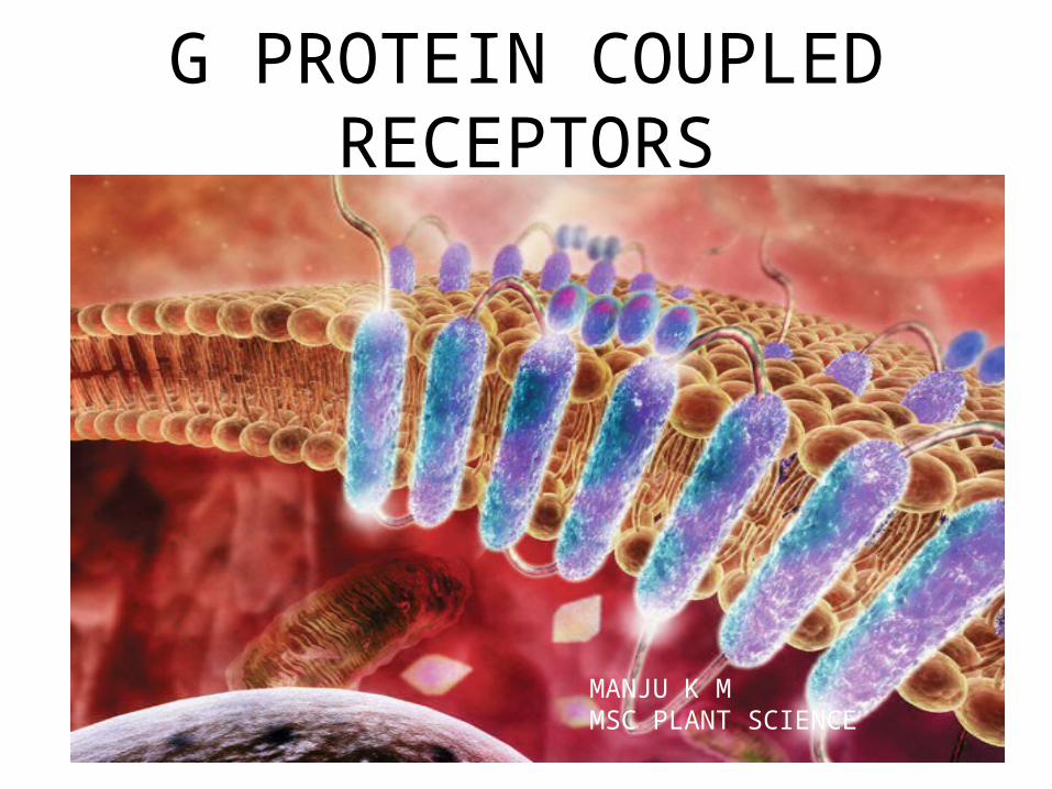
G PROTEIN COUPLED RECEPTORS
MANJU K M MSC PLANT SCIENCE
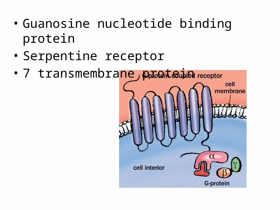
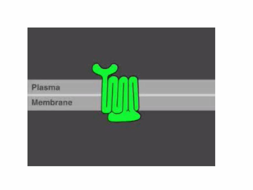
• Guanosine nucleotide binding protein• Serpentine receptor• 7 transmembrane protein
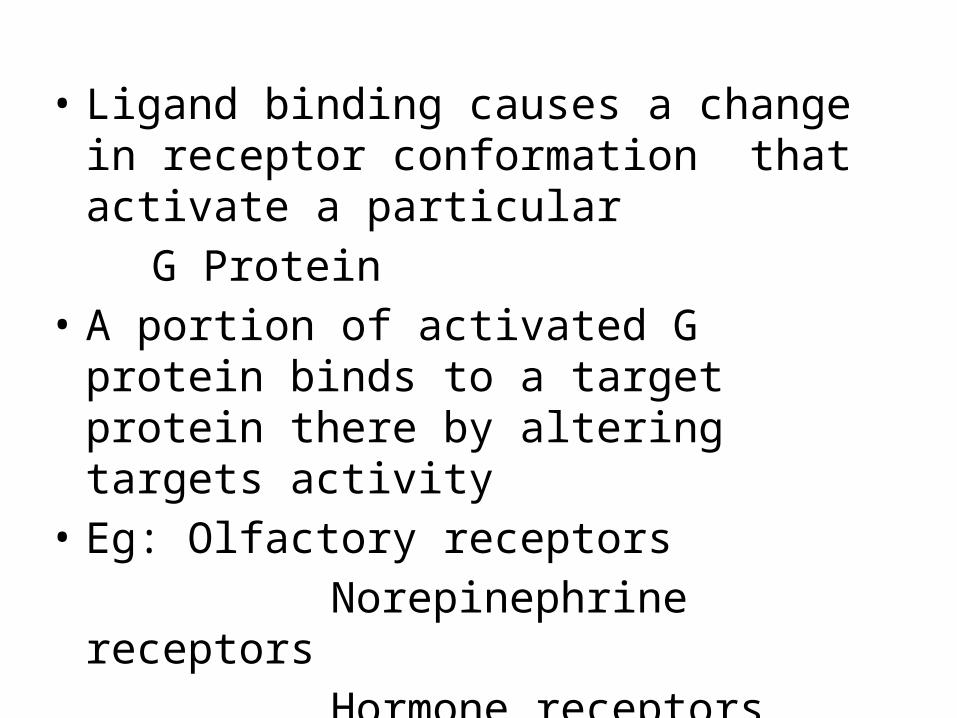
• Ligand binding causes a change in receptor conformation that activate a particular
G Protein• A portion of activated G protein binds to a
target protein there by altering targets activity• Eg: Olfactory receptors Norepinephrine receptors Hormone receptors
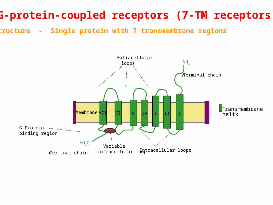
G-protein-coupled receptors (7-TM receptors) Structure - Single protein with 7 transmembrane regions
Transmembranehelix
C-Terminal chain
G-Proteinbinding region
Variableintracellular loop
Extracellularloops
Intracellular loops
N-Terminal chain
HO2C
NH2
VII VI V IV III II IMembrane

Structure of G protein

7 transmembrane helices connected by alternating cytosolic and extra cellular loop
C terminal: inside the cellN terminal : extra cellular regionExtra cellular portion has unique messenger
binding siteCytosolic loop allow receptor to interact with G
protein
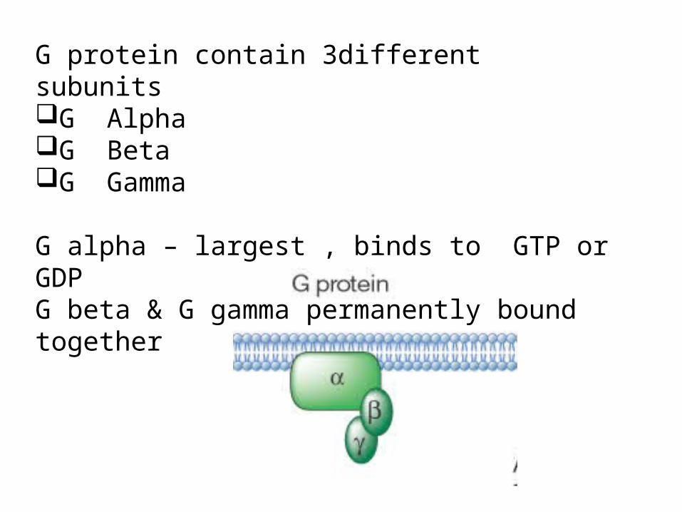
G protein contain 3different subunitsG AlphaG BetaG Gamma
G alpha – largest , binds to GTP or GDPG beta & G gamma permanently bound together

Act like MOLECULAR SWITCHESON – when bind to GTPOFF – when bind to GDPWhen G alpha bind to GTP it detaches from beta and gamma complex
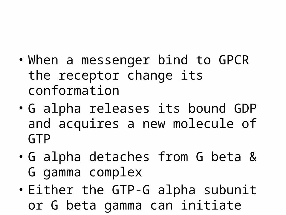
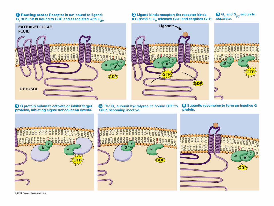
• When a messenger bind to GPCR the receptor change its conformation
• G alpha releases its bound GDP and acquires a new molecule of GTP
• G alpha detaches from G beta & G gamma complex
• Either the GTP-G alpha subunit or G beta gamma can initiate signal transduction
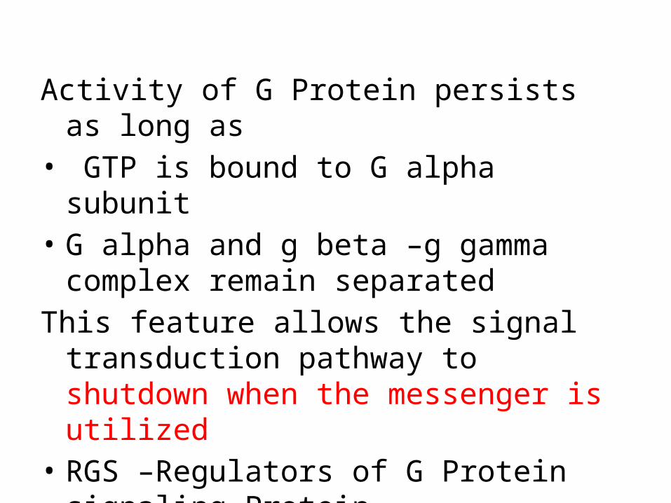
Activity of G Protein persists as long as• GTP is bound to G alpha subunit • G alpha and g beta –g gamma complex remain
separated This feature allows the signal transduction
pathway to shutdown when the messenger is utilized
• RGS –Regulators of G Protein signaling Protein
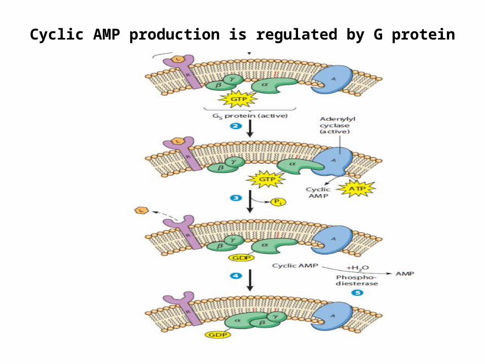
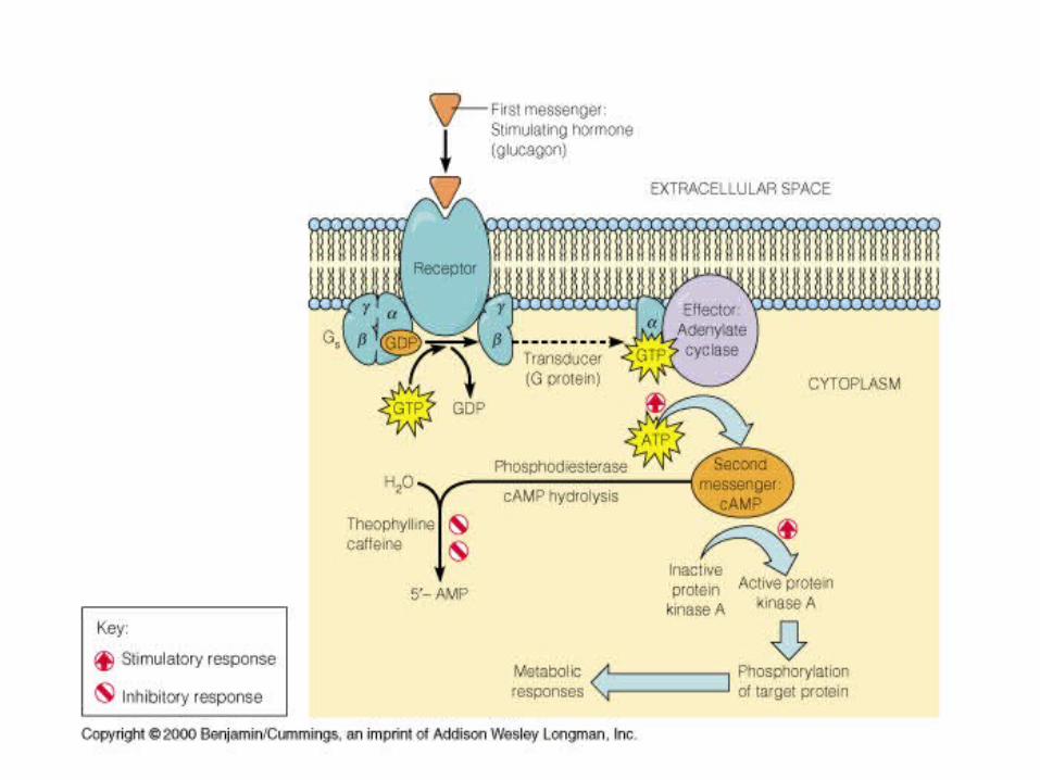
Cyclic AMP production is regulated by G protein
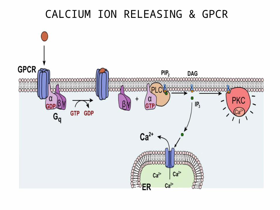
CALCIUM ION RELEASING & GPCR
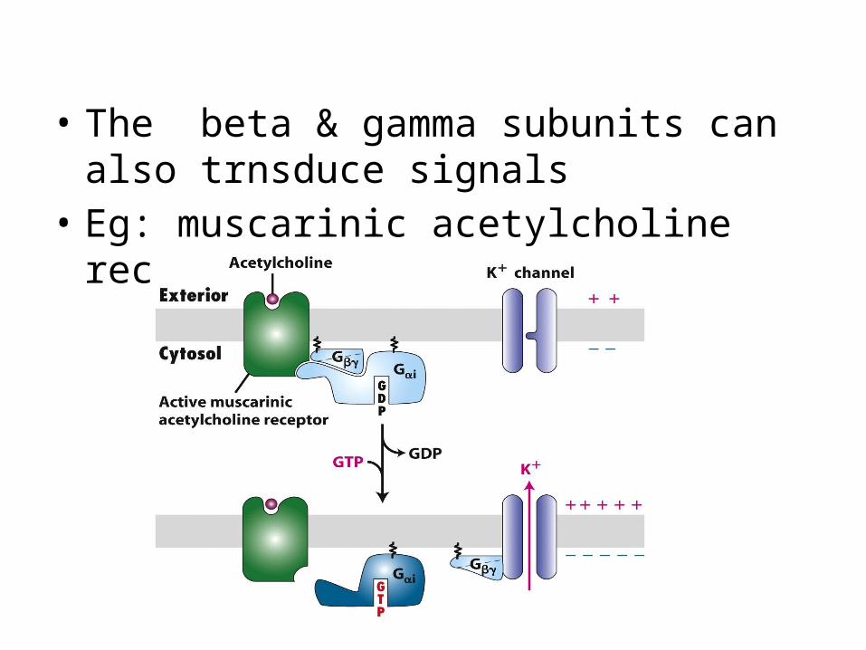
• The beta & gamma subunits can also trnsduce signals
• Eg: muscarinic acetylcholine receptors

Nobel prize for chemistry 2012 for studies of GPCR
ROBERT J. LEFKOWITZ BRIAN K . KOBILKA

THANK YOU