section i – ultrasound - trojanimaging.com images/documents/acr in service exam 20… ·...
TRANSCRIPT

Section I – Ultrasound
Figure 1
Figure 2
1. You are shown a sagittal image of the gallbladder (Figure 1) and a Doppler image of the main portal vein
(Figure 2). What is the MOST LIKELY diagnosis?
A. Acute cholecystitis B. Congestive heart failure C. Hepatitis D. Adenomyomatosis
Findings: Concentric gallbladder wall thickening and markedly pulsatile portal venous flow. Rationale: A: Acute cholecystitis is a common cause of gallbladder wall thickening, but gallbladder wall thickening is
a non-specific sign. B: Congestive heart failure can cause gallbladder wall thickening. The pulsatile portal flow in the Doppler
image indicates CHF is present C: Hepatitis can cause gallbladder wall thickening, but does not cause a pulsatile portal vein. D: Adenomyomatosis can cause various forms of gallbladder wall thickening but does not cause a pulsatile
portal vein.

Transabdominal image, right adnexa Endovaginal scan, right ovary grayscale Figure 3 Figure 4
Doppler endovaginal image of right ovary
Figure 5 2. You are shown three images of the right adnexa (Figures 3-5). What is the MOST LIKELY diagnosis?
A. Hemorrhagic cyst B. Dermoid C. Ovarian carcinoma D. Ovarian torsion
Findings: Avascular right adnexal mass with linear echogenic interfaces: the dermoid mesh Rationale: A: Hemorrhagic cyst can have a fishnet appearance, but the linear echoes are not echogenic B: The linear echogenic interfaces, or "dermoid mesh" represent hair fibers (Rumack, chapter 15
"Gynecologic sonography", p.570) C: Ovarian carcinomas may have septations and mural nodules but these will typically have flow.
(Rumack, chapter 15). D: A torsed ovary may appear enlarged and avascular, but the characteristic grayscale appearance has
multiple peripheral follicles, not a single large cyst cavity with echogenic linear interfaces. (Rumack, chapter 15)

Left CCA Left ICA Figure 6 Figure 7
3. You are shown a Doppler image of the left common carotid artery (Figure 6) and of the left internal
carotid artery (Figure 7). What is the MOST LIKELY diagnosis?
A. Subclavian steal syndrome B. Vasculitis C. Distal internal carotid artery stenosis or occlusion D. Aortic valve regurgitation
Findings: Absent diastolic flow in common and internal carotid arteries / High resistance waveform Rationale: A: Subclavian steal syndrome is diagnosed when there is reversal of flow in the vertebral artery. B: Vasculitis produces spectral broadening and thickened vessel walls, but not absent diastolic flow C: Absent diastolic flow suggests downstream high resistance as is seen in distal or intracranial stenosis or
occlusion. D: Aortic valve regurgitation can cause abnormalities of the carotid Doppler waveforms, but not absent
diastolic flow

Figure 8
4. You are shown a longitudinal image (Figure 8) of the right lower quadrant in a young man with pain and
anorexia. What is the MOST LIKELY diagnosis?
A. Intussusception B. Small bowel lymphoma C. Ureterolithiasis D. Appendicitis
Findings: Tubular, blind-ending appendix with appendicoliths. Rationale: A: The tubular structure in the image has a bowel signature, but not a "bowel-within-bowel" appearance
that would indicate intussusceptions. B: Lymphoma can cause bowel wall thickening but not luminal distention. The characteristic appearance of
lymphoma on ultrasound is a thickened hypo-echoic wall producing the "pseudo-kidney" sign. C: A distended ureter containing stones will appear tubular and echogenic calculi should be visible, but the
structure should not be blind-ending. D: This is a characteristic appearance for appendicitis, with a tubular, blind-ending structure in the right
lower quadrant containing echogenic foci (appendicoliths).

Transvaginal transverse image of uterus
Figure 9
Transvaginal sagittal image of uterus
Figure 10 5. A patient who is 10 weeks pregnant by dates presents to the ER because she is passing blood clots.
You are shown two pelvic ultrasound images (Figures 9 and 10). What is the MOST LIKELY diagnosis?
A. Ectopic pregnancy B. Molar pregnancy C. Spontaneous abortion D. Subchorionic hemorrhage
Findings: Tranvaginal images of the uterus demonstrate prominent amount of complex material within the endometrial canal but without a normal intrauterine pregnancy or fetal parts identified. Rationale: A: The complexity and amount of material in the endometrial canal is more than expected for
pseudogestational sac of ectopic pregnancy. B: Although the presented ultrasound appearance could be seen with molar pregnancy, this would not be
the most likely diagnosis in view of the history of passing blood clots. C: Although discrete fetal parts or gestational sac are not visualized, complex material within the
endometrial canal is most consistent with spontaneous abortion in view of the history of passing blood clots.
D: Subchorionic hemorrhage can range in size from a small loculated collection to complete separation of the membranes. Hemorrhage can enter the amniotic fluid and distend the endometrial canal. In the first trimester, a normal gestational sac is typically seen adjacent to the hematoma. A normal gestational sac is not present on the submitted images.

Transverse image of left superior kidney
Figure 11
Longitudinal image of left kidney
Figure 12 6. You are shown transverse (Figure 11) and longitudinal (Figure 12) images of the left kidney in a
70-year-old man. Which one of the following statements is CORRECT?
A. A focal parenchymal scar with invagination of perirenal fat is seen. B. The findings are most suggestive of prominent column of Bertin. C. Renal cell carcinoma is a significant diagnostic consideration. D. The findings are pathognomonic for angiomyolipoma.
Findings:
Large diffusely hyperechoic mass of the left superior kidney.
Rationale: A: Although a focal cortical insult can result in volume loss/scar which then contains adjacent fat, the images show a discrete focal hyperechoic mass. B: The location of the hyperechoic mass at the superior pole of the left kidney is not typical of the location for column of Bertin. Column of Bertin is typically located at the junction of the upper and middle third of the kidney. In addition, the degree of increased echogenicity and exophytic component of this mass are not typical of a prominent column of Bertin. C: Renal cell carcinoma can have a variable appearance including partially cystic, hypoechoic, isoechoic,
or even hyperechoic as in the above case. D: Although angiomyolipomas typically present as hyperechoic focal renal masses, differential considerations for hyperechoic renal mass also include renal cell carcinoma.
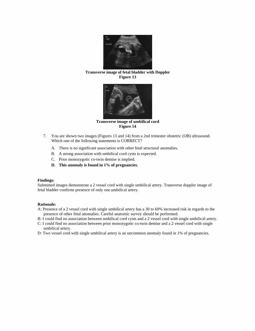
Transverse image of fetal bladder with Doppler
Figure 13
Transverse image of umbilical cord
Figure 14 7. You are shown two images (Figures 13 and 14) from a 2nd trimester obstetric (OB) ultrasound.
Which one of the following statements is CORRECT?
A. There is no significant association with other fetal structural anomalies. B. A strong association with umbilical cord cysts is expected. C. Prior monozygotic co-twin demise is implied. D. This anomaly is found in 1% of pregnancies.
Findings: Submitted images demonstrate a 2 vessel cord with single umbilical artery. Transverse doppler image of fetal bladder confirms presence of only one umbilical artery. Rationale: A: Presence of a 2 vessel cord with single umbilical artery has a 30 to 60% increased risk in regards to the
presence of other fetal anomalies. Careful anatomic survey should be performed. B: I could find no association between umbilical cord cysts and a 2 vessel cord with single umbilical artery. C: I could find no association between prior monozygotic co-twin demise and a 2 vessel cord with single
umbilical artery. D: Two vessel cord with single umbilical artery is an uncommon anomaly found in 1% of pregnancies.

Transverse Figure 15
Longitudinal
Figure 16 8. You are shown two images of the neck (Figures 15 and 16) from a patient with a palpable neck
mass. What is the MOST LIKELY diagnosis?
A. Parathyroid carcinoma B. Thyroid nodule C. Parathyroid adenoma D. Ectopic parathyroid tissue
Findings: Homogeneous, hypoechoic mass extrinsic to the thyroid gland. Rationale: A: Parathyroid carcinomas are usually lobular, heterogeneous, with a depth to width ration greater than or
equal to one B: The mass shown is extrinsic to the thyroid gland. C: Large, homogeneous, hypoechoic mass in the expected location of a left parathyroid gland. D: The mass is in the expected location of a left parathyroid gland and is also enlarged.

Sagittal
Figure 17
Transverse Figure 18
9. Based on the sagittal and transverse images (Figures 17 and 18) in a 37-year-old woman, what is
the MOST LIKELY diagnosis?
A. Multinodular thyroid B. Hashimoto’s thyroiditis C. Colloid cyst D. Normal thyroid
Findings: Diffusely heterogeneous thyroid gland. The right lobe is top normal in size. No discrete nodules. Rationale: A: The ultrasound images show a diffusely heterogeneous thyroid. The right lobe is top normal in size.
There are however no discrete nodules which would be required for a diagnosis of multinodular thyroid. B: Hashimoto thyroiditis presents as a diffusely heterogeneous appearance of the thyroid. As in this patient,
diffuse hypoechoic micronodules separated by echogenic septae can be seen. Thyroid size can be increased (in the acute phase), normal or decreased.
C: There are no discrete nodules seen. Colloid cysts present as predominantly cystic lesions in the thyroid, they may contain scattered echogenic foci associated with comet tail artifact.
D: The normal thyroid has a homogeneous ultrasound appearance with medium gray echotexture.

Sagittal
Figure 19
Duplex Doppler
Figure 20 10. You are shown ultrasound images (Figures 19 and 20) from a patient who had a liver transplant.
What is the MOST LIKELY diagnosis?
A. Liver abscesses secondary to hepatic artery stenosis B. Lymphoproliferative disorder C. Liver metastases with increased arterial flow to the transplanted liver D. Incidental liver cysts with normal arterial perfusion of the transplanted liver
Findings: The ultrasound images demonstrate a complex cystic mass in the liver. The duplex Doppler image of the main hepatic artery shows a parvus tardus waveform with increased diastolic flow and a resistive index less than 0.5. Rationale: A: The ultrasound images show a complex cystic mass in the liver. In a patient with fever and sepsis, the
primary concern should be liver abscesses. The Duplex Doppler image of the main hepatic artery at the porta shows a parvus tardus appearance of the waveform with increased diastolic flow with resistive index less then 0.5. This is a key finding indicating more proximal hepatic artery stenosis or thrombosis. Hepatic artery thrombosis or stenosis are the most common vascular complications in patients with liver transplants and can be associated with ichemic cholangiopathy and liver infarcts as well as abscesses.
B: Lymphoproliferative disorder (PTLD) is a know complication in patients with solid organ transplants and can certainly present as hypoechoic masses in the liver. However, the hepatic artery Doppler waveform should be normal.
C: Lymphoproliferative disorder (PTLD) is a know complication in patients with solid organ transplants and can certainly present as hypoechoic masses in the liver. However, the hepatic artery Doppler waveform should be normal.
D: The gray scale images show a complex cystic fluid collections in the liver. Cysts should not be this complex.

11. Regarding AIDS cholangitis, which of the following statements is TRUE?
A. It is usually due to retroviral therapy. B. It rarely occurs in patients with CD4 counts of less than 100. C. Patients are usually asymptomatic. D. Ultrasound findings may mimic primary sclerosing cholangitis.
Rationale: A: Retroviral therapy does not cause cholangiopathy. AIDS cholangiopathy is usually due to opportunistic
infection. B: AIDS cholangiopathy is most often due to opportunistic infection and therefore commonly occurs in
patients whose CD4 counts are less than 100. C: Patients usually have right upper quadrant pain and elevated alkaline phosphates. D: This is one of the well-established appearances of AIDS cholangiopathy.
12. A mirror image artifact is MOST LIKELY in which of the following imaging situations?
A. Extensive fatty moiety in the liver B. Air pockets in the soft tissues of the lung C. Echogenic tissues in the vicinity of the diaphragm D. Rapidly moving blood cells in the vasculature with Doppler acquisition
Rationale: A: Incorrect. B: Incorrect. C: Correct. D: Incorrect.
13. Renal vein thrombosis in a transplanted kidney is suggested by which one of the following
findings?
A. Decreased resistive index B. Prolonged acceleration time C. Aliasing at the arterial anastomosis D. Reversed diastolic flow in the main renal artery
Rationale: A: The resistive index is a commonly used measure of vascular function. However in renal vein
thrombosis, the resistive index will be increased. B: Prolonged acceleration time may be seen in the intra-renal vessels downstream from renal artery
stenosis, but not in renal vein thrombosis C: Aliasing at the arterial anastamosis may be seen in renal artery stenosis, but not in renal vein thrombosis D: Flow reversal in the main renal artery during diastole occurs because the normal egress of blood from
the transplant--the renal vein--is occluded. The renal artery becomes both an inflow and outflow vessel, with high resistance to downstream flow.

14. An ultrasound acquisition mode that helps improve resolution and reduce echogenic clutter
proximal to the tissues adjacent to the skin / transducer interface is known as:
A. time gain compensation imaging. B. multi-directional compound imaging. C. Doppler imaging. D. harmonic imaging.
Rationale: A: Incorrect. B: Incorrect. C: Incorrect. D: Correct. 15. Regarding peritoneal inclusion cysts, which of the following is MOST CORRECT?
A. Round or oval cyst located separate from the ovary B. Tubular cystic structure of adnexa adjacent to the ovary C. Round simple cyst arising from the ovary D. Fluid collection encasing the ovary with margins following the contour of the adjacent
pelvic cavity
16. Concerning interstitial ectopic pregnancy, which of the following statements is TRUE?
A. Its mortality rate is half the rate of other types of ectopic pregnancy due to surrounding myometrium.
B. It is the most common location for ectopic pregnancy. C. It is typically located eccentrically in the fundus of the uterus outside the expected region
of the endometrial canal with a thin rim of surrounding myometrium. D. It occurs most commonly in patients with a bicornuate uterus.
Rationale: A: Interstitial ectopic pregnancy has higher mortality rate due to later presentation and massive
hemorrhage. B: Majority of ectopic pregnancies occur at the ampullary or isthmic portion of the fallopian tube with only
2% to 4% located at the interstitial location. C: Interstitial ectopic pregnancies usually appear to be located eccentrically in fundus of uterus outside
expected region of endometrial canal with thin rim of surrounding myometrium. D: I can find no known association between interstitial ectopic pregnancy and bicornuate uterus.

17. Regarding the succenturiate lobe of the placenta, which of the following statements is MOST CORRECT?
A. A potential complication includes vasa previa. B. It occurs in one third of pregnancies. C. It appears as a rolled up edge or shelf at the edge of the placenta. D. It is associated with a high risk of placental abruption.
Rationale: A: Vasa previa is a serious complication associated with a succenturiate lobe in which the vasculature
extending to this accessory lobe from the main placenta extends across the cervix and can become entrapted or rupture during labor.
B: Succenturiate lobe is only present in 5% of pregnancies. C: A rolled up edge or shelf at the edge of the placenta describes the appearance of a circumvallate
placenta. D: A succenturiate lobe does not have a known association with a high risk for abruption.
18. Which of the following scrotal varicoceles requires evaluation for an underlying neoplasm?
A. Unilateral right-sided varicocele B. Unilateral left-sided varicocele C. Decompressible varicocele D. Any newly diagnosed varicocele
Rationale: A: Idiopathic varicoceles occur on the left side in 98% of the cases and are bilateral in 70% of the cases.
Idiopathic varicoceles are secondary to incompetent valves in the internal spermatic vein. It is thought that they occur much more commonly on the left because the venous drainage on the left is into the left renal vein.
B: Idiopathic varicoceles are usually left sided because of the venous drainage into the left renal vein. C: A varicocele that is nondecompressible should be evaluated for an underlying neoplasm as the cause of
the venous obstruction. D: Newly diagnosed varicoceles in men who are older than 40 years of age should be evaluated for an
underlying neoplasm as the source of obstruction of gonadal venous return.

19. Concerning polycystic ovary syndrome, which of the following statements is TRUE?
A. The diagnosis of polycystic ovary can be made with equal accuracy using transabdominal and endovaginal pelvic ultrasound.
B. Visualization of bilateral markedly enlarged ovaries with an ovarian volume greater than 50 cc with multiple cysts greater than 2.5 cm is highly suggestive of the diagnosis of polycystic ovary syndrome.
C. The ovaries in women with polycystic ovary syndrome have increased echogenic ovarian stroma.
D. A finding of increased ovarian vascularity by Doppler ultrasound is an integral part of the diagnosis of polycystic ovary syndrome.
Rationale: A: High frequency transducers used for endovaginal ultrasound afford better resolution then
transabdominal transducers and thus endovaginal ultrasound is more accurate for the diagnosis of polycystic ovar syndrome.
B: This appearance is usually seen with hyperstimulation syndrome or theca lutein cysts associated with molar pregnancy. The follicles in polycystic ovary syndrome typically measure 2 to 9mm and are peripherally placed.
C: The characteristic findings associated with polycystic ovary syndrome include: increase ovarian stroma which is abnormally echogenic and multiple (more then 12) peripherally placed follicles.
D: Reports regarding ovarian vascularity in patients with polycystic ovary syndrome have varied and increased vascularity is not part of the diagnostic criteria of polycystic ovary syndrome.
20. Concerning renal arteriovenous fistulas (AVFs), which of the following statements is TRUE?
A. AVFs are very uncommon in transplanted kidneys. B. Renal AVFs are usually associated with a systemic vascular disorder such as Rendu-Osler-
Weber syndrome. C. On color Doppler, AVFs are associated with a color bruit caused by vibration of the
adjacent renal parenchyma. D. All AVFs in transplanted kidneys should be managed with catheter embolization.
Rationale: A: AVF are found in up to 15% of patient following biopsy. As many patients with renal transplant
undergo biopsy to diagnose rejection, AVF are not uncommon in these patients. B: Most renal AVF, whether in native or transplanted kidneys are caused by prior renal biopsy. C: AVF are associated with high velocity low resistance flow, diagnosed by demonstration of a low
resistance (low resistive index) arterial flow pattern. The turbulent flow is usually also associated with a mosaic of color reverberating over the adjacent renal parenchyma creating a “color bruit”.
D: Many patients with renal AVF are asymptomatic and the AVF is found incidentally. Only patients with large AVF who have associated symptoms, such as significant hematuria need embolization.

21. Regarding evaluation of endometrial abnormalities in postmenopausal women, which of the following statements is TRUE?
A. Measurement of endometrial thickness is best performed on a coronal/transverse endovaginal ultrasound image of the midbody of the uterus.
B. Fluid within the endometrial canal is included in the measurement of the endometrium. C. All patients with postmenopausal vaginal bleeding should undergo histologic sampling,
regardless of endometrial thickness measurements on ultrasound to exclude endometrial cancer.
D. In a postmenopausal woman with no history of endometrial bleeding, an endometrial thickness of 7 mm is considered normal.
Rationale: A: Endometrial thickness should be measured on a sagittal endovaginal midline (or near midline) image of
the uterus B: If there is fluid present, it should be excluded from measurement of endometrial thickness. A very small
amount of simple fluid may be a normal finding in asymptomatic patients. Larger amount of endometrial fluid should raise the possibility of cervical stenosis or tumor.
C: Although endometrial cancer often presents with post menopausal vaginal bleeding, it is in fact not the most common cause of post menopausal vaginal bleeding. Large studies have shown that an endometrial thickness of 4mm or less in patients with post menopausal vaginal bleeding excludes endometrial cancer and these women do not require histologic sampling.
D: As mentioned above, several large studies comparing endometrial thickness as measured on endovaginal ultrasound and results from endometrial histologic sampling have shown that endometrial thickness of 8mm-9mm or less can be normal in asymptomatic postmenopausal women.

22. Concerning the ultrasound diagnosis of lower extremity deep vein thrombosis (DVT), which of the
following statements is TRUE?
A. Venous compression is best performed in the sagittal plane with the transducer oriented in a sagittal fashion over the longest segment of the vein being examined.
B. Loss of respiratory phasicity in the external iliac vein indicates more central iliac vein occlusion or compression by an adjacent mass.
C. Visualization of a thrombus in the greater saphenous vein is a sonographic finding indicating deep vein thrombosis.
D. Acute venous thrombi are always echogenic.
Rationale: A: Venous compression is a very important part of the sonographic examination of the veins of the lower
extremities. It should be performed on gray scale imaging in the transverse plane, so the entire cross section of the vein being examined is visualized. The normal vein should compress easily and completely with light pressure applied by the transducer. Lack of compressibility of the vein is the most important finding associated with deep vein thrombosis.
B: Loss of the normal respiratory phasicity in the external iliac vein is an important indirect sign of more central venous occlusion by thrombus or compression by adjacent mass such as adenopathy. Lack of the normal response to Valsalva maneuver is another indirect sign of more central DVT. Normally, The Valsalva maneuver produces an increase in intra abdominal pressure. During the Valsalva maneuver, normally, there is a short period of flow reversal followed by no flow. After release of the Valsalva maneuver, there is a transient surge in forward venous flow.
C: The greater saphenous vein (GSV) is part of the superficial venous system. However, the junction of the greater saphenous vein with the common femoral vein should be carefully examined and documented in every lower extremity venous study to exclude extension of a GSV thrombus in the common femoral vein.
D: The sonographic appearance of venous thrombi is variable and depends on the age of the thrombus. Acute thrombi may in fact be anechoic.