section 1: introduction to immunology and immunotherapy
TRANSCRIPT

Section 1: Introduction to immunology and immunotherapy
1

3
1.1 Immune Checkpoints L. Mahjoubi1
N. A. Rizvi2
A. Marabelle1,3
1 Gustave Roussy, Université Paris-Saclay, Département d’Innovation Thérapeutique et d’Essais Précoces, Villejuif, France
2Thoracic Oncology, Columbia University, New York, NY, USA3INSERM U1015, Gustave Roussy, Villejuif, France
DefinitionImmune checkpoint molecules are key modulators of the anti-tumour T cell immune response. They are present on T cells, antigen-presenting cells (APCs) and tumour cells; their interaction activates either inhibi-tory or activating immune signalling pathways. Examples of inhibitory immune checkpoints shown to induce a negative signal to T cells are: n Cytotoxic T-lymphocyte antigen 4 (CTLA-4, also known as CD152)n Programmed cell death protein 1 (PD-1, also known as CD279)n Lymphocyte-activation gene 3 (LAG-3)n T cell immunoglobulin and mucin domain 3 (TIM-3) n V-domain immunoglobulin suppressor of T cell activation (VISTA)
Inhibitory immune checkpoints play a vital role in maintaining immune self-tolerance. Indeed, negative co-stimulatory signals help to prevent T cells from showing autoimmune reactions.
On the other hand, co-stimulatory immune checkpoints have been shown to enhance T cell expansion and survival. Examples are: n CD40n OX40 (also known as CD134)

4
n 4-1BB (also known as CD137)n Glucocorticoid-induced tumour necrosis factor (TNF) receptor
(GITR)n Inducible T cell co-stimulator (ICOS, also known as CD278)
Other intracellular metabolic pathways play a critical role in the activation of immune cells and could, by extension, be considered as immune check-points. For instance, in tumour cells and myeloid cells, indoleamine 2, 3-dioxygenase (IDO) and arginase are key enzymes which, by depleting amino acids, can inhibit the effector functions of T cells. However, we will focus here on immune checkpoint molecules: membrane-expressed receptors and ligands, which determine, at the level of the intercellular synapse, if an immune cell becomes activated or inhibited. We will also concentrate on immune checkpoints involved in the activation of T cells, as they are of current clinical interest. However, other immune check-points play a critical role for the modulation of other subsets of immune cells (e.g. CD40 for B cells).
Immune SynapseImmune Recognition of Tumour Antigens by T Cells
During the priming phase of anti-tumour immunity, tumour antigens are presented to T cells via APCs, such as dendritic cells (DCs) or mac-rophages. The specificity of T cell activation against a tumour antigen relies on the cognate recognition of the antigen presented by the major histocompatibility complex (MHC) proteins on the surface of APCs and the T cell receptor (TCR). During the effector phase of the anti-tumour immune response, primed T cells will recognise the tumour antigens presented by MHC molecules expressed by the tumour cells. CD8+ and CD4+ T cells can recognise peptides presented by MHC-I and MHC-II molecules, respectively. This TCR/MHC interaction provides the first signal for T cell activation (signal 1).
Mahjoubi et al.

Co-stimulatory Molecules
The activation of a T cell also requires a second signal, provided by co-stimulatory molecules. The first co-stimulatory molecules historically identified belong to the immunoglobulin B7 superfamily. CD80 (also known as B7.1) and CD86 (also known as B7.2) are expressed at the surface of either APCs or cancer cells, and act as activating ligands of the co-stimulatory receptor CD28 expressed on the surface of T cells (signal 2). More recently, other co-stimulatory immune checkpoints have been described, such as OX40 (CD134), 4-1BB (CD137) or GITR (CD357). These TCRs belong to the TNF superfamily receptors (TNFSFRs) and their activation enhances T cell survival and effector functions. From the same family, CD40 is expressed on APCs and amplifies T cell acti-vation by increasing antigen presentations. Interestingly, co-stimulatory molecules are also highly expressed on immunosuppressive regulatory T cells (Tregs). The activation of Tregs favours immune self-tolerance. Defective Tregs have been associated with autoimmune disorders, while intratumoural Tregs have been associated with a worse prognosis in many cancers.
Co-inhibitory Receptors
Upon T cell activation, negative feedback loops can prevent overstimula-tion of self-reactivity. Like the CD28 receptor structure, but with oppo-site effects, the co-inhibitory receptor CTLA-4 has been shown to bind to CD80 and CD86 with a much higher affinity than CD28, delivering inhibitory signals to T cells and therefore blocking T cell activation. The membrane expression of CTLA-4 is mostly found on CD4+ T cells, nota-bly Tregs (Figure 1). Upon activation, the PD-1 receptor can be upregu-lated on T cells and can interact with two ligands: programmed death-ligand 1 (PD-L1) (also known as B7H1 or CD274) and PD-L2 (also known as B7DC or CD273). Once bound to its ligands, PD-1 confers a negative signal to effector T cells, thereby inhibiting their cytotoxic func-tions. CTLA-4 and PD-1 are usually highly expressed on intratumoural T cells and their stimulation is thought to contribute to the overall inhibi-tion of anti-tumour T cells.
51.1 Immune Checkpoints
6 Mahjoubi et al.
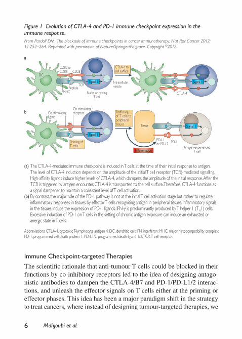
Figure 1 Evolution of CTLA-4 and PD-1 immune checkpoint expression in the immune response.From Pardoll DM. The blockade of immune checkpoints in cancer immunotherapy. Nat Rev Cancer 2012; 12:252–264. Reprinted with permission of Nature/Springer/Palgrave. Copyright ©2012.
Abbreviations: CTLA-4, cytotoxic T-lymphocyte antigen 4; DC, dendritic cell; IFN, interferon; MHC, major histocompatibility complex; PD-1, programmed cell death protein 1; PD-L1/2, programmed death-ligand 1/2; TCR, T cell receptor.
(a) The CTLA-4-mediated immune checkpoint is induced in T cells at the time of their initial response to antigen. The level of CTLA-4 induction depends on the amplitude of the initial T cell receptor (TCR)-mediated signalling. High-affinity ligands induce higher levels of CTLA-4, which dampens the amplitude of the initial response. After the TCR is triggered by antigen encounter, CTLA-4 is transported to the cell surface. Therefore, CTLA-4 functions as a signal dampener to maintain a consistent level of T cell activation.
(b) By contrast, the major role of the PD-1 pathway is not at the initial T cell activation stage but rather to regulate inflammatory responses in tissues by effector T cells recognising antigen in peripheral tissues. Inflammatory signals in the tissues induce the expression of PD-1 ligands. IFN-γ is predominantly produced by T helper 1 (TH1) cells. Excessive induction of PD-1 on T cells in the setting of chronic antigen exposure can induce an exhausted or anergic state in T cells.
Immune Checkpoint-targeted Therapies
The scientific rationale that anti-tumour T cells could be blocked in their functions by co-inhibitory receptors led to the idea of designing antago-nistic antibodies to dampen the CTLA-4/B7 and PD-1/PD-L1/2 interac-tions, and unleash the effector signals on T cells either at the priming or effector phases. This idea has been a major paradigm shift in the strategy to treat cancers, where instead of designing tumour-targeted therapies, we
a
b
DC
CD80 or CD86
Co-stimulating ligand
PD-L1 or PD-L2 PD-1
Co-stimulating receptor
Tissue
CD28
MHCPeptide
TCRIntracellular vesicle
CTLA-4 to cell surface
Trafficking of T cells to peripheral tissues
Priming of T cells
Signal 1 Signal 1
Signal 1
Signal 1
Inflammation
Naïve or resting T cell
Antigen-experienced T cell
CTLA-4

would now design immune-targeted therapies in order to break the cancer immune tolerance, restoring T cell recognition against tumour cells.
Technical ProceduresAs opposed to chemotherapies (ChTs) and tumour-targeted therapies, which aim to directly destroy cancer cells, immune checkpoint-directed therapies bind lymphocyte ligands or receptors to enhance the lympho-cyte activation and allow a cytotoxic anti-tumour immune response. The first immune checkpoint-targeted therapies developed in the clinic were humanised or fully human monoclonal antibodies selected for their antagonistic properties against immune checkpoints such as CTLA-4, PD-1 and PD-L1. They have demonstrated promising clinical activity in more than 30 different cancer types in early phase trials. Patients with a tumour response share a common feature: their response is more durable than has been observed to date with ChTs and tumour-targeted therapies. This durability of tumour response has translated into significant overall survival (OS) benefits in several phase III clinical trials. Another char-acteristic of these drugs is their safety profile: they can trigger autoim-mune and inflammatory toxicities in patients, so-called immune-related adverse events (irAEs).
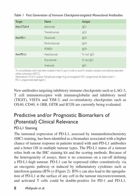
Different isotypes have been used so far in the clinic (Table 1). These antibodies usually have a long half-life and are usually infused intra-venously (i.v.) with varying intervals of administration. Anti-PD-1 and anti-PD-L1 antibodies were initially developed on weight-based dos-ing. However, results from several clinical trials have shown no corre-lation between dose, efficacy and toxicity for anti-PD-(L)1, and most compounds are now developed with a flat dose, sufficient to saturate the target. For the anti-CTLA-4 antibody ipilimumab, there was no dose-limiting toxicity (DLT) identified in early phase trials. However, a recent randomised study in patients with metastatic melanoma (MM) has shown that ipilimumab was more active and more toxic at 10 mg/kg than the approved dose of 3 mg/kg. This dose–efficacy relationship of ipilimumab currently raises questions about the mechanism of action of anti-CTLA-4 antibodies and the optimal dose to be used when combined with anti-PD-(L)1 antibodies.
71.1 Immune Checkpoints
8
New antibodies targeting inhibitory immune checkpoints such as LAG-3, T cell immunoreceptor with immunoglobulin and inhibitory motif (TIGIT), VISTA and TIM-3, and co-stimulatory checkpoints such as OX40, CD40, 4-1BB, GITR and ICOS are currently being evaluated.
Predictive and/or Prognostic Biomarkers of (Potential) Clinical RelevancePD-L1 Staining
The tumoural expression of PD-L1, assessed by immunohistochemistry (IHC) staining, has been identified as a biomarker associated with a higher chance of tumour response in patients treated with anti-PD-L1 antibodies and a better OS in multiple tumour types. The PD-L1 status of a tumour relies both on the IHC staining kit and the scoring methods. Because of the heterogeneity of assays, there is no consensus on a cut-off defining a PD-L1-high tumour. PD-L1 can be expressed either constitutively via an oncogenic pathway or induced by inflammatory cytokines such as interferon-gamma (IFN-γ) (Figure 2). IFN-γ can also lead to the upregula-tion of PD-L1 at the surface of any cell in the tumour microenvironment, and activated T cells could be double-positive for PD-1 and PD-L1.
Mahjoubi et al.
Table 1 First Generation of Immune Checkpoint-targeted Monoclonal Antibodies
Target Name Isotype
Anti-CTLA-4 Ipilimumab IgG1
Tremelimumab IgG2
Anti-PD-1 Nivolumab IgG4
Pembrolizumab IgG4
PDR001 IgG4
Anti-PD-L1 Atezolizumab Fc mut* IgG1
Durvalumab Fc mut IgG1
Avelumab IgG1*Fc mut: antibodies which have been mutated in their Fc part in order to avoid Fc receptor activation and antibody-dependent cellular cytotoxicity (ADCC).Abbreviations: CTLA-4, cytotoxic T-lymphocyte antigen 4; Ig, immunoglobulin; PD-1, programmed cell death protein 1; PD-L1, programmed death-ligand 1.
Pembrolizumab is, at present, the only anti-PD-1 antibody to be approved by the Food and Drug Administration (FDA) for a selected population of PD-L1-positive patients in non-small cell lung cancer (NSCLC) and gastric cancer.
Inflammatory Tumours and CD8+ T cells
In several tumour types, tumours with IFN-γ gene expression profile and a high level of tumour-infiltrative CD8+ T cells have better responses and survival following anti-PD-(L)1 therapy.
Mutational Load
Tumours with a high mutational load have been correlated with OS ben-efits following treatment with ipilimumab in MM, with pembrolizumab
91.1 Immune Checkpoints
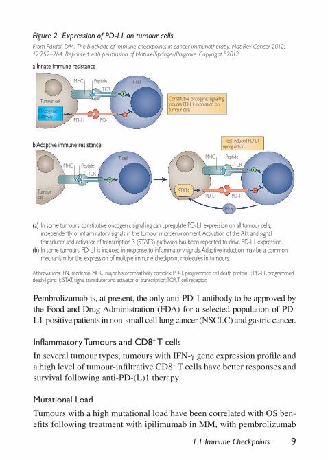
Figure 2 Expression of PD-L1 on tumour cells.From Pardoll DM. The blockade of immune checkpoints in cancer immunotherapy. Nat Rev Cancer 2012; 12:252–264. Reprinted with permission of Nature/Springer/Palgrave. Copyright ©2012.
Abbreviations: IFN, interferon; MHC, major histocompatibility complex; PD-1, programmed cell death protein 1; PD-L1, programmed death-ligand 1; STAT, signal transducer and activator of transcription; TCR, T cell receptor.
(a) In some tumours, constitutive oncogenic signalling can upregulate PD-L1 expression on all tumour cells, independently of inflammatory signals in the tumour microenvironment. Activation of the Akt and signal transducer and activator of transcription 3 (STAT3) pathways has been reported to drive PD-L1 expression.
(b) In some tumours, PD-L1 is induced in response to inflammatory signals. Adaptive induction may be a common mechanism for the expression of multiple immune checkpoint molecules in tumours.
Constitutive oncogenic signalling induces PD-L1 expression on tumour cells
T cell-induced PD-L1 upregulation
STATs
Oncogenic pathway
a Innate immune resistance
b Adaptive immune resistance
T cell
T cell
MHC
MHC
MHC
Peptide
Peptide
Peptide
TCR
TCR
TCR
PD-L1
PD-L1
PD-1
PD-1Tumour cell
Tumour cell
IFNγ

10
in NSCLC and with atezolizumab in bladder cancers. It is currently believed that a high tumour mutation burden (TMB) yields numerous immunogenic cancer cell neo-epitopes, that may be recognised by T cells upon presentation by MHC molecules. However, the TMB seems to be a prognostic marker independent of the intratumoural inflammatory gene expression profile. The assessment of TMB is currently evaluated from either tumour samples or circulating tumour DNA.
Mismatch Repair Status
Tumours with DNA mismatch repair deficiency (dMMR)/microsatellite instability-high (MSI-H) have shown great sensitivity to anti-PD-(L)1 therapies. It is currently believed that tumours harbouring an erroneous MMR system will accumulate DNA mutations, which can lead to the presence of high levels of mutation-associated neoantigens, most recog-nised by immune cells. Tumours identified as having a dMMR/MSI-H status are eligible for treatment with pembrolizumab in the USA.
Blood Biomarkers
A high neutrophil to lymphocyte ratio has been associated with poor outcomes in patients treated with ipilimumab and anti-PD-(L)1 therapy across different tumour types. High levels of serum lactate dehydroge-nase (LDH) or soluble CD25 have been associated with poor prognosis, although none is currently used in the clinic.
Microbiota
In preclinical models, the gut microbiota has been identified as a key modulator of the immune system by enhancing T cell activation and infiltration into tumours. The impact of the gut microbiome on anti-PD-(L)1 efficacy remains to be demonstrated in humans, but is currently under active investigation.
Clinical ResultsImmune checkpoint-targeted therapies have received FDA approvals in ten tumour types or categories of cancer between 2011 and 2017: MM, NSCLC, renal cell carcinoma (RCC), urothelial cancers, head and neck
Mahjoubi et al.

squamous cell carcinoma (HNSCC), Hodgkin lymphoma (HL), Merkel cell carcinoma (MCC), hepatocellular carcinoma, gastric cancer and a range of MSI-H cancers.
Anti-CTLA-4
The only anti-CTLA-4-blocking antibody that has received FDA approval is ipilimumab in MM patients, first as monotherapy in 2011, and in combi-nation with nivolumab in 2015. Approval was based on the pivotal data of the CheckMate 067 trial, with an objective response rate (ORR) of 72.1% with nivolumab plus ipilimumab versus 21.3% with ipilimumab alone and statistically significant updated OS results for the combination ver-sus ipilimumab (not reached [NR] versus 19.9 months in the ipilimumab group). Similarly, first-line combination therapy with nivolumab and ipil-imumab has recently demonstrated clinical benefit in patients with previ-ously untreated advanced or metastatic RCC. Results from the phase III CheckMate 214 trial showed significant improvement in OS (NR versus 26 months) and progression-free survival (PFS) (11.6 months versus 8.4 months) compared with sunitinib in intermediate- and poor-risk patients with metastatic RCC. In advanced NSCLC, the phase I CheckMate 012 trial showed significant clinical benefit for this combination, with an over-all response in up to 47% of the patients; a phase III trial (CheckMate 227) is currently ongoing to confirm these results. This combination is also currently being evaluated in patients with unresectable pleural meso-thelioma, in the CheckMate 743 study. In patients with advanced MM and patients with relapsed malignant mesothelioma, tremelimumab failed to demonstrate significant survival benefits compared with standard-of-care (SoC) ChT and placebo, respectively. Recently, the combination of durvalumab and tremelimumab did not reach the PFS outcome primary endpoint in the MYSTIC study in first-line treatment for patients with metastatic NSCLC, while the OS analysis is still pending.
Anti-PD-1
Nivolumab was first approved for patients with MM (CheckMate 066 and CheckMate 037) and for adjuvant therapy of resected stage III mela-noma (CheckMate 238). Nivolumab is approved for patients with squa-mous (CheckMate 017) and non-squamous (CheckMate 057) NSCLC,
111.1 Immune Checkpoints
12
RCC (CheckMate 025), HNSCC (CheckMate 141), urothelial carcinoma (CheckMate 275), dMMR metastatic colorectal cancer (CheckMate 142) and classical HL after failure of first-line therapies. Treatment in patients with HL must follow relapse after autologous haematopoietic stem cell transplantation and post-transplantation brentuximab vedotin (Check-Mate 205 and CheckMate 039). Most surprisingly, in NSCLC, nivolumab failed to demonstrate its superiority over ChT in the randomised phase III study CheckMate 026, in first-line treatment of patients with tumours with PD-L1 tumour expression ≥5%. Of note, there was imbalance in terms of TMB level between the two therapeutic arms, which could have contributed to this negative result. Indeed, patients with a high TMB showed a higher ORR and PFS when treated with nivolumab compared with ChT, and the inverse was shown in patients with low TMB. Inter-estingly, there was no correlation between the level of tumour PD-L1 expression and TMB.
Like nivolumab, pembrolizumab has been approved as second-line treat-ment of refractory/relapsing MM, NSCLC (with PD-L1 >1%), HNSCC, classical HL, urothelial carcinoma and any solid tumour expressing MSI-H status. In previously untreated advanced or metastatic NSCLC, pembrolizumab has been approved for patients harbouring PD-L1 expression on at least 50% of tumour cells, with an ORR of 44.8% versus 27.8% in the ChT group, from the pivotal phase III KEYNOTE-024 trial. Also, the combination of pembrolizumab with carboplatin and peme-trexed is now a SoC for patients with metastatic NSCLC, irrespective of PD-L1 expression, based on the results of the KEYNOTE-021 study (ORR 55% versus 29%). Pembrolizumab has also been approved as first-line treatment of cisplatin-ineligible urothelial carcinoma patients, thanks to the results of KEYNOTE-052 (ORR of 29%).
Anti-PD-L1
Anti-PD-L1-blocking antibodies have also been approved in certain other tumour types, such as advanced bladder carcinoma for durvalumab and atezolizumab, based on the results of the phase III DANUBE and phase II IMvigor 210 trials, respectively. In patients with metastatic NSCLC, atezolizumab has been approved based on the results of the
Mahjoubi et al.
phase II POPLAR and phase III OAK trials with OS of 12.6 months versus 8.9 months for second-line treatment. Recently, durvalumab has shown statistically significant improvement in PFS (16.8 months versus 5.6 months) after chemoradiotherapy in patients with locally advanced NSCLC (PACIFIC trial). A third anti-PD-L1 agent, avelumab, was approved in 2017 as both second-line treatment of metastatic urothelial carcinoma and first-line treatment of metastatic MCC.
Potential Future DevelopmentsBecause tumour-targeted therapies mostly confer improvements in PFS, and immune checkpoint-targeted therapies seem to provide greater OS benefits (at least for metastatic disease), the combination of the two cat-egories of agents may significantly improve both survival and durable responses in many cancer types. Also, the combination of immunothera-pies is currently investigated in many clinical trials in multiple tumour types. By boosting the efficacy of the immune system, co-stimulatory checkpoint agonists could also be of interest to enhance the anti-tumour response generated by immune checkpoint blockers. The modulation of innate immune cells with immune checkpoint antibodies, pattern recog-nition receptor agonists, or oncolytic viruses could also boost the adap-tive immune system. Another class of antibodies targeting both tumour cells and T cells (so-called bispecific T cell engager antibodies) is cur-rently being evaluated and could be of interest in combination with anti-PD-L1 antibodies.
Although immune checkpoint-targeted antibodies confer long-term durable responses, a greater understanding of primary and secondary resistance mechanisms to these agents is key for the future development of cancer immunotherapy and patient selection.
Declaration of Interest:
Dr Mahjoubi has reported no conflicts of interest.Dr Rizvi is an investigator and co-investigator of industry-sponsored clinical trials using immune checkpoint-targeted drugs. He has provided consulting services and has participated in scientific advisory boards of companies developing immune checkpoint-targeted therapies. He has
131.1 Immune Checkpoints

14 Mahjoubi et al.
served as a consultant to AstraZeneca, Lilly, Roche/Genentech, Pfizer, Novartis, Bristol-Myers Squibb and Merck Sharp & Dohme. He is a shareholder in Gritstone Oncology.Dr Marabelle is an investigator and co-investigator of industry-spon-sored clinical trials using immune checkpoint-targeted drugs. He has provided consulting services and has participated in scientific advisory boards of companies developing immune checkpoint-targeted therapies. He has received consultancy fees and honoraria from AstraZeneca, Merck Serono, Roche/Genentech, Pfizer, Novartis, Lytix Pharma, Bristol-Myers Squibb and Merck Sharp & Dohme. He has no stock ownership.
Further ReadingCarbone DP, Reck M, Paz-Ares L, et al. First-line nivolumab in stage IV or
recurrent non-small-cell lung cancer. N Engl J Med 2017; 376:2415–2426.Chen DS, Mellman I. Elements of cancer immunity and the cancer-immune set
point. Nature 2017; 541:321–330. Hodi FS, O’Day SJ, McDermott DF, et al. Improved survival with ipilimumab
in patients with metastatic melanoma. N Engl J Med 2010; 363:711–723. Le DT, Durham JN, Smith KN, et al. Mismatch repair deficiency predicts
response of solid tumours to PD-1 blockade. Science 2017; 357:409–413. Pardoll DM. The blockade of immune checkpoints in cancer immunotherapy.
Nat Rev Cancer 2012; 12:252–264. Peggs KS, Quezada SA, Chambers CA, et al. Blockade of CTLA-4 on both
effector and regulatory T cell compartments contributes to the antitumour activity of anti–CTLA-4 antibodies. J Exp Med 2009; 206:1717–1725.
Reck M, Rodríguez-Abreu D, Robinson AG, et al. Pembrolizumab versus chemotherapy for PD-L1–positive non-small-cell lung cancer. N Engl J Med 2016; 375:1823–1833.
Rizvi NA, Hellmann MD, Snyder A, et al. Cancer immunology. Mutational land-scape determines sensitivity to PD-1 blockade in non-small cell lung cancer. Science 2015; 348:124-128.
Watts TH. TNF/TNFR family members in costimulation of T cell responses. Annu Rev Immunol 2005; 23:23–68.
Wolchok JD, Chiarion-Sileni V, Gonzalez R, et al. Overall survival with com-bined nivolumab and ipilimumab in advanced melanoma. N Engl J Med 2017; 377:1345-1356.