secretin enhanced imaging of the pancreas - scbtmr.org enhanced... · s-mrcp: technique 1. soto ja,...
TRANSCRIPT

Secretin Enhanced Imaging of the Pancreas
Pablo R. Ros, MD
University Hospitals Case Medical Center Case Western Reserve University
SCBT-MR
Boston, MA October, 2012

Disclosures
• Consultant, Repligen Corporation • Member, Radiology Medical Advisory Network,
Philips

Acknowledgment
• Raj Paspulati, MD

ERCP
•Traditional Gold Standard for visualizing pancreatic and biliary ducts
•500,000 cases /year for diagnosis & therapy •Issues
• Technically difficult • Cost: >$2,000 + cost of complications • Safety
• Radiation exposure & sedation • Morbidity: ~10% = 50,000/yr • Mortality: ~0.5% = 2,500 deaths/yr
• NIH Consensus Statement (2002): ERCP NOT for diagnostic purposes
• Litigation
Confidential
MRCP
Confidential
• MRCP almost completely replaced ERCP for imaging diagnosis of the pancreatic duct
• Pancreatic duct diameter challenges the resolution of MRCP
• Benefit from increased pancreatic secretion

Secretin
• Hormone produced by duodenal epithelial cells under the stimulus of gastric acid • Produces secretion of fluid and bicarbonate by the exocrine pancreas • Increases the tone of the sphincter of Oddi
Matos C, et al. Pancreatic duct: morphologic and functional evaluation with dynamic MR pancreatography after secretin stimulation. Radiology 1997, 203:435-441

Secretin - Historical Perspective
• 1902: GI tract extract stimulates pancreas secretions; Secretin: first hormone discovered (Starling) • 1940: Use in pancreatic exocrine function testing • 1981: Extracted porcine secretin approved in US • 2002: Synthetic porcine secretin approved US (SecreFlo)
• 2004: Synthetic human secretin approved US (Chirostim)
• 1979: Specific binding in brain (Taylor) • 1998: Potential use in CNS disorders (Horvath)
Confidential
Secretin – Safety
• No deaths or drug-related SAEs • No anti-secretin antibody
formation (allergic reactions unusual)
• Most common side effects: • Transient increase in heart rate • Flushing • Transient, mild abdominal
discomfort
Confidential
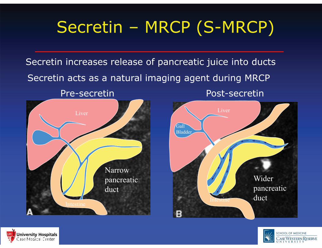
Pre-secretin Post-secretin
Secretin acts as a natural imaging agent during MRCP
Secretin increases release of pancreatic juice into ducts
Narrow pancreatic duct
Liver
Gall Bladder
Pancreas
Intestine
Pancreas
Liver
Gall Bladder
Intestine
Wider pancreatic duct
Secretin – MRCP (S-MRCP)
Confidential

S-MRCP: Literature Review
• S-MRCP well documented (off label) • Over 100 articles; 40+ safety; 20+ efficacy analyses
• Safety meta-analysis • Extent of exposure: 1,320 patients / 1,468 exposures • AE’s: only 9 reported, none serious (transient, mild)
• Efficacy meta-analysis • Duct segments, accessory and branch ducts
(p<0.001; 11 studies, 874 patients) • Duct diameters (p<0.001; 9 studies, 756 patients) • Image quality (p=0.01; 6 studies, 572 patients) • Diagnostic sensitivity (94% vs 53%)
Confidential http://www.smrcp.com/
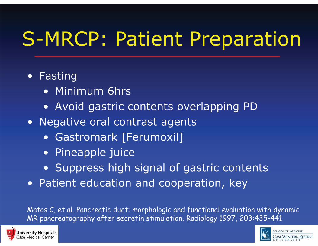
S-MRCP: Patient Preparation
Matos C, et al. Pancreatic duct: morphologic and functional evaluation with dynamic MR pancreatography after secretin stimulation. Radiology 1997, 203:435-441
• Fasting • Minimum 6hrs • Avoid gastric contents overlapping PD
• Negative oral contrast agents • Gastromark [Ferumoxil] • Pineapple juice • Suppress high signal of gastric contents
• Patient education and cooperation, key

S-MRCP: Technique
1. Soto JA, Barish MA, Yucel EK, et al. Pancreatic duct: MR cholangiopancreatography with a three-dimensional fast spin-echo technique. Radiology 1995; 196:459-464
• MR pancreatography – pres-secretin (20 min): • Breath-hold HASTE/SSFSE localizer + MIP
• Axial & coronal [3-5 mm] T2-weighted images • Thick slab breath-hold RARE
• Oblique coronal T2-slab, entire PD selected • Navigator controlled 3D images
• Secretin MRCP (S-MRP) • Post IV administration of Secretin (0.2 mg/kg body weight) • Dynamic imaging for 15 min (15-30 secs) • Test dose (?)
2. Matos C et al. Pancreatic duct: morphologic and functional evaluation with dynamic MR pancreatography after secretin stimulation. Radiology 1997, 203:435-441

S-MRCP: Technique
Matos C, et al. Pancreatic duct: morphologic and functional evaluation with dynamic MR pancreatography after secretin stimulation. Radiology 1997-2009
Technique of T2 TSE Coronal slabs
• Imaging plane- Coronal • Breath hold • Slab thickness – 20-50mm • No of signals acquired – 1 • FOV- 250mm(Rectangular) • Acquisition matrix- 256 • Flip angle – 150 degrees • TR - 2800 • Echo time - 1100
S-MRCP: Interpretation
• Pre-secretin MRCP:
• Ductal morphology • Post-secretin MRCP:
• Ductal morphology and distension • Characterization of filling defects • Duodenal distension, index of function
S-MRCP : Clinical Applications
• Congenital anomalies:
• Pancreas Divisum • Annular Pancreas • Ductal anatomical variations
• Acute Pancreatitis: • Ductal stricture, causing recurrent pancreatitis • Ductal involvement in pancreatic necrosis • Communicating vs noncommunicating pseudocyts • Planning interventional ERCP
• Chronic pancreatitis: • Staging, severity chronic • Number, length of strictures for possible intervention • Focal pancreatic mass evaluation • Assessment of exocrine function

S-MRCP : Clinical Applications
• Congenital anomalies: • Pancreas Divisum • Annular Pancreas • Ductal anatomical variations

S-MRCP : Clinical Applications
• Acute Pancreatitis: • Ductal stricture, causing recurrent
pancreatitis • Ductal involvement in pancreatic
necrosis • Communicating v noncommunicating
pseudocyts • Planning interventional ERCP

S-MRCP : Clinical Applications
• Chronic pancreatitis: • Staging, severity • Number, length of strictures for
possible intervention • Focal pancreatic mass evaluation • Assessment of exocrine function

S-MRCP : Clinical Applications
• Pancreatic focal lesions: • Differentiating side branch dilatation from cystic neoplasm • Differentiating side branch IPMT from nonductal cystic neoplasm • Differentiating pancreatic adenocarcinoma from chronic pancreatitis • Possible better delineation

S-MRCP : Clinical Applications
• Post surgical follow up:
• Post sphincterectomy • Post stent placement • Post Whipple pancreatectomy

Pancreatic MRI - Functional Imaging
• Parameters • Exocrine function • Sphincter of Oddi function • Pancreatic Fibrosis
• Methodology • Dynamic S-MRCP • Diffusion-weighted MR (DW-MR) • MR spectroscopy (MRS)
Confidential
Pre 5 min 10 min

Functional Imaging: Diffusion-weighted Secretin MR as a Proxy for Fibrosis
Time of Peak ADC Value
Erturk SM, Ichikawa T, Motosugi U, et al: Diffusion-weighted MR imaging in the evaluation of pancreatic exocrine function before and after secretin stimulation. Am J Gastroenterol. 2006 ;101(1):133-6.
0
0.5
1
1.5
2
2.5
3
3.5
0 1 2 3 4 5 6 7 8 9 10 Time after Secretin, in minutes
AD
C in
mm
2 /sec
(x10
-3)
Normals
At Risk (Alcohol Abuse)
Confidential

S-MRCP : Summary
• Detailed evaluation of the pancreatic ductal
morphology • Pancreatic exocrine function (functional
pancreatic MRI) • Patient education & cooperation, key to good
images • Radiologist supervision mandatory
Summary:
• Including secretin augmented MRCP in selected cases of pancreatic MR imaging provides more detailed evaluation of the pancreatic ductal morphology and also of pancreatic exocrine function
• Patient education & cooperation is the key to good images
• Radiologist supervision is mandatory