congenital lung lesions neonates to adults - scbtmr.org marilyn siegel md-ct... · congenital lung...
TRANSCRIPT

Marilyn J. Siegel, M.D.Mallinckrodt Institute of Radiology
Washington University School of MedicineSt. Louis, MO
and Visiting Scientist, AFIP, Washington, DC
Congenital Lung LesionsNeonates to Adults

Conflict of Interests
• None
Learning Objectives
• Describe imaging appearances of the common congenital anomalies
• Correlate with pathologic features• Emphasize “clues” to diagnosis

Congenital Lung Anomalies
• Normal vascularity– Lobar emphysema– Cystic adenomatoid malformation– Bronchogenic cyst– Bronchial atresia– Pulmonary agenesis, hypoplasia
• Abnormal vascularity– Scimitar syndrome– Sequestration– Arteriovenous malformation

Congenital Lobar Emphysema
• Misnomer; true emphysema not present• Lobar “emphysema” = “overinflation” from
bronchial obstruction–absence of bronchial cartilage–intraluminal web
• Onset: 50% 1st week–90% < 6 months–dyspnea, cyanosis, cough

Lobar Emphysema: LUL
• Lobar hyperinflation• Atelectatic adjacent lung• Mediastinal shift• Lobar predilection:
– LUL>RML >RUL

DDX: Swyer-James Syndrome
• Bronchiolitis obliterans– viral infection in childhood **
• Imaging findings–unilateral hyperlucent (low
attenuation) lung–small or normal size **–bronchiectasis **–air-trapping on expiration
** Helps to differentiate from CLE

Cystic Adenomatoid Malformation
• 90% symptomatic as neonates–Cyanosis, grunting, tachypnea
• 10% older children & adults–Presents as pneumonia or
recurrent infection• May be antenatal diagnosis

CCAM: Histologic Types• Type I: (50%) Large cyst(s) (> 2 cm)• Type II: (40%) Multiple cysts (< 2 cm) • Type III: (10%) Microcysts on cut-section
1 2 3
Stocker JT. Hum
Pathol 1977; 8:155-171

CCAM: Imaging
• Air-filled mass, mediastinal shift• Type I
–At least one dominant cyst• Type II
–Small cysts• Type III
–Solid mass, usually not imaged–High perinatal mortality
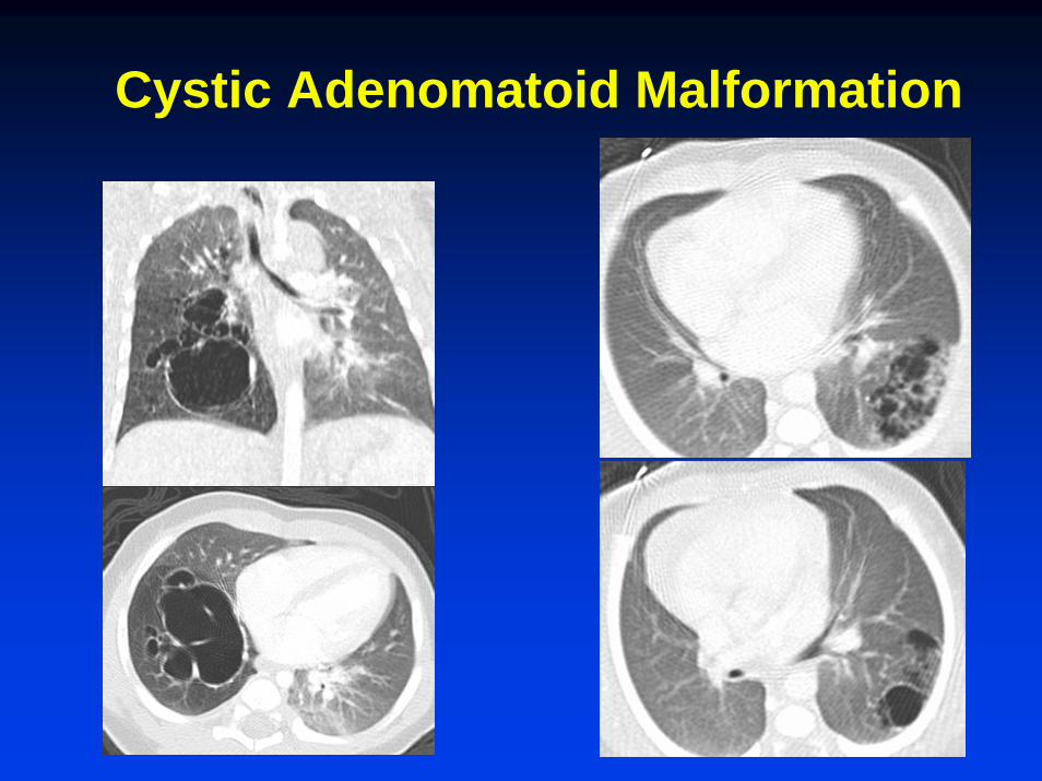
Cystic Adenomatoid Malformation

CCAM: Adults
• 10% diagnosed in adolescents & adults
• Infection common • Imaging studies
–Thick walled cavities–Fluid-fluid levels–Pneumonia

Bronchogenic Cyst
• Lung (30%) –mediastinum (70%)
• Incidental finding or symptoms from mass effect
• Path–Lined by respiratory mucosa–Clear or mucoid contents

Bronchogenic Cyst: Imaging• Round, unilocular mass• Air-filled +/- fluid

Bronchial Atresia(Congenital Mucocele)
• Failure of bronchial bud to maintain communication with airway
• Wheezing, cough or incidental detection• Imaging:
–nodular opacity»fluid-filled dilated bronchi
–surrounding emphysema (distal air drift)

Bronchial Atresia
Nodular/tubular opacity & overaeration

Arrested Pulmonary Development
• Agenesis: Complete absence of lung tissue, artery, & small or absent bronchus
• Hypoplasia: Small lung & bronchus (artery may or may not develop)

Adult: Lung Agenesis
Absent lung & artery--small bronchus

Pulmonary Hypoplasia Adult
Absent pulmonary artery- small lung & bronchus

Congenital Anomalies with Abnormal Vasculature
• Hypoplasia with anomalous venous return–Scimitar syndrome
• Pulmonary sequestration• Arteriovenous malformation

Hypogenetic Lung Syndrome
• AKA scimitar syndrome
• Lung hypoplasia with PAPVR
– RLL vein returns to IVC, portal or hepatic vein or RA
• May be symptomatic or incidental finding

Hypogenetic Lung Syndrome

Pulmonary Sequestration
• No normal connection with bronchial tree or pulmonary arteries
• Systemic blood supply• 2 types
–Intralobar (acquired)–Extralobar (congenital)

Own pleuraNeonate90% leftSyst. arterial supplySyst. venous drainage
Shared pleuraChildren & adults90% leftSyst. arterial supplyPulm. venous drainage
Extralobar Intralobar

Sequestration
• Intralobar (infected)–Symptomatic–Cough, recurrent pneumonia
• Extralobar (not infected)–Asymptomatic, incidental finding
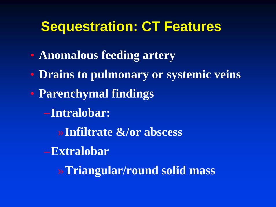
Sequestration: CT Features
• Anomalous feeding artery • Drains to pulmonary or systemic veins• Parenchymal findings
–Intralobar:»Infiltrate &/or abscess
–Extralobar»Triangular/round solid mass
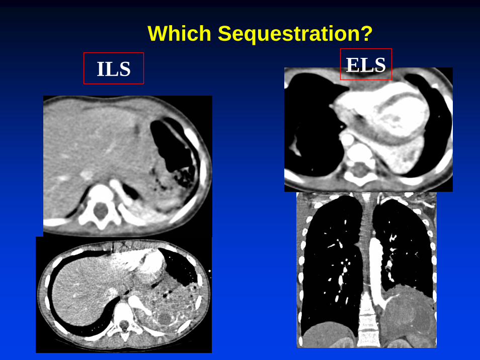
Which Sequestration?ILS ELS

Arteriovenous Malformation
• 80% Hereditary telangiectasia (OWR)–15% sporadic –5% cardiac surgeries (Glenn or Fontan)
• Symptomatic in older patients (cyanosis, polycythemia, dyspnea)
• 80-90% are simple AVMs –single feeding and draining vessel

Pulmonary AVM
Simple architecture

Congenital Lung Anomalies
ABNORMAL LUNGNORMAL VASCULATURE
NORMAL LUNGNORMAL LUNGABNORMAL VASCULATUREABNORMAL VASCULATURE
CLE Cyst CCAM Hypoplasia Sequestration Scimitar AVM