sebaceoma and related neoplasms with sebaceous …ahaq/muir torre syndrome/new/sebaceoma and... ·...
TRANSCRIPT

Sebaceoma and Related NeoplasmsWith Sebaceous DifferentiationA Clinicopathologic Study of 30 Cases
Noriyuki Misago, M.D., Ichiro Mihara, M.D., Shin-ichi Ansai, M.D.,and Yutaka Narisawa, M.D.
The classification of benign sebaceous neoplasms has beenchallenged both by the assertion that sebaceous adenomas arereally carcinomas and by difficulties in drawing the boundariesbetween sebaceomas and other lesions. We performed a clini-copathologic study of 30 cases of basaloid neoplasms withsebaceous differentiation, excluding cases of definite sebaceouscarcinoma with severe nuclear atypia invading deep within thesubcutaneous tissue and those of ocular sebaceous carcinoma.We tried to classify sebaceous neoplasms in six categories withdefined histopathologic criteria. All the neoplasms were char-acterized by aggregations of basaloid cells admixed with sebo-cytes and sebaceous duct-like structures located in the dermiswith or without connection to the epidermis. The categorieswere 1) sebaceoma (14 cases); 2) trichoblastoma with seba-ceous differentiation (3 cases); 3) apocrine poroma with seba-ceous differentiation (2 cases); 4) low-grade sebaceous carci-noma (6 cases); 5) sebaceous carcinoma (4 cases); and 6) basalcell carcinoma with sebaceous differentiation (1 case). Thesebaceoma was further subclassified as classic type (12 cases)or verruca/seborrheic keratosis type (2 cases). Although mostsebaceomas can be distinguished from other lesions, there areproblematic cases. We discuss the histopathologic diagnosticproblems associated with sebaceoma and also argue in favor ofthe concept of sebaceous adenoma.Key Words: Sebaceoma—Trichoblastoma with sebaceous dif-ferentiation—Apocrine poroma with sebaceous differentia-tion—Sebaceous carcinoma—Basal cell carcinoma with seba-ceous differentiation—Sebaceous adenoma—Sebaceousepithelioma.
In the past, many authors have suggested that the dis-tinction and classification of cutaneous neoplasms withsebaceous differentiation are often vague and difficultand that such classification and histopathologic diagnosismay occasionally be arbitrary (1–3). In addition, the cri-teria that have been used in such neoplasms with seba-ceous differentiation were often poorly defined, and thedefinitions of the terms used for these neoplasms havevaried among authors; a typical example of a confusedterm is sebaceous epithelioma. In 1984, Troy and Ack-erman (4) created a stir by proposing the term sebaceomafor what they considered a distinct benign neoplasm withsebaceous differentiation. Despite their proposal, someconfusion has remained concerning sebaceoma and itsrelated neoplasms with sebaceous differentiation (5–7).
We appreciate the fact that Troy and Ackerman’s pro-posal (4) provided a clear understanding of the classifi-cation of neoplasms with sebaceous differentiation andhoped to resolve some of the continuing confusion sur-rounding sebaceoma. We herein report a clinicopatho-logic study of 30 cases of basaloid neoplasms with se-baceous differentiation, in which we tried to classifythese neoplasms in six categories with defined histopath-ologic criteria to challenge the continuing confusion.
MATERIALS AND METHODS
We collected 30 cases of basaloid neoplasms with se-baceous differentiation from 29 patients in a multicentricstudy. We excluded cases of definite sebaceous carci-noma invading deep within the subcutaneous tissue withsevere nuclear atypia and those of ocular sebaceous car-cinoma. All the basaloid neoplasms were characterizedby aggregations of basaloid cells admixed with sebocytesand sebaceous duct-like structures located in the dermiswith or without connection to the epidermis. We classi-fied them in six categories: 1) sebaceoma; 2) trichoblas-
From the Division of Dermatology, Department of Internal Medicine(N.M., Y.N.), Saga Medical School, Saga; Mihara Clinic of Dermatol-ogy (I.M.), Tsuruoka; and the Department of Dermatology (S.A.),Akita University School of Medicine, Akita, Japan.
Address correspondence and reprint requests to Noriyuki Misago,M.D., Division of Dermatology, Department of Internal Medicine,Saga Medical School, Nabeshima 5-1-1, Saga 849-8501, Japan. E-mail:[email protected]
The American Journal of Dermatopathology 24(4): 294–304, 2002 © 2002 Lippincott Williams & Wilkins, Inc., Philadelphia
294 DOI: 10.1097/01.DAD.0000021434.86177.A0
toma with sebaceous differentiation; 3) apocrine poromawith sebaceous differentiation; 4) low-grade sebaceouscarcinoma; 5) sebaceous carcinoma; and 6) basal cellcarcinoma with sebaceous differentiation. Sebaceomawas further subclassified as the classic type or theverruca/seborrheic keratosis type. The histopathologiccriteria of each of the categories were as follows.
Sebaceoma (Classic Type)
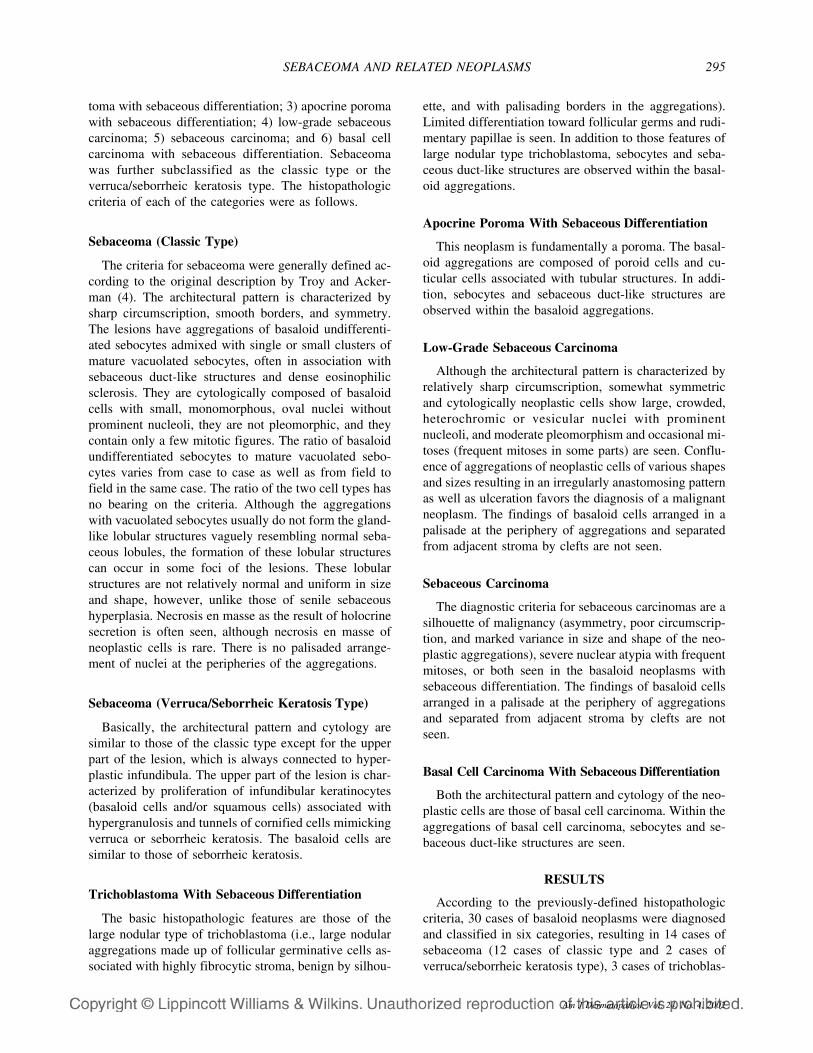
The criteria for sebaceoma were generally defined ac-cording to the original description by Troy and Acker-man (4). The architectural pattern is characterized bysharp circumscription, smooth borders, and symmetry.The lesions have aggregations of basaloid undifferenti-ated sebocytes admixed with single or small clusters ofmature vacuolated sebocytes, often in association withsebaceous duct-like structures and dense eosinophilicsclerosis. They are cytologically composed of basaloidcells with small, monomorphous, oval nuclei withoutprominent nucleoli, they are not pleomorphic, and theycontain only a few mitotic figures. The ratio of basaloidundifferentiated sebocytes to mature vacuolated sebo-cytes varies from case to case as well as from field tofield in the same case. The ratio of the two cell types hasno bearing on the criteria. Although the aggregationswith vacuolated sebocytes usually do not form the gland-like lobular structures vaguely resembling normal seba-ceous lobules, the formation of these lobular structurescan occur in some foci of the lesions. These lobularstructures are not relatively normal and uniform in sizeand shape, however, unlike those of senile sebaceoushyperplasia. Necrosis en masse as the result of holocrinesecretion is often seen, although necrosis en masse ofneoplastic cells is rare. There is no palisaded arrange-ment of nuclei at the peripheries of the aggregations.
Sebaceoma (Verruca/Seborrheic Keratosis Type)
Basically, the architectural pattern and cytology aresimilar to those of the classic type except for the upperpart of the lesion, which is always connected to hyper-plastic infundibula. The upper part of the lesion is char-acterized by proliferation of infundibular keratinocytes(basaloid cells and/or squamous cells) associated withhypergranulosis and tunnels of cornified cells mimickingverruca or seborrheic keratosis. The basaloid cells aresimilar to those of seborrheic keratosis.
Trichoblastoma With Sebaceous Differentiation
The basic histopathologic features are those of thelarge nodular type of trichoblastoma (i.e., large nodularaggregations made up of follicular germinative cells as-sociated with highly fibrocytic stroma, benign by silhou-
ette, and with palisading borders in the aggregations).Limited differentiation toward follicular germs and rudi-mentary papillae is seen. In addition to those features oflarge nodular type trichoblastoma, sebocytes and seba-ceous duct-like structures are observed within the basal-oid aggregations.
Apocrine Poroma With Sebaceous Differentiation
This neoplasm is fundamentally a poroma. The basal-oid aggregations are composed of poroid cells and cu-ticular cells associated with tubular structures. In addi-tion, sebocytes and sebaceous duct-like structures areobserved within the basaloid aggregations.
Low-Grade Sebaceous Carcinoma
Although the architectural pattern is characterized byrelatively sharp circumscription, somewhat symmetricand cytologically neoplastic cells show large, crowded,heterochromic or vesicular nuclei with prominentnucleoli, and moderate pleomorphism and occasional mi-toses (frequent mitoses in some parts) are seen. Conflu-ence of aggregations of neoplastic cells of various shapesand sizes resulting in an irregularly anastomosing patternas well as ulceration favors the diagnosis of a malignantneoplasm. The findings of basaloid cells arranged in apalisade at the periphery of aggregations and separatedfrom adjacent stroma by clefts are not seen.
Sebaceous Carcinoma
The diagnostic criteria for sebaceous carcinomas are asilhouette of malignancy (asymmetry, poor circumscrip-tion, and marked variance in size and shape of the neo-plastic aggregations), severe nuclear atypia with frequentmitoses, or both seen in the basaloid neoplasms withsebaceous differentiation. The findings of basaloid cellsarranged in a palisade at the periphery of aggregationsand separated from adjacent stroma by clefts are notseen.
Basal Cell Carcinoma With Sebaceous Differentiation
Both the architectural pattern and cytology of the neo-plastic cells are those of basal cell carcinoma. Within theaggregations of basal cell carcinoma, sebocytes and se-baceous duct-like structures are seen.
RESULTS
According to the previously-defined histopathologiccriteria, 30 cases of basaloid neoplasms were diagnosedand classified in six categories, resulting in 14 cases ofsebaceoma (12 cases of classic type and 2 cases ofverruca/seborrheic keratosis type), 3 cases of trichoblas-
SEBACEOMA AND RELATED NEOPLASMS 295
Am J Dermatopathol, Vol. 24, No. 4, 2002

toma with sebaceous differentiation, 2 cases of apocrineporoma with sebaceous differentiation, 6 cases of low-grade sebaceous carcinoma, 4 cases of sebaceous carci-noma, and 1 case of basal cell carcinoma with sebaceousdifferentiation. The clinical data of these 30 cases aresummarized in Table 1. Most of the lesions occurred onthe face and head in elderly people more than 50 yearsold, and there were no patent clinical differences be-tween the six categories except for larger size in thesebaceous carcinoma category and more frequent ulcer-ation in the low-grade sebaceous carcinoma and seba-ceous carcinoma categories than in the other categories.Neither recurrence nor metastasis of the neoplasm wasobserved during the follow-up period in any of the 30cases.
Sebaceoma (Classic Type)
Three cases occurred within a lesion of nevus seba-ceous, and the other nine cases had no associated disease.At scanning magnification, all the neoplasms showedsharply bordered basaloid aggregations in the dermismimicking trichoblastoma or cylindroma/spiradenoma.The dermal basaloid aggregations in eight cases showedno connection to the epidermis and those in the otherfour cases were connected to the epidermis. Scleroticstroma was often associated, and the basaloid aggrega-tion in three cases showed a reticular, cribriform, or even
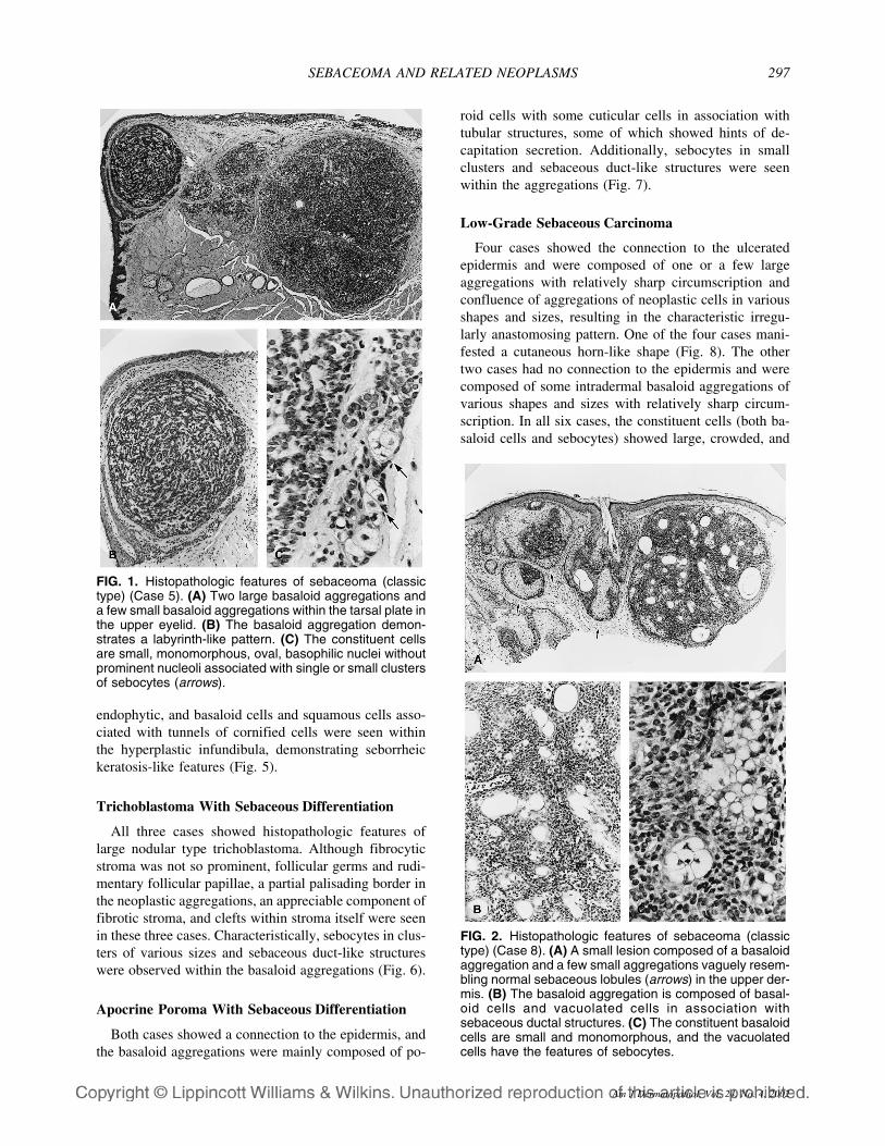
labyrinth-like pattern (Fig. 1). Another case showed arippled pattern, and this case has been reported elsewhere(8). The basaloid aggregations in all the cases containedsingle or small clusters of sebocytes and sebaceous duct-like structures to various degrees. One case associatedthe aggregations of sebaceoma with large clusters of se-bocytes vaguely resembling normal sebaceous lobules,demonstrating the features of so-called sebaceous ade-noma (Fig. 2). As a rule, the constituent cells had small,monomorphous, oval, basophilic nuclei without promi-nent nucleoli. In some cases (especially the cases asso-ciated with nevus sebaceus), however, the cells wererelatively large and showed vesicular nuclei with distinctnucleoli and occasionally some pleomorphism (Fig. 3).One case associated with nevus sebaceus had a focus oftrichoblastoma within the lesion of sebaceoma, and thiscase has been reported elsewhere (9).
Sebaceoma (Verruca/Seborrheic Keratosis Type)
Both of the two cases showed the connection to thehyperplastic infundibula and had typical histopathologicfeatures of classic type sebaceoma in the middle to lowerpart of the lesion. One case was an exoendophytic lesionand showed hypergranulosis, vacuolated cells, and largepale cells within the hyperplastic infundibula associatedwith capillaries in thin dermal papillae, demonstratingverruca-like features (Fig. 4). Another case was mostly
TABLE 1. Summary of clinicopathologic findings in 30 cases
Case DiagnosisAge
(years) Sex LocationSize(cm) Ulceration Association
1 Sebaceoma (classic) 85 F Scalp 2.5 + 2.0 −2 Sebaceoma (classic) 73 F Face 0.8 × 0.8 −3 Sebaceoma (classic) 78 M Nose 1.0 × 0.8 −4 Sebaceoma (classic) 71 F Eyelid 2.5 × 2.0 −5 Sebaceoma (classic) 52 M Eyelid 0.8 × 0.7 −6 Sebaceoma (classic) 62 M Head 6.0 × 4.0 −7 Sebaceoma (classic) 58 M Face 1.0 × 1.0 −8 Sebaceoma (classic) 48 F Face 0.4 × 0.3 −9 Sebaceoma (classic) (8) 71 F Head 1.2 × 1.0 +
10 Sebaceoma (classic) 70 F Head 1.8 × 0.8 − Nevus sebaceus11 Sebaceoma (classic) 48 F Head 1.8 × 0.9 − Nevus sebaceus12 Sebaceoma (classic) (9) 73 F Head 0.8 × 0.6 − Nevus sebaceus13 Sebaceoma (verruca/seborrheic keratosis) 87 F Face 0.5 × 0.4 −14 Sebaceoma (verruca/seborrheic keratosis) 65 F Face 1.1 × 1.0 −15 Trichoblastoma with sebaceous differentiation 85 F Face 1.4 × 1.2 −16 Trichoblastoma with sebaceous differentiation 57 F Face 1.6 × 1.4 −17 Trichoblastoma with sebaceous differentiation 51 M Face 2.0 × 1.0 −18 Apocrine poroma with sebaceous differentiation 22 F Face 0.2 × 0.2 −19 Apocrine poroma with sebaceous differentiation 74 F Face 0.6 × 0.5 −20 Low-grade sebaceous carcinoma 74 F Neck Cutaneous horn +21 Low-grade sebaceous carcinoma 74 F Face 0.8 × 0.5 −22 Low-grade sebaceous carcinoma 66 F Face 0.5 × 0.5 +23 Low-grade sebaceous carcinoma 58 M Face 0.5 × 0.5 −24 Low-grade sebaceous carcinoma (10) 59 F Face 1.5 × 1.2 + Muir-Torre syndrome25 Low-grade sebaceous carcinoma (11) 54 M Nose 2.5 × 2.0 +26 Sebaceous carcinoma 85 F Face 2.2 × 1.3 +27 Sebaceous carcinoma 61 F Face 2.2 × 2.0 +28 Sebaceous carcinoma 65 F Head 2.3 × 2.2 + Nevus sebaceus29 Sebaceous carcinoma (9) 73 F Head 2.2 × 2.5 + Nevus sebaceus30 Basal cell carcinoma with sebaceous differentiation 77 M Head 2.0 × 1.5 −
* Case 12 and case 29 are from the same patient.F, female; M, male.
N. MISAGO ET AL.296
Am J Dermatopathol, Vol. 24, No. 4, 2002
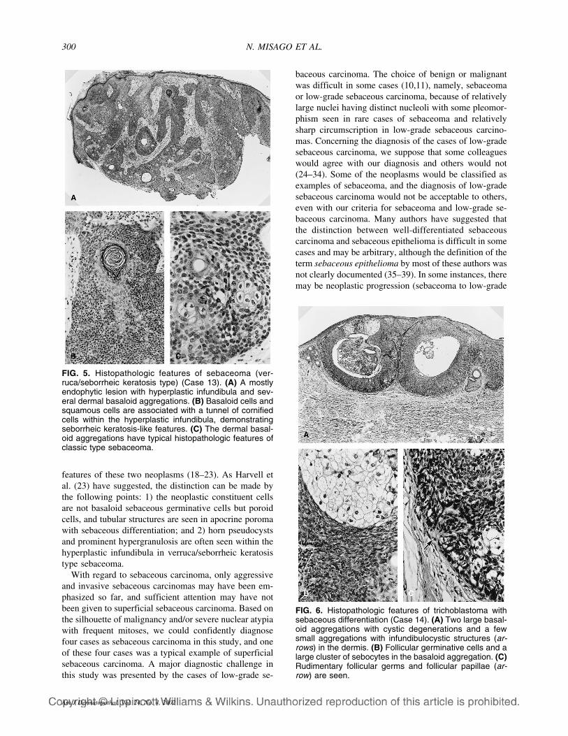
endophytic, and basaloid cells and squamous cells asso-ciated with tunnels of cornified cells were seen withinthe hyperplastic infundibula, demonstrating seborrheickeratosis-like features (Fig. 5).
Trichoblastoma With Sebaceous Differentiation
All three cases showed histopathologic features oflarge nodular type trichoblastoma. Although fibrocyticstroma was not so prominent, follicular germs and rudi-mentary follicular papillae, a partial palisading border inthe neoplastic aggregations, an appreciable component offibrotic stroma, and clefts within stroma itself were seenin these three cases. Characteristically, sebocytes in clus-ters of various sizes and sebaceous duct-like structureswere observed within the basaloid aggregations (Fig. 6).
Apocrine Poroma With Sebaceous Differentiation
Both cases showed a connection to the epidermis, andthe basaloid aggregations were mainly composed of po-
roid cells with some cuticular cells in association withtubular structures, some of which showed hints of de-capitation secretion. Additionally, sebocytes in smallclusters and sebaceous duct-like structures were seenwithin the aggregations (Fig. 7).
Low-Grade Sebaceous Carcinoma
Four cases showed the connection to the ulceratedepidermis and were composed of one or a few largeaggregations with relatively sharp circumscription andconfluence of aggregations of neoplastic cells in variousshapes and sizes, resulting in the characteristic irregu-larly anastomosing pattern. One of the four cases mani-fested a cutaneous horn-like shape (Fig. 8). The othertwo cases had no connection to the epidermis and werecomposed of some intradermal basaloid aggregations ofvarious shapes and sizes with relatively sharp circum-scription. In all six cases, the constituent cells (both ba-saloid cells and sebocytes) showed large, crowded, and
FIG. 2. Histopathologic features of sebaceoma (classictype) (Case 8). (A) A small lesion composed of a basaloidaggregation and a few small aggregations vaguely resem-bling normal sebaceous lobules (arrows) in the upper der-mis. (B) The basaloid aggregation is composed of basal-oid cells and vacuolated cells in association withsebaceous ductal structures. (C) The constituent basaloidcells are small and monomorphous, and the vacuolatedcells have the features of sebocytes.
FIG. 1. Histopathologic features of sebaceoma (classictype) (Case 5). (A) Two large basaloid aggregations anda few small basaloid aggregations within the tarsal plate inthe upper eyelid. (B) The basaloid aggregation demon-strates a labyrinth-like pattern. (C) The constituent cellsare small, monomorphous, oval, basophilic nuclei withoutprominent nucleoli associated with single or small clustersof sebocytes (arrows).
SEBACEOMA AND RELATED NEOPLASMS 297
Am J Dermatopathol, Vol. 24, No. 4, 2002

heterochromic or vesicular nuclei with prominentnucleoli in association with moderate pleomorphism andoccasional mitoses, and frequent mitoses were observedin some parts of the lesions (Fig. 8). Immature sebocytescharacterized by large, eosinophilic, granular cytoplasmwere often observed among the constituent cells in all sixcases (Fig. 8). Large clusters of sebocytes vaguely re-sembling normal sebaceous lobules were seen in threecases (Fig. 8). One case associated with the Muir-Torresyndrome was reported previously (10), and another casedemonstrating apocrine differentiation was also reportedelsewhere (11). We faced a diagnostic dilemma of be-nign or malignant (namely, sebaceoma or low-grade se-baceous carcinoma) in several of these six cases (10,11);the confluence of aggregations of neoplastic cells andulceration as well as more pleomorphic nuclei favoredthe latter diagnosis.
Sebaceous Carcinoma
Although the lesions were located in the dermis with-out invading deep within the subcutaneous fat, the sil-
houette and cytology demonstrated malignancy in allthree cases. One case was a broad and endophytic lesioncharacterized by many highly elongated and bulbous ag-gregations of various shapes and sizes oriented mostlyvertical to the skin surface and connected to the infun-dibula—a typical example of superficial sebaceous car-cinoma (Fig. 9). This case associated the aggregationswith large clusters of sebocytes vaguely resembling nor-mal sebaceous lobules, demonstrating the features of so-called sebaceous adenoma. Two other cases were com-posed of several intradermal basaloid aggregations ofvarious shapes and sizes associated with ulcerated epi-dermis (Fig. 10), and both were associated with nevussebaceous. One of the two cases was reported elsewhere(9). In another case, immature basaloid neoplastic cellswith severe pleomorphism and frequent mitoses werepredominant, but neoplastic sebocytes with vacuolatedand bubbly cytoplasm and sebaceous duct-like structurescould be seen in some parts (Fig. 10).
Basal Cell Carcinoma With Sebaceous Differentiation
Only one case showed histopathologic features ofbasal cell carcinoma. Within the aggregations of basalcell carcinoma, sebocytes and sebaceous duct-like struc-tures were seen. Apocrine duct-like structures were alsoseen, and precise histopathologic features will be dis-cussed elsewhere in detail (12).
DISCUSSION
Through diagnosis and classification of the 30 pre-sented cases of basaloid neoplasms with sebaceous dif-ferentiation in six categories, we confirmed the signifi-cance and usefulness of the concept and entity ofsebaceoma as a distinct benign neoplasm with sebaceousdifferentiation. Just as trichoblastoma is mostly com-posed of follicular germinative cells (13,14), sebaceomais considered to be mostly composed of sebaceous ger-minative cells. Both trichoblastoma and sebaceoma his-topathologically show a benign nature in their silhouette(4,13,14). As a rule, the nuclei of neoplastic cells intrichoblastoma are small, basophilic, and monomor-phous. Nevertheless, nuclei are occasionally large andeven pleomorphic (13,14). The current study suggeststhat the same can be said with regard to the nuclei ofneoplastic cells in sebaceoma. It may be argued that insebaceoma, constituent cells with small and monomor-phous nuclei are immature germinative cells in mantlesand those with relatively large and vesicular nuclei withconspicuous nucleoli are some of the mature germinativecells at the periphery of sebaceous lobules because ofsimilar cytologic features between the two cell types ineach of the events (8). Although the significance of theentity of sebaceoma was confirmed as a distinct benignneoplasm with sebaceous differentiation in this study, it
FIG. 3. Histopathologic features of sebaceoma associ-ated with nevus sebaceus (classic type) (Case 11). (A) Afew basaloid aggregations in the upper dermis. (B) Theconstituent basaloid cells are relatively large and vesicularnuclei with distinct nucleoli and have some pleomorphism.(C) Abnormal and primitive sebaceous budding adjacentto this sebaceoma within the lesion of nevus sebaceus.
N. MISAGO ET AL.298
Am J Dermatopathol, Vol. 24, No. 4, 2002

is also true that some difficulties were encountered in thediagnosis of the cases of sebaceoma and its relatedneoplasms.
All three cases of trichoblastoma with sebaceous dif-ferentiation were large nodular type trichoblastomaswithout prominent fibrotic stroma, which could not beeasily differentiated from sebaceoma. In these threecases, however, the presence of follicular germs and ru-dimentary follicular papillae, a partial palisading borderin the neoplastic aggregations, and an appreciable com-ponent of fibrotic stroma excluded a diagnosis of seba-ceoma. If no follicular differentiation and palisading bor-der in the neoplastic aggregations are seen in thissituation, sebaceoma is the preferred diagnosis. Diffi-culty and disagreement in the distinction between seba-ceoma and trichoblastoma (large nodular type) with se-baceous differentiation sometimes occurs (8,15,16),because sebaceoma and trichoblastoma are highly relatedneoplasms (9,17). Pluripotent stem cells in the folliculo-sebaceous-apocrine unit may give rise to follicular ger-
minative cells and sebaceous germinative cells (11). Insome instances, we believe that the distinction betweenthese two neoplasms was ambiguous.
It may be better that verruca/seborrheic keratosis typesebaceoma is called verruca vulgaris with sebaceous dif-ferentiation or seborrheic keratosis with sebaceous dif-ferentiation when the features of verruca vulgaris orthose of seborrheic keratosis, respectively, are prominent(18–20). In our two cases, however, the histopathologicfeatures in the middle to lower part of the lesion are thoseof sebaceoma; thus, we consider these two cases to beexamples of sebaceoma. There may be a difference inhistogenesis between the classic and verruca/seborrheictypes of sebaceoma, but we could not find any funda-mental histopathologic differences between the twotypes except in the superficial part of the lesion. Thedistinction between sebaceoma (especially of verru-ca/seborrheic type) and apocrine poroma with sebaceousdifferentiation has been controversial because of thesimilarity and degree of overlap in the histopathologic
FIG. 4. Histopathologic features of sebaceoma (verruca/seborrheic keratosis type) (Case 17). (A) An exoendophyticlesion with hyperplastic infundibula and several dermal basaloid aggregations. (B) Hypergranulomatosis, vacuolatedcells, and large pale cells within the hyperplastic infundibula associated with capillaries in thin dermal papillae, demon-strating verruca-like features. (C) The dermal basaloid aggregations have typical histopathologic features of classic typesebaceoma.
SEBACEOMA AND RELATED NEOPLASMS 299
Am J Dermatopathol, Vol. 24, No. 4, 2002
features of these two neoplasms (18–23). As Harvell etal. (23) have suggested, the distinction can be made bythe following points: 1) the neoplastic constituent cellsare not basaloid sebaceous germinative cells but poroidcells, and tubular structures are seen in apocrine poromawith sebaceous differentiation; and 2) horn pseudocystsand prominent hypergranulosis are often seen within thehyperplastic infundibula in verruca/seborrheic keratosistype sebaceoma.
With regard to sebaceous carcinoma, only aggressiveand invasive sebaceous carcinomas may have been em-phasized so far, and sufficient attention may have notbeen given to superficial sebaceous carcinoma. Based onthe silhouette of malignancy and/or severe nuclear atypiawith frequent mitoses, we could confidently diagnosefour cases as sebaceous carcinoma in this study, and oneof these four cases was a typical example of superficialsebaceous carcinoma. A major diagnostic challenge inthis study was presented by the cases of low-grade se-
baceous carcinoma. The choice of benign or malignantwas difficult in some cases (10,11), namely, sebaceomaor low-grade sebaceous carcinoma, because of relativelylarge nuclei having distinct nucleoli with some pleomor-phism seen in rare cases of sebaceoma and relativelysharp circumscription in low-grade sebaceous carcino-mas. Concerning the diagnosis of the cases of low-gradesebaceous carcinoma, we suppose that some colleagueswould agree with our diagnosis and others would not(24–34). Some of the neoplasms would be classified asexamples of sebaceoma, and the diagnosis of low-gradesebaceous carcinoma would not be acceptable to others,even with our criteria for sebaceoma and low-grade se-baceous carcinoma. Many authors have suggested thatthe distinction between well-differentiated sebaceouscarcinoma and sebaceous epithelioma is difficult in somecases and may be arbitrary, although the definition of theterm sebaceous epithelioma by most of these authors wasnot clearly documented (35–39). In some instances, theremay be neoplastic progression (sebaceoma to low-grade
FIG. 6. Histopathologic features of trichoblastoma withsebaceous differentiation (Case 14). (A) Two large basal-oid aggregations with cystic degenerations and a fewsmall aggregations with infundibulocystic structures (ar-rows) in the dermis. (B) Follicular germinative cells and alarge cluster of sebocytes in the basaloid aggregation. (C)Rudimentary follicular germs and follicular papillae (ar-row) are seen.
FIG. 5. Histopathologic features of sebaceoma (ver-ruca/seborrheic keratosis type) (Case 13). (A) A mostlyendophytic lesion with hyperplastic infundibula and sev-eral dermal basaloid aggregations. (B) Basaloid cells andsquamous cells are associated with a tunnel of cornifiedcells within the hyperplastic infundibula, demonstratingseborrheic keratosis-like features. (C) The dermal basal-oid aggregations have typical histopathologic features ofclassic type sebaceoma.
N. MISAGO ET AL.300
Am J Dermatopathol, Vol. 24, No. 4, 2002

sebaceous carcinoma to sebaceous carcinoma) (39)analogous to the morphologic progression according togenetic alterations in the colonic adenoma-carcinoma se-quence (small adenoma to large adenoma with a certaindegree of epithelial dysplasia-carcinoma) (40–44) asRütten et al. (45) have pointed out. Conversely, low-grade sebaceous carcinoma may be a malignant neo-plasm from the outset. Nevertheless, if the lesions arecompletely excised, there should be no clinical problemfor most patients with low-grade sebaceous carcinomaregardless of whether it is classified as benign or malig-nant. As Graham et al. (36) have suggested, however,when it is difficult to assign cases as benign or malignant(especially in cases occurring in the eyelids), we preferthe diagnosis of low-grade sebaceous carcinoma withoutrecourse to the term sebaceous epithelioma. We followup carefully after surgery, because cases diagnosed as
sebaceous epithelioma have shown malignant transfor-mation (46), recurred with cytologic atypia (47), or evenshowed metastasis (48).
The present study also revealed the rare existence ofbasal cell carcinoma with sebaceous differentiation,which should be differentiated from sebaceous carci-noma. This case was considered actually to be an ex-ample of basal cell carcinoma with folliculo-sebaceous-apocrine differentiation. However, there were somediagnostic problems in this case, therefore its precisehistopathologic features will be discussed in the “Casesin Consultation” section of the journal (12). To sum up,basal cell carcinoma with sebaceous differentiationshould have the histopathologic features of basal cellcarcinoma, whereas sebaceous carcinoma (including
FIG. 8. Histopathologic features of low-grade sebaceouscarcinoma (Case 20). (A) The confluence of aggregationsof neoplastic cells with an anastomosing pattern forming acutaneous horn-like shape in the epidermis and upperdermis. Large clusters of sebocytes vaguely resemblingnormal sebaceous lobules are seen (arrows). (B) Note theconfluence of aggregations of neoplastic cells of variousshapes and sizes with an irregularly anastomosing pat-tern. The confluent aggregations are composed of basal-oid cells, sebocytes, and sebaceous duct-like structures.(C) The constituent cells (both basaloid cells and sebo-cytes) show large, crowded, and heterochromic nucleiwith prominent nucleoli in association with moderate pleo-morphism. In this part, frequent mitoses are seen (smallarrows). Small clusters of immature sebocytes with eosin-ophilic and granular or vacuolated cytoplasm are oftenobserved (large arrow).
FIG. 7. Histopathologic features of apocrine poroma withsebaceous differentiation (Case 18). (A) Several basaloidaggregations are seen in the dermis, and some of themconnect to the epidermis. (B) An aggregation mainly com-posed of poroid cells with some cuticular cells in associa-tion with tubular structures, some of which showed hints ofdecapitation secretion, demonstrating both ductal andglandular differentiation. (C) Sebocytes in small clusters(arrows) and duct-like structures formed by cuticular cellswith keratohyalin granules (arrowhead), demonstratingdifferentiation toward apocrine acrosyringium within theaggregations of poroma.
SEBACEOMA AND RELATED NEOPLASMS 301
Am J Dermatopathol, Vol. 24, No. 4, 2002

low-grade sebaceous carcinoma) and sebaceoma neverhave these features (12).
If the term sebaceous adenoma is defined as neoplas-tic dermal lobules composed of sebocytes with an archi-tecture vaguely resembling that of normal sebaceous lob-ules, our study demonstrated that an aggregation ofsebaceous adenoma could be seen in sebaceoma, low-grade sebaceous carcinoma, and sebaceous carcinoma,suggesting that when sebaceous differentiation is ad-vanced in sebaceoma, low-grade sebaceous carcinoma,and sebaceous carcinoma, an architecture vaguely resem-bling that of normal sebaceous lobules can be observedin these neoplasms. We consider that sebaceous adenomais sebaceoma in some cases, because we regard mostsmall conventional sebaceous adenomas as sebaceomaswith advanced sebaceous differentiation. Nevertheless,we also agree with Nussen and Ackerman’s proposal that“sebaceous adenoma is sebaceous carcinoma” (7) in an-other set of cases, because we assume that most of the
cases diagnosed as large cystic sebaceous adenomas orbroad superficial sebaceous adenomas thus far are actu-ally well-differentiated sebaceous carcinomas (includinglow-grade carcinoma). Briefly, the cases diagnosed assebaceous adenoma hitherto include both sebaceomaswith advanced sebaceous differentiation and well-differentiated sebaceous carcinomas (including low-grade carcinomas). We disagree with Nussen and Ack-erman’s idea that all sebaceous adenomas are actuallysebaceous carcinomas (7). Many authors have noticedthat an aggregation of sebaceous adenoma can be ob-served within the lesions of sebaceoma or sebaceous epi-thelioma (1–3) and have pointed out the existence of aspectrum of neoplasms ranging from sebaceoma to se-baceous adenoma (5). The possibility of two spectra (be-nign and malignant), namely, from sebaceoma to seba-
FIG. 10. Histopathologic features of sebaceous carci-noma associated with nevus sebaceus (Case 28). (A) Alesion composed of several intradermal basaloid aggre-gations of various shapes and sizes associated with ul-cerative epidermis. (B) Immature basaloid neoplastic cellswith severe pleomorphism and frequent mitoses are pre-dominant. (C) In this part, the neoplastic sebocytes withvacuolated and bubbly cytoplasm (large arrow) and a se-baceous duct-like structure (small arrow) can be seen,demonstrating evidence of sebaceous differentiation.
FIG. 9. Histopathologic features of superficial sebaceouscarcinoma (Case 26). (A) A broad and endophytic lesioncomposed of many highly elongated and bulbous aggre-gations of varied shapes and sizes oriented mostly verti-cal to the skin surface and connecting to the infundibula.An aggregation vaguely resembling normal sebaceouslobules is observed (arrow). (B) The aggregation, consist-ing of basaloid cells, sebocytes, and sebaceous duct-likestructures, with connection to the infundibula. (C) Theneoplastic cells are large and heterochromic nuclei withprominent nucleoli in association with pleomorphism andfrequent mitoses.
N. MISAGO ET AL.302
Am J Dermatopathol, Vol. 24, No. 4, 2002
ceous adenoma in some cases and from sebaceouscarcinoma (including low-grade carcinoma) to sebaceousadenoma in a few other cases, has not been suggestedpreviously. If the term sebaceous adenoma is preserved,it should be used in a benign spectrum, namely, seba-ceoma with advanced sebaceous differentiation.
In closing this discussion, trichoblastoma is a neo-plasm with follicular differentiation, and apocrine po-roma is a neoplasm with apocrine differentiation. Basalcell carcinoma would be a neoplasm with follicular ger-minative differentiation (49). These three kinds of neo-plasms are not authentic neoplasms with sebaceous dif-ferentiation in contrast to the other neoplasms includedin this series. Practically, however, trichoblastoma withsebaceous differentiation, apocrine poroma with seba-ceous differentiation, and basal cell carcinoma with se-baceous differentiation are easily confused with the au-thentic neoplasms with sebaceous differentiation andhence were included in this study. �
Acknowledgments: The authors thank Mr. Toyoji Tanama-chi at the Photocenter of Saga Medical School (Saga, Japan) forperforming photomicrography. One of the authors (N.M.) alsothanks Dr. A. Bernard Ackerman (Director of Ackerman Acad-emy of Dermatopathology, New York, NY) for his kind answerto repeated questions from the author about sebaceoma whenthey studied together at the Institute for Dermatopathology,Jefferson Medical College (Philadelphia, PA).
REFERENCES
1. Zackheim HS. The sebaceous epithelioma. A clinical and histo-logic study. Arch Dermatol 1964;89:711–24.
2. Rulon DB, Helwig EB. Cutaneous sebaceous neoplasms. Cancer1974;33:82–102.
3. Finan MC, Connolly SM. Sebaceous gland tumors and systemicdisease: a clinicopathologic analysis. Medicine 1984;63:232–42.
4. Troy JL, Ackerman AB. Sebaceoma. A distinctive benign neo-plasm of adnexal epithelium differentiating toward sebaceouscells. Am J Dermatopathol 1984;6:7–13.
5. Sáchez Yus E, Requena L, Simón P, et al. Sebomatricoma: aunifying term that encompasses all benign neoplasms with seba-ceous differentiation. Am J Dermatopathol 1995;17:213–21.
6. Dinneen AM, Mehregan DR. Sebaceous epithelioma: a review oftwenty-one cases. J Am Acad Dermatol 1996;34:47–50.
7. Nussen S, Ackerman AB. Sebaceous “adenoma” is sebaceous car-cinoma. Dermatopathol Pract Concept 1998;4:5–14.
8. Misago N, Narisawa Y. Rippled-pattern sebaceoma. Am J Derma-topathol 2001;23:437–43.
9. Misago N, Kodera H, Narisawa Y. Sebaceous carcinoma, tricho-blastoma, and sebaceoma with features of trichoblastoma in nevussebaceus. Am J Dermatopathol 2001;23:456–62.
10. Misago N, Narisawa Y. Sebaceous neoplasms in Muir-Torre syn-drome. Am J Dermatopathol 2000;22:155–61.
11. Misago N, Narisawa Y. Sebaceous carcinoma with apocrine dif-ferentiation. Am J Dermatopathol 2001;23:50–7.
12. Misago N, Mochizuki Y, Narisawa Y. Basal cell carcinoma withsebaceous differentiation. Am J Dermatopathol. In press.
13. Ackerman AB, De Viragh PA, Chongchitnant N. Trichoblastoma.In: Neoplasms with follicular differentiation. Philadelphia: Lea &Febiger, 1993:357–422.
14. Ackerman AB, Reddy VB, Soyer HP. Trichoblastoma. In: Neo-
plasms with follicular differentiation. New York: Ardor Scribendi,2001:405–622.
15. Graham BS, Barr RJ. Rippled-pattern sebaceous trichoblastoma. JCutan Pathol 2000;27:455–9.
16. Yamamoto O, Hisaoka M, Yasuda H, et al. A rippled-patterntrichoblastoma: an immunohistochemical study. J Cutan Pathol2000;27:460–6.
17. Kaddu S, Schäppi H, Kerl H. Trichoblastoma and sebaceoma innevus sebaceus. Am J Dermatopathol 1999;21:552–6.
18. Steffen CH, Ackerman AB. Neoplasms with combined sebaceousand apocrine differentiation. In: Neoplasms with sebaceous differ-entiation. Philadelphia: Lea & Febiger, 1994:469–84.
19. Steffen CH, Ackerman AB. Verruca vulgaris with sebaceous dif-ferentiation In: Neoplasms with sebaceous differentiation. Phila-delphia: Lea & Febiger, 1994:279–327.
20. Steffen CH, Ackerman AB. Seborrheic keratosis with sebaceousdifferentiation. In: Neoplasms with sebaceous differentiation.Philadelphia: Lea & Febiger, 1994:433–47.
21. Hanau D, Grosshans E, Laplanche G. A complex poroma-likeadnexal adenoma. Am J Dermatopathol 1984;6:567–72.
22. Zaim MT. Sebocrine adenoma. An adnexal adenoma with seba-ceous and apocrine poroma-like differentiation. Am J Dermatopa-thol 1988;10:311–8.
23. Harvell JD, Kerschmann RL, LeBoit PE. Eccrine or apocrine po-roma? Six poromas with divergent adnexal differentiation. Am JDermatopathol 1996;18:1–9.
24. Ackerman AB. Serious limitations of a method. Am J Dermato-pathol 2001;23:242–3.
25. Cowen EW, Helm KF, Billingsley EM. An unusually aggressivetrichoblastoma. J Am Acad Dermatol 2000;42:374–7.
26. Helm KF, Cowen EW, Billingsley EM, et al. Trichoblastoma ortrichoblastic carcinoma? [letter] J Am Acad Dermatol 2001;44:547.
27. Chan JK, Ng CS, Tsang WY. Nodular desmoplastic variant oftrichoblastoma. Am J Surg Pathol 1994;18:495–500.
28. Ackerman AB. Nodular desmoplastic variant of trichoblastoma.Am J Surg Pathol 2000;24:1033–4.
29. Foucar E. ‘Individuality’ in the specialty of surgical pathology.Self-expression or just another source of diagnostic error? Am JSurg Pathol 2000;24:1573–6.
30. Nichols PW, Cote RJ. Diagnostic ‘individuality’ [letter]. Am JSurg Pathol 2001;25:1100.
31. Ackerman AB. ‘Individuality’ in the specialty of surgical pathol-ogy [letter]. Am J Surg Pathol 2001;25:1100–1.
32. Ioachim HL. On variability, standardization, and error in diagnos-tic pathology [letter]. Am J Surg Pathol 2001;25:1101–3.
33. Sridhar SR. ‘Individuality’ in surgical pathology [letter]. Am JSurg Pathol 2001;25:1103–4.
34. Foucar E. Author’s reply [letter]. Am J Surg Pathol 2001;25:1104–6.
35. Schwartz RA, Flieger DN, Saied NK. The Torre syndrome withgastrointestinal polyposis. Arch Dermatol 1980;116:312–4.
36. Graham RM, McKee PH, McGibbon D. Sebaceous carcinoma.Clin Exp Dermatol 1984;9:466–71.
37. Oka K, Katusmata M. Intraepidermal sebaceous carcinoma: casereport. Dermatologica 1990;180:181–5.
38. Elder D, Elenitsas R, Ioffreda M, et al. Sebaceous adenoma andsebaceous epithelioma (sebaceoma). In: Synopsis and atlas of Le-ver’s histopathology of the skin. Philadelphia: Lippincott Williams& Wilkins, 1999:304–5.
39. Kiehl P, Richter K, Erdelkamp J, et al. DNA image cytometry insebaceous tumors of the Muir-Torre syndrome. Br J Dermatol1998;138:706–8.
40. Fearon ER, Vogelstein B. A genetic model for colorectal tumori-genesis. Cell 1990;61:759–67.
41. Speroni AH, Vanzulli SI, Meiss RP. Adenomas of the colon: over-expression of p53 protein and risk factors. Endoscopy 1998;30:623–6.
42. Yang HB, Chow NH, Sheu BS, et al. The role of bcl-2 in theprogression of the colorectal adenoma-carcinoma sequence. Anti-cancer Res 1999;19:727–30.
SEBACEOMA AND RELATED NEOPLASMS 303
Am J Dermatopathol, Vol. 24, No. 4, 2002

43. Iwabuchi M, Sasano H, Hiwatashi N, et al. Serrated adenoma: aclinicopathologic, DNA ploidy, and immunohistochemical study.Anticancer Res 2000;20:1141–7.
44. Bennett MW, O’Connell J, Houston A, et al. Fas ligand upregu-lation is an early event in colonic carcinogenesis. J Clin Pathol2001;54:598–604.
45. Rütten A, Burgdorf W, Hügel H, et al. Cystic sebaceous tumors asmarker lesions for the Muir-Torre syndrome. Am J Dermatopathol1999;21:405–13.
46. Ohda C, Matsunaka M. A case of sebaceoma with malignant trans-formation [in Japanese]. Skin Research 1993;35:99–104.
47. Tsukada M, Ohara K. Sebaceous epithelioma [in Japanese]. RinshoDermatol (Tokyo) 1999;41:1325–7.
48. Burgdorf WHC, Koester G. Multiple cutaneous tumors: what dothey mean? J Cutan Pathol 1992;19:449–57.
49. Ackerman AB, Reddy VB, Soyer HP. Trichoblastic carcinoma. In:Neoplasms with follicular differentiation. New York: ArdorScribendi, 2001:625–1005.
N. MISAGO ET AL.304
Am J Dermatopathol, Vol. 24, No. 4, 2002