scanning electron microscope
TRANSCRIPT

1
SCANNING ELECTRON
MICROSCOPE
ANGEL ANNA LAL1st YEAR MSc Biotechnology

2
INTRODUCTION-TYPES

3
Scanning Electron Microscope

4
SCANNING ELECTRON MICROSCOPE (SEM) Invented by Max Knoll in 1935. Uses a focused beam of high-energy electrons to
generate images of a sample. 3-Dimentional images are obtained. Magnification ranging from 20X to
approximately 30,000X. Provides 250 times larger image than light microscope. Used in high or low vacuum in wet conditions and
even at wide range elevated temperatures.

5
PRINCIPLE The basic principle is that a beam of
electron is generated by a suitable source, typically a tungsten filament or a field emission gun.
The electron beam is accelerated through a high voltage[20 kV] and pass through a system of aperture and electromagnetic lenses to produce thin beam of electrons.
Then beam scans the surface of specimen.
Electrons are emitted from specimen by action of scanning beam and collected by suitably positioned detector.

6

7
SCHEMATIC OF AN SEM

8
SAMPLE PREPARATION
Appropriate size & should be dry. Specimens should be electrically
conductive. Coated with ultrathin layer of
electrically conducting method. Eg: Gold, Gold\Palladium
alloy,Platinum,Osmium,Tungsten,Graphite etc.

9
A SPIDER SPUTTER-COATED IN GOLD, HAVING BEEN PREPARED FOR VIEWING WITH AN SEM.

10
IMAGE FORMATIONElectron gun fitted with tungsten filament
Electron beam focused by one or two condenser, Passes through scanning coils\deflector plates
Primary electron beam interacts with sample-repeated random scattering.
Beam current absorbed by specimen is detected & electronic amplifier amplifies signal & image is displayed.

11
Signals:Secondary electrons (SE): mainly
topography Low energy electrons, high resolution Surface signal dependent on curvature
Backscattered electrons (BSE): mainly chemistry High energy electrons “Bulk” signal dependent on atomic number.
Sample
Secondary electrons Backscattered electrons
Incoming electrons
X-rays

12
Backscattered electron detector:
Secondary electron detector
13
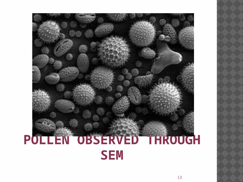
POLLEN OBSERVED THROUGH SEM

14
ADVANTAGES Most SEM's are comparatively easy to
operate, with user-friendly interfaces. Many applications require minimal sample
preparation. For many applications, data acquisition is
rapid [less than 5 minutes/image for SEI, BSE.]
Modern SEMs generate data in digital formats, which are highly portable.
15
LIMITATIONS Samples must be solid and they must fit
into the microscope chamber. Maximum size in horizontal dimensions
is usually on the order of 10 cm; vertical dimensions are generally much more limited and rarely exceed 40 mm.
For most instruments samples must be stable in a vacuum on the order of 10-5 - 10-6 torr.

16
THANK YOU!