rusting iron: an inorganic vs. (nanno)biological process · rusting of iron on this birdbath is a...
TRANSCRIPT

Page 1 of 7
Rusting Iron: an Inorganic vs.
(Nanno)biological process
R. L. FOLK UNIVERSITY OF TEXAS AT AUSTIN
In 2005 a brilliant pre-Med undergraduate,
Joey Carlin, with a double major in Geology and
Biology, came into my office asking if he could do
an informal research project. Hmm...I thought. Our
daughter just gave us an iron birdbath...what
happens when you fill it with water? Of course, it
will rust! Okay, is the oxidation of iron an
INorganic or organic process? Why is the planet
Mars red? It is due to the oxidation of iron minerals.
Is the process on Mars INorganic or biological –
before we can figure this out, we need to know how
it happens on our own earth. So I told Joey, let's
look at how this birdbath rusts and look for
evidence, pro or con, for (micro)biological
alteration – in reality, putative “nanno=bacterial”
activity.
The rust takes two forms. A soft pumpkin-
orange fluff forms on leaves that fall into the water
and also develops an iridescent scum on the water
surface. This must form from iron precipitated from
solution. The bottom of the birdbath gets covered
with a hard crust of dark chocolate-brown color,
formed essentially in situ.
The pumpkin-orange scum develops very
rapidly, in a few days, and within it are embedded
swarms of .05-.2 micron spheroids (50-200 nm),
along with a few “normal-sized” bacteria. Are the
spheroids biological or just the way that iron oxide
precipitates inorganically? To answer this question,
we hit the material with dilute hydrogen chloride
(HCl): the actual iron oxide dissolves and the
organic matter of the small bodies survives, so we
concluded that these are nannobacterial cells. When
seen in profile, some of these cells are hollow,
showing that they have not yet been mineralized.
The hard chocolate-brown crust shows
several morphologies: (1) strange aggregates of
nannoballs that look like flowers; (2) “euhedral”
hexagonal plates; (3) prismatic hexagonal crystals.
The “flowers” start out as 50-100 nm balls forming
a rosary-like chain, then these aggregate into a
monolayer of balls making a warped sheet, finally
these come together to form an object shaped like
the bloom of a carnation...amazing!
The hexagonal plates (1 x 10 microns) look
euhedral at low magnification, but at 50,000X one
can see that they are made up of parallel sheets of
50-100 nm balls with no apparent “glue” between
them, i.e. they are entirely balls. At first sight, one
reacts “must be hematite,” but ferrihydrite can also
form hexagonal plates.
The large hexagonal prisms (10 x 20
microns) are also strange. A thick hexagonal column
terminates in a wider hexagonal plate at each end;
and as a “belt” in the middle of the column there is
a bulging mass of much larger 500 nm balls. When
etched in 10% HCl for 24 hours, the column itself is
studded with 50-100nm balls making up about 40%
of its volume.
Iron oxides in natural situations are typically
a gamut of similar minerals that require detailed X-
ray study (and that not by amateurs) to unravel. Our
one X-ray showed dominant goethite (probably the
pumpkin-orange material), along with some
hematite (hexagonal plates) and maghemite
(magnetic phase).
But the primary conclusion is that the
rusting of iron on this birdbath is a predominantly
biological process carried out by nannobacteria.
Study of other iron oxides in my collection reveals
similar conclusions. Presumably the same applies to
oxidation of iron minerals on Mars, if so, indicating
that Mars indeed harbored nannobiological life.

Page 2 of 7
ACKNOWLEDGMENTS
In addition to Joey Carlin, I acknowledge SEM assistance by Isis Dlubac.
REFERENCES
Folk, R. L. and Carlin, J. P., 2006, Adventures in an Iron Birdbath: Nanostructure of iron oxides and a
nannobacterial connection. Geol. Soc. America, SE section, Knoxville, Tenn; Abs. w. Progr. v. 38
#3, p. 6.
And other references presaging this study:
Folk, R. L., 1997, Nannobacteria and the Oxidation of Iron on Earth (and perhaps Mars), Geol. Soc.
America Abs. w. Progr. v. 29 #6, p. 129
Folk, R. L. and Milliken, K. L., 2000, Morphological Evidence for microbes associated with iron
oxides...Geol. Soc. America SE section, Charleston, S.Car., Abs. w. Progr. v. 38 #3, p. 6.
LIST OF FIGURES
Figure 1. Orange iron (Fe) film on a leaf. Entirely nm-scale balls, 100-200 nm.
Figure 2. Orange film on another leaf, acidized to remove iron oxide. Surviving are nannobacterial rods, 50
nm X 200 nm because organic matter survives acid treatment.
Figure 3. Water-surface orange film, acidized to remove iron oxide. At right 5 “normal” bacteria; hordes of
~50 nm nannobacterial balls infect the rest of the photo.
Figure 4. Magnetic dark brown iron oxide; carnation-like structures are amazing! At left are sheets made of
50-100 nm nannobacterial balls.
Figure 5. Close-up of a “carnation,” each “petal” made of a warped sheet of 50-100 nm nannobacterial
cells.
Figure 6. Magnetic dark brown iron oxide – thin hexagonal plates about 10 microns wide look like
euhedral crystals – but see Fig. 7.
Figure 7. Corner of a hexagonal “crystal” from Fig. 6 – made of sheets of nannobacterial balls 50-100nm.
Figure 8. Hexagonal prisms of magnetic iron oxide acidized in HCl – a belt of 0.5 micron bacterial cells in
middle, and capping hexagonal plates at each end – amazing symmetry!
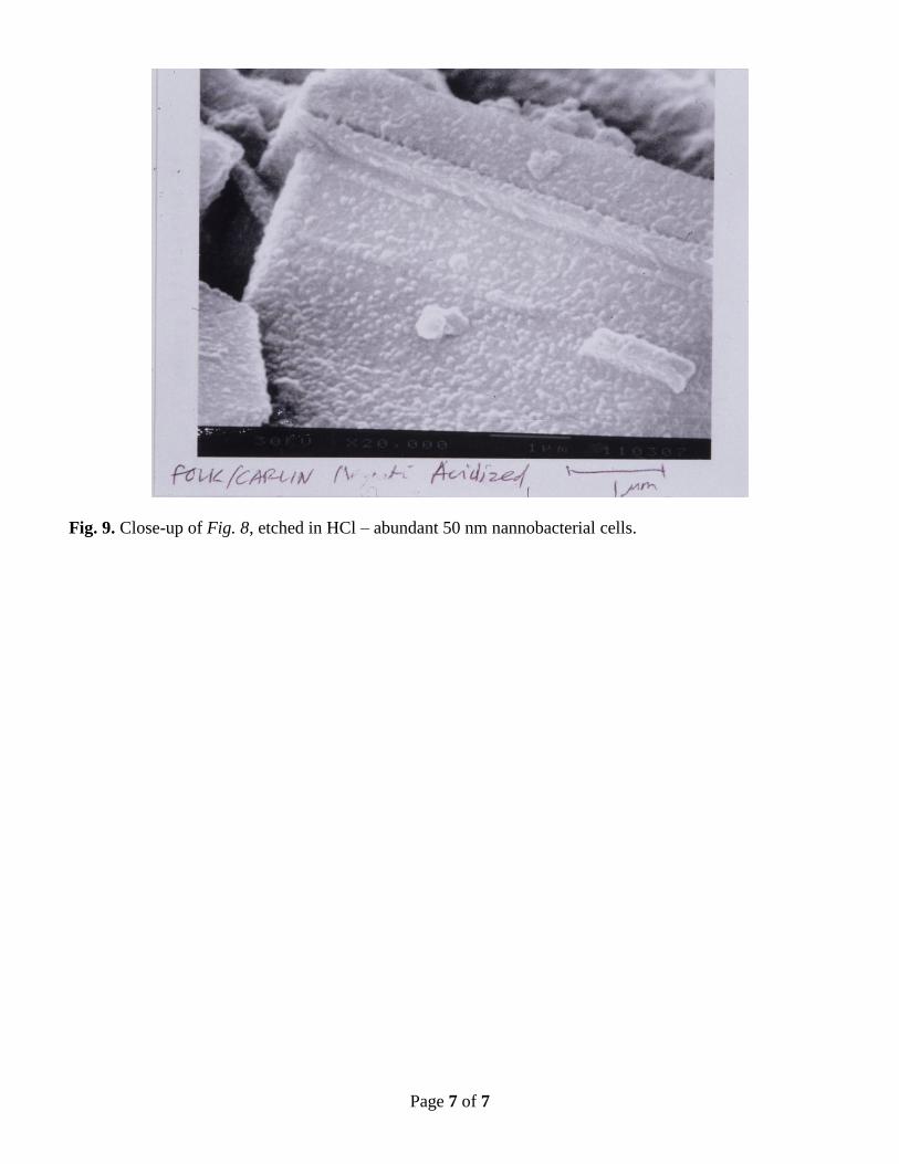
Figure 9. Close-up of Fig. 8, etched in HCl – abundant 50 nm nannobacterial cells.

Page 3 of 7
Fig. 1. Orange iron (Fe) film on a leaf. Entirely nm-scale balls, 100-200 nm.
Fig. 2. Orange film on another leaf, acidized to remove iron oxide. Surviving are nannobacterial rods, 50
nm X 200 nm because organic matter survives acid treatment.

Page 4 of 7
Fig. 3. Water-surface orange film, acidized to remove iron oxide. At right 5 “normal” bacteria; hordes of
~50 nm nannobacterial balls infect the rest of the photo.
Fig. 4. Magnetic dark brown iron oxide; carnation-like structures are amazing! At left are sheets made of
50-100 nm nannobacterial balls.

Page 5 of 7
Fig. 5. Close-up of a “carnation,” each “petal” made of a warped sheet of 50-100 nm nannobacterial cells.
Fig. 6. Magnetic dark brown iron oxide – thin hexagonal plates about 10 microns wide look like euhedral
crystals – but see Fig. 7.

Page 6 of 7
Fig. 7. Corner of a hexagonal “crystal” from Fig. 6 – made of sheets of nannobacterial balls 50-100nm.
Fig. 8. Hexagonal prisms of magnetic iron oxide acidized in HCl – a belt of 0.5 micron bacterial cells in
middle, and capping hexagonal plates at each end – amazing symmetry!
Page 7 of 7
Fig. 9. Close-up of Fig. 8, etched in HCl – abundant 50 nm nannobacterial cells.