rumen ciliates of white-tailed deer (odocoileus virginianus), axis deer (axis axis), sika deer...
TRANSCRIPT

J . Euk. Mkrohio l , 46(2), 1999 pp 125-131 0 1999 by the Society of Prolozoologisls
Rumen Ciliates of White-tailed Deer (Odocoileus virginianus), Axis Deer (Axis axis), Sika Deer (Cewus nippon) and
Fallow Deer (Dama dama) from Texas BURK A. DEHORITY,*,’ STEPHEN DEMARAIS**,2 and DAVID A. OSBORN**.3
*Department of Animal Sciences, Ohio Agricultural Research and Development Center, The Ohio State University, Wooster, Ohio 44691, USA, and
**Department of Range, Wildlife, and Fisheries, Texas Tech University, Lubbock, Texas 79409, USA
ABSTRACT. Samples of rumen contents from 33 white-tailed deer (Odocoileus virginianus), 31 axis deer (Axis axis), 26 sika deer (Cervus nippon), and 25 fallow deer (Dama duma) were collected from four study areas in central Texas. The geometric mean concen- tration of total protozoa was 50.2 X lo4 per ml, with no differences between species ( P > 0.36). White-tailed deer had a higher percentage of Enrodiniurn and lower percentage of Diplodiniinae ( P < 0.01) than the other deer species, which were not different from each other. Occurrence of Epidinium, Isotricha, and Dasytricha was sporadic and did not differ among deer species. Numerous new host records of protozoan species were observed: white-tailed deer-four; axis deer-five; sika deer-five; fallow deer-four. This brings the total number of protozoan species identified in each deer species to: white-tailed-eight; axis-12; sika-15; fallow-16. For all species combined, protozoan concentrations were 7.5 to I I-fold higher ( P < 0.01) from Area 4, which differed from the other three areas by having a stream that allowed deer to have free access to water. Criteria used for identification of medium-size Eudiplodinium species were evaluated.
Supplementary key words. Cell dimensions, Eudiplodinium impalae, protozoan concentrations, occurrence of species.
NLY a limited number of studies have been published on 0 the protozoan fauna of native and exotic deer in North America. Four published studies [6, 8, 21, 251 and a M.S. thesis (Sybert, V. L. 1990. A study of the ciliated protozoa of white- tailed deer (Odocoileus virginianus), axis deer (Axis axis), and sika deer (Cervus nippon) in the Edwards Plateau of Central Texas. Master’s thesis. Southwest Texas State University, San Marcos, Texas) have reported on the occurrence of protozoa in rumen contents of white-tailed deer. Fauna of the fallow deer have been investigated in three studies, one each from Germany [12], the Netherlands [22], and from Czechoslovakia [23]. However, protozoa were identified only to the generic level in the German and Netherland studies. Sybert’s thesis (1990, op. cit.) cites the only known information available about the fauna of axis deer. Fauna of sika deer have been reported by Sybert (1990, op. cit.) and by Imai et al. [14] from Japan.
The present study reports total protozoan concentrations as well as generic and species composition in white-tailed, axis, sika, and fallow deer from Texas. Rumen contents from all four deer species were collected from each of four study areas, pro- viding a better basis for between-species comparisons.
I To whom correspondence should be addressed. Telephone 330-263-
Current address: Department of Wildlife and Fisheries, Mississippi
’Current Address: Darrel B. Warnell School of Forest Resources,
3909; FAX 330-263-3949; Email: dehority. 1 @osu.edu
State University, Mississippi State, Mississippi 39762, USA.
University of Georgia, Athens, Georgia 30602, USA.
MATERIALS AND METHODS
Rumen contents were collected between 15 December 1988 and 15 January 1989 from adult female deer of four species. The rumen samples were taken as part of an overall project investigating inter-specific relationships of white-tailed deer with co-existing exotic species of deer. Deer species and num- ber of animals were: 33 white-tailed (Odocoileus virginianus), 31 axis (Axis axis), 26 sika (Cervus nippon), and 25 fallow (Duma duma). Following established animal care and use pro- tocol, almost all of the deer were harvested between sunset and sunrise, during their most active feeding periods. All species of deer had the same likelihood of being harvested at the same relative time in their feeding cycle. Fluctuations in protozoal concentrations are minimized the more frequently an animal eats, making sampling time of less importance for grazing an- imals. Four collection areas, representative of the Edwards Pla- teau Region of Texas, were designated as Areas 1, 3, 4, and 5. Rumen contents from all four deer species were collected from each area for comparison purposes. The study areas were heavi- ly grazed by wild ungulates (blackbuck antelope, Barbary sheep, and mouflon sheep), domestic sheep and cattle, and the four deer species. Due to overgrazing, vegetation consisted pri- marily of low quality browse and grass. Wire fencing limited movement of deer between the study areas. Areas 1, 3, and 5 had limited water available: only water tanks or small catch basins, while Area 4 had a small river.
Whole rumen contents were preserved by mixing with an equal volume of 18.5% formaldehyde. Protozoan concentra-
Table 1. Concentration of rumen protozoa in four species of deer grazing four different range areas in Texas.
Geometric mean of total protozoan concentrations X 104/mla Deer Area: 1 3 4 5 All areas Rangeh
Whi te-tailed 32.5 (8p 43.2 190.9 (5)Y 30.7 45.5 (33) 8-450 Axis 26.6 (5)” 33.4 (lo)% 340.2 (10)~ 21.8 (6p 62.7 (31) 12-574 Sikad 33.4 (3)” 45.8 (8)” 269.3 (4)Y 21.0 (10)” 42.9 (25) 13-424 Fallow 25.9 (6)” 33.6 (lop 330.2 ( 5 ) y 38.6 (4)” 50.9 (25) 7-63 I All deer 29.3 (22p 38.3 (38). 288.5 (24)Y 26.1 (30)x 50.2 (1 14) 7-63 1
a Calculated using natural logarithms. Observed range in actual numbers of protozoa X lo4 per ml. Number of animals in parentheses. One animal from Area 3 was protozoa-free and omitted from the calculations.
x ~ Y Means in the same row followed by different superscripts differ at P < 0.01.
125

126 J. EUK. MICROBIOL., VOL. 46, NO. 2, MARCH-APRIL 1999
Table 2. Percentage Entodinium and Diplodiniinae in four species of deer (white-tailed, axis, sika, and fallow) grazing four different areas in Texas.
Area
Deer 1 3 4 5 Al l areas Range
Percent Entodinium White-tailed 97.0 ? 1.5"" 89.6 2 3.2" 90.5 2 9.5 95.2 t 2.OX 93.2 2 1.8" 52-100 Axis 66.8 i 4.4Ey 64.2 2 4.6y 95.4 2 1.24 79.2 2 5.8"y 77.6 2 3.1y 43-100 Sika 75.8 2 5.0Y 80.0 t 10.4" 84.1 t 4.7 56.5 t 6.W 71.1 t 4 .8~ 0-100 Fallow 60.0 2 4.5") 67.0 t 5.3'" 97.7 2 1.5" 64.0 2 6 2 y L 71.0 2 3.7y 39-100 All deer 77.2 t 3.8 75.1 t 3 . 4 93.0 2 2.2" 75.0 t 3.9' 79.2 t 1.9 0-100
Percent Diplodiniinae White-tailed 2.8 i 1.6"" 9.2 2 2.W 0.3 2 0.3x 4.8 t 2.0" 5.0 t 1.2" 0-25 Axis 33.2 t 4.4cy 35.2 2 4.4y 4.6 t 1.2dx 20.3 t 5.YdX.y 22.1 t 3 .0~ 0-5 1 Sika 24.2 2 5.04 6.4 t 2.W 15.9 t 4 . 7 4 37.0 2 6.7"~ 21.7 2 3 .8~ 0-8 1 Fallow 37.5 i 3.2'y 30.7 2 5.4')' 2.3 2 1.5dx 33.2 t 5.1'~ 27.0 t 3 . 5 ~ 0-53 All deer 22.1 t 3.6 20.7 2 2.V 5.1 t 1.4d 22.4 2 3 .6 18.1 2 1.6 0-8 1
"Mean 2 SE. c,d Means in the same row with different superscripts differ at P < 0.01. x ~ w Means in the same column, either under Entodinium or Diplodiniinae, with different superscripts differ at P < 0.01.
tions were determined according to the procedures previously described by Dehority [4]. Briefly, a I-ml aliquot of the for- malinized rumen contents was stained 4 h to overnight with Brilliant Green dye (2 g of Brilliant Green and 2 ml of glacial acetic acid diluted to 100 ml with distilled water). Because of the particulate matter in rumen contents, samples were pipetted with a 1.0-ml-wide orifice (3 mm) pipette (Bellco Glass Inc., no. 1231-01001, Vineland, NJ, USA). After staining, 9 ml of 30% glycerol solution were added and the diluted sample was pipetted into a Sedgewick-Rafter counting chamber (Thomas Scientific, no. 9851 C20, Swedesboro, NJ, USA). Further di- lutions, if required, were made with 30% glycerol. Protozoa were counted at a magnification of lOOX with a 0.5-mm square grid in the microscope ocular. Using a calibrated stage, 50 grids, evenly spaced over the 1,000-mm2 chamber, were counted twice, rotating the chamber 180" between counts.
Detailed studies of cell anatomy were made by light micros-
copy. Acidified methylene blue and methyl green were used to stain nuclei; skeletal plates were stained with Lugol's iodine. All measurements were made with a calibrated eyepiece micro- meter. Photographs were taken with a Polaroid camera, model ED-10. Identification of species was based primarily on the de- scriptions of Dogie1 [ l l ] , Kofoid and MacLennan [15], Lubin- sky [17-191, Sladecek [23], and Dehority [7]. Terminology used in describing Eudiplodinium species follows the system pro- posed by Lubinsky [18]. To distinguish generic names, the fol- lowing abbreviations are used: Entodinium (En.), Eudiplodi- nium (Eu.), Epidinium (Ep.), Diplodinium (Di.), Dasytricha (Da.), and Zsotricha (Z.).
Data were analyzed as a 4 X 4 factorial using GLM proce- dures to identify significant main effects and interactions [20]. One-way analysis of variance was performed when effects were significant and means were separated using Fisher's pairwise comparisons. Protozoan concentrations were converted to nat-
Table 3. Percent species occurrence of ciliate protozoa in rumen contents of 33 white-tailed, 3 1 axis, 26 sika, and 25 fallow deer grazing four different areas in Texas.
% Occurrence
Species Deer species: White-tailed Axis Sika Fallow
Entodinium: caudatum groupa dubardi grouph exiguum laterospinum longinucleatum
Diplodinium dogieli Eudiplodinium:
irnpalae maggii
Epidinium: caudatum ecaudatum
Isotricha intestinalis Dasytricha ruminantium Total no. of soecies obs.
0.0 t w 100.0 t 0
3.0 2 3.W 0.0 2 0 0.0 t W 0.0 t 0
45.4 2 8.W 3.0 2 3.OX
12.1 2 5.8" 12.1 t 5.W 0.0 t 0 0.0 t_ 0
6
9.7 i 5.41y 100.0 2 0
6.4 2 4.5' 0.0 ? 0 6.4 2 4.5xY 3.2 2 3.2
54.8 2 9.1% 87.1 2 6.1Y
9.7 2 5.4x 6.4 2 4.5' 0.0 2 0 0.0 5 0
9
11.5 2 6.4xy 92.3 2 5.3 26.9 2 8.9x 7.7 2 5.3 3.8 2 3.gXy 0.0 2 0
69.2 t 9.Py 50.0 2 10.0'
26.9 2 8.9^ 26.9 t 8.9x 3.8 ? 3.8
11.5 2 6.4 I I
24.0 2 8.7y
56.0 2 1O.OY 8.0 2 5.5
20.0 2 8.2y 4.0 2 4.0
88.0 t 6.6y 64.0 2 9.W
60.0 2 10.0~ 60.0 t 1O.Oy
8.0 t 5.5 4.0 t 4.0
12
100.0 2 0
Includes En. loboso-spinosum, see Lubinsky [17]. Includes En. dubardi, En. birnastus, En. caudatum dubardi, En. elongatum, En. nanellum, En. patvum, and En. simplex, see Dehority [7]. ' Mean t SE. x ~ Y J Means in the same row followed by different superscripts differ at P < 0.01.

DEHORITY ET AL.-RUMEN CILIATES IN TEXAS DEER 127
Fig. 1-4. Photomicrographs of Eudiplodinium impalae. 1, 3. Eudiplodinium impalae from white-tailed deer, stained with Lugol’s iodine. 2. E. impalae from fallow deer, stained with methylene blue. 4. E. impalne from sika deer, stained with Lugol’s iodine. Arrows show location of the micronucleus. Cells varied in length (L) from 65-79 pm; L/W ratios were between 1.40 and 1.49; all cells had a cuticular groove on upper left and a posterior right lobe. The location of the micronucleus varied within the middle third of macronucleus while the extension of the endoplasmic sac posteriorly to anterior end of cytoproct was also variable. Bar = 20 pm.
ural logarithms for statistical analyses and calculation of geo- metric means.
RESULTS Total protozoan concentrations, for each species of deer in-
dividually and all species combined, were higher (P < 0.01) in those animals harvested from Area 4 (Table 1). There was no interaction between species of deer and study area nor were there any differences between deer species in an individual study area or all areas combined.
There were differences both between deer species and study area as well as an interaction (P < 0.01) in percent Entodinium occurring in the fauna (Table 2 ) . For axis and fallow deer, as well as all deer combined, higher percentages of Entodinium occurred in Area 4 (P < 0.01). The percentage of Entodinium was higher for the white-tailed deer in Area 1, lower for axis deer in Area 3, and similar between species in Area 4. In Area 5 , the percentage of Entodinium in white-tailed deer was higher than in sika and fallow deer; however; the percentage of En- todinium in axis deer was only higher than in sika deer. Across all areas, white-tailed deer had a higher percentage of Entodi- nium ( P < 0.01).
There were also differences in the percent occurrence of pro- tozoa in the subfamily Diplodiniinae (Table 2 ) among deer spe- cies, pasture study area, and an interaction between the two ( P < 0.01). Diplodiniinae percentages were lower in fallow deer and in all species combined in Area 4, lower in Area 4 than Areas 1 and 3 for axis deer and higher in Area 5 for sika deer
than in Areas 1 and 3 (P < 0.01). Percent Diplodiniinae was lower in white-tailed deer in Area 1 , both white-tailed and sika were lower than axis and fallow in Area 3, sika was higher in Area 4, white-tailed was lower than sika and fallow in Area 5 and lower in all areas combined ( P < 0.01).
Except for a higher percentage of Epidinium in sika deer in Area 5 (P < O.Ol), there were no differences among species or study areas in the percentages of Epidinium. Mean ? SE for percent Epidinium were: white-tailed, 1.8 5 1.4%; axis, 0.3 ? 0.2%; sika, 2.6 t 1.1%; and fallow, 1.9 ? 0.6%, with an overall mean of 1.6 ? 0.5%. Isotricha occurred in countable numbers only in sika deer with a mean value of 0.1 1 ? 0.1 1 %. However, a few cells of Isotricha intestinalis were observed in two of the 25 fallow deer. Dasytricha was present in both sika and fallow deer constituting 0.64 ? 0.44% and 0.09 ? .09% of the pro- tozoan population, respectively.
Six ciliate species were observed in white-tailed deer, nine species in axis, 11 species in sika, and 12 species in fallow deer (Table 3). In general, the highest percentages of occurrence for most protozoan species were in fallow deer. When percent spe- cies occurrence of protozoa was evaluated by pasture study area (data not shown): En. exiguum was not found in Area 4, En. laterospinum occurred only in Area 4, Di. dogieli was present only in deer from Area 3, Da. ruminantium only in deer from Area 5 and I. intestinalis was absent from all deer in Area 4.
Considerable difficulty was encountered in identification of the medium-sized Eudiplodinium species present in at least half of all the deer examined. This variable species (Fig. 1-4) was
Table 4. Dimensions of Eudjpiodi~ium impalae from four species of deer in Texas“.
Deer species
White-tailed Axis Sika Fallow Mean ~~ ~~ ~~ ~ ~
Length (L) 70.3 Z 0.ghx 58.9 t 1.2) 69.1 -+ 1.7x 63.4 2 1.1) 65.5 ? 0.7 (55.0-80.3)’ (42.9-79.2) (49.5-97.9) (5 1 .I-79.2) (42.9-97.9)
Width (W) 46.5 ? 0.6x 44.8 2 1.Y 52.4 -+ I.5Y 44.0 ? 0.7x 47.0 2 0.6 (39.6-52.8) (30.8-61.6) (39.6-69.3) (34.1-52.8) (30.8-69.3)
LAV 1.51 ? 0.02^ 1.33 ? 0.02) 1.34 Z 0.02y 1.44 -+ 0.01’ 1.40 2 0.01 ( I .32-1.81) (1.06- I .60) ( I . 16-1.57) ( 1.22- 1.67) ( 1.06-1.8 1 )
li Forty specimens were measured for each deer species, 10 from each of four animals. All dimensions are in km. hMean ? SE.
Range. Means in the same row followed by different superscripts differ at P < 0.01.

128 J . EUK. MICROBIOL., VOL. 46, NO. 2, MARCH-APRIL 1999
subsequently classified as Eu. impalae. In general, these cells were longer in the white-tailed and sika deer, wider in sika deer, and had a larger L/W ratio in white-tailed and fallow deer ( P < 0.01) (Table 4).
Ten cells of Eudiplodinium maggii were measured from one animal in each of the four deer species. The overall mean, SE, and range (in pm) were as follows: Length = 110.0 t 2.5 (82.5-137.2); Width = 78.9 ? 1.5 (57.2-99.0); LengtWidth = 1.39 2 .015 (1.23-1.59). Although the size of Eu. maggii in the present material is about half of that reported previously [ 151, the characteristic shape of the macronucleus, skeletal plate, prominant cuticular fold, and pointed posterior end clear- ly indicate classification to this species (Fig. 5, 6).
Forty specimens of En. laterospinum (10 cells from each of two sika and two fallow deer) were also measured. Dimensions (in pm) were: Length = 38.1 +- 0.7 (31-47); Width = 22.6 t 0.4 (18-28); L e n g w i d t h = 1.69 t 0.03 (1.32-2.12); Mac- ronucleus = 22.6 2 0.6 (15-31); Posterior right spine = 1.43 t 0.2 (0-3.3).
DISCUSSION Data on the concentration of total rumen protozoa in these
four species of deer is extremely limited. In addition, compar- isons may be difficult because both geometric and arithmetic means have been reported in the literature. On this basis, only the range in concentrations will be discussed. For white-tailed deer, Dehority [6] has reported values ranging from 0.2-725 X lo4 from 23 animals in Ohio. He has also reported a concen- tration of 105 X 104 from a single animal in Montana [8]. Es- timates from the graphic data of Pearson [21] on concentrations from animals in south Texas are approximately 20-350 X lo4 per ml, while Sybert (Sybert, 1990, op. cit.) reported values ranging from 60-310 X lo4 per ml from 149 deer harvested from central Texas. Concentrations in the present study ranged from 7.6-450 X lo4 per ml, similar to previously reported val- ues.
The only data in the literature for total concentration of pro- tozoa in axis deer are from Sybert (Sybert, 1990, op. cit.) who reported a range between 86-217 X 104/ml, compared to 12- 574 X 104/ml in the present study.
For sika deer, reported concentrations range of 71-224 X lo4/ ml in deer from central Texas (Sybert, 1990, op. cit.), and a range of 0.7-58.1 X 10⁴/ml for Japanese sika deer. Values were somewhat higher in our sika deer, 13-424 X 104/ml.
The range of total concentration of protozoa measured in 16 fallow deer from the Netherlands [22] was 11-223 X lo4 per ml, which is slightly lower than the range in this study, 7-631 X 104 per ml. No ranges were reported in the studies from Germany [12] nor from Czechoslovakia [23].
Description of the rangeland pastures in the study by Sybert (Sybert, 1990, op. cit.) suggests they are of somewhat higher quality than those in this study. However, protozoan concentra- tions from all four deer species were markedly higher from our Area 4, and in the same general range as previous studies. Since all four study areas had browse of similar quality, it can only be assumed that unlimited access to water had a major effect on food intake by the deer. Restricted intake would obviously decrease energy intake which in turn should be reflected in lower total protozoan concentrations [3]. However, it should be noted that when Lugol’s iodine was added to the samples for staining of skeletal plates, almost all rumen contents from Area 4 gave a deep purple to red color. Since this color reaction is indicative of the presence of starch, the possibility exists that a concentrate-type feed may have unknowingly been present in Area 4, which would help explain the higher protozoan con- centrations. The higher percentage of Entodinium in Area 4
Fig. 5, 6. Photomicrographs of Eudiplodinium maggii from axis deer. Both cells stained with Lugol’s iodine. Although the cells of this species, observed in all four deer species, were about one-half the size of most previous descriptions, they possessed the distinguishing char- acteristics for E. maggii: a pistol-shaped macronucleus, prominent cu- ticular fold, large cytoproct, and pointed posterior end. Bar = 20 bm.
would also be expected with an increased intake of readily available carbohydrate [24].
Across all areas, the percentage Entodinium was higher and Diplodiniinae lower ( P < 0.01) in white-tailed deer. It is of interest that in five previous studies on the protozoa in white- tailed deer, involving a total of 287 animals, only one animal contained any genera in addition to Entodinium and that single animal contained 99.1 % Entodinium and 0.9% Diplodiniinae. Sybert (Sybert, 1990, op.cit.) did not report percent composition

DEHORITY ET AL.-RUMEN CILIATES IN TEXAS DEER 129
Table 5. A summary of ciliate species observed in rumen contents of white-tailed, axis, sika and fallow deer in Texas.
White-tailed Axis Sika Fallow
Litd Presh Lit Pres Lit Pres Lit Pres ~
Entodinium abruptum bicarinutum bimastus caudutum group' dami dilobum dubardi groupd exiguum laterospinum longinucleatum minimum ovule quadricuspis
Diplodinium dogieli rangiferi
impalae (neglectum type)' maggii (bursu type)s
caudatum ecaudatum
intestinalis
ruminantium
Eudiplodinium
Epidinium
Isotricha
Dasytricha
Total number of species
+
X
+ ++
+ + + + +r ++
+ ++ ++ ++
+ + + +
+ +
12
++
+ +
++ ++
+ +
+
+ + +
+ ++ ++ +
+ + + +
+ + + + +
+ ++ + + +
+ + + +
++ ++ +
++ ++ +
++ ++ 15 16
Species previously reported in the literature. References for white-tailed deer are: Zielyk, 1961 [25]: Pearson, 1965 [21]; Dehority, 1990 [ 6 ] ; Sybert, 1990 (Sybert, 1990. M. S. Thesis) and Dehority, 1995 [XI . For axis deer: Sybert, 1990 (Sybert, 1990. M. S. Thesis). For sika deer: Sybert, 1990 (Sybert, 1990. M.S. Thesis), Imai et al., 1993 [14]. For fallow deer: Sladecek, 1946 [23]; Prins & Geelen, 1971 [22]; Drescher-Kaden & Seifelnasr, 1977 [ 121.
Present study. Includes Eu. loboso-spinosum, see Lubinsky [17]. Includes En. dubardi Buisson, 1923; En. caudatum duburdi Lubinsky, 1957; En. convexum MacLennan, 1935, En. elongatum Dogiel, 1927;
++ indicates a new host record. En. nanellum Dogiel, 1922; En. parvum Buisson, 1923; and En. simplex Dogiel, 1927 (see Latteur [16] and Dehority 171).
' All Eu. neglectum type species have been grouped under Eu. impalue (see Discussion). 8 All species previously identified as Eu. bursu have been identified as Eu. maggii (see Discussion).
data, but Diplodiniinae species were not observed in white- tailed deer and only in a limited number of axis and sika deer. Prins and Geelen [22] reported a range of 84.5-100% Enrodi- nium in 16 fallow deer. Dresher-Kaden and Seifelnasr [12], found a mean value of 79% Entodinium in 10 fallow deer and 84.7% Entodinium from one additional fallow deer found in a separate area. These values are fairly close to the 71.0% mean found in the present study. The range in percent Entodinium for the 25 fallow deer in this study was 39.4-100% (Table 2).
Percent occurrence of individual species in the four deer spe- cies (Table 3) revealed that En. dubardi was present in all an- imals except two sika deer. One of the sika deer was protozoa- free and the other contained only En. caudatum. Except for a low percentage of Eu. maggii in the white-tailed deer (3.0%), both Eudiplodinium species occurred in 45% or more of the animals. Occurrence of Epidinium species was quite high in fallow deer.
Previous reports on the protozoan fauna from these four deer species, along with the present results, are summarized in Table 5. New host records were established as follows: white-tailed deer-four; axis deer-five; sika deer-five; and fallow deer-four. These observations increased the total number of protozoan species identified in each deer species to: white-tailed-eight, axis-12, sika-15, and fallow-16.
The relative similarity of En. ovule, reported by Sybert (Sy- bert, 1990, op. cit.) in axis and sika deer, to several species included in our En. dubardi group should also be mentioned. For example, the drawing presented by Sybert quite closely resembles En. nanellum or En. pawum, and does not show any marked bending of the anterior end of the macronucleus toward the left. Thus, it appears quite possible that we would have classifed the organism depicted by Sybert as En. ovule in the En. dubardi group. Lubinsky [18] and Williams and Coleman [24] suggest that En. ovule and En. damae may be the same organism.
A similar question might be raised about En. minimum, also reported by Sybert in both axis and sika deer. Entodinium min- imum is somewhat similar in shape to En. laterospinum; how- ever, En. laterospinum has a shorter macronucleus, may have a short posterior right spine, and the left body side is not as con- vex. Dimensions of En. laterospinum in our material were quite close to those for En. minimum reported by Sybert (Sybert, 1990, op. cit.) and her drawing appears relatively similar to the cells identified as En. laterospinum in this study. Lubinsky [ 181 describes En. minimum as the whole cell bending to the left and with a higher L/W ratio, 2.3 (1.9-2.6).
Morphological characteristics and size for the medium-sized Eudiplodinium species were similar to the descriptions for a
I30 J. EUK. MICROBIOL., VOL. 46, NO. 2, MARCH-APRIL I999
Table 6 . Comparison of those characteristics used to identify medium-sized Eudiplodinium species between previously described ciliate species and those in the present study.
Charactera
Protozoal species Animal host Reference 1 2 3 4 5 6 Total
Eu. neglectum Antelope [91 0 0 0 1 0 0 1
ELI. bovis Bos indicus ~ 1 5 1 0 0 0 1 0 1 2 Eu. monolobum Cattle [111 1 1 0 1 0 1 4 Eu. rotundum Bos indicus [I51 0 1 0 0 0 0 1 Eu. spertubile Reindeer [93 0 1 0 1 0 0 2 Eu. spectabile Reindeer [I91 0 1 1 1 0 I 4 Eu. impalue Reindeer [91 0 1 1 1 1 0.5 4.5 Eu. impalae Reindeer ~ 9 1 1 1 1 1 1 0.5 5.5 Eu. impalae Antelope [91 1 0 0 1 1 1 4
Eu. impalae Musk-ox [L 51 1 1 1 1 1 0.5 5.5 Eu. impalae White-tail deer [81 1 1 1 1 0.5 0.5 5 Eu. impalae White-tail, Axis, Sika and Fal- Present study 1 1 1 1 0 0.5 4.5
Eu. neglect. cervi Red deer [231 1 1 1 0.5 0.5 0.5 4.5 Eu. bovis Cattle, sheep [ I l l 0 0 0 1 0 0.5 1.5
Eu. impulue Caribou P I 0 1 1 1 1 0.5 4.5
low deer
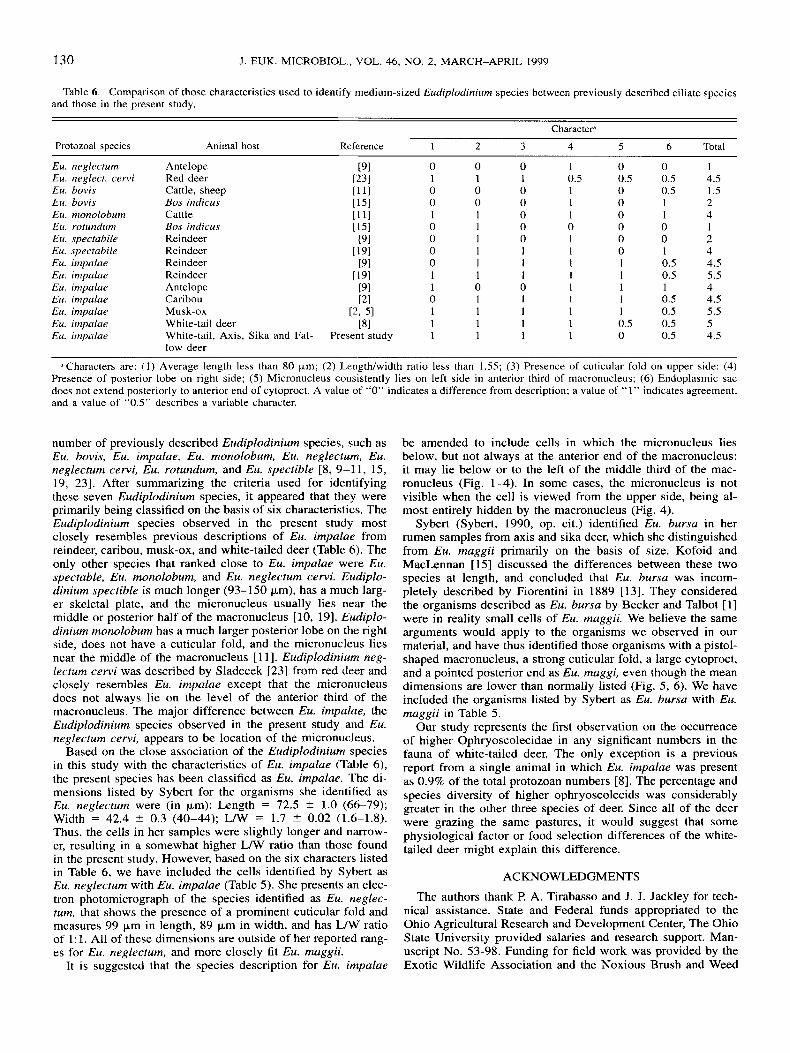
a Characters are: (1) Average length less than 80 pm; (2) Length/width ratio less than 1.55; (3) Presence of cuticular fold on upper side: (4) Presence of posterior lobe on right side; ( 5 ) Micronucleus consistently lies on left side in anterior third of macronucleus; (6 ) Endoplasmic sac does not extend posteriorly to anterior end of cytoproct. A value of “0” indicates a difference from description; a value of “1” indicates agreement, and a value of “0.5” describes a variable character.
number of previously described Eudiplodinium species, such as Eu. bovis, Eu. impalae, Eu. monolobum, Eu. neglectum, Eu. neglectum cervi, Eu. rotundurn, and Eu. spectible [8, 9-11, 15, 19, 231. After summarizing the criteria used for identifying these seven Eudiplodinium species, it appeared that they were primarily being classified on the basis of six characteristics. The Eudiplodinium species observed in the present study most closely resembles previous descriptions of Eu. impalae from reindeer, caribou, musk-ox, and white-tailed deer (Table 6). The only other species that ranked close to Eu. impalae were Eu. spectable, Eu. monolobum, and Eu. neglectum cewi . Eudiplo- dinium spectible is much longer (93-150 pm), has a much larg- er skeletal plate, and the micronucleus usually lies near the middle or posterior half of the macronucleus [lo, 191. Eudiplo- dinium monolobum has a much larger posterior lobe on the right side, does not have a cuticular fold, and the micronucleus lies near the middle of the macronucleus [ 1 11. Eudiplodinium neg- lectum cewi was described by Sladecek [23] from red deer and closely resembles Eu. impalae except that the micronucleus does not always lie on the level of the anterior third of the macronucleus. The major difference between Eu. impalae, the Eudiplodinium species observed in the present study and Eu. neglectum cewi , appears to be location of the micronucleus.
Based on the close association of the Eudiplodinium species in this study with the characteristics of Eu. impalae (Table 6), the present species has been classified as Eu. impalae. The di- mensions listed by Sybert for the organisms she identified as Eu. neglectum were (in Fm): Length = 72.5 2 1.0 (6679); Width = 42.4 ? 0.3 (40-44); L/W = 1.7 ? 0.02 (1.6-1.8). Thus, the cells in her samples were slightly longer and narrow- er, resulting in a somewhat higher L/W ratio than those found in the present study. However, based on the six characters listed in Table 6, we have included the cells identified by Sybert as Eu. neglectum with Eu. impalae (Table 5). She presents an elec- tron photomicrograph of the species identified as EM. neglec- turn, that shows the presence of a prominent cuticular fold and measures 99 pm in length, 89 pm in width, and has L/W ratio of 1 : 1. All of these dimensions are outside of her reported rang- es for Eu. neglectum, and more closely fit Eu. maggii.
It is suggested that the species description for Eu. impalae
be amended to include cells in which the micronucleus lies below, but not always at the anterior end of the macronucleus: it may lie below or to the left of the middle third of the mac- ronucleus (Fig. 1-4). In some cases, the micronucleus is not visible when the cell is viewed from the upper side, being al- most entirely hidden by the macronucleus (Fig. 4).
Sybert (Sybert, 1990, op. cit.) identified Eu. bursa in her rumen samples from axis and sika deer, which she distinguished from Eu. maggii primarily on the basis of size. Kofoid and MacLennan [ 151 discussed the differences between these two species at length, and concluded that Eu. bursa was incorn- pletely described by Fiorentini in 1889 [13]. They considered the organisms described as Eu. bursa by Becker and Talbot [ 11 were in reality small cells of Eu. maggii. We believe the same arguments would apply to the organisms we observed in our material, and have thus identified those organisms with a pistol- shaped macronucieus, a strong cuticular fold, a large cytoproct, and a pointed posterior end as Eu. maggi, even though the mean dimensions are lower than normally listed (Fig. 5, 6). We have included the organisms listed by Sybert as Eu. bursa with Eu. maggii in Table 5 .
Our study represents the first observation on the occurrence of higher Ophryoscolecidae in any significant numbers in the fauna of white-tailed deer. The only exception is a previous report from a single animal in which Eu. impalae was present as 0.9% of the total protozoan numbers [8]. The percentage and species diversity of higher ophryoscolecids was considerably greater in the other three species of deer. Since all of the deer were grazing the same pastures, it would suggest that some physiological factor or food selection differences of the white- tailed deer might explain this difference.
ACKNOWLEDGMENTS
The authors thank F? A. Tirabasso and J. J. Jackley for tech- nical assistance. State and Federal funds appropriated to the Ohio Agricultural Research and Development Center, The Ohio State University provided salaries and research support. Man- uscript No. 53-98. Funding for field work was provided by the Exotic Wildlife Association and the Noxious Brush and Weed

DEHORITY ET AL.-RUMEN CILIATES IN TEXAS DEER 131
Control Program, Department of Range, Wildlife, and Fisheries Management, Texas Tech University.
LITERATURE CITED I . Becker, E. R. & Talbot, M. 1927. The protozoan fauna of the
rumen and reticulum of American cattle. Iowa State Coll. J. Sci., 1:
2. Dehority, B. A. 1974. Rumen ciliate fauna of Alaskan moose (Al- ces americana), musk-ox (Ovibos moschatus) and Dall mountain sheep (Ovis dalli). J . Protozool., 21:26-32.
3. Dehority, B. A. 1978. Specificity of rumen ciliate protozoa in cat- tle and sheep. J. Protozool., 25509-5 13.
4. Dehority, B. A. 1984. Evaluation of subsampling and fixation pro- cedures used for counting rumen protozoa. Appl. Environ. Microbiol., 48: 182-185.
5. Dehority, B. A. 1985. Rumen ciliates of musk-oxen (Ovibos mos- chatus) from the Canadian Arctic. J. Protozool., 32:246-250
6. Dehority, B. A. 1990. Rumen ciliate protozoa in Ohio white-tailed deer (Odocoileus virginianus). J. Protozool., 37:473-475.
7. Dehority, B. A. 1994. Rumen ciliate protozoa of the blue duiker (Cephalophus monticola), with observations on morphological variation lines within the species Entodinium dubardi. J , Euk. Microbiol., 41: 103-1 I I .
8. Dehority, B. A. 1995. Rumen ciliates of the pronghorn antelope (Antilocapra americana), mule deer (Odocoileus hemionus), white- tailed deer (Odocoileus virginianus) and elk (Cervus canadensis) in the northwestern United States. Arch. Protistenkd., 146:29-36.
9. Dogiel, V. A. 1925. Nouveaux infusoires de la famille des ophryoscolecides parasites d’antilopes africaines. Ann. Parasitol., 3: 116-142.
10. Dogiel, V. A. 1925. Neue parasitische infusorien aus dem magen des renntieres (Rangifer tarandus). Arch. Russ. Protistol., 4:43-65.
1 1. Dogiel, V. A. 1927. Monographie der Familie Ophryoscolecidae. Arch. Prorisfenkd., 59: 1-288.
12. Dresher-Kaden, V. U. & Seifelnasr, E. A. 1977. Untersuchungen am verdauungstrakt von reh, damhirsch, und mufflon. Mitteilung 3: Mikroorganismen im pansen von reh, damhirsch und mufflon. Z. Jagd- wiss., 23:64-69.
345-372.
13. Fiorentini, A. 1889. Inrorno ai Protisti Stomaco dei Bovini. Frat Fusi, Pavia, Italy.
14. Imai, S . , Matsumoto, M., Watanabe, A. & Sato, H. 1993 Rumen ciliate protozoa in Japanese sika deer (Cervus nippon centralis). Anim. Sci. Technol., W.578-583.
15. Kofoid, C. A. & MacLennan, R. E 1932. Ciliates from Bos in- dicus Linn., 11. A revision of Diplodinium Schuberg. Univ. Calf$ Publ.
16. Latteur, B. 1968. Revision systematique de la famille des Ophryoscolecidae Stein, 1858. Sous-famille des Entodiniinae Lubinsky, 1957. Genre Enfodinium Stein. Ann. SOC. Roy. Zool. Belg., 98: 1-41,
17. Lubinsky, G. 1957. Studies on the evolution of the Ophryosco- lecidae (Ciliata: Oligotricha). 1. A new species of Entodinium with “caudatum”, “loboso-spinosum”, and “dubardi” forms. with some evolutionary trends in the genus Entodinium. Can. J . Zool.. 35:111- 133.
18. Lubinsky, G. 1958. Ophryoscolecidae (Ciliata: Entodiniomor- phida) of reindeer (Rangifer tarandus L.) From the Canadian Arctic. 1. Entodiniinae. Can. J . Zool., 365319435,
19. Lubinsky, G. 1958. Ophryoscolecidae (Ciliata: Entodiniomor- phida) of reindeer (Rangifer rarandus L.) from the Canadian Arctic. 11. Diplodiniinae. Can. J. Zool., 36:937-959.
20. MINITAB Reference Manual. 1991. PC version, release 8. Quickset Inc. Rosemont, Pennsylvania.
21. Pearson, H. A. 1965. Rumen organisms in white-tailed deer from south Texas. J. Wildl. Mgmt., 29:493-496.
22. Prins, R. A. & Geelen, M. J. H. 1971. Rumen characteristics of red deer, fallow deer, and roe deer. J. Wildl. Mgmr., 35:673-680.
23. Sladecek, E 1946. Ophryoscolecidae from the stomach of Cervus elaphus L., Duma dama L., and Capreolus capreolus L. Vestn. Cesk. Spol. Zool., 10:201-231 (in Czech with English summary).
24. Williams, A. G. & Coleman, G. S. 1992. The Rumen Protozoa. Springer-Verlag, Inc., New York. Pp. 4-85.
25. Zielyk. M. W. 1961. Ophryoscolecid fauna from the stomach of the white-tailed deer (Odocoileus virginianus borealis), and observa- tions on the division of Entodinium dubardi Buisson 1923 (Ciliata En- todiniiomorpha). J. Prorozool., 8:33-41,
ZOO^., 37:53-152.
Received 6-2-98, 10-30-98: accepted 12-5-98
UPCOMING MEETING
5th International Chrysophyte Symposium
July 26-31, 1999 Edwardsville, Illinois Southern Illinois University
For more information, contact: Jim Wee
Department of Biological Sciences Loyola University
6363 St. Charles Avenue New Orleans, LA 701 18-6195, USA
Email: wee @ loyno.edu FAX: 504-865-2920