rri~###: o/ - shodhgangashodhganga.inflibnet.ac.in/bitstream/10603/14916/12/12... · 2015-12-04 ·...
TRANSCRIPT

rri~###:
~~ o/ f!7UMu:u£ed ~ o/ :Yt;([JJ [ll; in £ ~ fRiww
r!J. .

CHAPTER III
6.A INTRODUCTION
The function of any protein depends on the three dimensional in
vwo structure of the protein along with the various post-translational
modifications it may be undergoing. Important information about the
structural aspects of the protein can be derived by the studies on the '
interaction of the protein in solution and by solving the crystal structure.
The crystal structure of HABPl has been solved and results shows it to
be a trimeric protein with each monomer consisting of one N-terminal a
(aA) helix followed by seven anti-parallel ·~ strands forming a highly
twisted~ sheet followed by the two C-terminal a helices (as and ac) (Diag.
lA). All the three helices lie on the same side of ~ sheet with as being
parallel to the p sheet but perpendicular to the orientation of the
individual p strands. The helix as and the N-terminal portion (4 turns) of
the helix ac make extensive hydrophobic contacts with the p-sheet that
seems to be important for the structural stability of the monomer. TheN
terminal helix aA does not contact the p sheet within the monomer but
forms an anti-parallel coiled coil with the C-terminal portion of ac. This
region is important for protein-protein interactions and is required for
oligomerization.
The trimer forms a doughnut shaped homo-trimer, with an unique
non-crystallographic three fold axis of symmetry, a structure unobserved
till date in any other protein (Jiang et al., 1999). The crystal structure of
this protein also displays a solvent exposed HA-binding motif in the
trimer.
Recent studies have shown that the HABPl trimer forms cysteine
mediated dimer of trimers {Jha et al., 2002). This oligomerization has
functional implications as this makes the whole structure more compact
which shows greater affinity for HA than a more relaxed structure. It is
101

gC1q BINDING MOTIF
N
a-B a-C ~~~~----~--~c a-A
HABINDING MOTIF
/ ' 119 * 121 KLVRKVAGEK
ERK PHOSPHORYLATION SITE •
/ ' "' * 155 PELTSTP
N
c
A
B
~----------~----~~~.a--------------------~~~NHABP1
II ~CHABP1
Diag 1 :(A) Schematic representation of a monomeric unit of the HABPl crystal showing the a.- helices and the ~-strands making up the ~-sheet. (B) Schematic representation of the allignments of the N-terminal and C-terminal deleted mutants with respect to the mature HABPl
CHAPTER III
also observed that there is asymmetric charge distribution in the
protein- the positive charges are present on one face while the negative
changes being present on the other. This allows for greater
conformational flexibility to the protein, which could be the reason for
multi-ligand interaction and multi-compartmental localization of HABP1.
Since both the monomeric and trim eric forms of HABP 1 are active
mainly through the interactions of the terminal a helices, truncated
variants of HABP 1 were generated to check their structural and
functional relationships (Sengupta, 2003). For the generation of N
terminal deleted clone the amino acids 7 4 to 105 have been removed
from the mature HABP1 which consists of the 74 to 282 amino acids.
This deletes the N-terminal UA helix and a part of the loop connecting the
131 strand. Since the N-terminal aA seemed to be important for protein
protein interactions, it was hypothesized that elimination of this helix
would substantially alter the structure and thus may affect the
interaction of the truncated protein (~N.HABP1) with other proteins in S.
pombe (Diag. 1B).
For the generation of C-terminal mutant clone, the ammo acids
267 to 282 have been deleted thereby resulting in removal of half of the
ac helix along with the tail end of the C-terminal to generate the
truncated protein ~C.HABPl. The terminal helices of HABP1 were
considered to be important for the structural integrity of HABP1 and
hence for its functions. A schematic representation of the secondary
structure of the HABP1 monomer along with the generation of the N
terminal and C-terminal deleted mutants are given in the panels A and B
of Diagram 1 respectively. The N-terminal and C-terminal deleted
mutants so generated (Sengupta., 2003) were subcloned into the shuttle
vector pRep 1 and the clones generated were named pRH~N 1 and
pRH~C 1 respectively and were used to transform S. pombe cells.
102

CHAPTER III
It was hypothesized that such critical structural changes in HABPl
molecule would alter its behaviour in S. pombe a..."ld would help in
interpreting the observed aberrant morphological changes and growth
inhibition displayed by S. pombe cells on expression of HABPl.
103
CHAPTER III
6.B RESULTS
6.B.i SUBCLONING OF ~N AND 8C FRAGMENTS INTO THE SHUTTLE VECTOR pREPl
The gel purified ~N and ~C fragments (Sengupta, 2003) were
ligated with gel purified Nde1, BamH1 double digested pRep1 vector.
Since ~N and ~C were also cloned between the Ndel and BamH1 sites,
the sub-cloning would be directional. Ligation was done at 16oC for 16
chours with T4 DNA ligase (NEB) in the appropriate buffer. After the
ligation step, the ligation mixture was used to transform competent E.
coli DH5a cells and the colonies were screened for the proper clone.
Cloning was confirmed by restriction digestion with Nde1 and BamH1
(Fig. 1) and the correct size of the insert was checked. The ~N fragment is
of 900 base pairs while the ~C fragment was of 600 base pairs.
6.B.ii EXPRESSION OF THE TRUNCATED VARIANTS OF HABPl IN S. Pombe MBY 624 CELLS
S. pombe transformed pRHP 1, pRH~N 1, pRH~C 1 were grown to
saturation and lysed. The extracted proteins are estimated a~d equal
amounts (100 Jlgm) were resolved in a 12.5% SDS-PAGE, transferred
onto a PVDF membrane and probed with monoclonal anti-HABP1
antibody (Fig. 2). As can be seen, the expression of the mature HABP1,
~N. HABP1 and .8C.HABP1 show a migration of 34, 27 and 32 kDa
respectively on the immunoblot. This confirms the expression of HABP1,
pRH~N1 and pRH~C1 inS. pombe (MBY 624) cells.
6.B.iii CELL GROWTH ASSAY OF THE. pRH~Nl AND pRH~Cl TRANSFORMED S. pombe CELLS
pRH~N 1 and pRHL1C 1 were used to transform S. pombe cells as
described in Material and Methods. Following transformation the cells
were grown in EMM leu-medium along with pRepl and pRHPl
transformed cells. and their growth rates measured at different time
104

Q..
CQ -< :I: :...
Fig I : Shows the mature and truncated fragments, cloned in pRepl, with Nde I and BamHI restriction endonucleases and resolved on a 0.7 % Agarose Gei.The fragment of HABP1 was 1030 BP in length, while that of tlN was 900 BP and tlC was 600 BP in length respectively.
....., u z <J <J Q..
:I: :I: :I: 0:: 0:: 0:: 0.. 0.. 0.. I<Da
97.0
67.0
45.0
30.1
u z <J <J Q..
:I: :I: :I: 0:: 0:: 0:: 0.. 0.. 0..
J, J, J,
0..
"' 0:: 0..
J,
:...
"' ..:.: :... (<:!
:;; J, bp
10000
3000
2000 1650
1000
500
Fig 2:The mature protein and its truncated variants were immunodetected using monoclonal anti-HABPI antibody after resolving them on a 12.5 u;;, SDS-PAGE The truncated proteins can be detected by the anti-HABP I antibody. The N-
20.1 terminal deleted mut:::nt shows a migration of approximately 28 kDa and the C-terminal shows a migration of30 kDa in SDS-PAGE.
7
6
5 E c: m 4
iV 3 c 0
2
0
00 vsTime
10 20 30 40 50
Time In hrs
60 70 80
--pRHP1
---- pRep1
___..___ pRHdeiN1
--pRHdeiC1
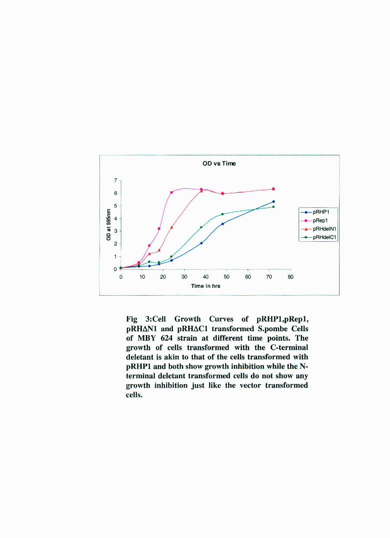
Fig 3:Cell Growth Curves of pRHPl,pRepl, pRIMNl and pRIMCl transformed S.pombe Cells of MBY 624 strain at different time points. The growth of cells transformed with the C-terminal deletant is akin to that of the cells transformed with pRHPl and both show growth inhibition while the Nterminal deletant transformed cells do not show any growth inhibition just like the vector transformed cells.
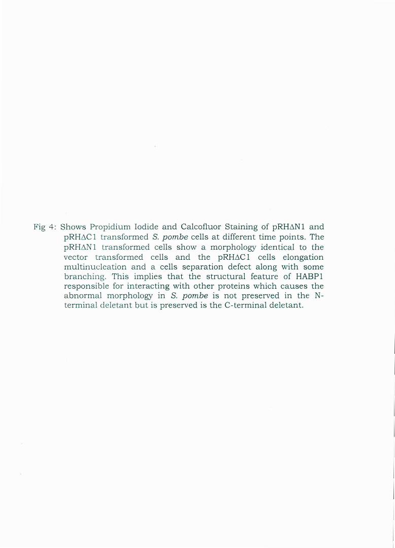
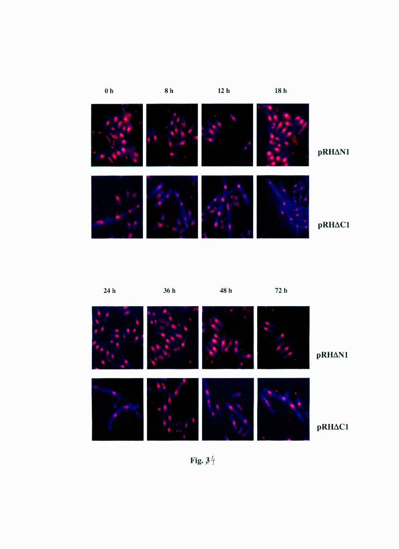
Fig 4 : Shows Propidium Iodide and Calcofluor Staining of pRHt-.N1 and pRHt-.C 1 t ra nsformed S. pombe cells at different time points. The pRHtlN 1 tra nsformed cells show a morphology identical to the vector t ransformed cells and the pRHt-.C 1 cells elongation multinucleation and a cells separation defect along with some bra nch ing. This implies that the structural feature of HABP1 responsible for interacting with other proteins which causes the a bnormal morphology in S. pombe is not preserved in the Ntermina l de leta nt but is preserved is the C-terminal deletant.
I
I
I
I
I
I
I
I
I
I
I
I
I
I
I
I
I
I
I
I
I
I
Oh 8h 12 h 18 h
pRH~Nl
pRH~Cl
24h 36 h 48 h 72h
pRH~Nl
pRH~Cl
Fig. p'f

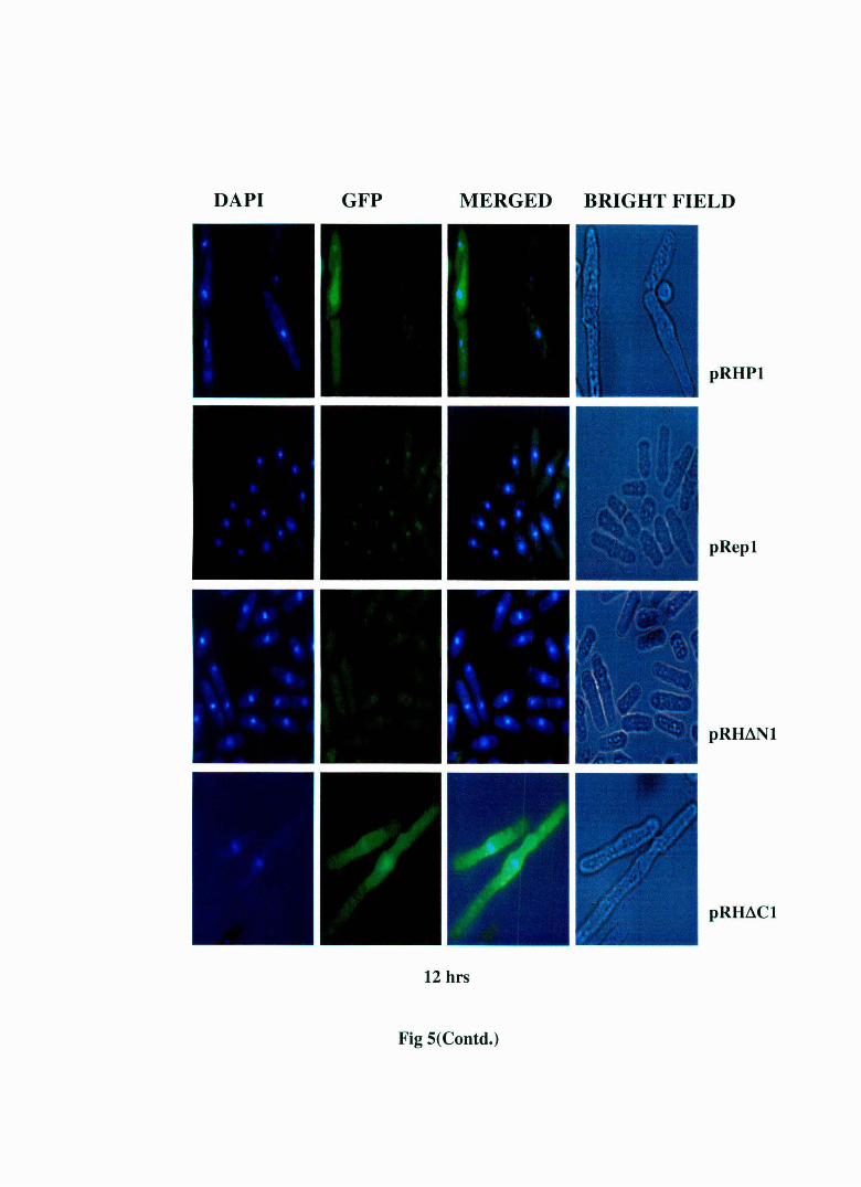
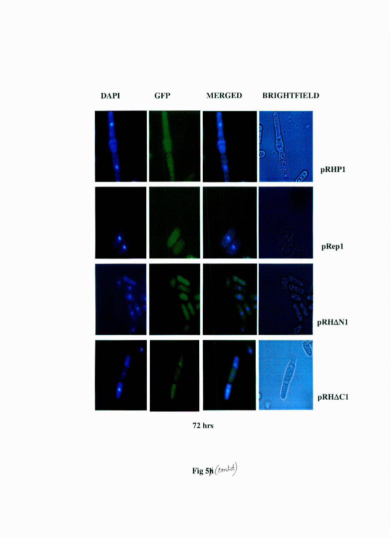
Fig 5: Live cell staining of S. pombe MBY 624 strain transformed with pRHP1 and pRep1, pRH~N1 and pRH~C1 at different time points. The regulatory light chain (rlc) of myosin has been tagged with GFP in this strain and the nucleus has been stained with DAPI. The pRHP 1 and pRH~C 1 transformed cells display elongation, multinucleation and abnormal cell septum formation . The pRep 1 and pRH~N 1 cells show normal morphology. The cell septa in pRHP1 and pRH~C1 transformed cells do not show the green fluorescence indicating it's the primary septum. This indicates that this is a cell separation defect.

DAPI GFP MERGED BRIGHT FIELD
pRepl
pRHPl
pRH~Nl
pR~Cl
0 hrs
Fig 5

DAPI GFP MERGED BRIGHT FIELD
pRHPl
pRepl
pRH~N
pRH~C J
6 Hrs
Fig S(Contd.)
DAPI GFP MERGED BRIGHT FIELD
~
pRHPl
--.
pRepl
I . I
\ .
' pRH~Nl -·
-
• ..t· , I
pRH~Cl
I I I
12 hrs
Fig S(Contd.)

DAPI GFP MERGED BRIGHT FIELD
pRHPl
pRepl
pRmNl
pRH~Cl
18 hrs
Fig S(Contd.)

DAPI GFP MERGED BRIGHT FIELD
pRHPl
pRepl
pRHdNl
pRHdCl
24 hrs
Fig S(Contd.)

DAPI GFP MERGED BRIGHT FIELD
pRHPl
pRepl
pRHdNl
pRH~Cl
36 hrs
Fig S(Contd.)

DAPI GFP MERGED BRIGHT FIELD
pRHPl
pRepl
pRH~Nl
pRH~Cl
47 hrs
Fig S(contd.)
DAPI GFP MERGED BRIGHTFIELD
pRHPl
pRepl
pRH~l
pRH.::lCl
72 hrs
FigS~{~

CHAPTER III
points and plotted graphically. As seen in Fig. 3, the growth pattern of
pRHt.N 1 follows that of the vector tra.."'1sformed cells while that of
pRHt.C 1 strain is similar to that of pRHP1 transformed cells. Also, it was
evident that the growth inhibition d isplayed by pRHt.C 1 transformed
cells is less than the pRHP1 transformed cells.
6.B.iv PROPIDIUM IODIDE/CALCOFLUOR STAINING OF S. pombe SHOW pRHt.Nl TRANSFORMED CELLS HAVING A NORMAL MORPHOLOGY WHILE pRHt.Cl TRANSFORMED CELLS WITH A MULTI-NUCLEATED MORPHOLOGY WITH SOME HYPHAL PROJECTIONS
pRHt.N1 and pRH~C1 transformed S. pombe (MBY 6 24) cells a re
grown in EMM leu - medium and their growth rates measured at different
time points (0, 8 , 12, 18, 24, 36, 48, 72 hrs respectively)
spectrophotomeLrically. Samples of 107 cells were accordingly collected at
suc h time points and flxed with ethanol. Later these cells were
rehydrated and were stained with Propidium iodide (for nucleus) and
Calcofluor (for cell wall and medial septa). The results shown in flgure 4
demonstrate that the pRHt.N 1 transformed cells exhibit a morphology
similar to that of pRep1 transformed cells (see Fig. 5, Chapter 1) while
pRHt.C 1 transformed cells show an aberrant morphological features like
multiple nuclei, abnormal septa and in a few cases hyphal projections at
all time points. This implies that the a bnormal morphological features
seen in HABP 1 expressing cells are also present in pRH6C 1 transformed
ce lls.
6.B .v LIVE CELL STAINING OF TRANSFORMED S. pombe STRAIN MBY 624
To eliminate the possibility of any artifact resulting due to the use
of e thanol fixed cells for microscopic analysis, live cell staining at
different time points (0,6, 12, 18,24,36,4 7 and72 hrs respectively) wa s
performed with S. pombe MBY 624 cells transformed with pRHL'lN1 and
105

Fig 6 : Shows the ultrastructural analys is of pRep 1, pRHP1 , pRHL'l N 1 a nd pRHL'lC 1 transformed S. pombe cells at d iffe rent time points . In the panel to the right the observed m orphological abnorma lities of multiple cell septum increase in vacu oles and elongation in pRHPl transformed S .pombe cells a re clearly visible vis-a-vis th e pRep 1 transformed cells. Similar abnormalities are a lso seen in pRHL'lC 1 transformed cells too in the right most panel. The pRHL'lNl transformed cells a s s een in the second pa n el from th e right display normal morphology like th a t of the pRep 1 transformed cells shown in the second panel from the left.

6h
12h
24h
36h
48h
pRHPI pRep I pRHLlNI
Fig 6
pRHL:lCI

CHAPTER III
pRH~C 1. The regulatory light chain (RLC) of myosin is tagged with GFP
in this strain. The nuclei of the living cells were stained with DAPI. The
results (Fig. 5) show that pRH~N 1 transformed cells display a normal
morphology just like pRep 1 transformed cells while pRH~C 1 transformed
cells show the same morphological abnormalities as pRHPl transforming
cells. Therefore the results obtained from ethanol fixed cells were in
complete agreement with the results obtained using live cells ruling out
the possibility that the abnormal morphology observed in ethanol fixed
cells are artefactual in nature.
6.B.vi ULTRA MICROSCOPY OF pRH~Nl
TRANSFORMED CELLS AND pRH~Cl
For studies with ultra microscopy S. pombe MBY 624 cells
transformed with pRHP1, pRep1, pRH~N1 and pRH~C1 were grown and
processed at differer..t time points as described in Material and Methods.
The morphological features of the cells were examined with an electron
microscope. The results in figure 6 show that while pRH~N 1 transformed
cells have complete similarity with the pRep 1 transformed cells, pRH~C 1
transformed cells show a morphological changes that were similar to
pRHP1 transformed cells.
106

CHAPTER III
6.C DISCUSSION
The results presented show that the growth inhibition shown by
pRH~N 1 expressing cells is negligibly small and cells maintain a normal
morphology. However the pRH~C1 expressing cells show significantly
high inhibition of cell growth similar to pRHP1 transformed cells. The
morphological abnormalities exhibited by pRH~C 1 cells are quite
identiCal to that shown by pRHP1 transformed cells. In fact as evident
from the pictures, pRH~C 1 transformed cells cannot be differentiated
from pRHP1 transformed cells. This observation suggests that the in S.
pombe cells expressing ~C.HABP1 are capable of retaining the necessary
structure to discharge the same cellular functions as mature HASP 1
albeit not as efficiently. Since the expression of ~N.HABP1 does not have
any significant effect on the growth and morphology of the S. pombe cells,
it could be that the ~N.HABP1 does not retain the necessary structure.
However the biophysical analysis of recombinant HABP1, ~N.HABP1 and
~c:HABP1 indicate that the truncated mutants fail to trimerize but can
form cysteine. mediated dimers and does not display any change in their
ability to bind HA. This implies that the structural attributes responsible
for the HA binding property of HABP1 is not important for the rendering
of its functions in S. pombe. Incidentally there are other structural
changes due to the indicated truncations in HABP1 as have been
observed by Circular Dichorism experiments (Sengupta, 2003), the
functional implications of which are not yet fully understood.
The N-terminal of HABP1 shows great similarity to the WD 40
family of regulatory proteins which entails its having a signature
sequence of G-H and W-D/E di-peptide usually separated by 25-27
amino acids. Such sequences predominantly form a a helix: and
biochemical evidence suggests that this signature motif/fold are critical
for protein-protein interactions. Since in ~N.HABP1 the entire aA helix is
107

CHAPTER III
cleaved off. This destroys the coiled-coil interaction aA has with as of an
adjacent monomer, which is extremely important for protein-protein
interactions. The hypothesis that the N-terminal a helix (aA) is very
important for protein-protein interactions in S. pombe is also borne out
by the observed results as its seen that deletion of aA just about
abolishes the interaction of this truncated protein with other proteins in
S. pombe. Also since the N-terminal sequence bears the WE signature
motif, which is very significant for protein-protein interactions, the
importance of the aA cannot be over-estimated. However an alternative
possibility is there that the loss of aA reduces the structure of the N
terminal deleted mutant to a random coil. Further investigations are
required to specify which is the correct explanation.
In case of the C-terminal deleted mutant, only half of the ac helix
has been removed. So it is possible that this deletion does not cause
much change in the structure of the monomer and the aA helix is also
intact in this mutant. This may be the reason why it shows a morphology
and growth patterns similar to that of pRHPl transformed cells.
In conclusion, it is rational to state that the N-terminal helix of the
HABPl bearing WE signature is necessary for the changes it produces in
S, pombe cells. Further studies need to be pursued to understand the
structural differences between ~N.HARPl and ~C.HABPl and analyze
why the two truncated proteins impart dissimilar response when
expressed in S. pombe cells and whether that is by losing its ability to
interact with other proteins like CDC25.
6.0. Summary
To summarize:
1) The two truncated mutants of HABPl expressed inS. pombe have
the same affinity for HA. Hence the HA binding property of
108

CHAPTER III
HABPl may not be important for its causing the growth and
morphological abnormalities in S. pombe.
2) The N-terminal deleted mutant shows a growth pattern and
morphology identical to the pRep 1 transformed cells. The
structure of HABPl responsible for causing growth inhibition and
morphological abnormalities is disrupted in the N-terminal
deetant implying the great importance of the N-terminal UA helix
in protein-protein interactions of HABPl.
3) The C-terminal deleted mutant shows a growth pattern and
morphology very similar to pRHPl transformed cells implying
that the structure of HABPl responsible for causing the aberrant
growth and morphology in S. pombe cells is intact in the C
terminal deletant.
4) The N-terminal a helix is important protein-protein interactions
may be responsible for the abolition of abnormal behaviour
caused by the expression of mature HABPl in S. pombe.
109